UNIVERSITÉ DE MONTRÉAL
FLEXIBLE HARDWARE ARCHITECTURES FOR RETINAL IMAGE ANALYSIS
HAMZA BENDAOUDI
DÉPARTEMENT DE GÉNIE INFORMATIQUE ET GÉNIE LOGICIEL ÉCOLE POLYTECHNIQUE DE MONTRÉAL
THÈSE PRÉSENTÉE EN VUE DE L’OBTENTION DU DIPLÔME DE PHILOSOPHIAE DOCTOR
(GÉNIE INFORMATIQUE) AVRIL 2017
UNIVERSITÉ DE MONTRÉAL
ÉCOLE POLYTECHNIQUE DE MONTRÉAL
Cette thèse intitulée:
FLEXIBLE HARDWARE ARCHITECTURES FOR RETINAL IMAGE ANALYSIS
présentée par: BENDAOUDI Hamza
en vue de l’obtention du diplôme de : Philosophiae Doctor a été dûment acceptée par le jury d’examen constitué de :
M. BOIS Guy, Ph. D., président
M. LANGLOIS J.M. Pierre, Ph. D., membre et directeur de recherche Mme CHERIET Farida, Ph. D., membre et codirectrice de recherche M. BILODEAU Guillaume-Alexandre, Ph. D., membre
DEDICATION
ACKNOWLEDGEMENTS
I would like to express my sincere and greatest gratitude to my supervisors, Dr. Pierre Langlois and Dr. Farida Cheriet. I am thankful for their constant support and encouragement and for their constructive advice and comments.
I am also thankful for my colleagues in the LASNEP and LIV4D, for the good times, mutual sup-port and encouragement that we shared.
I will always be thankful to the faculty, staff and students of the department who helped me to expand my knowledge and expertise and appreciate my work even more. I would also like to thank them for every bit of help they provided each in his way.
RÉSUMÉ
Des millions de personnes autour du monde sont touchées par le diabète. Plusieurs compli-cations oculaires telle que la rétinopathie diabétique sont causées par le diabète, ce qui peut con-duire à une perte de vision irréversible ou même la cécité si elles ne sont pas traitées. Des examens oculaires complets et réguliers par les ophtalmologues sont nécessaires pour une détection précoce des maladies et pour permettre leur traitement. Comme solution préventive, un protocole de dépis-tage impliquant l'utilisation d'images numériques du fond de l'œil a été adopté. Cela permet aux ophtalmologistes de surveiller les changements sur la rétine pour détecter toute présence d'une maladie oculaire. Cette solution a permis d'obtenir des examens réguliers, même pour les popula-tions des régions éloignées et défavorisées. Avec la grande quantité d'images rétiniennes obtenues, des techniques automatisées pour les traiter sont devenues indispensables. Les techniques automa-tisées de détection des maladies des yeux ont été largement abordées par la communauté scienti-fique. Les techniques développées ont atteint un haut niveau de maturité, ce qui a permis entre autre le déploiement de solutions en télémédecine.
Dans cette thèse, nous abordons le problème du traitement de volumes élevés d'images réti-niennes dans un temps raisonnable dans un contexte de dépistage en télémédecine. Ceci est requis pour permettre l'utilisation pratique des techniques développées dans le contexte clinique. Dans cette thèse, nous nous concentrons sur deux étapes du pipeline de traitement des images rétiniennes. La première étape est l'évaluation de la qualité de l'image rétinienne. La deuxième étape est la segmentation des vaisseaux sanguins rétiniens.
L’évaluation de la qualité des images rétinienne après acquisition est une tâche primordiale au bon fonctionnement de tout système de traitement automatique des images de la rétine. Le rôle de cette étape est de classifier les images acquises selon leurs qualités, et demander une nouvelle acquisition en cas d’image de mauvaise qualité. Plusieurs algorithmes pour évaluer la qualité des images rétiniennes ont été proposés dans la littérature. Cependant, même si l'accélération de cette tâche est requise en particulier pour permettre la création de systèmes mobiles de capture d'images rétiniennes, ce sujet n'a pas encore été abordé dans la littérature. Dans cette thèse, nous ciblons un algorithme qui calcule les caractéristiques des images pour permettre leur classification en mau-vaise, moyenne ou bonne qualité. Nous avons identifié le calcul des caractéristiques de l'image
comme une tâche répétitive qui nécessite une accélération. Nous nous sommes intéressés plus par-ticulièrement à l’accélération de l’algorithme d’encodage à longueur de séquence (Run-Length
Ma-trix – RLM). Nous avons proposé une première implémentation complètement logicielle mise en
œuvre sous forme d’un système embarqué basé sur la technologie Zynq de Xilinx. Pour accélérer le calcul des caractéristiques, nous avons conçu un co-processeur capable de calculer les caracté-ristiques en parallèle implémenté sur la logique programmable du FPGA Zynq. Nous avons obtenu une accélération de 30,1 × pour la tâche de calcul des caractéristiques de l’algorithme RLM par rapport à son implémentation logicielle sur la plateforme Zynq.
La segmentation des vaisseaux sanguins rétiniens est une tâche clé dans le pipeline du trai-tement des images de la rétine. Les vaisseaux sanguins et leurs caractéristiques sont de bons indi-cateurs de la santé de la rétine. En outre, leur segmentation peut également aider à segmenter les lésions rouges, indicatrices de la rétinopathie diabétique. Plusieurs techniques de segmentation des vaisseaux sanguins rétiniens ont été proposées dans la littérature. Des architectures matérielles ont également été proposées pour accélérer certaines de ces techniques. Les architectures existantes manquent de performances et de flexibilité de programmation, notamment pour les images de haute résolution. Dans cette thèse, nous nous sommes intéressés à deux techniques de segmentation du réseau vasculaire rétinien, la technique du filtrage adapté et la technique des opérateurs de ligne. La technique de filtrage adapté a été ciblée principalement en raison de sa popularité. Pour cette technique, nous avons proposé deux architectures différentes, une architecture matérielle person-nalisée mise en œuvre sur FPGA et une architecture basée sur un ASIP. L'architecture matérielle personnalisée a été optimisée en termes de surface et de débit de traitement pour obtenir des per-formances supérieures par rapport aux implémentations existantes dans la littérature. Cette implé-mentation est plus efficace que toutes les impléimplé-mentations existantes en termes de débit. Pour l'ar-chitecture basée sur un processeur à jeu d’instructions spécialisé (Application-Specific
Instruction-set Processor – ASIP), nous avons identifié deux goulets d'étranglement liés à l'accès aux données
et à la complexité des calculs de l'algorithme. Nous avons conçu des instructions spécifiques ajou-tées au chemin de données du processeur. L'ASIP a été rendu 7.7 × plus rapide par rapport à son architecture de base.
La deuxième technique pour la segmentation des vaisseaux sanguins est l'algorithme détec-teur de ligne multi-échelle (Multi-Scale Ligne Detector – MSLD). L'algorithme MSLD est choisi
en raison de ses performances et de son potentiel à détecter les petits vaisseaux sanguins. Cepen-dant, l'algorithme fonctionne en multi-échelle, ce qui rend l’algorithme gourmand en mémoire. Pour résoudre ce problème et permettre l'accélération de son exécution, nous avons proposé un algorithme efficace en terme de mémoire, conçu et implémenté sur FPGA. L'architecture proposée a réduit de façon drastique les exigences de l’algorithme en terme de mémoire en réutilisant les calculs et la co-conception logicielle/matérielle.
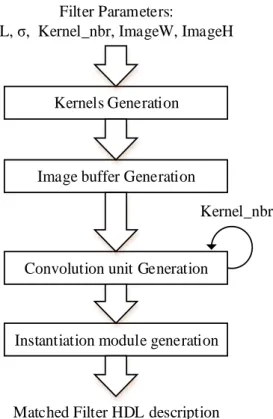
Les deux architectures matérielles proposées pour la segmentation du réseau vasculaire réti-nien ont été rendues flexibles pour pouvoir traiter des images de basse et de haute résolution. Ceci a été réalisé par le développement d'un compilateur spécifique capable de générer une description HDL de bas niveau de l'algorithme à partir d'un ensemble de paramètres. Le compilateur nous a permis d’optimiser les performances et le temps de développement. Dans cette thèse, nous avons introduit deux architectures qui sont, au meilleur de nos connaissances, les seules capables de trai-ter des images à la fois de basse et de haute résolution.
ABSTRACT
Millions of people all around the world are affected by diabetes. Several ocular complica-tions such as diabetic retinopathy are caused by diabetes, which can lead to irreversible vision loss or even blindness if not treated. Regular comprehensive eye exams by eye doctors are required to detect the diseases at earlier stages and permit their treatment. As a preventing solution, a screening protocol involving the use of digital fundus images was adopted. This allows eye doctors to monitor changes in the retina to detect any presence of eye disease. This solution made regular examinations widely available, even to populations in remote and underserved areas. With the resulting large amount of retinal images, automated techniques to process them are required. Automated eye de-tection techniques are largely addressed by the research community, and now they reached a high level of maturity, which allows the deployment of telemedicine solutions.
In this thesis, we are addressing the problem of processing a high volume of retinal images in a reasonable time. This is mandatory to allow the practical use of the developed techniques in a clinical context. In this thesis, we focus on two steps of the retinal image pipeline. The first step is the retinal image quality assessment. The second step is the retinal blood vessel segmentation.
The evaluation of the quality of the retinal images after acquisition is a primary task for the proper functioning of any automated retinal image processing system. The role of this step is to classify the acquired images according to their quality, which will allow an automated system to request a new acquisition in case of poor quality image. Several algorithms to evaluate the quality of retinal images were proposed in the literature. However, even if the acceleration of this task is required, especially to allow the creation of mobile systems for capturing retinal images, this task has not yet been addressed in the literature. In this thesis, we target an algorithm that computes image features to allow their classification to bad, medium or good quality. We identified the com-putation of image features as a repetitive task that necessitates acceleration. We were particularly interested in accelerating the Run-Length Matrix (RLM) algorithm. We proposed a first fully soft-ware implementation in the form of an embedded system based on Xilinx's Zynq technology. To accelerate the features computation, we designed a co-processor able to compute the features in parallel, implemented on the programmable logic of the Zynq FPGA. We achieved an acceleration of 30.1× over its software implementation for the features computation part of the RLM algorithm.
Retinal blood vessel segmentation is a key task in the pipeline of retinal image processing. Blood vessels and their characteristics are good indicators of retina health. In addition, their seg-mentation can also help to segment the red lesions, indicators of diabetic retinopathy. Several tech-niques have been proposed in the literature to segment retinal blood vessels. Hardware architec-tures have also been proposed to accelerate blood vessel segmentation. The existing architecarchitec-tures lack in terms of performance and programming flexibility, especially for high resolution images. In this thesis, we targeted two techniques, matched filtering and line operators. The matched filter-ing technique was targeted mainly because of its popularity. For this technique, we proposed two different architectures, a custom hardware architecture implemented on FPGA, and an Application Specific Instruction-set Processor (ASIP) based architecture. The custom hardware architecture area and timing were optimized to achieve higher performances in comparison to existing imple-mentations. Our custom hardware implementation outperforms all existing implementations in terms of throughput. For the ASIP based architecture, we identified two bottlenecks related to data access and computation intensity of the algorithm. We designed two specific instructions added to the processor datapath. The ASIP was made 7.7× more efficient in terms of execution time com-pared to its basic architecture.
The second technique for blood vessel segmentation is the Multi-Scale Line Detector (MSLD) algorithm. The MSLD algorithm is selected because of its performance and its potential to detect small blood vessels. However, the algorithm works at multiple scales which makes it memory intensive. To solve this problem and allow the acceleration of its execution, we proposed a memory-efficient algorithm designed and implemented on FPGA. The proposed architecture re-duces drastically the memory requirements of the algorithm by reusing the computations and SW/HW co-design.
The two hardware architectures proposed for retinal blood vessel segmentation were made flexible to be able to process low and high resolution images. This was achieved by the develop-ment of a specific compiler able to generate low-level HDL descriptions of the algorithm from a set of the algorithm parameters. The compiler enabled us to optimize performance and develop-ment time. In this thesis, we introduce two novel architectures which are, to the best of our knowledge, the only ones able to process both low and high resolution images.
TABLE OF CONTENTS
DEDICATION ... III ACKNOWLEDGEMENTS ... IV RÉSUMÉ ... V ABSTRACT ... VIII TABLE OF CONTENTS ... X LIST OF TABLES ... XIII LIST OF FIGURES ... XIV LIST OF SYMBOLS AND ABBREVIATIONS... XVICHAPTER 1 INTRODUCTION ... 1
1.1 Overview and motivation ... 1
1.2 Problem statement ... 4
1.3 Research contributions ... 5
1.4 Thesis organization ... 7
CHAPTER 2 LITERATURE REVIEW ... 8
2.1 Diabetic retinopathy ... 8
2.2 Retinal image processing pipeline ... 11
2.2.1 Quality assessment ... 11
2.2.2 Retinal image enhancement ... 12
2.2.3 Features extraction ... 15
2.3 Public datasets and metrics ... 20
2.4 Architectural considerations for the implementation of image processing algorithms .. 22
2.4.1 Data access for image processing ... 22
2.4.3 Processor architecture for image processing ... 24
2.5 Tools for HDL description generation for image processing ... 27
2.6 Summary and research objectives ... 29
CHAPTER 3 CO-PROCESSOR FOR RUN LENGTH ENCODING ALGORITHM ... 31
3.1 Introduction ... 31
3.2 Run-Length Matrix and RLM Features ... 33
3.2.1 Image acquisition ... 36
3.2.2 Mask generation ... 36
3.2.3 Run-Length Matrix computation ... 36
3.2.4 Features computing ... 36
3.3 Zynq based architecture ... 37
3.3.1 Data analysis ... 37
3.3.2 RLM features co-processor ... 38
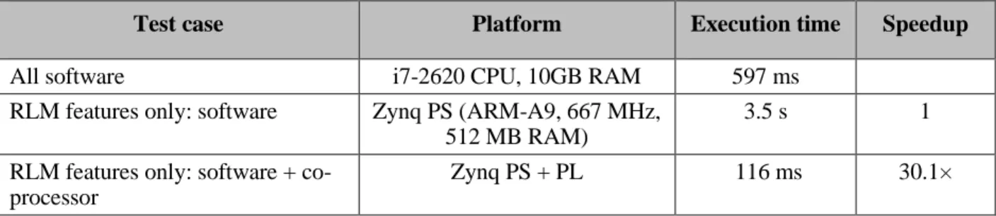
3.4 Results and discussion ... 39
3.5 Conclusion ... 42
CHAPTER 4 FLEXIBLE ARCHITECTURES FOR RETINAL BLOOD VESSEL SEGMENTATION USING MATCHED FILTERING ... 43
4.1 Introduction ... 43
4.2 Matched filter algorithm description ... 44
4.3 Proposed architectures for the matched filter algorithm ... 46
4.3.1 Scalable hardware matched filter architecture ... 46
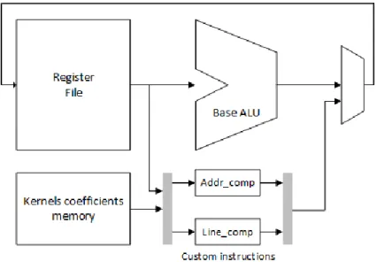
4.3.2 Proposed ASIP for matched filter algorithm ... 50
4.3.2.1 Custom instruction Line_comp ... 51
4.3.2.2 Custom instruction Addr_comp ... 52
4.5 Verification of the matched filter designs ... 59
4.6 Conclusion ... 59
CHAPTER 5 MEMORY-EFFICIENT ARCHITECTURE FOR RETINAL BLOOD VESSEL SEGMENTATION ... 61
5.1 Introduction ... 61
5.2 MSLD algorithm description ... 62
5.3 Proposed MSLD memory-efficient architecture ... 63
5.3.1 Memory-efficiency ... 64
5.3.2 Line buffers ... 66
5.3.3 Line Response Computing Module (LRCM) ... 67
5.3.4 Raw Response Computation Module (RRCM) ... 67
5.3.5 Mean and standard deviation values computation ... 69
5.3.6 Standardized and Combined Response Computation ... 70
5.3.7 Operations scheduling ... 71
5.3.8 MSLD architecture scalability ... 72
5.4 MSLD implementation results ... 74
5.5 Verification of the MSLD designs ... 80
5.6 Conclusion ... 80
CHAPTER 6 GENERAL DISCUSSION ... 82
CHAPTER 7 CONCLUSION AND FUTURE WORK ... 86
LIST OF TABLES
Table 3.1. Programmable logic resources usage ... 41
Table 3.2. Execution time under Zynq PS for several image sizes ... 41
Table 3.3. Execution time under Zynq PS for several image sizes ... 42
Table 4.1. Parameters of the basic architecture of the Xtensa processor ... 51
Table 4.2. Quality measures of blood vessel detection using DRIVE dataset. ... 54
Table 4.3. Estimated design and verification efforts ... 59
Table 5.1. Quality measures of blood vessel segmentation for DRIVE database images – CPU and FPGA implementations ... 76
Table 5.2. FPGA versus CPU implementation performances ... 78
Table 5.3. Logic utilisation for the MSLD algorithm for DRIVE database images ... 78
Table 6.1. Summary of existing implementations of Retinal Blood Vessel Detection Algorithms ... 83
LIST OF FIGURES
Figure 2-1. Centralized processing system overview ... 3
Figure 2-1. Normal retina anatomical parts ... 9
Figure 2-2. a- Normal vision, b- Vision with diabetic retinopathy [12] ... 10
Figure 2-3. Diabetic retinopathy symptoms: a- Microaneurysms, b- Haemorrhages, c- Hard exudates, d- Soft exudates, e- Neovascularisation [11]. ... 10
Figure 2-4. Main steps of an automated eye disease detection system ... 11
Figure 3-1. Examples of bad quality retinal images ... 32
Figure 3-2. a. Image and b. its corresponding Run Length Matrix ... 33
Figure 3-3. Different steps for computing the image features ... 35
Figure 3-4. Original image and the corresponding mask ... 37
Figure 3-5. Overview of the Zynq-based system ... 37
Figure 3-6. RLM features co-processor architecture ... 39
Figure 3-7. State machine sequencer of the co-processor ... 40
Figure 3-8. Overview of the proposed system for RLM features computing ... 41
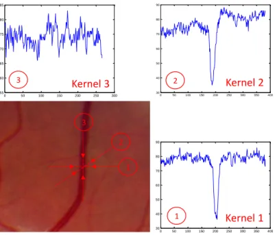
Figure 4-1. Retinal blood vessel (a) and the intensity profile of its cross section for different orientations ... 45
Figure 4-2. 2D kernel of the matched filter ... 46
Figure 4-3. Retinal blood vessel detection system overview ... 47
Figure 4-4. The matched filter scalable architecture overview ... 47
Figure 4-5. A simplified architecture of the Convolution Unit ... 48
Figure 4-6. Matched filter generation steps ... 49
Figure 4-7. Replacing multiplication by left shifts and additions ... 50
Figure 4-8. Datapath of the proposed ASIP ... 51
Figure 4-10. Original image and the corresponding mask ... 54
Figure 4-11. Green channel of a retinal image with the matched filter response ... 54
Figure 4-12. FPGA resources utilization as a function of the number of kernels of size 15 × 15 ... 55
Figure 4-13. FPGA maximum frequency as a function of the number of kernels of size 15 × 15 ... 56
Figure 4-14. FPGA resources utilization as a function of kernel size (for 12 kernels) ... 56
Figure 4-15. FPGA maximum frequency as a function of kernel size (for 12 kernels) ... 57
Figure 4-16. Number of cycles for the Xtensa processor for each TIE ... 58
Figure 4-17. Comparison between the designed TIE and the generated TIEs in terms of Speed-up and additional gates ... 58
Figure 5-1. A 13 × 13 window with its different scales and orientations ... 63
Figure 5-2. MSLD architecture overview ... 65
Figure 5-3. a. CPU and b. Custom parallel architecture flows ... 66
Figure 5-4. Structure of the line buffers for window pixels access ... 67
Figure 5-5. Line response computing module ... 68
Figure 5-6. Raw response computation module architecture ... 69
Figure 5-7. Mean and standard deviation values computation module... 70
Figure 5-8. Principle of re-computing raw responses to avoid saving them ... 71
Figure 5-9. Combined response computation architecture ... 72
Figure 5-10. Original image and the corresponding mask ... 75
Figure 5-11. Original retinal image and binarized MSLD ... 76
Figure 5-12. Quality measures with respect to precision of fixed point computation ... 77
Figure 5-13. DSP blocks and circuit frequency as function of the window size ... 79
LIST OF SYMBOLS AND ABBREVIATIONS
ACC Accuracy
ALU Arithmetic Logic Unit
ASIC Application-Specific Integrated Circuit ASIP Application-Specific Instruction-set Processor AUC Area Under the Curve
AXI Advanced Extensible Interface Brosters Brothers and sisters
CPU Central Processing Unit DMA Direct Memory Access DSP Digital Signal Processor FFT Fast Fourier Transform FIFO First In First Out
FIR Finite Impulse Response
FPGA Field Programmable Gate Array GOPs Giga Operations Per second GPP General Purpose Processor GPU Graphics Processing Unit HDL Hardware Description Language HLS High Level Synthesis
LUT Look Up Table
MSLD Multi-Scale Line Detector
PL Programmable Logic
RAM Random Access Memory
RISC Reduced Instruction Set Computer RLM Run Length Matrix
ROI Region Of Interest RTL Register Transfer Level
SE Sensitivity
SIMD Single Instruction Multiple Data
SoC System on Chip
SP Specificity
TIE Tensilica Instruction Extension VLIW Very Long Instruction Word
CHAPTER 1
INTRODUCTION
1.1 Overview and motivation
An estimated 285 million people worldwide are affected by diabetes [1]. With a further 7 million people developing diabetes each year, this number is expected to reach 439 million by 2030. Today, more than 2 million Canadians live with diabetes. According to data from the Na-tional Public Health Institute of Quebec, 376,000 people had diabetes in 2003-2004 in the province. The most serious ocular complication of this disease is diabetic retinopathy, a leading cause of blindness and partial sightedness in Canadians under the age of 50. Diabetic retinopathy is an eye condition where elevated blood sugar levels cause blood vessels in the eye to swell and leak in the retina. Without treatment, the condition can result in irreversible vision loss or even blindness. With regular, comprehensive eye exams by an eye doctor, diabetic retinopathy can be detected early and treated.
Blindness and visual loss can be prevented and avoided through early detection using digital fundus imaging. This approach involves taking color images of the retina, which allows eye doctors to monitor changes in the retina and to detect the presence of diabetic retinopathy. This solution has been proposed to make regular examination widely available, even to population in remote and underserved areas. Thus, millions of retinal images would potentially require evaluation in Canada and in the world (retinal images for all people with diabetes, and at least two images per eye).
The current challenge is to make early detection using digital fundus imaging more accessible by reducing the cost and manpower required with an improved detection accuracy. Automated detection is a solution to meet this challenge. Most early detection programs use fundus images examined by human experts to detect the presence of specific lesions (microaneurysms, haemor-rhages, exudates and cotton-wool spots) indicative of diabetic retinopathy. If such abnormalities are found −typically in 10% of the examined images− the patient is referred to an ophthalmologist or retinal specialist.
The major weakness of the traditional solution is the human expert, who is expensive to train. Evaluating hundreds if not thousands of images per day is a tedious task, and most images have no abnormalities. In addition, the delay between taking the images and the result of the reading can be very long. This makes it impractical to inform the patients of the test result immediately at the point
of service. Thus, using an automated system to analyse retinal image abnormalities and to assist human experts to detect diabetic retinopathy is a very attractive approach that addresses the weak-nesses of the traditional one.
In 1987, Baudoin et al. [2] were the first to propose an image analysis method for microan-eurysms detection, and since, several algorithms and approaches have been developed to detect other disease indicators (microaneurysms, haemorrhages …). To assist the development of more accurate detection algorithms and to determine how these different algorithms rank in terms of performance, publicly available annotated image databases have been established, and quality met-rics have been proposed [3] to serve in algorithms comparison. Recent works have proved that automated detection of early diabetic eye disease can be as good as highly trained human experts [4], they have proved also that the existing algorithms are mature, and significant improvements in performance are difficult to achieve [3].
Several companies have started commercializing solutions to help and assist ophthalmolo-gists. With the aim to make eye-health care accessible in low-income countries, Peek Vision de-veloped the Portable Eye Examination Kit (Peek). It’s a clip-on hardware adapter for smartphones with an application. Peek is designed to be affordable and portable, and to be used by non-experts with minimal training to carry out eye-health checks. CARA of Diagnos Inc is also one of the commercialized solutions. CARA is a software solution and a Tele-ophthalmology platform that uses enhanced digital images to support the early detection of diabetic retinopathy. Statistics show that 50% of people with diabetes in the USA do not have access to any form of regular examination [3]. Tele-ophthalmology platform are a good alternative to solve the care-accessibility problem. Another advantage, analysis of cost-effectiveness automated detection systems showed that their cost is lower when compared to the manual methods [3].
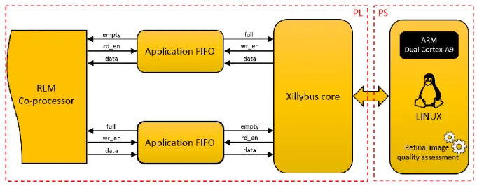
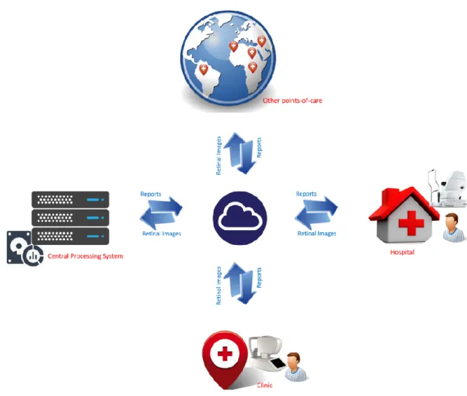
Tele-ophthalmology system as novel tool takes advantage of technology to offer more op-portunities for people with diabetes, and to provide to the community outreach better services. A tele-ophthalmology platform is based on a centralized processing system. Figure 1-1 shows the principle of a centralized processing system. This system would receive retinal images to be pro-cessed via the Internet. The images could be sent from hospitals, clinics, and/or points-of-care. After processing, the centralized system would return back the examination results to the source.
Figure 1-1. Centralized processing system overview
We can estimate that in the USA, if all underserved populations were to be provided with digital imaging, 32 million retinal images would require evaluation annually. This number assumes that 40% of the population affected by diabetes requires fundus images examination and that two images per eye are taken [3]. In other regions in the world, a greater number of images would require evaluation.
Existing algorithms for automated detection of diabetic retinopathy require several minutes to process images of public databases when using general processors. Unfortunately, the resolution of these public databases images is much smaller than the one provided by recent retinal cameras (768 × 584 pixels images). Recent cameras provide high resolution images (4288 × 2848 pixels images), which implies a higher processing time to obtain a diagnostic result.
1.2 Problem statement
In this thesis, we are addressing the problem of processing, in a reasonable time in the context of telemedicine, the large number of retinal images expected to be collected from a centralized processing system. This will require efficient processing systems. Moreover, accelerating the pro-cessing will allow the realization of embedded propro-cessing systems that can be integrated with the retinal camera to allow on-site diagnostic. Hardware platforms (FPGAs or ASICs) are very ade-quate for image processing algorithms acceleration. The aim of the proposed research is to intro-duce suitable hardware architectures to accelerate the retinal image algorithms, which presents several important challenges.
First, we consider the problem of retinal images quality assessment. This task is necessary for automated detection systems since it represents the first task of retinal image processing pipe-line. This task is responsible of identifying images of good quality after acquisition and reject im-ages of poor quality. This task require the computation of several image features which include complex algorithms difficult to parallelize by their nature. Accelerating such algorithms is very useful and favorable, which will allow the creation of mobile retinal capturing systems. Many ap-proaches and algorithms for retinal image quality assessment have been proposed in the literature. However, there are no existing hardware implementations in this context.
Secondly, we select the problem of retinal blood vessel segmentation. This is a key and com-plex task in the process of eye disease detection. This task gains its importance because vessel features are good indicators of various pathologies. Detecting the blood vessels allows the evalua-tion of these features that subsequently allows to detect the pathologies. Indeed, in CAD systems we usually remove segmented vessels and consider other structures as lesions. Selecting the ade-quate algorithm is very important to get better results in terms of blood vessel detection quality.
A third problem is to propose adequate architectures for the targeted algorithms. The com-plexity of the architecture is very dependent to the algorithm comcom-plexity. Thus, selecting hardware friendly algorithms or making them hardware friendly is very important. A hardware friendly al-gorithm is an alal-gorithm that can take advantage of the hardware platform structure. It can be par-allelized easily, and that can avoid complex computations. In other words, making an algorithm hardware friendly is to make its conception suitable for hardware implementation. We can achieve this goal by proposing several simplifications and modifications of the existing algorithms while
maintaining a good compromise between simplicity and accuracy. This should take in considera-tion the algorithm requirements in terms of data access and funcconsidera-tional units.
Furthermore, existing retinal image datasets include images of low-resolution while recent advances in retinal imaging and cameras technology promote the use of image of higher resolution. Proposed hardware architectures should take this in consideration to solve the problem of architec-ture scalability. The same architecarchitec-ture should be able to scale according to the image resolution with less development effort and minor modifications. Generally, optimized hardware architectures require the use of very-low level programming models, which increases the amount of needed ef-forts. A task such as blood vessel segmentation require high computations precision to achieve the best algorithm accuracy in terms of blood vessel detection quality. Functional units and their opti-mizations are not easy to scale, which represent an additional challenge.
1.3 Research contributions
The main objective of this research is to design and implement retinal image processing al-gorithms on hardware platforms with the aim to achieve high throughput with a certain level of design flexibility. This section reviews the contributions of the different parts of this thesis.
We started our work by analyzing the context of retinal image processing and their hardware implementations in order to identify opportunities for acceleration and efficient implementation. We identified two main tasks to be addressed. The first task was the retinal image quality assess-ment. We thus proposed an embedded system to compute the RLM features for retinal images. The RLM features provide quantitative information describing textural properties of retinal images, which eventually allows to differentiate images according to their quality. Our first solution was completely software. In order to accelerate the execution of the algorithm, we proposed a hardware co-processor to be implemented on the programmable fabric of Zynq FPGA. We designed, imple-mented and tested the system with the co-processor. Our results show improvements over the soft-ware implementation. This contribution was published in a conference paper entitled "A run-length encoding co-processor for retinal image texture analysis," presented in the International Confer-ence on ReConFigurable Computing and FPGAs (ReConFig) in 2015 [5].
The second task identified to be accelerated was the retinal image blood vessel segmentation. To that effect, we targeted two different techniques. The first technique is based on matched filter-ing. There exist several hardware implementations of this algorithm, however there are several limitations in terms of performances and programming flexibility. In this thesis, we designed a scalable and optimized architecture of the matched filter. The adopted low-level programming model with several optimizations allowed us to achieve better performances in terms of execution time in comparison with existing implementations. To overcome the problem of programming flex-ibility and to keep low level optimizations, we proposed a tool able to generate automatically a low-level hardware description of the algorithm based on a set of its parameters. With the aim of comparing two implementation strategies, we proposed a novel architecture based on an ASIP. This second architecture was improved by designing two specific instructions integrated into the Xtensa processor datapath, which allowed the improvement of the ASIP performances when exe-cuting the matched filter algorithm. These contributions were published first in a conference paper entitled "A scalable hardware architecture for retinal blood vessel in high resolution fundus im-ages," presented in the Conference on Design & Architectures for Signal & Image Processing in 2014 [6], and in an extended version in a paper entitled "Flexible architectures for retinal blood vessel segmentation in high-resolution fundus images," published in the Journal of Real-Time Im-age Processing in 2016 [7].
The second targeted technique for retinal blood vessel segmentation is based on the MSLD algorithm. In this thesis, we introduced an optimized memory-efficient architecture for the MSLD algorithm. This architecture was designed and implemented in a Zynq FPGA. It benefits from HW/SW co-design to satisfy the memory requirements of the algorithm. Results in terms of blood vessel segmentation accuracy are shown in addition to acceleration results over CPU and GPU implementations. To be able to target low and high resolution images, the automatic generation of low-level HDL description of the algorithm was made possible by the development of a tool that takes as input the algorithm parameters. In addition, we provide also a full comparison table of our implementations with existing ones. These contributions were published first in a conference paper entitled "Memory Efficient Multi-Scale Line Detector Architecture for Retinal Blood Vessel Seg-mentation" presented in the Conference on Design & Architectures for Signal & Image Processing in 2016 [8], and have been submitted in an extended version to the IEEE Transactions on Circuits
and Systems for Video Technology in a paper entitled "Memory Efficient Flexible Architecture for Retinal Blood Vessel Segmentation Using a Multi-Scale Line Detector," Jan. 2017 [9].
1.4 Thesis organization
This thesis is divided into 6 chapters. Chapter 2 reviews the important background material and related works that are used in this thesis. In Chapter 3, we present an implementation of a co-processor to accelerate a feature extraction algorithm. In chapter 4, we present flexible architectures for matched filtering technique for retinal blood vessel segmentation. Chapter 5 presents a memory-efficient architecture for a multi-scale algorithm for retinal blood vessel segmentation. Chapter 6 presents a general discussion where we present the novelty of our work and its limitations. Chapter 7 concludes the thesis by summarizing our contributions and outlining our recommendations and future research directions
CHAPTER 2
LITERATURE REVIEW
In this chapter, we examine the relevant literature for our research subject. Two topics which are directly related to our research were selected: algorithmic aspects of the automated detection of diabetic retinopathy and their hardware architectures and implementations. For the automated detection of diabetic retinopathy, we start by describing the eye anatomy and the manifestations of eye diseases. Then, we present the different parts of an automated detection system using digital fundus images and the existing approaches. For the second topic, we review the proposed hardware architectures and implementations of retinal image processing algorithms. High level synthesis of image processing algorithms is also presented.
Accordingly, this chapter is organized as follows. Section 2.1 introduces the diabetic reti-nopathy disease and its symptoms. In section 2.3, the retinal image processing pipeline and its hardware implementations are presented and described. Section 2.2 presents the public databases and metrics established by the scientific community to evaluate the effectiveness and the advances in disease detection quality. Section 2.4 reviews the architectural considerations for the implemen-tation of image processing algorithms. This include the data access, hardware function evaluation and processor architectures for image processing applications. Section 2.5 reviews the existing tools for HDL description generation for image processing algorithms. Finally, section 2.6 defines our research objectives.
2.1 Diabetic retinopathy
The human eye is the most powerful sensor for the perception of its environment. By analogy, it’s often compared to a camera, and the camera sensor can be seen analogous to the retina. The retina is the inner surface of the eye and consists of transparent tissue of several layers of cells designated to absorb and convert light into neural signals [10]. Once converted to neural signals and collected to the optic nerve in the optic disc, the impulses are transmitted to the brain. The nutritional support of the retina is provided by blood vessels. Figure 2-1 highlights the different anatomical parts of the retina (Macula, fovea, optic disc and the blood vessels).
In most cases, visual disorders are consequences of vascular changes that diabete causes to the eye. Neovascular glaucoma, diabetic neuropathies, cataract and diabetic retinopathy are the
most common diabetic eye diseases. In this section and in the rest of this proposal we will concen-trate on diabetic retinopathy.
Figure 2-1. Normal retina anatomical parts
Diabetic retinopathy is the most prevalent ocular complication of diabetes, and a leading cause of blindness in American adults [3]. It occurs when high blood sugar levels provoke the abnormal growth of blood vessels. These new blood vessels are weak. They can swell and leak fluid or even close off completely and damage the cells of the retina. A healthy retina is necessary for good vision. Patients with diabetic retinopathy first start noticing changes in vision. But over time, diabetic retinopathy can get worse and cause vision loss. Usually, diabetic retinopathy affects both eyes. Figure 2-2 shows the difference between the normal vision and vision with diabetic retinopathy.
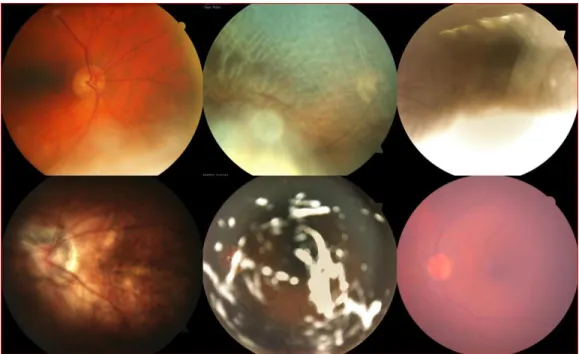
The detection of diabetic retinopathy is possible by detecting its symptoms in fundus images. These symptoms are: microaneurysms, haemorrhages, hard exudates, soft exudates and neovascu-larisation. Microaneurysms appear as small red dots in the retina. Due to blood vessel damage, small blood vessels may rupture and cause intraretinal haemorrhages that appear either as small red spots or larger round-shaped blots. The hard exudates are lipid formations leaking from the weakened blood vessels and appear yellowish with well-defined borders. Soft exudates are small
microinfarcts pale areas with diffuse borders. Neovascularisation is the growth of new fragile ves-sels due to the extensive lack of oxygen and obstructed capillary in the retina [11]. The different cited symptoms are shown in Figure 2-3.
-a- -b-
Figure 2-2. a- Normal vision, b- Vision with diabetic retinopathy [12]
-a- -b- -c-
-d- -e-
Figure 2-3. Diabetic retinopathy symptoms: a- Microaneurysms, b- Haemorrhages, c- Hard exudates, d- Soft exudates, e- Neovascularisation [11].
2.2 Retinal image processing pipeline
In any complete automated system for eye diseases detection, four steps are necessary as shown in Figure 2-4. These four steps are: retinal images quality assessment, retinal images en-hancement, features extraction (blood vessels, lesions, microaneurysms …) and disease detection.
Figure 2-4. Main steps of an automated eye disease detection system
2.2.1 Quality assessment
Image quality evaluation is a common problem in image processing systems. For medical imaging and especially diabetic retinopathy detection, sufficient image quality is necessary for fur-ther processing to ensure reliable diagnosis. The quality determines the ability of a human expert or an automated system to correctly detect disease symptoms from the image. When the image quality is not sufficient, it becomes difficult or impossible to make a reliable clinical judgment on the presence or absence of any eye disease. Several published works report that 10% of the acquired mydriatic (pupil dilation) images are rejected [13] due to insufficient quality. For single field non-mydriatic (no pupil dilation), the rejection rate can be up to 20.8% [14]. Assessing the retinal image quality represents an important limiting factor for automated diabetic retinopathy detection. To avoid the costs associated with processing useless images, which must be replaced in a second patient sitting, the research community started to develop vision-based solutions to assess the qual-ity of the retinal images [15, 16]. The qualqual-ity of the acquired retinal image should be assessed as a first step, and this task should be executed immediately after the acquisition of the images. Retinal fundus images quality assessment algorithms can be grouped in three different categories: histo-gram based methods, retina morphology methods and “bag-of-words” methods [17].
In histogram-based methods, the quality of a given image is determined through the differ-ence between its histogram of certain features and the mean histogram of a set of good-quality images as reference. Based on this, Lee and Wang [18] were the first to address the problem of automatic evaluation of fundus image quality. They employed the global histogram of the image intensities approximated by a Gaussian distribution. This approach was extended by Lalonde et al.
[19] using two different sets of features: the edge magnitudes distribution and the local pixel inten-sity distribution.
In retina morphology methods, features describing the retinal structure are used to evaluate their quality. A vessel segmentation algorithm was used by Usher et al. [20] to estimate the image blurring. This algorithm uses the area of the detected vessels as a specific feature for the retinal fundus images. Fleming et al. [15] presented a method that evaluates the image clarity using vessel area in the macula region, and the field definition using the relative position of the fovea and the length of the main vessel arcades. The image clarity and field definition are finally combined to generate a global quality metric.
In “bag-of-words” methods, a pattern recognition classifier (Support Vector Machine, Naïve Bayes, etc.) is employed to classify the occurrence of some common words automatically generated from the raw features in the test set. Niemeijer et al. [21] employed this approach to evaluate the fundus image quality. The authors use the color and second order statistics of the image as two sets of raw features to provide a compact representation of the structures found in an image. A different set of features was employed by Paulus et al. [22] : the pixel gray levels and the Haralick texture features.
The quality assessment of retinal images is still an open topic for research. There are several algorithmic challenges to be addressed such as the creation of public datasets with images of dif-ferent quality. Without public datasets, it would be difficult to classify the algorithms based on their performances. Even if the quality assessment is the most critical step in terms of execution time since it’s the first task of the pipeline, the hardware implementation and acceleration of its algorithms in the context of the retina have not been addressed yet.
2.2.2 Retinal image enhancement
Acquired images through cameras may be contaminated by a variety of noise sources (e.g. photon or on chip electronic noise), distortions, shading or improper illumination. Eventually, the image quality is reduced, which affects the performance and efficiency of any subsequent pro-cessing algorithms. A pre-propro-cessing and enhancement step is necessary. Image enhancement al-gorithms consist of a collection of techniques which aim to improve the visual appearance of an image.
For retinal fundus image systems, image enhancement is the second step for an automated detection system. In the retina context, the aim is to reduce the noise, to correct the non-uniform illumination and to improve the contrast. The illumination can be non-uniform in retinal images due to the variation of the retina response and the imaging systems non-uniformity. To correct the non-uniform illumination, Yang et al. [23] proposed to divide the image by an osmoothed ver-sion of it using a spatially large median filter. Many authors employ standard local or global histo-gram equalization techniques as Sopharak et al. [24].
In an RGB fundus image, the green channel is always considered the best to exhibit a good contrast of the structures of interest according to the background. Hence, it is a common practice in the literature to use the green channel for segmentation purposes. This has a major advantage, instead of processing the three channels, the calculation efforts are reduced to the processing of the green channel.
The background subtraction technique is one of the popular techniques for image enhance-ment in the literature. This technique is based on the estimation of the retina background with a large median filter applied on the green channel of the image [25]. This technique was refined by Cree et al. [26] to reduce the inter-patient color variability assuming that the background-less fun-dus image has colors normally distributed. The color of the new image is equalized to a reference one instead of simple histogram equalization. A hardware implementation of the background tech-nique can be parallelized to improve the execution of the algorithm.
The contrast enhancement is a popular technique in biomedical image processing. This tech-nique is very effective in making interesting silent parts more visible. Walter et al. [27] proposed to enhance the contrast of fundus images by applying a gray level transformation to the original grayscale image. Contrast limited adaptive histogram equalization (CLAHE) [28] is very popular. In this technique, the image is split into disjoint regions. In each region a local histogram equali-zation is applied. The boundaries between the regions are eliminated with a bilinear interpolation.
Several hardware architectures have been proposed to implement image enhancement algo-rithms. Tsutsui et al.[29] proposed hardware architecture for real-time Retinex video image en-hancement. In order to efficiently reduce the enormous computational cost required for high reso-lution image enhancement, processing layers and repeat counts of iterations are determined accord-ing to a software evaluation result. The implemented architecture is able to support WUXGA
(1920×1200) at 60 fps.Hanumantharaju et al. [30] proposed hardware architecture for a new algo-rithm for adaptive color image enhancement based on Hue-saturation value (HSV) color space. This algorithm uses a saturation feedback to enhance contrast and luminance of the color image, while the saturation component is enhanced by stretching its dynamic range to get rich color dis-play. This algorithm was implemented on Xilinx Virtex II FPGA.
Bo-Hyun et al. [31] proposed a new algorithm for image enhancement with less computa-tional complexity. This new algorithm consists of a decimation filter, contrast enhancement and color enhancement blocks. Simulation and experimental results show better performances with lower complexity when compared to conventional algorithms. Zhang et al. [32] proposed digital color enhancement architecture based on reflectance/illumination model. The approach uses the approximation techniques for efficient estimation of log2 and its inverse to promote the log-domain computation and to eliminate multiplications, divisions and exponentiations. With effective color space conversion, the HSV-domain image enhancement architecture is able to achieve a through-put rate of 182.65 (MOPS) on Xilinx Virtex II FPGA at a clock frequency of 182.65MHz.
Image filtering is an essential part of image enhancement techniques and plays an important role in image processing. Azizabadi et al. [33] proposed hardware architectures for image filters including Gaussian, median and weighted median filters. The aim of the implemented architecture is to optimize the speed and the area. The proposed architectures are implemented and synthesized in ASIC with 65 nm technology. The authors report a maximum frequency of 1 GHz for median filter and 666.67 MHz for both Gaussian filter and weighted median filter. An FPGA implementa-tion of median and weighted median filter for image processing was presented in [34] by Fahmy et al. The input samples are first used to construct a cumulative histogram, which is used to find the median value. The resource usage of the design is kept independent of the window size, but dependant on the number of bits of the data samples, which offers an efficient implementation of large-windowed median filtering. This method was extended to the weighted median filter. Reza et al. [35] proposed a suitable architecture for high speed VLSI and FPGA of the contrast limited adaptive histogram equalization (CLAHE) algorithm. The goal of this implementation is to mini-mise the latency without sacrificing precision. The maximum latency encountered in this approach is about half frame.
2.2.3 Features extraction
One goal of computer image processing is the efficient and effective visual features extrac-tion. Deciding which visual features to extract and choosing the best way to extract them are crucial problems in many image processing tasks. Retinal image features extraction is a main step in many automated detection system. In this step, the aim is to detect retina anatomy and disease symptoms. Extracting retina anatomy facilitates the task of identifying disease symptoms. In the literature, many works target the retina anatomy, this includes the optical disk, fovea and blood vessels. Dis-ease symptoms are also targeted, such as microaneurysms, lesions and exudates. In this section we will review two of the most important features of retinal images, blood vessels and microaneu-rysms.
2.2.3.1 Blood vessel extraction
The ability to distinguish retinal blood vessels from other structures is a step of great im-portance for diabetic retinopathy detection and in many retinal imaging applications. For diabetic retinopathy detection, the aim is to extract the retinal blood vessels to remove them and to identify red lesion candidates which are symptoms of diabetic retinopathy. A large number of algorithms have been published relating to the detection of retinal blood vessels. A complete review of the existing methods for retinal blood vessels detection can be found at [36]. There exist other methods based on tracking techniques [37] and supervised methods [38, 39] to deal with the retinal blood vessel detection problem. In this section we will present some existing algorithms and their hard-ware implementations.
Matched filtering is a popular technique for vessels detection in retinal images. It is based on the convolution of a 2-D kernel with the retinal image. The kernel is designed to model a feature based on its properties in the image at some unknown position and orientation. The matched filter response indicates the presence of the feature. For retinal images, the properties of the blood vessels are exploited to design the matched filter kernels. Three main properties are usually used: blood vessels can be approximated by a piece-wise linear segments since they have a limited curvature, the diameter of the vessels decrease as they move radially outward from the optic disc, the intensity profile of the cross section of the vessel approximates a Gaussian curve. The matched filtering technique can benefit greatly from a parallelized hardware implementation.
Chaudhuri et al. [40] were the first to propose a two-dimensional linear kernel with a Gauss-ian profile to segment the retinal blood vessels. The kernel is rotated 12 times with an increment of 15° to fit into vessels of different orientations. The highest response of the filter in each pixel of the image is selected and is thresholded to provide a binary vessel tree image. Some problems of the proposed matched filter are noticed. The kernel may be quite large and needs to be applied at several orientations especially for high resolution images, which result in a computational over-head. The kernel responds optimally to vessels that have the same form as the proposed kernel, and may not respond to those vessels with a different profile. It’s also possible that the matched filter respond to some present pathologies in the image, especially with the retinal background variations, which increases the number of false detections.
Al-Rawi et al. [41] proposed an improved matched filter to detect the retinal blood vessels. They used an optimization method based on exhaustive search to find the best parameters of the filter. Then they proposed an automated method to find the best threshold to segment the retinal blood vessels based on the number of connected components and Euler number. Zhang et al. [42] proposed to use the matched filter with the first-order derivative of a Gaussian to reduce the false detections produced by the original matched filter and to detect the missed fine vessels. Dalmau et al. [43] proposed to combine the matched filter with a segmentation strategy by using a Cellular Automata, while Zolfagharnasab et al. [44] proposed a new kernel function with Cauchy distribu-tion to improve the accuracy of the retinal vessel detecdistribu-tion. These works introduce new segmenta-tion methods or propose new kernel funcsegmenta-tions for the blood vessel cross secsegmenta-tion intensity approxi-mation to improve the original matched filter.
Mathematical morphology is also used for vessels detection. The basic morphology of the retinal blood vessels is known a priori to be comprised of connected linear segments [45]. Zana et al. [46] proposed a method based on the fact that vessels are piecewise linear and connected. Hence, mathematical morphological operators are used to differentiate the vessels from the background. Jiang et al. [47] propose to threshold the image at different levels by multi-threshold probing tech-nique, and uses a verification procedure to detect the vessels in the segmented images. The final segmentation is obtained by a combination of those segmented images returned in each step [45]. Lam et al. [48] proposed to employ the normalized gradient vector field to detect the centerlines after the use of the gradient vector field to detect vessel like objects. To reduce the falsely detected vessels, the authors proposed to use a pruning step to remove all vessel pixels that are far away
from the centerlines. The same authors proposed a new method in [49]. This method deals with the bright and dark lesions. The authors proposed the use of three different concavity measures to detect the vessels and to distinguish them from the lesions.
Supervised methods have been widely proposed because of their performance [50-53]. A weakness of supervised methods is that they depend on the training set. Thus, in a system where images of different resolutions must be processed, the supervised methods must be trained with a set of representative images of each resolution. In addition, these methods are generally imple-mented on CPUs or GPUs because any update to the classifier may require a new implementation and major modifications, and are thus not suitable for custom processor implementations unless great efforts are paid.
Line operators detectors are an unsupervised method used to segment retinal blood vessels. These methods were previously used in mammography, were introduced by Ricci et al. [52] as a feature extractor with a classifier for retinal blood vessels detection [52]. Multi-scale line operators were also introduced and used in [54-56]. Nguyen et al. [45] proposed the MSLD algorithm for retinal blood vessel detection. By changing the length of a basic line detector, line detectors of varying scales are achieved, and their responses at different scales are linearly combined to produce a final segmentation. Multi-scale operation is achieved by changing the length of a basic line de-tector, which is more suitable for hardware implementation than Gaussian pyramids as in [55]. Contrary to supervised methods, a single parameter change makes this algorithm suitable for im-ages of different resolutions.
Three main implementation strategies are followed to implement blood vessel segmentation algorithms. These strategies are based on the use of CPUs, GPUs or custom processors in FPGAs. Most of the existing approaches are implemented in software in CPUs [45-49]. Palomera-Perez et al. [44] proposed a parallel multiscale feature extraction and region growing algorithm applied for high-resolution images. The algorithm is implemented on 14 parallel CPUs. The proposed ap-proach is able to process images of 2890 × 2308 pixels 9× faster in comparison with a serial im-plementation with 8% less accuracy. Becker et al. [57] proposed an approach that both maps and localizes retinal blood vessels in real-time on a video targeting an intraocular surgery application. The algorithm was implemented in an Intel i7 CPU. It processes 30-40 frames per second with an image resolution of 400 × 304. The presented CPU implementations give satisfactory results in
terms of segmentation quality but they are computationally intensive due to their high complexity and they are too slow to achieve onsite, real-time processing during the patient’s visit. While CPUs present the most flexible development and implementation process, they still do not have enough computation power to deal with large number of high-resolution images.
Many GPU implementations are also presented in the literature. Krause et al. [58] imple-mented a local Radon transform based algorithm on GPU. This implementation is able to process images of size 4288 × 2848 pixels in 1.2 s on an NVIDIA Geforce GTX680. Argüello et al. [59] proposed a hybrid strategy based on global image filtering and contour tracing for retinal blood vessel extraction. Its GPU implementation processes images of the DRIVE database in an average of 0.014 s. For high-resolution images of 4288 × 2848 pixels, the execution time is 0.753 s. Sava-rimuthu et al. implemented a matched filter for blood vessel detection in human forearms [60]. When implemented in GPU, the algorithm is able to process 44 images of 640 × 480 pixels each second. This implementation does not consider high-resolution images.
Several features extraction algorithms in general are implemented in FPGA such as in [61] [62] [63] [64]. Many others are proposed for retinal blood vessel segmentation. Savarimuthu et al. [60] have also implemented their algorithm as a custom processor in FPGA. For the same image size, this implementation is able to process 215 images per second. In [65], an FPGA implementa-tion of a programmable SIMD architecture for vessel tree extracimplementa-tion in retinal images was pre-sented. The implemented algorithm is based on an active contour technique called Pixel-Level Snakes (PLS) and morphological operations. This implementation requires a total of 1.349 s to extract the vessel tree from 768 × 584 pixels images. Koukounis et al. [66] presented a hardware architecture implemented in FPGA for retinal vessel segmentation targeting portable embedded systems. The implemented architecture processes images of 768 × 584 pixels in 0.0523 s. Alonso-Montes et al. [67] presented a hardware approach for an authentication system based on the retinal blood vessels extraction using local dynamic convolutions and morphological operations. This ap-proach was implemented and tested in a fine-grained single instruction multiple data (SIMD) pro-cessor array. The execution time required to process a 768 × 584 pixels image is 0.1925 s excluding the I/O operations. These implementations only consider images of low-resolution.
The previous cited implementations achieve significant improvements over software imple-mentations in terms of execution time. Some of them are partially able to process low resolution
images of public databases in a reasonable time. However, in a telemedicine context, more im-provements are necessary especially when processing high-resolution images. This is especially true for hardware implementations where the memory size and bandwidth are real challenges [68]. CPU and GPU implementations are flexible for parameters tuning when changing the image reso-lution. However, custom architectures are specific to a given set of algorithm parameters and image resolution, and tend to be inappropriate in situations where images of different resolutions must be processed such as in telemedicine. This is true unless the problem of ease of hardware architecture customization is resolved.
2.2.3.2 Microaneurysms detection
Microaneurysms are one of the symptoms of diabetic retinopathy, and their detection is es-sential in the process of diabetic retinopathy detection and grading. Detecting the microaneurysms in fundus images is a very vivid field for the automatic detection of eye diseases community. The first automated approach for the segmentation of retinal microaneurysms was described by Bau-doin et al. [2]. Since then, many approaches have been proposed in the literature. The two most relevant approaches are: Morphological approach and Region growing approach [69].
Morphological processing is a collection of techniques used for image component extraction. This approach is most commonly used for microaneurysms detection. Niemeijer et al.[70] used a hybrid approach to detect the red lesions by combining the prior works by Spencer et al. [25] and Frame et al. [71]. Spencer et al. used morphological processing to detect microaneurysms. A bilin-ear top-hat transformation and matched filtering were used to provide an initial segmentation. Frame et al. proposed a list of features to characterise microaneurysms. These features are used by a classifier to decide which candidates are microaneurysms. Kande et al. [72] used a matched filter for contrast enhancement of red lesions, the enhanced lesions are then segmented by relative en-tropy based thresholding. A top-hat transformation is applied to supress the enhanced blood ves-sels. The candidates red lesions are classified from other dark segments using a support vector machine. The linear filtering and thresholding are employed by Matei et al. [73] to identify the presence of specific retinal lesions like microaneurysms.
Spencer et al. [25] and Frame et al. [71] have used region growing algorithm to find final candidate object set after the detection of the retinal vessel tree. Cree et al. [74] proposed a method for microaneurysms detection based on region growing algorithm to find the underlying candidate
morphology. Feature classification is used to distinguish microaneurysms from other spurious ob-jects. Usher et al. [75] used region growing algorithm to detect the microaneurysms. After prepro-cessing, microaneurysms are extracted using recursive region growing and adaptive intensity thresholding. The classification of the detected lesions is done by neural network. Streeter et al. [76] proposed a microaneurysms detection algorithm in color images based on region growing. After preprocessing, the blood vessel tree are extracted and removed. Thresholding and region growing are applied by taking a candidate seed image, after the region growing, the features are extracted.
Neural networks are also used and considered because they are able to detect the regions that contain the microaneurysms and reject other regions. To achieve this goal, the image is divided into several windows. According to a multi-stage training, the presence of microaneurysms is de-tected in these windows by the neural network. Generally, neural networks, support vector machine (SVM) and naïve Bayes techniques are used for the classification of the detected candidates.
2.3 Public datasets and metrics
Automated detection systems can significantly decrease the manual labour in diagnosing large quantities of retinal fundus images. Several algorithms have been developed in the literature. Annotated data is necessary to test and evaluate algorithms. There are some publicly available annotated databases of retinal fundus images. These databases are different in goals, characteristics and completeness level. Their main goals are vessel segmentation, diabetic retinopathy and micro-aneurysms detection. Each public database provides a gold standard reference which is necessary for algorithm training and testing, also called Ground Truth data. The most known public databases are: ROC [77], DRIVE [51] and STARE [78].
The ROC (Retinopathy Online Challenge) is a microaneurysms dataset. This database is part of a multi-year online competition of microaneurysms detection that was arranged by the Univer-sity of Iowa in 2009. This database contain 50 training images with available annotated images and 50 test images where the annotated images were withheld by the organizers. The images are JPEG compressed and were acquired using a Topcon NW100, a Topcon NW200 and Canon CR5-45NM non-mydriatic camera at 45° field of view. There are three different image sizes present in the
database: 768 × 576, 1058 × 1061 and 1389 × 1383 pixels [77]. The DRIVE (Digital Retinal Im-ages for Vessel Extraction) consist of a total of 40 color JPEG compressed fundus imIm-ages. Between the 40 images, 7 contain pathology (exudates, hemorrhages and pigment epithelium changes). The images were acquired using a Canon CR5 non-mydriatic 3-CCD camera with 45° field of view. The images are 8 bits per color with a resolution of 768 × 584 pixels [51]. The STARE database contains 20 images for blood vessel segmentation. 10 among these images contain pathology. The images were captured using a Topcon TRV-50 fundus camera with 35° field of view. The resolu-tion of the images is 605 × 700 pixels with 8 bits per color. All the images were segmented man-ually by two observers [78].
Metrics have also been developed to compare and evaluate algorithms. Algorithms can be evaluated against a ground truth image dataset using sensitivity, specificity and accuracy metrics. The sensitivity is a ratio between 0 and 1, which is the number of true-positives (TP) divided by the sum of the total number of false-negatives (FN) (incorrectly missed) and true-positives as shown in Equation (2-1). Specificity is also a ratio between 0 and 1, which is the number of true-negatives (TN) divided by the sum of the total number of false-positives (FP) (incorrectly thought to have disease) and true-negatives as shown in Equation (2-2). The accuracy metric is computed as shown in Equation (2-3). 𝑆𝑒𝑛𝑠𝑖𝑡𝑖𝑣𝑖𝑡𝑦 = 𝑇𝑃 𝑇𝑃 + 𝐹𝑁 (2-1) 𝑆𝑝𝑒𝑐𝑖𝑓𝑖𝑐𝑖𝑡𝑦 = 𝑇𝑁 𝐹𝑃 + 𝑇𝑁 (2-2) 𝐴𝐶𝐶 = 𝑇𝑃 + 𝑇𝑁 𝑇𝑃 + 𝑇𝑁 + 𝐹𝑃 + 𝐹𝑁 (2-3)
An automated detection system can be better evaluated through a receiver operating charac-teristics (ROC) curve. This curve is obtained by setting, for example, 100 different thresholds, and obtaining sensitivity and specificity pairs of the algorithm at each of these thresholds. The resulting sensitivity/specificity pairs are plotted in a curve which represents the sensitivity on the vertical axis and the complement to 1 of the specificity on the horizontal axis. A compact representation is possible by reducing the curve to a single number, the area under the ROC curve or AUC, which is a number between 0 and 1, where 1 denotes perfect performance.
2.4 Architectural considerations for the implementation of image
processing algorithms
One of the main goals of image processing is to extract useful information from images and video. This task is difficult and many different algorithms are proposed. Image processing algo-rithms are often applied on large volumes of data, which implies high memory bandwidth require-ments, and real-time processing implies high computational loads. The insufficient performance observed in GPP (general purpose processors) and GPU (Graphical Processing Unit) implementa-tions has led to the use of hardware architecture and reconfigurable computing. Hardware archi-tectures are often considered to accelerate their execution, especially to meet to main requirements: needs for data, and computationally needs. In this section we review some aspects of hardware implementations of image processing algorithms. First, the aspect of data access and processor architectures for image processing is discussed. Existing hardware implementations for image quality assessment, image enhancement and features extraction are reviewed with more attention to retinal image processing algorithms.
2.4.1 Data access for image processing
Data access is an important problem in image/video processing application. Image processing applications are both computationally and data intensive. Based on data dependencies, some algorithms require data from a relativelly small neighborhood, this includes point or pixel operators (such as gamma correction, threshoding) and window operators (such as 2-D convolution, erosion and dilation). Some other algorithms depend on data from the entire image like the Fast Fourier Transform and histogram techniques, or several images like motion estimation.
To allow efficient access to structured data such as in 2D or 3D images, Larabi et al. [79] proposed a low-cost n-dimensional cache architecture for FPGA-based image and signal pro-cessing systems on chip. They developed a theoretical model for the architecture and a methodol-ogy to define the cache’s practical implementation based on the application and system parameters. Using this solution, numerical results indicate that 50% improvement in run-time performance can be achieved.
![Figure 2-2. a- Normal vision, b- Vision with diabetic retinopathy [12]](https://thumb-eu.123doks.com/thumbv2/123doknet/2324361.29948/27.918.119.777.219.457/figure-normal-vision-b-vision-diabetic-retinopathy.webp)