HAL Id: hal-02678074
https://hal.inrae.fr/hal-02678074
Submitted on 31 May 2020HAL is a multi-disciplinary open access archive for the deposit and dissemination of sci-entific research documents, whether they are pub-lished or not. The documents may come from teaching and research institutions in France or abroad, or from public or private research centers.
L’archive ouverte pluridisciplinaire HAL, est destinée au dépôt et à la diffusion de documents scientifiques de niveau recherche, publiés ou non, émanant des établissements d’enseignement et de recherche français ou étrangers, des laboratoires publics ou privés.
Identification of Na+/H+ exchange as a new target for
toxic polycyclic aromatic hydrocarbons.
Laurence Huc, Lydie Sparfel, Mary Rissel, Marie-Thérèse Dimanche-Boitre,
André Guillouzo, Olivier Fardel, Dominique Lagadic-Gossmann
To cite this version:
Laurence Huc, Lydie Sparfel, Mary Rissel, Marie-Thérèse Dimanche-Boitre, André Guillouzo, et al.. Identification of Na+/H+ exchange as a new target for toxic polycyclic aromatic hydrocarbons.. FASEB Journal, Federation of American Society of Experimental Biology, 2004, 18 (2), pp.344-346. �10.1096/fj.03-0316fje�. �hal-02678074�
The FASEB Journal express article 10.1096/fj.03-0316fje. Published online December 4, 2003.
Identification of Na
+/H
+exchange as a new target for toxic
polycyclic aromatic hydrocarbons in liver cells
Laurence Huc, Lydie Sparfel, Mary Rissel, Marie-Thérèse Dimanche-Boitrel, André Guillouzo, Olivier Fardel, and Dominique Lagadic-Gossmann
INSERM U456, Détoxication et Réparation Tissulaire, Faculté de Pharmacie, Univ. Rennes 1, 2 av du Professeur Léon Bernard, 35043 RENNES Cedex, FRANCE
Corresponding author: Dr. Dominique Lagadic-Gossmann, INSERM U456, Faculté de
Pharmacie Université Rennes 1, 2, avenue du Professeur Léon Bernard 35043 RENNES cedex, France. E-mail: [email protected]
ABSTRACT
The ubiquitous environmental pollutants polycyclic aromatic hydrocarbons are responsible for important carcinogenic and apoptotic effects, whose mechanisms are still poorly understood, owing to the multiplicity of possible cellular targets. Among these mechanisms, alterations of ionic homeostasis have been suggested. In this work, the effects of benzo(a)pyrene [B(a)P] on pHi were tested in the rat liver F258 epithelial cell line, using the fluoroprobe
carboxy-SNARF-1. After a 48-h treatment, B(a)P (50 nM) induced an alkalinization, followed by an acidification after 72 h and the development of apoptosis. Determinations of pHi recovery following an acid
load showed an increased acid efflux at 48 h. Cariporide inhibited both the early alkalinization and the increased acid efflux, thus suggesting the involvement of Na+/H+ exchanger 1 (NHE1). Besides, α-naphtoflavone (α-NF), an inhibitor of CYP1A1-mediated B(a)P metabolism, prevented all pHi changes, and NHE1 activation was blocked by the antioxidant thiourea, which
inhibited CYP1A1 metabolism-dependent H2O2 production. Regarding B(a)P-induced apoptosis,
this was prevented by α-NF and bongkrekic acid, an inhibitor of mitochondria-dependent apoptosis. Interestingly, apoptosis was significantly reduced by cariporide. Taken together, our results indicate that B(a)P, via H2O2 produced by CYP1A1-dependent metabolism, induces an
early activation of NHE1, resulting in alkalinization; this appears to play a significant role in mitochondria-dependent B(a)P-induced apoptosis.
Key words: intracellular pH • benzo(a)pyrene • apoptosis • metabolism
olycyclic aromatic hydrocarbons (PAHs), such as benzo(a)pyrene [B(a)P], are ubiquitous environmental pollutants notably formed during the burning of fossil fuels and cigarette smoking products (e.g., tobacco) or during cooking and to which humans are commonly exposed (1). Due to their high lipophilicity, they can easily enter cells, where they are usually metabolized into reactive metabolites by xenobiotic metabolizing enzymes, especially cytochrome P450 1A1. Note that the produced metabolites include the formation of proposed electrophilic ultimate intermediates, such as the B(a)P 7,8 diol- 9,10 epoxide, which has been shown to form mutagenic DNA adducts in various target organs (2).
Partly on the basis of the genotoxic properties described above, PAHs are now recognized to be complete carcinogens exhibiting both tumor-initiating and tumor-promoting properties (3). In addition to these carcinogenic properties, PAHs may also exert major immunosuppressive effects (4–7) and possible effects on, for example, the endocrine system (8) and the cardiovascular system (9). In vitro exposure to PAHs also results in major toxicity in some cell types, especially through induction of cell cycle alteration, oxidative damage, or apoptosis. For example, B(a)P has been shown to trigger apoptosis in cultured liver cells as well as in progenitor B cells and oocytes (10) and to decrease growth of isolated lymphocytes (11).
The aryl hydrocarbon receptor (AhR), a ligand-activated transcription factor of the basic helix-loop-helix family, is considered to play a major role in many toxic effects due to PAHs. It displays high affinity binding to various PAHs and, upon activation by these compounds, can act on transcription of various PAH-responsive genes. Thus, CYP1A1 is usually markedly up-regulated in an AhR-dependent manner by PAHs, resulting in an increased formation of mutagenic and toxic metabolites of PAHs. AhR-unrelated mechanisms may however also account for some properties of PAHs (12).
Regarding molecular events underlying PAH toxicity, several studies have implied protein kinases (p38, c-jun NH2-terminal kinase 1 JNK), proteins of the Bcl-2 family, and caspases to be important elements of the PAH-induced apoptotic cascade (10, 13, 14). However, early cellular signaling pathways responsible for the AhR-dependent and -independent toxicity of PAHs remain largely unknown. Depending on the concentration and the experimental system used, they might involve multiple and various cellular targets such as mechanisms implicated in the regulation of intracellular ionic concentrations. Indeed, works by Pessah et al. (15) have demonstrated that B(a)P metabolites alter ryanodine receptor function, thereby modulating Ca2+ transport across microsomal membrane. Interestingly, studies dealing with intracellular Ca2+ have further shown that an early increase of intracellular Ca2+ concentration is associated with the tumor-promoting effect of PAHs in human mammary epithelial cells (16) or with immunotoxicity of B(a)P metabolites in human B cells (17). Although calcium might represent one important determinant of PAH toxicity, a role for other ions cannot be excluded yet.
In the present study, we focused on cellular H+ homeostasis, that is, pHi regulation, in response
to PAHs, because (i) some crosstalks between Ca2+ and H+ regulatory pathways have been previously described (18) and because (ii) cellular H+ concentrations, notably through Na+/H+
exchange 1 (NHE1) modulation, are affected by or affect several situations that also deal with PAHs such as apoptosis, cell growth, lymphocyte activation, and neoplastic transformation (19– 22). Thus, NHE1 was described to be activated or inhibited during apoptosis, leading to alkalinization or acidification, respectively (23, 24). Using the rat liver F258 epithelial cell line known to be sensitive to PAHs (25), we provide evidence that acute exposure to PAHs such as B(a)P induces a biphasic variation of pHi through alterations of NHE1 activity, therefore
pointing to intracellular H+ homeostasis as a new cellular PAH target. Moreover, we show that these PAH-dependent pHi changes require CYP1A1 activity and related reactive oxygen species
(ROS) and are likely involved in and/or reflect induction of apoptosis in F258 cells, thus linking pHi regulation and toxicity due to PAHs.
MATERIALS AND METHODS
Chemicals
B(a)P, α-naphtoflavone (α-NF), dimethylbenz(a)anthracene (DMBA), benzanthracene (BA), and bongkrekic acid (BgA) were purchased from Sigma (St Louis, MO). 2,3,7,8-Tetrachlorodibenzo-p-dioxin (TCDD) was obtained from Cambridge Isotope Laboratories (Cambridge, MA). Thiourea was purchased from Prolabo (Saint Herblain, France). Cariporide was a kind gift from Aventis (Francfort, Germany). All these products were used as a stock solution in dimethyl sulfoxide (DMSO); final concentration of this vehicule in culture medium was <0.05% (v/v), and control cultures received the same concentration of vehicle as treated cultures. Hoechst 33342, dihydroethidine (DHE), and dichlorodihydrofluorescein diacetate (H2-DCF-DA) were purchased
from Molecular Probes (Eugene, OR). DEVD-AMC (Asp-Glu-Asp-7-amino-4-methylcoumarin) was purchased from Bachem (Voisins, France) and Z-Asp-2,6-dichlorobenzoyloxymethyl-ketone (Z-Asp) from Alexis (Lausanne, Switzerland).
Cell culture
The F258 rat liver epithelial cell line was cultured in Williams’ E medium supplemented with 10% fetal calf serum, 2 mM L-glutamine, 5 UI/mL penicillin, and 0.5 mg/ml streptomycin at
37°C under a 5% CO2 atmosphere as described previously (25). Cells were passaged every week
using 0.1% trypsin-EDTA solution. F258 cells, growing in exponential phase, were treated 24 h following seeding (at 2.5×10³ cells/cm2) with B(a)P and/or the different inhibitors tested (applied
to cells 2 h before PAH exposure) for various treatment times. In control cells, confluence was observed only at day 5 after seeding.
Experimental solutions
HCO3–-buffered solution contained (in mM): NaCl 111.8, KCl 4.7, MgCl2 1.0, KH2PO4 1.2,
CaCl2 1.0, glucose 10.0, and NaHCO3 23.0, pH adjusted to 7.4 at 37°C using 5% CO2/95% O2.
HEPES-buffered solution contained (in mM): NaCl 134.8, KCl 4.7, MgCl2 1.0, KH2PO4 1.2,
CaCl2 1.0, glucose 10.0, and HEPES (N-2-hydroxyethylpiperazine-N'-2-ethanesulphonic acid)
10, pH adjusted to 7.4 at 37°C with NaOH. In Na+-free medium, NaCl was replaced with 134.8
mM choline, and the pH was adjusted to 7.4 at 37°C with KOH. When ammonium chloride (NH4Cl 20 mM; Sigma) was used, 20 mM NaCl were removed from the medium to avoid any
change of osmotic force. NH4Cl was added to solutions shortly before use. We added and then
removed NH4Cl to induce an acid load in order to activate the pHi-regulatory mechanisms (26).
Cariporide (30 µM) (a known inhibitor of Na+/H+ exchange isoform 1) was added to solution shortly before use. Nigericin (Sigma) calibration solutions used in this study have been described elsewhere (27).
Measurement of pHi
The pHi of F258 cells cultured on glass coverslips was monitored using the pH-sensitive
fluorescent probe carboxy-SNARF-1 (carboxy-seminaphtorhodafluor; Molecular Probes) (28). Cells were loaded with SNARF by incubating them in a 5 µM solution of the acetoxy-methyl ester for 20 min at 37°C, just before performing the pHi recording. SNARF-loaded cells were
placed in a continuously perfused recording chamber (at a temperature of 36±1°C) mounted on the stage of an epifluorescent microscope (Nikon Diaphot). F258 cells were then excited with light at 514 nm, and fluorescence from the trapped probe was measured at 590 and 640 nm. The necessary setup to produce and detect the fluorescence has been described previously (29). Emitted fluorescence signals were recorded every 12 s and originated from a small area of coverslip representing ~10 cells in the field of view. All the selected cells exhibited an intact plasma membrane, which is an absolute requirement when performing pHi measurements using
carboxy-SNARF-1; that is why a sufficient SNARF loading was always looked for before the experiment, as estimated by the intensity of the fluorescence light emitted. The emission ratio 640/590 (corrected for background fluorescence) detected from intracellular SNARF was calculated and converted to a linear pH scale using in situ calibration obtained by nigericin technique described elsewhere (28). In the set of experiments aimed at validating the concentration-dependent kinetic effects of B(a)P, about 12 measurements per coverslip (10 coverslips tested in each case) were carried out in order to estimate the percentage of pHi
measurements given similar values.
Estimation of intracellular intrinsic buffering power at different pHi
The method used to estimate intracellular intrinsic buffering power (βi) has been described
previously (27). In brief, a stepwise reduction of external NH4Cl (from 20 mM) was applied to
selected cells. Each reduction in NH4+ resulted in the generation of intracellular H+, due to the
dissociation of NH4+ into H+ ions and NH3, with subsequent efflux of NH3. The resultant changes
(∆) in pHi were used to estimate βi as follows: βi = ∆[NH4+]i/∆pHi, where [NH4+]i = [NH4+]e ×
10(pHi-pHe). In this latter equation, [NH4+]i and [NH4+]e were intracellular and extracellular
ammonium ion concentrations, respectively; pHi and pHe were intracellular and extracellular pH,
respectively. The experiments were carried out in the absence of extracellular Na+ in order to prevent acid extrusion and in the presence of barium (1 mM) to reduce NH4+ efflux through
potassium channels (30).
Calculation of sarcolemmal acid efflux
Details of the method for calculating acid efflux (JeH) during pHi recovery in cells have been
described previously (27). In brief, acid efflux was estimated using the following equation: JeH =
βT × dpHi/dt, where βT is the total intracellular buffering power and dpHi/dt is the rate of pHi
recovery at any given pHi. In HEPES-buffered medium, βT equals the intrinsic buffering power
βi. In the present study, when effects of B(a)P treatments on JeH were studied, we used the
constant value βi given in the Results, because we found that βi was insensitive to pHi under such
conditions. Following determinations of the pHi dependence of acid effluxes from control and
B(a)P-treated cells, the kinetic enzymatic parameters (Vmax, K'[H+]) of a data fit curve realized
according to an allosteric model were estimated using a computer program designed for nonlinear regression data analysis (GraphPad Software, Prism 3.02).
Microscopical detection of apoptosis
Microscopical detection of apoptosis was performed in both floating and adherent cells, using Hoechst 33342 labeling. In brief, culture media and trypsinized-adherent cells were collected and then washed with phosphate-buffered saline (PBS), stained with Hoechst 33342 (0.5 µg/ml) in
PBS, and inspected using a fluorescence Olympus BX60 microscope. Cells with apoptotic nuclei, that is, condensed or fragmented, were then counted, in comparison with total population (n>200 nuclei).
Caspase activity assay
Caspase activity assay has been described previously (31). In brief, F258 cells were lysed in the caspase activity buffer. Crude cell lysates (40 µg) were incubated with 80 µM DEVD-AMC for 2 h at 37°C. Caspase-mediated cleavage of DEVD-AMC was measured by spectrofluorimetry (Spectromax Gemini, Molecular Devices) at the excitation/emission wavelength pair of 380/440 nm. The caspase activity was given in % of fluorescence arbitrary units compared with control.
Membrane isolation and Western blotting
Following treatments, cells were harvested, washed with PBS, collected, and placed in a buffer containing 120 mM NaCl, 10 mM Tris, pH 7.4, 0.1 mM PMSF, 0.1 mM benzamidine, 37.5 µM ALLN (calpain I inhibitor), and a proteinase inhibitor cocktail. Samples were homogenized at 4°C for 30 s, incubated on ice for 30 s, and then homogenized again for 30 s with mechanical homogenizer. To obtain crude membrane fractions, we subjected homogenates to a series of centrifugation steps as described previously (32). Total protein was quantitated using the Bio-Rad (Hercules, CA) protein assay kit. Samples were heated for 5 min at 37°C and then analyzed by 9% SDS-PAGE and blotted onto nitrocellulose membranes. In brief, nitrocellulose membranes were incubated first in 10% Tris-buffered solution (TBS) and milk overnight at 4°C and then with primary antibody anti-NHE1 (N1P1) raised against a fusion protein encoding the last 157 amino acids of the human NHE1 C terminus (kindly given by Josette Noël, Montréal, Canada) diluted in TBS overnight, at 4°C, with constant rocking on a shaker. After three washes with TBS, the blots were incubated with horseradish peroxidase-conjugated secondary goat vs. rabbit antibody at room temperature. Revelation was performed by chemiluminescence. Densitometry data analysis was performed using the software Bio-Profil Bio1D (Vilber-Lourmat, France); to normalize the NHE1 signals, we used β-catenin (mouse monoclonal antibody, sc-7963, Santa Cruz Biotechnology, Santa Cruz, CA) as an invariant protein marker.
Reactive oxygen species (ROS) detection
Production of ROS was assessed fluorometrically using oxidation-sensitive fluorescent probes H2-DCF-DA and DHE to detect H2O2 and O2.–, respectively. F258 cells were pretreated with
α-NF, thiourea, or vehicule and H2-DCF-DA (1 µM) or DHE (5 µM) for 2 h, and then B(a)P was
added. After a 24-h treatment, cells were collected and samples analyzed using a FACSCalibur flow cytometer (Becton-Dickinson, Franklin Lakes, NJ). Fluorescence emission from oxidized H2-DCF-DA was detected at 525 nm and oxidized DHE at 605 nm. Prooxidants were used as
positive controls: H2O2 (10 mM) for H2-DCF-DA and menadione (100 µM) for DHE. Each
measurement was conducted on 10,000 cells and analyzed on Cell Quest software (Becton-Dickinson). Four independent experiments were carried out.
Statistical analysis
All data are quoted as mean ± SE along with number of observations (n) corresponding, if not
otherwise stated, to the number of separate cultures used. ANOVA followed by Newman-Keuls test was used to test the effects of B(a)P. Differences were considered significant at the level of P<0.05. Statistical analysis using a one-way ANOVA was performed to test whether there were differences in the estimated values of Vmax and K'[H+] between control and treated groups.
RESULTS
Effects of PAHs on pHi of F258 cells
In the first set of experiments, we tested the effects of a low dose (50 nM) and a high dose (5 µM) of B(a)P on resting pHi in F258 cells following various times of treatment, in the presence
of HEPES-buffered medium (Fig. 1A). Whereas no effect of B(a)P was detected upon an acute exposure (<1 h) with 5 µM or 50 nM (data not shown), we have found that using 50 nM B(a)P, a significant alkalinization was induced after 48 h when compared with untreated cells, followed by an acidification after a 72-h and 96-h treatment. Such pHi alterations were also detected when
a more physiological buffer (HCO3–/CO2) was used in the extracellular medium (Fig. 1B).
Similar biphasic variations also occurred in 5 µM B(a)P-treated cells but were detected earlier. Indeed, alkalinization developed after a 12-h treatment, whereas no variation was observed following a 24-h treatment with the low dose. Experiments performed to test the number of cells recruited for alkalinization at 12 h for 5 µM B(a)P and at 48 h for 50 nM B(a)P (i.e., at the times alkalinization was detected) pointed to a very probable dose-dependent kinetic effect of this PAH on pHi because, in both cases, ~80% of pHi measurements gave an alkaline shift of similar
amplitude (data not shown).
In an attempt to elucidate the systems responsible for such pHi changes, we measured the rate of
pHi recovery following an acid load induced by an NH4+ prepulse, in F258 cells treated or not
with 50 nM or 5 µM B(a)P. As shown in Figure 2A and 2B, the rate of pHi recovery was
markedly increased after a 48-h exposure to 50 nM B(a)P; similar results were obtained after a 12-h exposure to 5 µM B(a)P (data not shown). Such an increase might be due either to a decrease of the intrinsic buffering power (βi) or an increase of H+-equivalent extrusion.
Estimations of βi in F258 cells showed that this parameter was unchanged by treatment with
B(a)P, whatever the concentration and the time of treatment, and gave a mean βi value of 13.13 ±
4.03 mM (n=113 estimations from 53 coverslips), which was not dependent on pHi (data not
shown). Estimations of the mean H+-equivalent efflux, JeH (dpHi/dt×βi), at a pHi of 6.9 following
various times of B(a)P treatment clearly indicated an increase of the acid extrusion at 48 h and moreover showed that this up-regulation progressively returned toward control values after 72 h (Fig. 2C). In another set of experiments, we verified that the rate of background acid loading in F258 cells, that is, the loading detected when inhibiting pHi-regulating mechanisms operative in
HEPES-buffered solution (a Na+-free solution was then used) remained unchanged upon treatments with B(a)P (data not shown). Taken together, these data point to the activation of one or several alkalinizing transporter systems that might be responsible for both the transient alkalinization and the transient increase of acid efflux.
To identify the alkalinizing transporter responsible for the effects of B(a)P on H+ homeostasis described above, we tested the involvement of the Na+/H+ exchanger 1, because in HEPES-buffered medium, this transporter has been shown to carry most of acid extrusion, all HCO3–
-dependent systems being inhibited (27). The following experiments demonstrated the constitutive presence of NHE activity in untreated F258 cells. Indeed, pHi recovery following the
acid load was found to be strongly inhibited upon removal of extracellular Na+; moreover, upon readdition of extracellular Na+, acid extrusion was rapidly reactivated (data not shown). The use of the NHE1-specific inhibitor cariporide (30 µM) (33) allowed us to identify the Na+-dependent
transporter as the isoform 1 of NHE (Fig. 3B).
We next tested the role of NHE1 in the alkalinization recorded after a 48-h treatment with 50 nM B(a)P. This concentration was chosen because it is possibly encountered in the environment. Cotreatments with 50 nM of B(a)P and 30 µM of cariporide were thus performed. As illustrated in Figure 3A, measurements of resting pHi after a 48-h exposure showed an inhibition of
B(a)P-induced alkalinization, as well as acidification, under such conditions. After an acid load B(a)P-induced by NH4+ removal, the presence of cariporide significantly blocked pHi recovery in B(a)P-treated
cells (Fig. 3C) similarly to what occurred in control cells (Fig. 3B). Taken together, these results pointed to NHE1 as the B(a)P- activated alkalinizing system responsible for the intracellular alkalinization measured after a 48-h treatment.
Role of CYP1A1-dependent metabolism and related ROS production in pHi variations The direct involvement of AhR was analyzed by exposing F258 cells to TCDD (50 nM), which binds to AhR without being metabolized by CYP1A1 (34). Measurements of pHi were
performed following 48 and 72 h of treatment and showed no effect of this molecule (Fig. 4A). Similar results were obtained with BA (50 nM), whereas DMBA (50 nM) induced both an alkalinization at 48 h (∆pHi =+0.125±0.05, n=11) and an acidification at 72 h (∆pHi=–
0.150±0.05, n=10).
To test the involvement of B(a)P metabolism through CYP1A1 in pHi variations, we next
cotreated F258 cells with 50 nM B(a)P and 10 µM α-NF, known as a CYP1A1 and AhR antagonist (35), and then measured resting pHi following 48 h and 72 h of treatment. As shown
in Figure 4B, both the alkalinization at 48 h and the acidification at 72 h were prevented under such conditions.
In Figure 5, we tested the effects of α-NF on the up-regulation by B(a)P of the acid efflux. Similar to what was observed with resting pHi, Figure 5A clearly shows that in the presence of
α-NF, B(a)P (50 nM) was no longer capable of up-regulating acid extrusion, which was confirmed when determining the mean pHi dependence of JeH from several experiments (Fig. 5B). Taken
together, these results confirm the crucial involvement of CYP1A1-dependent metabolism in the activation of NHE1 by B(a)P.
To determine the level of NHE1 regulation by B(a)P, we carried out kinetic analysis of the exchanger activity following 48 h of treatment. In Figure 6A, no significant change in Vmax was
observed, whereas K'[H+]i was decreased by half upon B(a)P treatment (control, 65.55±1.57 nM
vs. B(a)P, 31.10±7.12 nM; n=7), suggesting that B(a)P modulated Na+/H+ exchange activity by increasing its affinity for H+. The results given in Figure 6B point to no change in NHE1 protein
expression by B(a)P. The fact that xenobiotic metabolism can induce a production of ROS such as H2O2 and O2.– and that such species are also known to regulate NHE1 activity (36, 37)
prompted us to study ROS production and its impact on NHE1 activation. Figure 7A shows that an H2O2 (detected by H2-DCF-DA) rather than an O2.– production (detected by DHE) occurred
following a 24-h treatment with 50 nM B(a)P, that is, before NHE1 activation. This H2O2
production was found to be dependent on CYP1A1, since inhibited by 10 µM α-NF. In the presence of the antioxidant molecule thiourea, at a concentration (10 mM) capable of preventing any H2O2 production (Fig. 7A), no more alkalinization was observed following a 48-h treatment
with B(a)P (Fig. 7B). Furthermore, thiourea blocked any up-regulation of acid extrusion following B(a)P treatment, as shown in Figure 7C. Taken together, these results show that the activity of NHE1 is modulated by B(a)P likely via H2O2 produced by CYP1A1-dependent
metabolism.
Relationship between PAH-mediated alterations of pHi and toxicity
PAHs are known to elicit cell death through an apoptotic mode in hepatic cells (10). Therefore, we tested whether the pHi variations detected in the present study might play a role in the toxic
effects of these compounds. We first verified that B(a)P was indeed toxic for F258 cells. Thus, a decrease of cell viability was observed upon B(a)P as estimated by MTT assay (IC50=37.7±16.3
nM after 96-h treatment). As illustrated in Figure 8A, B(a)P applied at a concentration of 50 nM for 48 and 72 h was found to increase the number of apoptotic nuclei compared with control conditions, as estimated by chromatin staining with Hoechst 33342. Interestingly, similar results were obtained with DMBA but not with BA (Fig. 8A). Moreover, cotreatments of F258 cells with 50 nM of B(a)P (Fig. 8B) or DMBA (data not shown) and 10 µM of α-NF prevented any toxicity of these PAHs. This therefore indicates that besides affecting pHi, metabolites of B(a)P
and DMBA are the most likely responsible for induction of apoptosis.
Xenobiotic-induced apoptosis is often reported to be mediated by mitochondria (38) and to be associated with a mitochondria-dependent acidification (39). To test whether similar effects were occurring here, we used BgA, known to inhibit the breakdown of the mitochondrial transmembrane potential (∆Ψm) and hence apoptosis (40). Figure 8B shows that BgA markedly reduces the apoptosis percentage, pointing to a role for mitochondria in B(a)P apoptotic signaling. With the aim of testing the possible involvement of mitochondria in pHi variations, we
next measured resting pHi in cells cotreated with B(a)P and BgA. The results, given in Figure
8C, showed that whereas alkalinization was maintained after 48 h of treatment with B(a)P (50 nM), late acidification was prevented, most likely indicating that mitochondria were indeed involved in the development of B(a)P-induced secondary cytosolic acidification.
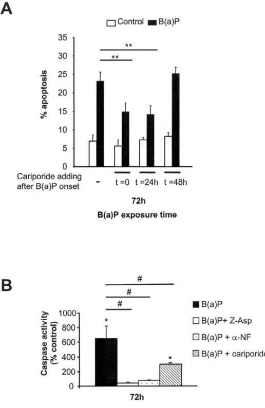
To analyze the role of NHE1-dependent alkalinization in PAH-induced toxicity, we next cotreated F258 cells with B(a)P or DMBA (50 nM) and cariporide (30 µM) and estimated the number of apoptotic nuclei under such conditions. Figure 9A shows that the specific inhibitor of NHE1 significantly reduced toxicity of B(a)P; similar protection was observed for DMBA (data not shown), especially at 72 h (reduction by 36.4% and 34.8%, respectively, compared with control PAH-treated cells; Fig. 9A). Furthermore, such a protective effect of cariporide was also detected when cariporide was added 24 h after the onset of PAH treatment (i.e., before alkalinization occurrence) but not when this addition was performed 48 h later (i.e., after alkalinization had developed) (Fig. 9A). Figure 9B further shows that cariporide also reduced the
caspase activity induced by B(a)P. We have verified that cariporide had no effect on CYP1A1 activity by EROD activity measurements as well as on the production of either B(a)P metabolites or H2O2 (unpublished data), therefore ruling out any direct inhibitory action of cariporide on the
metabolism of B(a)P. The present results therefore strongly suggest a link between PAH-induced toxicity and NHE1 activation and/or alkalinization detected at 48 h. In this context, we made a hypothesis that late acidification, which occurred after a 72-h treatment, would have been a mere consequence of prior alkalinization and hence of apoptosis signaling. This was the case because under conditions that inhibited alkalinization, we found that late intracellular acidification was significantly inhibited, as stated above (Fig. 4A).
DISCUSSION
PAHs are environmental pollutants exhibiting potent carcinogenic and immunotoxic effects. Whereas alterations of Ca2+ homeostasis have been shown to be partly responsible for such effects (16), up to now, to our knowledge, no information was available regarding putative effects of PAHs on another biologically important ion, namely H+. The present work indicates for the first time to the best of our knowledge that PAHs, following oxidative metabolism by CYP1A1, are capable of modifying cellular H+ homeostasis in liver epithelial cells through alterations of Na+/H+ exchanger located at the plasma membrane and following mitochondrial targeting. Moreover, these pHi changes were shown to be closely associated with the induction
of PAH-related toxic effects.
Exposing F258 cells to B(a)P induced biphasic pHi changes with first an alkalinization followed
by an acidification, in both CO2/HCO3–- and HEPES-buffered solutions. Moreover, we observed
that the greater the concentration of B(a)P was, the faster pHi changes appeared. Concerning the
early alkalinization, the hypothesis put forward to explain such a pHi variation was that B(a)P
was increasing an acid extrusion pathway. Following acid loads, we found that the resulting pHi
recovery was increased at the times corresponding to pHi alkaline shift, as compared with
control. This up-regulation, however, appeared to be transient because, following longer treatments with B(a)P, no increase in the rate of pHi recovery was detected. Because the
intracellular intrinsic buffering power and the background acid loading (due to acid influx and/or metabolic acid production) remained unchanged upon B(a)P, the present data were therefore in favor of a transient activation by B(a)P of one or several acid extruders in F258 cells.
As pHi variations were shown as still occurring in the presence of HEPES as extracellular buffer,
we next examined whether the ubiquitous Na+/H+ exchange 1 (NHE1) might be one such transporter. Using a specific inhibitor of this transporter, we showed that NHE1 was indeed active in F258 cells and that, following 48-h treatments with 50 nM B(a)P, both the alkalinization and the increase of acid extrusion were strongly inhibited. Taken together, the present results demonstrate that upon B(a)P treatment, the activity of NHE1 undergoes two phases: first, an up-regulation, which underlies the increase in resting pHi, and second, a
recovery toward basal level. When seeking the level of B(a)P-induced regulation on NHE1, we found that NHE1 protein expression was unaffected. Moreover, modulation of NHE1 activity seemed to be due to an increase of its H+ affinity; indeed, K'[H+]i was diminished by half upon a
48-h treatment with 50 nM B(a)P, without any change in Vmax, this latter result being in
Based on the fact that most of the PAH carcinogenic and toxic effects rely on the AhR (41) and that the AhR is expressed in F258 cells (25), its possible involvement in the above alterations of the H+ homeostasis was tested in this study by using the very potent AhR ligand TCDD. TCDD failed to induce any pHi change, whereas α-NF—a partial antagonist of this receptor but overall,
in the present case, a CYP1A1 metabolism inhibitor (data not shown)—completely prevented the resting pHi changes as well as the NHE1 up-regulation induced by B(a)P. Taken together, these
results therefore suggest that direct activation of AhR is not sufficient for inducing pHi
alterations. It might be argued that, as recently shown for the AhR-driven Bax gene expression (14), whereas the B(a)P-activated AhR would be capable of eliciting the presently detected pHi
changes, the TCDD-activated AhR would not, due to different AhR response elements (AHREs) present in the target genes. However, the PAH benzanthracene, which binds AhR and whose metabolism leads to nontoxic products (42), was also found to be ineffective on pHi, even at a
high concentration (5 µM; data not shown). In this context, the inhibitory effects of α-NF strongly point to the involvement of bioreactive PAH metabolites in these alterations. Nevertheless, despite its nondirect implication, AhR very likely contributed in this scheme, due to its determinant role in activating the expression of CYP1A1 gene (43).
Because PAH metabolism appeared to be determinant and considering that very acute treatment (<1 h) with high concentrations of B(a)P (5 and 10 µM) had no effect on pHi (data not shown),
the delay between B(a)P application and the appearance of H+ homeostasis changes might then be necessary for bioreactive metabolites and ROS to be formed and accumulate inside the cells so as to elicit their effects on pHi. In support of this hypothesis, we observed that a dose of 5 µM
B(a)P elicited an earlier alkalinization compared with 50 nM. With respect to a role for B(a)P metabolites in the present effects, note that a single o-quinone metabolite of B(a)P, B(a)P-7,8-dione, has recently been shown to be responsible for time-dependent biphasic alterations in the function of the ryanodine receptor 1 (RyR1), first activating then inhibiting channel activity (15). To explain their results, the authors have then proposed that the activation of RyR1 was due to production of H2O2 and the inhibition, to direct binding of metabolites to the protein. This has
led us to suppose that B(a)P metabolism was affecting NHE1 activity via ROS production. We first showed that in B(a)P-treated F258 cells, H2O2 production was dependent on CYP1A1
metabolism and occurred before the alkalinization onset. Experiments performed with the antioxidant molecule thiourea further demonstrated the involvement of these ROS in the modulation by B(a)P of NHE1 activity. Concerning a role for H2O2 in NHE1 regulation, note
that in cardiac cells, treatment with exogenous H2O2 has been shown to be capable of stimulating
sarcolemmal NHE activity, notably through an increase in phosphorylation of the exchanger by action of diverse protein kinases, including mitogen-activated protein kinases (MAPKs) (44, 45). As PAH metabolism is known to activate the MAPKs c-jun NH2-terminal kinase 1 (JNK) and p38 (10, 46, 47), these might be involved in the transient B(a)P-induced NHE1 up-regulation. This remains to be tested.
As reported above, B(a)P-induced intracellular alkalinization was followed by a significant intracellular acidification. Based on the fact that after longer treatments, the activity of NHE1 did not differ between treated and untreated cells and therefore would not be involved, we tested whether mitochondria might be responsible for this late acid load by using BgA (to target the adenine nucleotide translocase of mitochondria [48]). Our results showed that BgA prevented intracellular acidification without any effect on prior alkalinization, thus indicating that B(a)P
induced late acidification results from an effect on mitochondria. An important observation in this study was that the occurrence of prior alkalinization appeared to be necessary for the acidification to develop, because inhibited by NHE1 inhibitors. Taken together, these results strongly suggest that, regarding the alterations in H+ homeostasis presently described, metabolism of B(a)P (and probably that of DMBA) essentially targets NHE1 located at the plasma membrane, thus inducing alkalinization, whereas acidification would be a mere consequence of prior NHE1 activation. In this context, due to the presence of other important pHi-regulating mechanisms in the plasma membrane (eg Na+-HCO3– symport, Cl–/HCO3–
inhibited in HEPES-buffered solutions), it might then be possible that B(a)P metabolism would also affect such transporters. This has to be tested yet.
Following demonstration of PAH-induced alterations in H+ homeostasis in F258 cells, the question remained as to whether these were relevant to the well-known carcinogenic or toxic effects of these chemicals. Indeed, NHE-dependent pHi change has been shown by several
groups to play an important role in proliferating processes (19, 21, 22) as well as in inducing cell death (24, 49, 50), and intracellular acidification is often related to the mitochondrial pathway of apoptosis (51, 52). In our cell model, a B(a)P-induced toxicity was found to be via apoptosis, as already observed in other cell types (10, 53, 54), depending on CYP1A1-related metabolism (since inhibited by α-NF) and mitochondrial dysfunction (since inhibited by BgA). Regarding pHi, we found that only AhR ligands producing pHi alterations, that is, B(a)P and DMBA, were
capable of inducing cell toxicity, unlike TCDD or BA. Moreover, a high dose of B(a)P (5 µM), which induced fast variations of pHi, triggered a rapid and massive apoptosis in F258 cells. In
this context, the role of pHi alterations in PAH-induced toxicity was tested by using cariporide to
inhibit NHE1-dependent alkalinization.
We found that cotreatment with B(a)P and cariporide significantly reduced toxicity through a specific effect on pHi. We suppose at this stage that the remaining apoptosis in the presence of
cariporide might rely on other pathways, and especially the p53 pathway known to be triggered by the binding of B(a)P-derived reactive metabolites to DNA (55). Taken together, these results are the first to demonstrate the crucial role for NHE1-dependent alkalinization in the development of PAH-induced toxicity (Fig. 10).
According to recent studies, Bax, known to lead to mitochondria dysfunction after translocation, represents one possible target for intracellular alkalinization during apoptotic processes (24, 52, 56). Because a recent study has shown that Bax transcription is increased upon PAH exposure, thereby inducing cell death (14, 57), we might then suppose that the NHE1-dependent alkalinization would create a permissive environment favoring translocation of Bax to mitochondria and hence mitochondrial dysfunction and related cytosolic acidification, all these leading to activation of caspases and ultimately to cell death. Analysis of the precise sequence of events is currently under investigation. Finally, considering the well-described carcinogenic effects of B(a)P (2), the proapoptotic role of NHE1 presently shown might be viewed as possibly affording some protection against neoplastic transformation induced by PAHs, through an elimination process of cells exhibiting DNA attack by reactive metabolites and ROS and hence possible DNA mutations.
In conclusion, the present study demonstrates alterations in H+ homeostasis upon PAH exposure,
transient activation of NHE1 might constitute an important early signal in the development of the apoptotic cascade induced by toxic chemicals such as PAHs.
ACKNOWLEDGMENTS
This work was supported by grants from the Institut National de Recherche et de Sécurité (INRS) and the Ministère de l’Aménagement du Territoire et de l’Environnement.
REFERENCES
1. Zedeck, M. S. (1980) Polycyclic aromatic hydrocarbons: a review. J. Environ. Pathol. Toxicol. 3, 537–567
2. Pelkonen, O., and Nebert, D. W. (1982) Metabolism of polycyclic aromatic hydrocarbons: etiologic role in carcinogenesis. Pharmacol. Rev. 34, 189–222
3. Ethier, S. P., and Ullrich, R. L. (1982) Induction of mammary tumors in virgin female BALB/c mice by single low doses of 7,12-dimethylbenz[a]anthracene. J. Natl. Cancer Inst.
69, 1199–1203
4. Laupeze, B., Amiot, L., Sparfel, L., Le Ferrec, E., Fauchet, R., and Fardel, O. (2002) Polycyclic aromatic hydrocarbons affect functional differentiation and maturation of human monocyte-derived dendritic cells. J. Immunol. 168, 2652–2658
5. Woods, G. M., Doherty, K. V., Malley, R. C., Rist, M. J., and Muller, H. K. (2000) Carcinogen-modified dendritic cells induce immunosuppression by incomplete T-cell activation resulting from impaired antigen uptake and reduced CD86 expression. Immunology 99, 16–22
6. Burchiel, S. W. (1999) The effects of environmental and other chemicals on the human immune system: the emergence of immunotoxicology. Clin. Immunol. 90, 285–286
7. van Grevenynghe, J., Rion, S., Le Ferrec, E., Le Vee, M., Amiot, L., Fauchet, R., and Fardel, O. (2003) Polycyclic aromatic hydrocarbons inhibit differentiation of human monocytes into macrophages. J. Immunol. 170, 2374–2381
8. Santodonato, J. (1997) Review of the estrogenic and antiestrogenic activity of polycyclic aromatic hydrocarbons: relationship to carcinogenicity. Chemosphere 34, 835–848
9. Mennear, J. H. (1993) Carbon monoxide and cardiovascular disease: an analysis of the weight of evidence. Regul. Toxicol. Pharmacol. 17, 77–84
10. Lei, W., Yu, R., Mandlekar, S., and Kong, A. N. (1998) Induction of apoptosis and activation of interleukin 1beta-converting enzyme/Ced-3 protease (caspase-3) and c-Jun NH2-terminal kinase 1 by benzo(a)pyrene. Cancer Res. 58, 2102–2106
11. Black, K. A., McFarland, R. D., Grisham, J. W., and Smith, G. J. (1989) S-phase block and cell death in human lymphoblasts exposed to benzo[a]pyrene diol epoxide or N-acetoxy-2-acetylaminofluorene. Toxicol. Appl. Pharmacol. 97, 463–472
12. Dertinger, S. D., Lantum, H. B., Silverstone, A. E., and Gasiewicz, T. A. (2000) Effect of 3′-methoxy-4'-nitroflavone on benzo[a]pyrene toxicity. Aryl hydrocarbon receptor-dependent and -independent mechanisms. Biochem. Pharmacol. 60, 189–196
13. Page, T. J., O'Brien, S., Jefcoate, C. R., and Czuprynski, C. J. (2002) 7,12-Dimethylbenz[a]anthracene induces apoptosis in murine pre-B cells through a caspase-8-dependent pathway. Mol. Pharmacol. 62, 313–319
14. Matikainen, T., Perez, G. I., Jurisicova, A., Pru, J. K., Schlezinger, J. J., Ryu, H. Y., Laine, J., Sakai, T., Korsmeyer, S. J., Casper, R. F., et al. (2001) Aromatic hydrocarbon receptor-driven Bax gene expression is required for premature ovarian failure caused by biohazardous environmental chemicals. Nat. Genet. 28, 355–360
15. Pessah, I. N., Beltzner, C., Burchiel, S. W., Sridhar, G., Penning, T., and Feng, W. (2001) A bioactive metabolite of benzo[a]pyrene, benzo[a]pyrene-7,8-dione, selectively alters microsomal Ca2+ transport and ryanodine receptor function. Mol. Pharmacol. 59, 506–513 16. Tannheimer, S. L., Lauer, F. T., Lane, J., and Burchiel, S. W. (1999) Factors influencing
elevation of intracellular Ca2+ in the MCF-10A human mammary epithelial cell line by carcinogenic polycyclic aromatic hydrocarbons. Mol. Carcinog. 25, 48–54
17. Mounho, B. J., and Burchiel, S. W. (1998) Alterations in human B cell calcium homeostasis by polycyclic aromatic hydrocarbons: possible associations with cytochrome P450 metabolism and increased protein tyrosine phosphorylation. Toxicol. Appl. Pharmacol. 149, 80–89
18. Vaughan-Jones, R. D., Lederer, W. J., and Eisner, D. A. (1983) Ca2+ ions can affect intracellular pH in mammalian cardiac muscle. Nature 301, 522–524
19. Putney, L. K., Denker, S. P., and Barber, D. L. (2002) The changing face of the Na+/H+ exchanger, NHE1: structure, regulation, and cellular actions. Annu. Rev. Pharmacol. Toxicol. 42, 527–552
20. Benedetti, A., Di Sario, A., Casini, A., Ridolfi, F., Bendia, E., Pigini, P., Tonnini, C., D'Ambrosio, L., Feliciangeli, G., Macarri, G., et al. (2001) Inhibition of the Na(+)/H(+) exchanger reduces rat hepatic stellate cell activity and liver fibrosis: an in vitro and in vivo study. Gastroenterology 120, 545–556
21. Grinstein, S., Smith, J. D., Onizuka, R., Cheung, R. K., Gelfand, E. W., and Benedict, S. (1988) Activation of Na+/H+ exchange and the expression of cellular proto- oncogenes in mitogen- and phorbol ester-treated lymphocytes. J. Biol. Chem. 263, 8658–8665
22. Reshkin, S. J., Bellizzi, A., Caldeira, S., Albarani, V., Malanchi, I., Poignee, M., Alunni-Fabbroni, M., Casavola, V., and Tommasino, M. (2000) Na+/H+ exchanger-dependent
intracellular alkalinization is an early event in malignant transformation and plays an essential role in the development of subsequent transformation-associated phenotypes. FASEB J. 14, 2185–2197
23. Li, J., and Eastman, A. (1995) Apoptosis in an interleukin-2-dependent cytotoxic T lymphocyte cell line is associated with intracellular acidification. Role of the Na(+)/H(+)-antiport. J. Biol. Chem. 270, 3203–3211
24. Khaled, A. R., Moor, A. N., Li, A., Kim, K., Ferris, D. K., Muegge, K., Fisher, R. J., Fliegel, L., and Durum, S. K. (2001) Trophic factor withdrawal: p38 mitogen-activated protein kinase activates NHE1, which induces intracellular alkalinization. Mol. Cell. Biol. 21, 7545– 7557
25. Payen, L., Courtois, A., Langouet, S., Guillouzo, A., and Fardel, O. (2001) Unaltered expression of multidrug resistance transporters in polycyclic aromatic hydrocarbon-resistant rat liver cells. Toxicology 156, 109–117
26. Boron, W. F., and De Weer, P. (1976) Intracellular pH transients in squid giant axons caused by CO2, NH3, and metabolic inhibitors. J. Gen. Physiol. 67, 91–112
27. Lagadic-Gossmann, D., Buckler, K. J., and Vaughan-Jones, R. D. (1992) Role of bicarbonate in pH recovery from intracellular acidosis in the guinea-pig ventricular myocyte. J. Physiol. 458, 361–384
28. Buckler, K. J., and Vaughan-Jones, R. D. (1990) Application of a new pH-sensitive fluoroprobe (carboxy-SNARF-1) for intracellular pH measurement in small, isolated cells. Pflugers Arch. 417, 234–239
29. Lagadic-Gossmann, D., Rissel, M., Galisteo, M., and Guillouzo, A. (1999) Intracellular pH alterations induced by tacrine in a rat liver biliary epithelial cell line. Br. J. Pharmacol. 128, 1673–1682
30. Vaughan-Jones, R. D., and Wu, M. L. (1990) Extracellular H+ inactivation of Na(+)-H(+) exchange in the sheep cardiac Purkinje fibre. J. Physiol. 428, 441–466
31. Gilot, D., Loyer, P., Corlu, A., Glaise, D., Lagadic-Gossmann, D., Atfi, A., Morel, F., Ichijo, H., and Guguen-Guillouzo, C. (2002) Liver protection from apoptosis requires both blockage of initiator caspase activities and inhibition of ASK1/JNK pathway via glutathione S- transferase regulation. J. Biol. Chem. 277, 49220–49229
32. Rieder, C. V., and Fliegel, L. (2002) Developmental regulation of Na(+)/H(+) exchanger expression in fetal and neonatal mice. Am. J. Physiol. Heart Circ. Physiol. 283, H273–H283 33. Scholz, W., Albus, U., Lang, H. J., Linz, W., Martorana, P. A., Englert, H. C., and
Scholkens, B. A. (1993) Hoe 694, a new Na+/H+ exchange inhibitor and its effects in cardiac ischaemia. Br. J. Pharmacol. 109, 562–568
34. Wilson, C. L., and Safe, S. (1998) Mechanisms of ligand-induced aryl hydrocarbon receptor-mediated biochemical and toxic responses. Toxicol. Pathol. 26, 657–671
35. Sparfel, L., Loewert, M., Huc, L., Payen, L., Guillouzo, A., Lagadic-Gossmann, D., and Fardel, O. (2002) Acute cytotoxicity of the chemical carcinogen 2-acetylaminofluorene in cultured rat liver epithelial cells. Toxicol. Lett. 129, 245–254
36. Rothstein, E. C., Byron, K. L., Reed, R. E., Fliegel, L., and Lucchesi, P. A. (2002) H(2)O(2)-induced Ca(2+) overload in NRVM involves ERK1/2 MAP kinases: role for an NHE-1-dependent pathway. Am. J. Physiol. Heart Circ. Physiol. 283, H598–H605
37. Flowers, L., Bleczinski, W. F., Burczynski, M. E., Harvey, R. G., and Penning, T. M. (1996) Disposition and biological activity of benzo[a]pyrene-7,8-dione. A genotoxic metabolite generated by dihydrodiol dehydrogenase. Biochemistry 35, 13664–13672
38. Robertson, J. D., and Orrenius, S. (2000) Molecular mechanisms of apoptosis induced by cytotoxic chemicals. Crit. Rev. Toxicol. 30, 609–627
39. Matsuyama, S., and Reed, J. C. (2000) Mitochondria-dependent apoptosis and cellular pH regulation. Cell Death Differ. 7, 1155–1165
40. Henderson, P. J., and Lardy, H. A. (1970) Bongkrekic acid. An inhibitor of the adenine nucleotide translocase of mitochondria. J. Biol. Chem. 245, 1319–1326
41. Delescluse, C., Lemaire, G., de Sousa, G., and Rahmani, R. (2000) Is CYP1A1 induction always related to AHR signaling pathway? Toxicology 153, 73–82
42. Lewis, D. F., and Parke, D. V. (1995) The genotoxicity of benzanthracenes: a quantitative structure-activity study. Mutat. Res. 328, 207–214
43. Dertinger, S. D., Nazarenko, D. A., Silverstone, A. E., and Gasiewicz, T. A. (2001) Aryl hydrocarbon receptor signaling plays a significant role in mediating benzo[a]pyrene- and cigarette smoke condensate-induced cytogenetic damage in vivo. Carcinogenesis 22, 171– 177
44. Wei, S., Rothstein, E. C., Fliegel, L., Dell'Italia, L. J., and Lucchesi, P. A. (2001) Differential MAP kinase activation and Na(+)/H(+) exchanger phosphorylation by H(2)O(2) in rat cardiac myocytes. Am. J. Physiol. Cell Physiol. 281, C1542–C1550
45. Snabaitis, A. K., Hearse, D. J., and Avkiran, M. (2002) Regulation of sarcolemmal Na(+)/H(+) exchange by hydrogen peroxide in adult rat ventricular myocytes. Cardiovasc. Res. 53, 470–480
46. Yoshii, S., Tanaka, M., Otsuki, Y., Fujiyama, T., Kataoka, H., Arai, H., Hanai, H., and Sugimura, H. (2001) Involvement of alpha-PAK-interacting exchange factor in the PAK1-c-Jun NH(2)-terminal kinase 1 activation and apoptosis induced by benzo[a]pyrene. Mol. Cell. Biol. 21, 6796–6807
47. Kwon, Y. W., Ueda, S., Ueno, M., Yodoi, J., and Masutani, H. (2002) Mechanism of p53-dependent apoptosis induced by 3-methylcholanthrene: involvement of p53 phosphorylation and p38 MAPK. J. Biol. Chem. 277, 1837–1844
48. Scarlett, J. L., Sheard, P. W., Hughes, G., Ledgerwood, E. C., Ku, H. H., and Murphy, M. P. (2000) Changes in mitochondrial membrane potential during staurosporine- induced apoptosis in Jurkat cells. FEBS Lett. 475, 267–272
49. Barriere, H., Poujeol, C., Tauc, M., Blasi, J. M., Counillon, L., and Poujeol, P. (2001) CFTR modulates programmed cell death by decreasing intracellular pH in Chinese hamster lung fibroblasts. Am. J. Physiol. Cell Physiol. 281, C810–C824
50. Rich, I. N., Worthington-White, D., Garden, O. A., and Musk, P. (2000) Apoptosis of leukemic cells accompanies reduction in intracellular pH after targeted inhibition of the Na(+)/H(+) exchanger. Blood 95, 1427–1434
51. Hirpara, J. L., Clement, M. V., and Pervaiz, S. (2001) Intracellular acidification triggered by mitochondrial-derived hydrogen peroxide is an effector mechanism for drug-induced apoptosis in tumor cells. J. Biol. Chem. 276, 514–521
52. Matsuyama, S., Llopis, J., Deveraux, Q. L., Tsien, R. Y., and Reed, J. C. (2000) Changes in intramitochondrial and cytosolic pH: early events that modulate caspase activation during apoptosis. Nat. Cell Biol. 2, 318–325
53. Revel, A., Raanani, H., Younglai, E., Xu, J., Han, R., Savouret, J. F., and Casper, R. F. (2001) Resveratrol, a natural aryl hydrocarbon receptor antagonist, protects sperm from DNA damage and apoptosis caused by benzo(a)pyrene. Reprod. Toxicol. 15, 479–486
54. Chin, B. Y., Choi, M. E., Burdick, M. D., Strieter, R. M., Risby, T. H., and Choi, A. M. (1998) Induction of apoptosis by particulate matter: role of TNF-alpha and MAPK. Am. J. Physiol. 275, L942–L949
55. Wu, J., Gu, L., Wang, H., Geacintov, N. E., and Li, G. M. (1999) Mismatch repair processing of carcinogen-DNA adducts triggers apoptosis. Mol. Cell. Biol. 19, 8292–8301 56. Khaled, A. R., Kim, K., Hofmeister, R., Muegge, K., and Durum, S. K. (1999) Withdrawal
of IL-7 induces Bax translocation from cytosol to mitochondria through a rise in intracellular pH. Proc. Natl. Acad. Sci. USA 96, 14476–14481
57. Matikainen, T. M., Moriyama, T., Morita, Y., Perez, G. I., Korsmeyer, S. J., Sherr, D. H., and Tilly, J. L. (2002) Ligand activation of the aromatic hydrocarbon receptor transcription factor drives Bax-dependent apoptosis in developing fetal ovarian germ cells. Endocrinology
143, 615–620
Fig. 1
Figure 1. pHi variations following B(a)P exposure. F258 cells were treated with 50 nM or 5 µM B(a)P for the different
times indicated in the graphs. Resting pHi measurements were monitored by microspectrofluorimetry using the pH-sensitive fluorescent probe, carboxy-SNARF-1. Cells were superfused with HEPES-buffered medium (A) or 5% CO2/23 mMHCO3–-buffered medium (B)(extracellular pH of 7.4 in both conditions). pHi values were derived from a pH calibration curve (as described in Materials and Methods), and ∆pHi were calculated by substracting pHi values in
untreated cells from those in treated cells. *P < 0.05, **P < 0.01. Number in brackets given next to each bar represents the number of coverslips used (i.e., the number of performed measurements) from at least 3 independent experiments. Mean values correspond to the average of all pHi determinations.
Fig. 2
Figure 2. Determination of acid extrusion in cells treated with B(a)P. F258 cells were treated with 50 nM B(a)P for
various exposure times. Subsequently, pHi recovery rate was monitored following an acid load induced upon removal of NH4Cl (20 mM) from extracellular medium (see Materials and Methods). A) Example of pHi-recovery recording in both untreated and treated cells during 48 h. Only pHi recovery recorded in control F258 cells is illustrated. B) Apparent dpHi/dt (pH unit/min) estimated from the recordings illustrated in A. C) Mean apparent acid efflux J
e
H calculated from 4– 10 independent experiments, at pHi = 6.9 (see Materials and Method for calculation). *P < 0.05 treated vs. control.
Fig. 3
Figure 3. Effects of the NHE1-inhibitor, cariporide, on resting pHi and on pHi recovery in B(a)P-treated cells. A)
Resting pHi measurements recorded in F258 cells treated or not with B(a)P (50 nM) and/or cariporide (30 µM) for the times indicated in HEPES-buffered medium. Number in brackets given next to each bar represents the number of coverslips used (i.e., the number of performed measurements) from at least 3 independent experiments. Mean values correspond to the average of all pHi determinations. Basal pHi was 7.30 ± 0.04 (n=9) and 7.31 ± 0.03 (n=11) in control untreated and cariporide-treated cells, respectively. *P < 0.05 treated vs. control. B, C) Representative pHi recordings from 12 independent experiments in cells treated with control + cariporide (B) and B(a)P + cariporide (C). Cells were superfused with HEPES-buffered medium with 30 µM cariporide and submitted to a NH4
+
(20 mM) prepulse to activate acid extrusion.
Fig. 4
Figure 4. Role of CYP1A1-dependent metabolism of PAHs in pHi variations. A) F258 cells were treated with either
50 nM DMBA or 50 nM BA (two PAHs), or a specific AhR-ligand, 50 nM TCDD, during 48 h and 72 h. B) F258 cells were cotreated with 50 nM B(a)P and 10 µM α-NF during 48 h and 72 h. Subsequently, resting pHi was measured, and ∆pHi between treated and control cells was calculated. Basal pHi was 7.31 ± 0.04 (n=12) and 7.31 ± 0.03 (n=12) in control untreated and α-NF-treated cells, respectively. Number in brackets next to each bar represents the number of coverslips used (i.e., the number of performed measurements) from at least 3 independent experiments. Mean values correspond to the average of all pHi determinations. *P < 0.05 treated vs. control.
Fig. 5
Figure 5. Effects of α-NF on the increase in acid extrusion elicited upon B(a)P. F258 cells were cotreated with 50
nM B(a)P and 10 µM α-NF for 48 h. pHi recovery rate was monitored following an acid load induced upon NH4Cl (20 mM) addition and subsequent removal. A) Recordings obtained from B(a)P + α-NF treated cells vs. control. B) Acid effluxes were calculated as described in Materials and Methods and averaged from 10 independent experiments.
Fig. 6
Figure 6. Kinetic analysis of NHE1 activity (A) and NHE1 protein expression (B) following B(a)P exposure of F258 cells. A) Treated and untreated F258 cells were acidified with NH4Cl (20 mM) following a 48-h treatment with 50 nM of B(a)P. NHE1 efflux rates were calculated as described in Materials and Methods and averaged from 7 independent experiments. Curves were fitted to data by using a sigmoidal dose-response nonlinear regression. Kinetic parameters K'[H+] and Vmax were estimated for control and B(a)P (50 nM)-treated F258 cells. ***P < 0.001, B(a)P vs. control. B) Western blot on membrane total proteins is performed with NHE1-specific antibody and β-catenin antibody (used as an invariant protein marker). Both the mature and the precursor forms of NHE1 are shown. The histogram showed the quantification of relative amounts of NHE1 protein. Results are expressed as means ± SE of 3 independent experiments, standardized by β-catenin and to respective controls.
Fig. 7
Figure 7. Involvement of CYP1A1-dependent ROS production in B(a)P-induced NHE1 activity. A) Production of ROS assessed by flow cytometry analysis. F258 cells were either untreated or treated with 50 nM B(a)P with or without a CYP1A1 inhibitor (10 µM α-NF) or an antioxidant (10 mM thiourea ). H2O2 or O2–. were detected with H2-DCF-DA or DHE probes, respectively. H2O2 (10 mM) and Menadione (100 µM) were used as positive control of H2-DCF-DA or DHE probes, respectively. B) F258 cells were cotreated with 50 nM B(a)P and 10 mM thiourea, during 48 h and 72 h.
Subsequently, resting pHi was measured and ∆pHi between treated and control cells was calculated. Basal pHi was 7.37 ± 0.05 (n=6) and 7.39 ± 0.06 (n=9) in control untreated and thiourea-treated cells, respectively. Number in brackets next to each bar represents the number of coverslips used (i.e., the number of performed measurements) from at least 3
independent experiments. Mean values correspond to the average of all pHi determinations. *P < 0.05 treated vs. control. C) F258 cells were cotreated with 50 nM B(a)P and 10 mM thiourea during 48 h. pHi recovery rate was monitored following an acid load induced upon NH4Cl (20 mM) addition and subsequent removal. Acid effluxes were calculated as described in Materials and Methods and averaged from 8 independent experiments. *P < 0.05, B(a)P-treated vs. control.
Fig. 8
Figure 8. Role of metabolites and mitochondrial targeting in PAH-induced toxicity. F258 cells were treated during
48 h and 72h, subsequently collected, and stained with Hoechst 33342. Chromatin fragmentation and condensation were analyzed with a fluorescent microscope. A) Treatments with 50 nM BA, DMBA, or B(a)P *P < 0.05, **P < 0.01 treated vs. control (n=6). B) Cotreatments with 50 nM B(a)P and 10 µM α-NF or 10 µM Bongkrekic acid (BgA). *P < 0.05, cotreated B(a)P vs. control B(a)P (n=5). C) Resting pHi was measured in B(a)P + BgA treated cells.Mean values correspond to the average of all pHi determinations (n=9 performed measurements from at least 3 independent
experiments). pHi was 7.40 ± 0.05 (n=7) and 7.44 ± 0.03 (n=9) in control untreated and BgA-treated cells, respectively. *P < 0.05, **P < 0.01 treated vs. appropriate control.
Fig. 9
Figure 9. Role of NHE1 activation in PAH- induced toxicity. A) F258 cells were treated with 50 nM B(a)P alone or in
the presence of 30 µM cariporide added at 0, 24, or 48 h after the onset of B(a)P treatment. Treatments with B(a)P were performed during 72 h. Subsequently, apoptotic nuclei were analyzed by Hoechst 33342 staining. *P < 0.05, **P < 0.01, cariporide and PAH-treated cells vs. control PAH-treated cells (n=7 independent experiments). B) F258 cells were treated with 50 nM B(a)P alone or in the presence of 30 µM cariporide, 10 µM α-NF or 10 µM Z-Asp (a global caspase
inhibitor). After a 72-h treatment, cells were harvested and washed with phosphate-buffered saline, before being lysed as described in Materials and Methods. Caspase activities (as detected by cleavage of DEVD-AMC) were measured by spectrofluorimetry and averaged from 5 independent experiments. *P < 0.05, B(a)P ± inhibitor vs. control ± inhibitor; #
Fig. 10
Figure 10. Recapitulative model of NHE1-dependent, B(a)P-induced apoptosis. B(a)P, a lipophilic molecule, goes
across the bilayer membrane. In the cytosol, B(a)P is metabolized by CYP1A1 into reactive metabolites, which target NHE1 via H2O2 production, inducing an alkalinization. This activation favors the apoptotic signaling via mitochondria, leading to a late acidification and caspase activity corresponding to the effector phase of apoptosis. Other pathways (such as the p53 pathway) are likely to be involved in this model and are represented by a dotted arrow.