Université de Montréal
EXPRESSION AND LOCALIZATION 0f MT1-MMP AND ITS ACTIVATING ENZYME FURIN IN TUE GLOMERULAR WALL 0F SHORT AND LONG
TERM DIABETIC RATS
EXPRESSION ET LOCALISATION DE LA MT1-MMP ET DE LA FURINE DANS LA PAROI GLOMÉRULAIRE DE RATS DIABÉTIQUES
Par
Emmanuelle Boucher
Département de pathologie et biologie cellulaire Faculté de Médecine
Mémoire présenté à la Faculté des études supérieures en vue de l’obtention du grade de Maître ès sciences (M.Sc.)
en biologie cellulaire
MAI 2005
LI
Direction des bibliothèques
AVIS
L’auteur a autorisé l’Université de Montréal à reproduire et diffuser, en totalité ou en partie, par quelque moyen que ce soit et sur quelque support que ce soit, et exclusivement à des fins non lucratives d’enseignement et de recherche, des copies de ce mémoire ou de cette thèse.
L’auteur et les coauteurs le cas échéant conservent la propriété du droit d’auteur et des droits moraux qui protègent ce document. Ni la thèse ou le mémoire, ni des extraits substantiels de ce document, ne doivent être imprimés ou autrement reproduîts sans l’autorisation de l’auteur.
Afin de se conformer à la Loi canadienne sur la protection des renseignements personnels, quelques formulaires secondaires, coordonnées ou signatures intégrées au texte ont pu être enlevés de ce document. Bien que cela ait pu affecter la pagination, il n’y a aucun contenu manquant.
NOTICE
The author of this thesis or dissertation has granted a nonexclusive license allowing Université de Montréal to teproduce and publish the document, in part or in whole, and in any format, solely for noncommercial educational and research purposes.
The author and co-authors if applicable retain copyright ownership and moral rights in this document. Neither the whole thesis or dissertation, nor substantial extracts from it, may be printed or otherwise reproduced without the author’s permission.
In compliance with the Canadian Privacy Act some supporting forms, contact information or signatures may have been removed from the document. While this may affect the document page count, it does not represent any loss of content from the document.
Identification du jury
Université de Montréal Faculté des études supérieures
Ce mémoire intitulé
EXPRESSION AND LOCALIZATION 0f MT1-MMP AND IlS ACTIVATING ENZYME FURIN IN TUE GLOMERULAR WALL 0f SHORT AND LONG
TERM DIABETIC RATS
Présenté par Emmanuelle Boucher
a été évalué par un jury composé des personnes suivantes
Dr Guy Boileau président-rapporteur Dr Moïse Bendayan directeur de recherche Dr Louis Gabomy membre du jury
SUMMARY
Diabetic glomerulopathy has been traced to shifts in balance between the synthetic and degradative pathways of the glomerular basernent membrane, a key player in the permselectivity of macromolecules. The goal of this study was to trace the expression and iocalization of MT 1 -MMP and its activating enzyme furin, key proteins involved in basement membrane turnover, in short- and long-term diabetic rat renal tissues.
To verify these assumptions, the expression of MT1-MMP and furin was evaluated in young and old, control and diabetic rat renal tissues. Immunoelectron microscopy revealed that the overail expression of MTI-MMP and ftirin is reduced in plasma membranes of ceils of old normoglycemic animais, a phenomenon that is exacerbated in long-term diabetic animais. This observation supports the prevailing theory that diabetes fosters an acceleration in the aging process. Western biots were also performed on glomenilar lysates. Interestingiy, while biochemicai resuits confirmed a decrease in MTY-MMP expression, an increase in furin was observed. Immunocytochemical studies resolved this discrepancy by tracing the increased furin expression in Golgi and ER membranes of podocytes, indicating that furin rnight be retained in the biosynthetic pathway in a diabetic environment. This suggests that whiie furin is overexpressed in diabetes, it is unabie to reach the ccii surface to contribute to extraceliular matrix turnover.
SOMMAIRE
Les glomérulopathies diabétiques sont caractérisées par un déséquilibre entre les voies de synthèse et de dégradation de la membrane basale gloméntiaire (MBG), une structure essentielle à la perrnsélectivité des macromolécules. L’objectif était de suivre l’expression et la localisation de la MTY-MMP et de son enzyme d’activation la furine, deux protéines essentielles pour le renouvellement de la MBG, dans les reins de rats diabétiques.
Pour vérifier cela, l’expression de la MT1-MMP et de la furine a été évaluée dans les tissus rénaux de rats jeunes et âgés, contrôles et diabétiques. L’expression de la Mil MMP et de la furine semble être réduite dans les membranes plasmiques des cellules glomérulaires des animaux âgés, mais par ailleurs normaux, un phénomène qui est aggravé chez les animaux diabétiques. Cette observation appuie la théorie selon laquelle le diabète accélère le processus normal de vieillissement. L’immunobuvardage sur des lysats glomérulaires a effectivement confiniié une baisse d’expression de la Mil-MMP. En revanche, en immunocytochimie, une hausse de niveau d’expression de la furine a été observée. Cette discordance a été résolue par immunocytochimie par la détection accrue de furine dans les membranes du Golgi et du réticulum endoplasmique. Cette observation suggère que la furine est retenue dans la voie biosynthétique des podocytes lors du diabète. Malgré sa sur-expression, la furine semble incapable d’atteindre la surface cellulaire pour contribuer au renouvellement de la MBG.
Table of contents
Titie page I Identification of jury II Sommaire III Summary Iv Table of contents V List of tables VIList of figures VII
List of symbols ami abbreviations VIII
Foreword XI Dedications XII Introduction 1 1. The Kidney 1 1.1 The nepliron 2 1.2 The glomerulus 3
2. The Glomerular WaII 4
2.1 The endothelïum 5
2.2 Basement membranes 6
2.3 The glomerular basement membrane 7
2.4 Podocytes 11
2.5 Sut Diaphragm 12
2.6 Basal Membrane 14
2.7 Apical Membrane 14
3. Diabetic Nephropathy 16
4. Membrane Type-I Matrix Metalloprotease 21
5. Furin 25 Objective of study 27 Article 30 Discussion 72 Conclusion 85 Bibliography 89 Acknowledgements XIII
List of Tables
ARTICLE
Page
Table 1. 61 Quantitative Evaluation of Immunogold Labeling for Membrane Type-I Metalloprotease over the Glomerulus
Table 2. 62 Quantitative Evaluation of Immunogold Labeling for Furin over the Glomerulus
Table 3. 63 Quantitative Evaluation oflmmunogold Labeling for Membrane Type-I Metalloprotease over Podocytic Cellular Compartments
Table 4. 64 Quantitative Evaluation of Immunogold Labeling for Furin over Podocytic Cellular Compartments
List of Figures
THESIS
Page
Figure 1. 4 Electron micrograph ofthe glornerular wall.
Figure 2. 18 Comparison between glomerular walls of normal and diabetic animais.
Figure 3. 23 Domain structure ofMTl-MMP.
Figure 4. 28 Dissected areas of the glomerular wall for morphometrical evaluation.
ARTICLE
Page
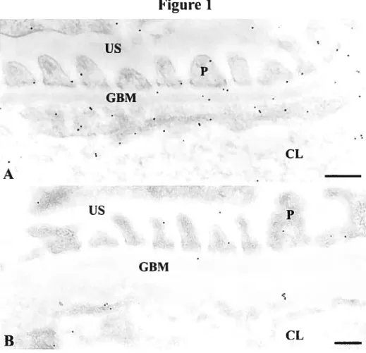
Figure 1. 67 Immunocytochemical localization of MT1-MMP in the glomerular wall of (A) short-term control, and (B) long-term diabetic rat renal tissues.
Figure 2. 68 Immunocytochemical localization of fririn in the glomerular wall of (A) short-terni control, and (B) long-term diabetic rat renal tissues.
Figure 3. 69 Western blot analysis of MT1-MMP in glomenilar lysates: ta) 2-months control, (b) 2-2-months diabetic, (c) 12-2-months control, (d)
Y 2-months diabetic.
Figure 4. 69 Western blot analysis of furin in glomenilar lysates: (a) 2-months control, (b) 2-months diabetic, (e) months control, (d) 12-months diabetic.
Figure 5. 70 Immunocytochemical localization of MT1-MMP over Golgi cisternae of a podocyte in normal rat renal tissue.
Figure 6. 71 Immunocytochemical localization of furin in the RERof (A) short term control and (B) long-term diabetic rat podocytes, and in the Golgi cisternae of (A) short-terni control and (B) long-term diabetic rat podocytes.
List of Symbols and Abbreviations
THESIS
Alpha
(3 Beta
À
ArigstrômAGE Advanced glycation end products CL Capillary lumen
CTL Control
GBM Glomerular basement membrane ECM Extracellular matrix
END Endothelium
HSPG Heparan sulfate proteoglycan kDa kilo-Dalton
mRNA Messenger ribonucleic acid
nm Nanometer
P Podocyte
R-H-R-R Arginine-Lysine-Arginine-Arginine STZ Streptozotocin
TGF-(3 Transforniing growth factor beta US Urinaiy space
ARTICLE % Percent Degree Celsius 11g Microgram Micrometer CL Capillary lumen G Golgi
GBM Glomerular basement membrane ECM Extracellular matrix
EM Electron microscope ER Endoplasmic reticulum
Fig Figure
fur Furin gene
kDa kilo-Dalton kg kilogram L Liter mg Milligram min Minute mol Mole mmol Millimolars MMP Matrix metalloprotease mRNA Messenger ribonucleic acid
MT 1 -MMP Membrane type-I matrix metalloprotease
nm Nanorneter
N-terminal Amino terminal
P Podocytes
pH Hydrogen cation concentration RER Rough endoplasmic reticulum RHRR Arginine-Lysine-Arginine-Arginine rpm Rotations per minute
SDS-PAGE Sodium dodecyl sulfate polyacrylamide gel electropheresis SEM Standard error to the mean
TGf-fl Transforming growth factor beta US Urinary space
Foreword
Portions ofthe resuits ofthis thesis were presented at the following meetings
BOUCHER, E., MAYER, G., BENDAYAN, M. LOCALISATION
ULTRASTRUCTURALE DE LA MTJ-MMP DANS LA MEMBRANE BASALE
GLOMÉRULAIRE DE RATS DIABÉTIQUES. 22e journée scientifique du Département de pathologie et biologie cellulaire, Montréal (Québec), Mai 2004.
BOUCHER, E., MAYER,G., BENDAYAN, M. EXPRESSION 0F GLOMERULAR MEMBRANE TYPE-I METALLOPROTEINASE IN RENAL TISSUE 0F
DIABETIC RATS. 1 2th International Congress of Histochemistry and Cytochemistry, La Joua (California), July 2004.
BOUCHER, E., MAYER, G., BENDAYAN, M. LOCALISATION
ULTRASTRUCTURALE DE LA MTJ-MMP DANS LA MEMBRANE BASALE GLOMÉRULAIRE DE RATS DIABÉTIQUES. Congrès annuel de l’Association Diabète Québec (ADQ), Québec (Québec), Novembre 2004.
BOUCHER, E., MAYER, G., BENDAYAN, M. EXPRESSION ET LOCALISATION DE MT1-MMP ET FUffiNE DANS LA PAROI GLOMÉRULAIRE DE RATS DIABÉTIQUES. 23e journée scientifique du Département de pathologie et biologie cellulaire, Montréal (Québec), Juin 2005.
Dedications
À
mes parents qui m’ont envoyé leur amour sure une brise tunisienne durant ces deux dernières années.Introduction
The human kidney is a veritable marvel of creation. There is no need to question the absolute indispensability of kidneys once we leam that the totalvolume of our circulating bÏood passes through the kidneys once every five minutes! This amounts to a rough total of seventy liters of blood plasma per day! The kidneys not only inspire us to cite staggering statistics that require exclamation points, but also perform the extraordinary and vital tasks of overseeing water and electrolyte homeostasis, regulating blood and extracellular fluid chemical composition, maintaining blood pressure through release of the renin hormone, stimulatingred blood celi synthesis through release of erythroprotein, and simply being an ail
around great pair oforgans (Leeson and Leeson, 1981).
Anatomically speaking, the individual kidney resembles a bean...or is it the bean
that resembles a kidney? Regardless of any phulosophical debate, the kidneys are located in the posterior part ofthe upper abdomen, and each kidney is about 10-12 centimeters in iength and 2.5 centimeters thick (Leeson and Leeson, 1981). The three most prominent anatomical features seen in a transverse kidney section are the outer cortex, the inner medulla, and the pelvis, a hollow inner structure that joins with the ureters (Tisher and Madsen, 1986). In the medial sections of each kidney is an indentation calied the hilum which serves as an exit point for the ureter and gives way to nerves, blood and lymphatic vessels entering andleaving (Leeson and Leeson, 1981). Other distinguishing features include the renal
capsule serving as a protective membrane, as welÏ as right and left adrenal glands which, as the name implies, sit snugly on top ofeach kidney.
The Nephron
The interior medulla of each kidney is adomed with 8-18 pyramids that are striated in appearance. These are positioned with their tips, the renal papillae, facing towards the hulum and their bases aligned with the edge of the renal cortex (Leeson and Leeson, 1981). The renal cortex extends betweeneach renal pyramid creating structures calÏed renal columns. If we were to zoom into the medulla and cortex we would encounter a myriad of tiny microscopic convoluted structures called nephrons. At last, with the acquaintance of the nephron, we make the leap from structure to function and are doser to understanding the essence of the kidney. Also known as the functional units of the kidney, nephrons number slightÏy over one million in each kidney (Tisher and Madsen, 1986). Embedded in both the cortex and medulla, they are assigned the delicate task of filtering the blood plasma, that is, producing a filtrate while retaining the cellular elements of the blood and plasma proteins in circulation (Farquhar, 1991). The nephron may well exhibit a baffling hodge-podge of loops and twists and tums reminiscent of a Cirque du Soleil contortionist, but within this seemingly disorderly arrangement exists an highly organised and complex system of gradients through which altemating functions of secretions and reabsorptions result in the production of urine.
The essential components of the nephron are the glomerulus and Bowman’s capsule, the proximal convoluted tubule, the ascending and descending limbs of the loop of Henle, and the distal convoluted tubule (Tisher and Madsen, 1986). The glomerulus and Bowman’s capsule form the renal corpuscule which lies in the cortex while the renal tubule comprising of the thin limbs and connecting segment, the proximal and distal tubules, extends into the medullar pyramid (Leeson and Leeson, 1981). Given that we are interested in the production ofthe ultrafiltrate of blood plasma, the following information will focus solely on the
renal glomerulus where this process takes place.
Ihe Glomerulus
The glomerulus has been accurately yet perhaps unglamorouslydescribed as a tufi of capillaries. Within the layers of the Bowman’s capsule, blood enters the glomerulus through the afferent arteriole and exits through the same opening by the efferent arteriole. The afferent arteriole branches into a capillary network where hydrostatic blood pressure forces the glomerular filtrate out into the capsular space to eventually be funneled into the renal tubule where reabsorption of most ftuids and saits take place (Farquhar, 1991). A transversal cut of the glomerular capillary tuft unveils where ultraffitration takes place. The different structures observed, collectively called the glomerular wall, impose a precise permselectivity size and charge barrier to the blood plasma and will be
The Glomerular WaII
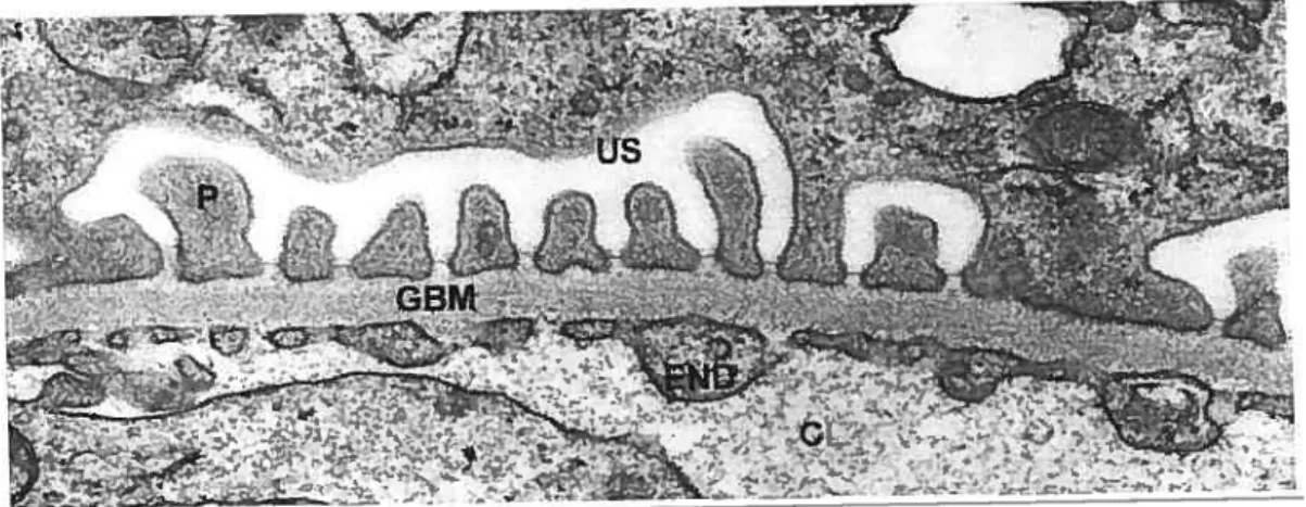
In a remarkable feat of precision, the glomerular capillary wall allows water and small solutes to pass readily into Bowman’s space whule rejecting albumin and other large proteins with great efficiency. In its entirety, this meticulous engine of ultrafiltration is composed of three noteworthy parts- endothelial ceils, extracellular basement membrane, and epithelial ceils (Figure 1).
FIGURE 1: The glomerular wall. Electron micrograph of the glomerular wall of tissues fixed in osmium and embedded in Epon. Endothelium (END) and podocytes (P) flank the glomerular basement membrane (GBM) which together make up the filtration barrier to the blood plasma. The filtrate moves from the capillary lumen (CL) and into the urinary space (US) by way of the sut diaphragms at the junction of GBM and podocytes. X40 000 (Courtesy of I. Londo?io)
Endothelium
The endothelium of glomerular blood capillary vessels represents the first physical barrier in blood filtration. The most salient feature of this endothelial layer is the presence of fenestrae that are relatively large in size (50-100 nm) and are open, that is, lack the usual diaphragms that are present in other fenestrated capillaries (Farquhar, 1991). Whule it may seem like these large open windows offer littie or no resistance to the passage of blood plasma, recent evidence emphatically argues that heavily fenestrated endothelial ceils are important elements of the highly permselective glomerular barrier (Haraldsson and Sirensson, 2004). Admittedly, the actual fenestrae are too large to effectively sieve macromolecules (Deen, 2004), but the celi coat or glycocalyx secreted by endotheliaÏ ceils themseÏves lias been under recent scrutiny for its potential size and charge-selective properties (Haraldsson and Srensson, 2004). To date it lias been established that this layer coating endothelial celi surfaces is made up of
glycosaminoglycans and proteoglycans in combination with plasma proteins, but further investigations on its composition and turnover rate are needed before defining its precise role in glomerniar permselectivity (Haraldssonand S6rensson, 2004). One well-established fact, however, is the presence of the negatively charged protein podocalyxin at the ceil surface of endothelial ceils (Dekan et al., 1991; Kerjaschki et al., 1984; Latta et al., 1975). Podocalyxin reportedly pushes away other negatively charged molecules such as albumin. The fact that endothelial fenestrae are open and larger than those of other fenestrated capillaries makes them highly efficient for filtration, but this also renders the GBM
vuinerable to toxic injury such as that caused by hyperglycemia in the blood, a phenomenon we wiÏl explore in detail shortly.
Basement Membranes
The eariiest histologicai descriptions of basement membranes date back to 1857 from the laboratories of Todd and Bowman (Kefalides, 1973). In broad tenns, basement membranes are recognized as extraceliular matrices associated with the epithelial and endotheliai iinings of almost ail organs of the body. They are found wherever celis (other than connective tissue ceils) meet connective tissue (Farquhar, 1991). A century afier having identified these characteristics, great leaps in studying tissue morphology were made with the invention ofthe electron microscope and the ultrastructure of the basement membrane was described (Kefalides, 1973). Visualization at unprecedentedly high magnificationsunveiied the lamina densa, 20-50 nm in thickness, running parallel to the basai ceil membranes of either epithelium or endothelium, and separated from these by the lamina lucida, a lighter, 10 nm layer (Farquhar, 1991). Histochemical studies in the 1960s revealed the collagenous and noncollagenous glycoprotein and proteoglycan content of basement membranes with various chemicai staining experiments. For example, Kefalides and Denduchis (1969) used pronase on canine basement membranes to demonstrate for the first time the overwhelming presence of coliagen. Before that, Leblond (1957) indicated the presence of carbohydrates with the periodic anti-Schiff reagent. Further biochemical investigations throughout the sixties by several research groups characterised the
complete amino acid and carbohydrate composition of basement membranes, underlining their high content of hydroxyproÏine, glycine, and hydroxylysine as well as glucose, galactose, fucose and mannose (Kefalides, 1973). The general concensus is that components of basement membranes are secreted by ceils lying adjacent and do flot differ significantly in content from organ to organ but rather in quantity and structural organization, reflecting tissue function and specificity (Kefalides and Denduchis, 1969). The participation of basement membranes in processes such as tissue repair and regeneration, as weÏÏ as cellular attachment, migration and development, has provided important insights into research on cancer and diabetes (Erickson and Couchman, 2000; Timpi and Brown, 1996).
The Glomerular Basement Membrane
An extensively studied and long-time favorite of basement membrane-aficionados is the highly specialized basement membrane of the glomerular capillaries of mammalian kidneys. Pioneering the ultrastructural description of the glomerular basement membrane under electron microscopy were Rhodin (1955), Yamada (1955), Kurtz and McManus (1959), and farquhar (1960) among others. Their ground-breaking studies paved the way for more involved histochemical and biochemical analyses of the glomerular basement membrane, analyses that eventually ascribed it its two main functions of support and selective filtration (Kefalides, 1973).
The glomerular basement membrane bears certain distinguishing features that sets
it apart from other basement membranes. It is thicker (350 nm in humans) and
more compact, it is formed by fusion of endothelial and epithelial basement membranes, and it faces endothelial and epithelial celi layers on both surfaces (Farquhar, 1991; Desjardins and Bendayan, 1991). The particular structure fiinction relationship of the glomerular basement membrane in relation to other extracellular matrices was well defined. Desjardins et al. demonstrated that the specific spatial distributions of the main components of the GBM were instrumental in detemiining its filtration properties. This was done by showing how the passage of albumin is increasingly restricted to the subendothelial layer
as maturation ofthe GBM progresses (Desjardins and Bendayan, 1991; Bendayan
et al., 1986). It has thus become dogma that the structural and chemical
composition of the mature glomerular basement membrane is what renders it its unique filtration properties.
Throughout the 1970s, the works of Spiro and Kefalides with purified GBM fractions provided a great wealth of information on the fine architecture of the glomerular basement membrane (Farquhar, 1991). The 1980s ushered in the era of immunolabeling in electron microscopy which also proved to be a valuable tool in elucidating not only the content but also the distribution of GBM constituents (Courtoy et al., 1982; Stow et al., 1985; Kerjaschki et al., 1986). This was done through the purification of proteins from renal glomeruli and tubules which were then used as antigens to prepare specific antibodies (Kerjaschki et al., 1986). The
culmination of this work, mostly carried out in Farquhar’s laboratory, revealed that the major components of the GBM are collagen type W, laminin, lieparan sulfate proteoglycan, nidogen or entactin, and BM4O (farquhar, 1991). Electron microscopy uncovered specific pattems of distribution of collagenous components, proteoglycans and glycoproteins in laminae rarae and laminae densae, eventually leading to sophisticated three-dimensional models of glomentiar basement membrane architecture. The sturdy scaffolding networks created from the binding affinities existing between type-IV collagen, heparan sulfate, laminin, nidogen and entactin underlines the basement membrane’ s tough supportive qualities (f arquhar, 1991).
The highly selective filtering properties of the GBM are dependent on the quality, the quantity, and the distribution of its constituents. Increasingly sophisticated physiological blood plasma clearance studies have demonstrated that the glomerular basement membrane serves primarily as a charge- and size-selective barrier (farquhar, 1991). for example, the presence ofhighly negatively charged regions of glycosaminoglycans containing heparan sulfate impose a significant charge barrier (Farquhar, 1991), conferring them the role ofthe main anionic site for GBM charge selectivity (Akthar, 2004). It lias been speculated that the size selective sieving properties of the GBM are a property of the highly compact meshwork of type-W collagen (Kanwar, 1984). Collagen is composed of three alpha-chains forming triple-helical molecules, which form a tightly cross-linked network structure. It is chiefly concentrated in the lamina densa of the GBM
(Kanwar, 1984). Although the exact size of the pores formed by networks of collagen and other GBM proteins have flot been precisely determined, it is widely accepted that size restriction is an inherent quality of the highly specialized meshwork ofthe GBM. Morphological approaches using the large (60
À)
ferritin molecule as a tracer revealed that much of the size-selection occurs at the level of the lamina rara interna (Farquhar et al., 1961) while smaller molecules like horseradish peroxidase can traverse the membrane (Kefalides, 1973). The significance of the GBM as an important size-barrier was also highlighted by f arquhar in an experiment involving dextran tracer molecules (f arquhar, 1975), a finding which actually shified the attention of the primary filtration barrier from podocytes to the GBM. Albumin lias also played a central role in identifying the specific size retention areas in the glomerular basement membrane (Bendayan et al., 1986; Londoflo et al., 2003). Immunocytochemistry revealed the presence of albumin at the level of the endothelial celi basal plasma membrane and on the subendothelial side of the lamina densa of the GBM, indicating that albumin retention occurs at these sites (Bendayan et al., 1986). following up on these experiments, it was shown that glycated albumin, once in circulation, penetrates the glomerular wall of normal animais deeper than nonglycated albumin and eventualiy reaches the urinary space. This provided evidence that albumin normally does not pass through the glomerular wall while an abnormai glycated version ofthe protein is able to modify the glomerular filtration properties in such a way as to be able to reach the urinary space (Londoflo et al., 2003).Podocytes
The epithelial ceils of the glomerular wall are called podocytes which separate the endothelial capillary vessels from the urinary space ofthe Bowman’s capsule. In scanning electron microscopy it is possible to visualize their unique cell body prolongations which resemble interlacing fingers covering the entire surface area of capillary vessels (Guyton and Hall, 2000; Tisher and Madsen, 1986). from these elongations emerge smaller secondary projections called pedicels. Pedicels interdigitate with other pedicels from neiglibouring podocytes and firmly anchor themselves in the lamina rara externa of the GBM (Tisher and Madsen, 1986; Pihiajaniemi, 1996). It is interesting to note that adjacent pedicels are most likely projections from different podocytic cell bodies (Tisher and Madsen, 1986).
The podocytes are the most voluminous cells in the glomerulus (Guyton and Hall, 2000). Along with a well-developed Golgi, they contain free ribosomes as well as rough and smooth ER cistemae (Tisher and Madsen, 1986), which indicates their marked propensity for protein synthesis. Kurtz and f eldman (1962) pointed out that podocytes are largely responsible for the biosynthesis of the glomerular basement membrane. In fact, it has been widely documented that enzymes responsible not only for GBM synthesis but also turnover and maintenance, are largely synthesized within podocytic ce!! bodies (Lee et al., 1993; McCarthy, 1997). 0f these enzymes, the family of matrix metalloproteases play a crucial role as will be seen shortly.
In the past decade, attention has been shifted to podocytes for a clearer understanding of permselectivity. The spaces existing between pedicel interdigitations vary between 25 and 60 nm and are called filtration siits (Pihiajaniemi, 1996), while the porous thin membranes that traverse the filtration slits are called sut diaphragms (Leeson and Leeson, 1981). The sut diaphragms of adjacent pedicels are linked to each other by specialized plasma membrane adherens junctions (Guyton and Hall, 2000; Tisher and Madsen, 1986), and the integrity of this arrangement is largely dictated by the presence of a sialic acid rich glycocalyx (Farquhar, 1991).
It was long believed that the primary filtration barrier of the glomerular wall resided in the filtration sut regions ofpodocytic interdigitations. Jndeed, this area was shown to exhibit size-restrictive properties (Rodewald and Kamovsky, 1974), and is also essential for glomerular wall reinforcement in the face of high hydrostatic blood plasma pressures (Keijaschki, 2001). It is only with the recent molecular era that our understanding of podocytes and their role in filtration has greatly advanced. Kerjaschki greatly facilitated our comprehension of the increasingly complex molecular characteristics of the podocytes by separating its plasma membrane area in three distinct microdomains: the sut diaphragms at the junction of adjacent podocytes, the basal membrane which touches the GBM, and
Sut Diaphragm
The most influential study ofthe fine structure ofthe sut diaphragm was provided by Rodewald and Kamovsky back in 1974 (Rodewald and Kamovsky, 1974). They interpreted electron microscopy images of sut diaphragms as zipper-like structures that are arranged in sucli a way to restrict passage of molecules. The zipper resembles a central dense region with openings roughly the size of albumin (Deen, 2000). The novel application of electron tomography was able to show an increased complexity of the pores, more tortuous and irregular than previously supposed (Deen, 2000). Characterization of the molecular composition of slit diaphragms boomed in recent years with the identification of nephrin (HoÏzman et al., 1999; Kerjaschki, 2001), NEPH-1 (Keijaschki, 2001), CD2AP (Keijaschki, 2001; Li et al., 2000), and podocin (Keijaschki, 2001; Schwarz et al., 2001). The newly discovered protein that received the most attention was the transmembranous nephrin, found to be an adhesion protein necessary for the formation ofjunctions (Holzman et al., 1999; Kerjaschki, 2001). It was recently demonstrated that the fibers constituting the slit diaphragm appear to be formed largely by the association of extracellular strands of nephrin (Deen, 2000). The exact mechanism by which nephrin controls permselectivity is not yet clear but the Finnish congenital nephrotic syndrome emphasized the importance of nephrin which is absent in this disease (Akthar, 2004). Tiideed, in nephrin-knockout mice, ordered slit diaphragm structures are no longer evident, and proteinuria resuits (Deen, 2000). Increasing evidence only ftirther confirms that disruption ofthe sut
diaphragms is sufficient to cause abnormal loss of proteins in the urinary space (Hamano et al., 2002).
Basal Membrane
The basal plasma membrane of podocytes is anchored to the GBM through adhesion proteins, most notably integrin a331 (Kerjaschki et al., 1989). a3t31 bridges podocytes and GBM through association with paxillin, talin and vinculin on the cytoplasmic podocytic side (Drenckhahn and Francke, 1988), and collagen, fibronectin, laminin and entactin on the GBM extracellular matrix side (Kretzler, 2002). Another noteworthy constituent of the podocyte basal membrane is dystroglycan which is believed to regulate the position and spacing of proteins in the extracellular matrix. This would confer it a role in porosity and GBM permeability. Indeed, a fali in dystroglycan expression has been shown to coincide with podocyte effacement and proteinuria (Kerjaschki, 2001).
Apical Membrane
The main role of the apical membrane region of podocytes seems to be in the maintenance of structure and cohesion of podocytes (Kerjaschki, 2001). This function has been linked to the presence of podocalyxin, a highly negativeÏy charged protein also present on the endothelium, as was mentioned earlier. This glycoprotein was found to be abundant on the apical surface of podocytes by Sawada et al. in 1986 who also discovered that neutralizing the charges on podocalyxin leads to alterations in podocytic processes and slit diaphragm
organisation, a finding which underlines the importance of its presence in podocyte maturation. Recent evidence also indicates that podocalyxin can also link itself to cytoskeletal actin by virtue of NHERV-2 and ezrin (Kerjaschki, 2001), which confers to podocyte projections their particular “feet”-like architecture.
Together, the endothelium, glomerular basement membrane and podocytes interact to foi-m an highly intricate sieve designed to select molecules on the basis of size, shape and charge. Despite the comprehensive and compelling evidence offered on the structural and biochemical composition of the glomerular wall, the specifics of ultrafiltration stiil remain nebulous. Initially it was widely believed that the primary filtration barrier was located exclusively at the interdigitations of podocytes (Kamovsky and Ainsworth, 1972) but Bendayan and f arquhar produced persuasive evidence highlighting the size-restrictive properties of the glomerular basement membrane (f arquhar, 1975; Bendayan et al., 1986). Recently, more weight has been given to the epithelial sut diaphragms due to important advances in molecular biology and the identification of nephrin. A comprehensive study has shown how decreased expression of nephrin correlates with a loss in glomerular filtration integrity (Hamano et al., 2002). finally, in 2004, S5rensson and Haraldsson argued that too much attention has been given to the GBM and epithelial podocytes in particular, while the endothelium probably has a much greater role in glomerular permselectivity than we believe. This is in light of new information conceming the cell surface coat covering endothelial
ceils called the glycocalyx, and the identification ofproteins within it that seem to be of vital importance for capillar permeability (S5rensson and Haraldsson, 2004). The debate rages on, yet one thing remains certain: the proper functioning of the glomerural wall is a function of the integrity, interdependence, and proper communication of ail its parts.
Diabetic nephropathy
Whether we like it or flot, our understanding of how something works is greatly advanced when that thing doesn’t work. Such is the case with the glomemiar filtration apparatus; rnuch of our knowledge of its function cornes from observation of pathophysiological situations which surround glomerulosclerosis. Malfunctioning as a resuit of diabetes leads to generalized proteinuria or abnormal loss of proteins into the urinary space due to an imbalance in the size and charge-selective barrier properties of the glomerular wall. The relevance of diabetes mellitus in today’s society cannot be overemphasized: there is a 2-5% increase in the incidence of type-I diabetes but the real concem is the global increase in type-II, largely due to childhood obesity (Silink, 2002). Diabetic nephropathy, a condition that develops afler exposures of tissues to chronic hyperglycemia, is one of the leading causes of renal failure in Western countries (Mason and Wahab, 2003). Diabetic patients account for nearly haif of ah patients on haemodialysis, the kidneys being home to the gravest pathophysiological consequences of this disease (Del Prete et al., 1998). The complications arising from these lesions lead to glomerular fibrosis which
progressively destroys the renal filtration unit and may eventually cause renal failure (Del Prete et al., 1998). Our project deals specifically with changes, structural and compositional, in the glomerular wall at the molecular level that accompany the onset ofthis devastating disease.
As early as 1959, morphological changes ofthe glomerulus in diabetic conditions were observed by electron and light microscopy (farquhar et al., 1959). Studies by several different groups concurred that the most striking feature of a glomerular wall exposed to an hyperglycemic environment is the significant thickening of the glomerular basement membrane (Figure 2) (Kimmelstiel et al., 1962; Bloodworth, 1963; Østerby and Lundbaeck, 1970). Further investigations into this morphological discrepancy revealed an important correlation between GBM thickening and biochemical changes in the amino acid and sugar composition ofthe basement membrane (Spiro and Spiro, 1968; Beisswenger and Spiro, 1970; 1973; Beisswenger, 1976; Spiro, 1976). Pioneering work conceming the effect of diabetes on the biochemical composition of the basement membrane was thus undertaken. Kanwar demonstrated by autoradiography and biochemical analysis that proteoglycan synthesis decreased by 40% in diabetic rats relative to normal rats (Kanwar et aI., 1983). In an elegant experiment employing glucosyltransferase, an enzyme involved in the synthesis of certain GBM components, Spiro and Spiro demonstrated increased levels of this signaling protein in diabetic kidneys relative to normal ones (Spiro and Spiro, 1971). Indicating that increased basement membrane synthesis in diabetes might be a
resuit of the overexpression of this protein, they also noted that this process is reversible through insulin administration. Additional studies signaled the effect of diabetes on the metabolism of the GBM, as well as the significantly reduced level of heparan sulfate, the aforementioned major player enforcing charge selectivity, in the GBM under diabetic conditions (Parthasarathy and Spiro, 1982; Brownlee and Spiro, 1979). Employing ultrastructural analyses, other studies also confirmed a decrease in heparan sulfate immunolabeling in the lamina rara interna of the GBM in diabetic rats (Desjardins and Bendayan, 1990; Vemier et al, 1992).
While Spiro focused on biochemical modifications, the works of Østerby for the past three decades were instrumental in elucidating those structural changes that characterise glomerulopathy (Østerby and Gundersen, 1980; Gundersen and
f •1.,
-4
FIGURE 2: Comparison between gtomerular walls of normal and diabetïc animais. Electron micrograph of glomerular walls of rat renal tissues, 2-month old control (CTL) and 6-month old diabetic (STZ) animals. Significant thickening is observed in the glomerular basement membrane of the diabetic animal due to increased deposition of extracellular matrix components. X60 000 (Courtesy of T. Londoflo)
Østerby, 1977; Østerby et al., 1993; Østerby et al., 1987; Østerby et al., 1988). Using streptozotocin-induced diabetes in rats, it was shown that generalized glomerular enlargement was apparent only four days afler injection (Østerby and Gundersen, 1980). Progressive expansion of the mesangial matrix, and thickening of the glomerular and tubular basement membranes thus became universal hallmarks of human and experimental diabetic nephropathy (Del Prete et al., 1998). These findings provided the firm basis upon which countless experiments were inspired to supply additional information on the characteristics of diabetic nephropathy.
The technique of high-resolution immunocytochemistry using electron microscopy and immuno-biochemical quantifications offered great insights into the expression and distribution of structural proteins in the GBM. In diabetic patients with only mild glomerulosclerosis, increases in collagens type-W, ai, a2, cr3 and cr4, laminin, and fibronectin were reported while levels of perlecan decreased (Kim et al., 1991; Mohan et al., 1990; Shimohura and Spiro, 1987). Other factors found to be abundant in glomerular diabetic tissues were advanced glycated end products (Gugliucci and Bendayan, 1996; Bendayan, 1998). The main cuiprit ofbasement membrane expansion, however, is the high deposition of collagen. Increases in collagen deposition in GBM and mesangial matrix (Regoli et al., 1998; Nerlich et al., 1994) as well as an augmentation in collagen mRNA (Park et al., 1997; Fukui et al., 1992) have been well documented in advanced diabetic tissues. Desjardins and Bendayan brought to our attention the
modification in the spatial distribution of collagen type-W in fong-term diabetic animais (Desjardins and Bendayan, 1990). Normaliy concentrated in the lamina densa, collagen type-W was found to be relocalized to the subendothelial side of the GBM in diabetic animais (Bendayan, 1985; Desjardins and Bendayan, 1990). No changes were observed in the distributions of laminin, entactin and heparan sulfate in those same animais (Desjardins and Bendayan, 1990). It thus became apparent that any modifications conceming the expression and distribution of structural proteins in the GBM were reflected in the three-dimensional scaffolding networks. lndeed, through immunocytochemistry in electron microscopy, it was possible to observe an alteration at the level of structural organisation of GBM components during the course ofdiabetes. Specificaliy, the endothelial side ofthe GBM gave rise to dense bundles of fibrils identified as basotubuies which were found to be iargeiy associated with type-W coilagen in diabetic animais (moue and Bendayan, 1995).
Our project focuses on specific factors leading to the occurrence of the most prominent morphologicai feature of giomeruiopathy: the thickening of the giomerular basement membrane, a phenomenon which has been associated with a decrease in filtration area, abnormal loss of proteins in the urinary space and ultimately end-stage renai failure. With this important observation, it became apparent that perpetual maintenance and turnover of the molecules that make up the giomeruiar basement membrane is sine qua none for proper permseiectivity. It is now widely accepted that the amount and composition of the GBM
extraceliular matrix relies on the delicate interactions existing between synthetic and degradative pathways, and that various forms of giornerular diseases are characterized by shifts in this balance.
Membrane Type-I Metalloprotease
Nowadays, when someone says “tissue remodeling”, the matrix metalloproteases (MMP5) imrnediateiy corne to mmd (well, arnong us molecular biologists anyway). This growing famiiy of zinc-dependent metalloendopeptidases is the object of rnany studies dealing with extraceliular rnatrix turnover. This is due to their prominent role as cleavers of practically ail components of the extracellular matrix such as collagen, proteoglycans, fibronectin, and larninin (Bode et al., 1999). MMPs have thus been shown to be key players in processes involving degradation of pericellular proteins, thereby assurning pivotai roles in normal and pathological processes (Stemlicht and Werb, 2001).
Historically, MMPs were classified as collagenases, gelatinases, stromelysins, and rnatrilysins based on their cleaving specificities, but the growing number ofMMP substrates has prompted us to group them according to structure (Egeblad and Werb, 2002). The 21 MMPs are divided into eight structural classes; five secreted and three membrane-type MMPs (Egeblad and Werb, 2002). The degradative functions of metalloproteases are iargeiy kept in check by their endogenous tissue inhibitors (TIMPs) and disrnption of this balance has been documented to resuit in serious diseases such as arthritis, turnor growth and
metastasis (Bode et al., 1999). Thus MMPs are abie to regulate many biological processes and are closely regulated themselves.
MMPs share several structural and functional properties: they are synthesized as inactive zymogens containing a secretory signal sequence, a propeptide hinge, as well as hemopexin-like and catalytic zinc-binding domains (Lenz et al., 2000; Stamenkovic, 2003). Activation requires proteolytic removal of the propeptide prodomain (Egeblad and Werb, 2002). 0f the currently known MMPs, MMP-1, MMP-13, MMP-3, MMP-2, MMP-9, and MT1-MMP ail share the characteristic of being collagenases and thus have been extensively studied in the glomerulus (Lenz et al., 2000). In this project, we chose to study the membrane type-I matrix metalloprotease (MT1-MMP) due to its unique properties conferred by its location at the cellular membrane.
The membrane type-I metalloprotease distinguishes itself as a membrane-bound enzyme capable of exerting its cleavage activities from the vantage point of the celi surface (Bode et al., 1999). MT1-MMP possesses an additional stretch of around 20 hydrophobic amino constituting a transmembrane domain, followed by a short cytoplasmic tau (Figure 3) ($ato et al., 1994). Active MT1-MMP inserts itself into the plasma membrane with the catalytic domain facing the extracellular space, where it can cleave substrates in the extracellular matrix (Osenkowski et al., 2004). Localization of MT1-MMP at the ceil membrane is perfect for pericellular proteolysis allowing for a new set of substrate targets, distinctive
interactions with T1MPs and a non-conventional mechanism of regulation involving enzyme intemalization, processing and ectodomain shedding (Toth et al., 2002; Osenkowski et al., 2004).
MOPl3
FIGURE 3: Domain structure of MI1-MMP. Signal sequence (PRE), propeptide (PRO) with a free zinc-ligating thiol (5H) group, furin-susceptible site (F), zinc-binding site (Zn), hinge region (H), transmembrane region (TM), and cytoplasmic tau (C). The hemopexin domain contains four repeats, the first and last are linked by a disulfide bond. (Illustration not to scale by Emmanuelle Boucher, adapted ftom Sternlicht and Werb, 2001: How matrix metalloproteinases regulate ccli behavior. Ann Rev Ceit Dey Bio! 17: 463-5 16)
Current evidence indicates that MT1-MMP is able to regulate matrix turnover due to its ability to degrade matrix-associated molecules, either directly or via activation of downstream MMPs (Yana and Weiss, 2000). The dual role of Mil MMP as activator of other MMPs capable of initiating activation of zymogen cascades, and as direct cleaver of ECM components, only underscores its vital function as an extracellular matrix remodeler and singles it out as a potential therapeutic target (Lenz et al., 2000; Ohuchi et al., 1997). The importance of MT1-MMP’s role as a collagenase was demonstrated in mice deficient in Mil MMP which suffered severe complications in remodeling of skeletal connective tissues, resulting in earlier death (Holmbeck et al., 1999). It has been speculated that MT1-MMP’s strategic location confers it an advantage over other
collagenases in that it can accornplish localized and efficient collagen degradation at the celi surface (Hotary et al., 2000), as well as initiate zymogen activation cascades (Toth et al., 2003). One such cascade is the activation of pro-MMP-2 which was actually the mechanism by which MT1-MMP was first identified (Overail and LopezOtin, 2002). Here, active MT1-MMP binds TIMP-2 which generates a surface receptor for pro-MMP-2 (Osenkowski et al., 2004). The resulting MT 1 -MMP/TIMP-2/pro-MMP-2 ternary complex presents the bound pro-MMP-2 to a neighbouring TTMP-2-free MT1-MMP which will initiate the activation of pro-MMP-2. Once proteolytically processed by MT1-MMP, active MMP-2 is released into the extracellular space where it can carry out its cleavage activities (Bemardo and Fridman, 2003). This effectively changed our traditional view of TJMPs as inhibitors to that of versatile molecules capable of acting in the promotion ofpericellular proteolysis. Indeed, MT1-MMP plays a dual role in the pathophysiological digestion of extracellular matrix through direct cleavage of the substrates and the indirect activation of pro-MMP-2 as well as other zymogen cascades (Coweil et al., 1998; Toth et al., 2003). MT1-MMP’s wide substrate range specificity includes collagen types I, II and III (into typical 3/4 and Y4 length fragments), laminin types 1 and 5, vitronectin, gelatin, proteoglycan fibrin, and aggrecan (Ohuchi et al., 1997). It is thus evident that appropriate expression and activity of this endoprotease is a mainstay for basement membrane maintenance and turnover.
Furin
As mentionned earlier, cleavage of the propeptide region is essential for MT1-MMP activation and subsequent proteolytic activity. Yana and Weiss demonstrated the existence of a proprotein convertase-MT1-MMP axis allowing for the unmasking of MT1-MMP’s catalytic domain and conversion into a catalytically active species (Yana and Weiss, 2000). This was done through the identification of the RX(KIR)R basic motif in the propeptide region of MT1-MMP, a sequence of amino acids that can potentially be recognized by the proprotein convertase family of subtilisin-like proteases. Jndeed in vitro studies using MT1-MMP deletion mutants confirmed that furin is capable of cleaving the proMTl-MMP zymogen (Yana and Weiss, 2000). It is thus apparent that cooperative interactions between proprotein convertases and membrane-anchored MMP’s play an important role in regulating the remodeling ofECM.
furin is a caïcium-dependent endoprotease involved in the proteolytic activation of a large variety of proprotein substrates in secretory pathway compartments (Thomas, 2002). The importance of furin as a proprotein convertase capable of processing a wide range of precursors cannot be overemphasized as it has been shown to be ubiquitous in most ceil types (Blanchette et al., 1997). Also, knockout of furin gene is embryonic lethal (Roebreck et al., 199$), underlining its importance in the maturation, function and activation of several hormones, growth factors, and ceil surface receptors (Molloy et al., 1999). Specific amino acid motifs identified in furin’s cytoplasmic tau have shown to target furin to different
cellular compartments; a finding which has shed light on furin trafficking between the trans-Golgi network, endosomal compartments and the ceil surface (Plaimauer et al., 2001; Molloy et al., 1994; Mayer et al., 2003).
The physiological role of furin in glomerular celis was well characterized by Mayer et al.. Double immunogold labelings revealed that furin was co-localized along with pro-MT1-MMP in the biosynthetic pathway, and was also observed beyond the trans-Golgi network and on the cell surface of podocytes (Mayer et al., 2003). Mayer et al. propose an MT1-MMP/furin activation axis at podocytic and endothelial abluminal membranes facing the GBM for a type of focalized pericellular proteolysis essential for GBM turnover (Mayer et al., 2003). These novel findings on the cellular biology of podocytes prompted us to investigate whether the expression of these proteins was altered in a hyperglycemic environment. This would shed some light on the elements that characterize the integrated functions of cellular and extracellular elements involved in tissue remodeling, and their disregulation in diabetes.
Objective of $tudy
The aim of our project is to characterize some specific molecular changes in the glomerular wall that accompany diabetic glomerulosclerosis. The possibility that the expression andlor localization of proteins involved in extracellular matrix turnover is modified is highly suggested by the thickening of the glomerular basement membrane. This is why the membrane type-I matrix metalloprotease, one such endoprotease involved in the cleaving of many ECM components, and its activating enzyme furin, were elected for study. It was decided that valuable comparisons couid be made between short- and long-term diabetic animais and their age-matched controls. Rats were thus injected with streptozotocin and sacrificed afler 2 and 12 months of maintaining a hyperglycemic state. Comparitive evaluations could then be undertaken at a morphological and biochemical level.
Immunocytochemical techniques with colloidal gold immunolabeling offer distinct advantages in evaluating these enzymes’ expression and localization in the renal tissues of normal and diabetic animais. Antigenic sites of MTY-MMP and furin can be detected with their corresponding polyclonal antibodies which in mm can be revealed by the protein-A colloidai gold complex (Bendayan, 1995). This high resolution technique flot only allows us to view the localizations of MT1-MMP and fiirin, but also to quantify their intensity in labeling.
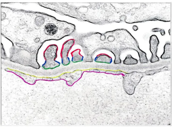
Morphometncal analyses were greatly facilitated by virtual dissection of the glomerular wall into distinctive structures (Figure 4). It is thus possible to quantitatively evaluate selected portions of glomerular cell surfaces suspected to harbor these matrix-remodeling enzymes.
-
—e-Ç?
FIGURE 4: Dïssected areas of the glomerular walI for morphometrical evaluation. The plasma membrane surfaces of the glomerular wall were virtually separated into five distinct regions to facilitate morphometncal analysis: the abluminal (red) and luminal (green) podocyte membranes, the sut diaphragm (blue), and the abluminal (yellow) and luminal (pink) endothelial membranes. Gold particles per micrometer were measured. X40 000 (Courtesy of I. Londoflo)
To further substantiate immunocytochemical data, we perform western blots on glomerular lysates, once again using polyclonal antibodies against MT1-MMP
‘4:
morphological resuits, immunocytochemistry once again proved to be an invaluable tool in resolving these inconsistencies by revealing changes in localization of the studied enzymes within the glomerular wall. The western blotting and immunogold techniques thus complement each other well, rendering it possible to determine overall expression and specific placement of proteins in the glomerular wall.
Article
EXPRESSION AND LOCALIZATION 0F MT1-MMP AND ITS ACTIVATING ENZYME FURIN IN TUE GLOMERULAR WALL 0F
SHORT AND LONG TERM DIABETIC RATS
Emmanuelle BOUCHER, Gaétan MAYER, and Moïse BENDAYAN
Department ofPathology and Celi Biology, University ofMontreal, Montreal, Quebec, Canada, H3C 317
Under review in:
ABSTRACT
Diabetic glomerulopathy has been linked to shifts in balance between the synthetic and degradative pathways of the glomerular basement membrane, a key player in the permselectivity of macromolecules. The goal of this study was to trace the expression and localization of MT1-MMP and its activating enzyme
fin-in, key proteins involved in basement membrane turnover, in short and long
term diabetic rat renal tissues.
Quantitative immunogold was carried out for MT1-MMP and finin and their expression was evaluated in renal tissues of young and old, control and diabetic rats. To corroborate immunocytochemical findings, western blots were performed on glomerular lysates.
Electron microscopy revealed that the overali expression of MT 1 -MMP and furin
is reduced in plasma membranes of celis of old normoglycemic animais, a
phenomenon that is exacerbated in long-terni diabetic animais. This observation supports the prevailing theory that diabetes fosters an acceleration in the aging process. Interestingly, while biochemical resuits confirmed a decrease in Mil MMP expression, an increase infurinwas observed.
Immunocytochemical studies resolved this discrepancy by tracing the increased furin expression in ER and Golgi membranes of podocytes, indicating that furin miglit be retained in the secretory pathway in a diabetic environment.
Disturbances at the moledular level of the otherwise tightly regulated MT1-MMP/furin interactions found at the ceil surface must account for a lack in extracellular matrix remodeling, increased deposition of GBM material, and loss of glomerular filtration integrity.
INTRODUCTION
As the ultimate site of plasma filtration, the glomerular wall plays a vital role in maintaining proper permeability for the production of primary urine (1). The integrity of the glomerular wall in terms of structural organization and protein composition is the comerstone of intact permselectivity. Obvious morphological changes associated with the onset of diabetes and loss of permselectivity include thickening of the glomerular basement membrane (GBM), expansion of the mesangial matrix, and podocyte effacement (2-4). The molecular era of the past two decades has allowed us to witness a wave of information conceming the proteinacious composition of the main filtrating constituents of the glomerular wall, notably the endothelial fenestrations, the GBM, the sut diaphragms at the junction ofpodocytes, and the mesangial matrix (1,5-6). It has thus recently been possible to identify the molecular mechanisms responsible for progressive glomerulosclerosis at an ultrastructural level.
Electron microscopy employing the immunogold technique as an investigative tool has played an essential role tracing the modifications in molecular composition of glomerular wall components in hyperglycemic environments (7-12). Desjardins et al. in particular, followed the expression and localization of certain key molecular constituents of the GBM and reported that while most molecules remained intact, there were changes in type W collagen distribution and expression in diabetic rats (12,13). Indeed the importance of collagen deposition and accumulation in the thickening of the GBM cannot be
overemphasized (5,14-15). One of the consequences was a race to identify and locate the enzymes responsible for basement membrane turnover, a process which proves integral to the maintenance of intact glomerular filtration. We then came to realize that the amount and composition of extracellular matrix depends on the delicate interactions existing between synthetic and degradative pathways, and that various forms of glomerular disease are characterized by shifis in this balance (16-17).
The matrix metalloproteases are a large family of zinc endopeptidases widely recognized as crucial mediators in extracellular matrix turnover (18). The membrane type-I metalloprotease distinguishes itself as a membrane-bound enzyme capable of exerting its cleavage activities from the vantage point of the celi surface (19). Osenkwoski et al. underline the regulatory benefits of Mil MMP’s strategic positioning at the membrane. Localization of MT1-MMP at the celi membrane is perfect for pericellular proteolysis allowing for a new set of substrate targets, distinctive interactions with TIMPs and a non-conventional mechanism of regulation involving enzyme internalization, processing and ectodomain shedding (20-2 1).
The primary function of MT1-MMP is to degrade matrix-associated molecules either directly or via the activation of downstream MMPs (22). Animal knockouts have shown that MT1-MMP is the only MMP known to be essential for suwival (20), a fact that is emphasized by its wide array of substrates (22).
The dual role of MT1-MMP as activator of other MMPs capable of initiating activation of zymogen cascades, and as direct cleaver of ECM components, only underscores its vital function as an extracellular matrix remodeler and singles it out as a potential therapeutic target (17,22).
Cleavage of the propeptide region is sine qua non for MT1-MMP activation and subsequent proteolytic activity. Yana and Weiss demonstrated the existence of a proprotein convertase-MMP axis allowing for the unmasking of MT1-MMP’s catalytic domain and conversion into a catalytically active species (23). This was done through the identification of the RX(K!R)R basic motif in the propeptide region of MT1-MMP, a sequence of amino acids that can potentially be recognized by the proprotein convertase family of subtilisin-like proteases. Jndeed in vitro studies using MT1-MMP deletion mutants confirmed that furin is capable of cleaving the proMTl-MMP zymogen (23). It is thus apparent that cooperative interactions between proprotein convertases and membrane-anchored MMP’s play an important role in regulating the remodeling ofECM.
Furin is an endoprotease involved in the proteolytic activation of a large variety of proprotein substrates in secretory pathway compartments (24). The importance of furin as a proprotein convertase capable of processing a wide range of precursors cannot be overemphasized as it has been shown to be ubiquitous in most celi types (25). Specific amino acid motifs identified in furin’s cytoplasmic tail have shown to target furin to different cellular compartments; a finding which has shed
light on furin trafficking between the trans-Golgi network, endosomal compartments and the celi surface (26-28). The physiological role of furin in glomerular celis was well characterized by Mayer et al. who co-localized the protein with pro-MT1-MMP in the biosynthetic pathway and traced their transiocation beyond the trans-Golgi network onto the celi surface (28). Mayer et al. propose an MT1-MMP activation axis at podocytic and endothelial abluminal membranes facing the GBM for a type of focalized pericellular proteolysis which characterizes the integrated functions of cellular and extracellular elements involved in tissue-remodeling (28). The purpose of our study was to extend this knowledge of interactions among ceil surface proteins mediating GBM turnover in normal celis, to their potential modification of expression and/or deregulation in diabetic rat renal tissues.
RESEARCH DESIGN AND METHODS
Antibodïes
The rabbit anti-matrix metalloproteinase-14 (MMP-14 or MT1-MMP) was obtained from Sigma-Aidrich (Oakville, ON, Canada). Raised against the N-terminal portion of the enzyme, the antibody tags both mature and pro forms of MT1-MMP. Rabbit anti-fin-in antibody was supplied by Alexis Biochemicals (Axxora, LLC). The antibody was raised against the N-terminal portion (amino acid sequence 187-198).
Animais
Experimental diabetes was induced in 150g male Sprague-Dawley rats by an intraperitoneal streptozotocin injection (70 mg/kg body weight dissolved in 10 mM/l citrate buffer, pH 4.5). Within 48 hours, animals developed a hyperglycemic state which lasted throughout their lifetime. Glycemia was assessed on a monthly basis with Accu-Check test strips and blood glucose meters (Roche Diagnostics) while glycosuria measurements were carried out weekly using UriScan test strips (YD Diagnostics). Glycemic states at death of 2-month and 12-month old diabetic rats averaged 25.3+4.3 and 27.4±4.0 mmolIL respectively versus 5.6±0.8 mmol/L and 6.7±1.2 mmol/L for their age-matched controls. Four experimental groups were created, each comprising of four animais. Two groups were composed of streptozotocin-injected animals which maintained hyperglycemia for 2 and 12 months, and two were their age-matched
controls. Animais were housed and handled according to the guidelines from the Canadian Council on Animal Care (CCAC).
Glomerular isolation
Animais were anaesthetized with urethane. Upon excision, entire kidneys were immediateiy recovered in PBS and subjected to sequential sieving methods to separate giomeruli (29). Isoiated glomeruli were washed and suspended in freshiy-made lysis buffer (29), homogenized, and kept on ice for 1 hour. Homogenates were then centriftiged (2500 rpm) for 20 min at 40
C to remove non-soiubiiized materiai. The supematant carrying membranous proteins MT1-MMP and furin were used for our studies. Protein quantitation was carried out using the bicinchoninic acid method (BCA Protein Concentration Assay, Pierce) with bovine serum aibumin as a standard. Sampies were aiiquoted at concentrations of 40 ig and stored at 80° C untii needed.
Immunocytochemistry
SmaiÏ tissue fragments measuring 1 mm3 were rapidiy sampied from excised kidneys and fixed by immersion in a 4% paraformaldehyde-iysine-periodate solution for two hours at 4°C. Tissue fragments were rinsed in 0.1 mol/i phosphate buffer, dehydrated in methanoi and embedded at -30° C in Lowicryi K4M (30). Ultrathin sections were mounted on Parlodion-carbon-coated nickel grids for immunocytochemistry.
MT1-MMP and furin antigenic sites were revealed using the corresponding polyconal antibodies in combination with the protein A-gold complex on a post embedding immunocytochemical approach as described previously (30). The anti-MT1-MMP was used at 1:50 and anti-furin at 1:10, both ovemight at 4°C. Incubation with protein A-gold (10 nm) was carried out for 30 min at room temperature. The tissue sections were stained with uranyl acetate and observed with a Philips 41OSL electron microscope. Specificity of immunolabelings was assessed by incubating tissue sections with the protein-A gold complex, omitting the primary antibody step. Competition experiments were also carried out using antigen-adsorbed antibodies.
Precise localization and quantification ofboth MT1-MMP and furin antigens over glomerular tissue were carried out on electron micrographs with Clemex Vision Analysis unit. Pictures were recorded at X 21 000. Forty pictures were taken for each animal in each experimental group. The glomerular wall was morphologically separated into 7 distinct regions for counting: abluminal and luminal endothelial membranes, abluminal and luminal podocyte membranes, slit diaphragm, mesangial celi plasma membrane, and glomerular basement membrane. In addition, labelings over the rough endoplasmic reticulum and Golgi regions of podocytes, as well as mitochondria were evaluated. The length of these various plasma membrane domains as well as those delineating the cellular compartments were measured and gold particles associated with these membranes counted. Values are expressed in particles per micrometer. Mean
values were calculated along with their standard deviations. Statistical evaluations were carried out using the Student’s T-test.
Western blotting
Glomerular protein samples were boiled for 4 minutes in reducing SDS-sample buffer, separated by SDS-PAGE on 10% handmade polyacrylamide gels, and then electrophoretically transferred to nitrocellulose membranes. For MT1-MMP and furin immunodetection, membranes were blocked in TBS (50 mM) containing 0.05% Tween 20 and 1% milk for 1 hour and 3 hours respectively, and then incubated ovemight at 40
C with the appropriate antibodies. Antibodies were revealed with Lumi-Light Plus chemilurninescence detection kit and resuits were exposed with Kodak X-Omat-AR films.
RESULIS
Under examination by electron microscopy, distinct structural features of the renal tissue can be observed. At low magnification it is possible to view the Bowman’s capsule surrounding the renal corpuscule composed of endothelial cells facing the capillary lumen and epithelial podocytes facing the urinary space. Mesangial cells are at the junction of podocytes and are embedded within the extracellular matrix while the epithelial podocytic celi bodies project onto the GBM. Most striking observations were podocyte effacement and pronounced thickening ofthe GBM in tissues of 12-month oid hypergiycemic animais and to a iesser degree in tissues of their age-matched normoglycemic animais. No prominent morphological differences were noted between 2-month old animals, normai as weii as diabetic.
Immunotabeting
In ail tissues, gold particles revealing membrane type-I metalloprotease antigenic sites were found over luminal and abluminal podocyte plasma membranes, in particular at the sut diaphragms, luminal and abluminal endothelial celi membranes (Fig. 1), as weii as over mesangiai ceil plasma membranes. However a specific look at the sut diaphragm region unmistakably revealed a drop in MT1-MMP labeling from tissues of 2-month old control and diabetic rats to tissues of their 12-month counterparts. Gold particles were seen labeling the GBM. Labeling was virtually absent in the urinary space and capillary luminae (Fig. 1).
Morphometrical analyses confirm subjective observations (Table 1). There is an overall decrease in MT1-MMP labeling with age and this decline is most striking in diabetic conditions. Number of gold particles in podocytic abluminal and luminal plasma membranes and endothelial abluminal and luminai membranes of old diabetic animals is significantly less than in young control animais. Overali abiuminal and luminal podocyte membrane labelings appear to decrease by littie less than half in tissues of old versus young animais. Similariy, there is a steady decline in MT1-MMP labeling in the sut diaphragm regions along with age and diabetes; 2-month oid control animals displaying the greatest number and 12-month oid diabetic animais exhibiting less than haif that number. Labeling in the mesangiai ceil plasma membranes of young normal and diabetic rats is quite low but increases in old rat tissues, especially in the old diabetic ones. GBM immunoiabelings aiso exhibit a steady increase in MT1-MMP labeling, from young non-diabetic rat tissues to old diabetic ones. Antibody control test measurements reveaied no significant labeling (0.0 14 ± 0.00 1 particies/Lm),
confirming the specificity of our resuits.
Furin immunoiabeiing was also found over both iuminai and abluminal membranes of podocytes and endotheiial ceiis as well as over the mesangiai celi plasma membranes (Fig. 2). Labeling of furin in luminal and abiuminal endothelial plasma membranes of diabetic rat tissues seemed to be lower than in the non-diabetic ones. There also appeared to be iess furin over luminal and abluminai podocytic ceil plasma membranes, and in the sut diaphragm regions of
12-month old versus 2-month old rat tissues. Furin in the glomerular wall was in higher concentrations in luminal plasma membranes of podocytes and slit diaphragm junctions, especially in normal rat tissues, while other analyzed regions seemed to be more sparsely decorated. Particles were also found in mesangial cell plasma membranes but they were few in number.
Morphometrical evaluations are in agreement with above observations (Table 2). Gold particles numbered over one particle per micron in 2-month and 12-month old, abluminal and luminal endothelia of control rat and decreased in diabetic age matched counterparts. Labeling densities over abluminal and luminal podocytic plasma membranes are considerably less in old and diabetic rat tissues when compared to tissues of 2-month old animals. In the slit diaphragms, a two-fold decrease occurred between young normal rats and old diabetic ones while GBM measures oscillate within the same narrow range. Total number of gold particles in mesangial plasma membranes remained low in both normal and diabetic animals. Antibody control testing yield very low values (0.017 ± 0.001
particles/m) which confirms the specificity of labelings.
Westerit Btotting
Membranes incubated with anti-MT 1 -MMP revealed three bands of different molecular weights (Fig. 3). The heaviest band at 63 kDa represents the latent precursor proprotein before activation by cleavage (31). Bands at 57 and 44 kDa were also observed, the former being the active and membranous version of the
protein and the latter representing the inactive ectodomain form (20). Heavy signaling of the 63 kDa latent precursor is seen in young, non-diabetic rat tissue lysates. The intensity of the signal decreases sharply in samples from young diabetic animais and nearly disappears in oid diabetic and non-diabetic ones. A similar pattem is observed for the 57 kDa active form ofMTl-MMP. In contrast, the 44 kDa hand signaling intensity steadiiy increases from young controi to old diabetic animai giomerular lysates, suggesting a marked propensity for autocatalytic degradation of the protein into its inactive form.
Western blotting of glomerular lysates reveaied a hand at 9$ kDa which corresponds to mature rat furin (Fig. 4) (32). Only very faint signaling could be observed in tissues of the 2-month old control and diabetic animais while denser hands appeared in 12-month old control and diabetic rat tissue samples. This steady signaied a discrepancy between electron microscopic observations and biochemical resuits involving furin. This prompted us to carry on an additional morphological investigation of the intraceliular expression of funn.
MT1-MMP and Furin in the ER and Golgi
MT1-MMP imrnunoiabeling was found over ER and Goigi membranes (Fig. 5). There were greater amounts of gold particles in 2-month old normoglycemic and hyperglycemic rat tissues than in their 12-month old counterparts and a quantitative evaluation of these regions reveals a two-foid decrease in labeling (Table 3).