Fatty acid metabolism and modulation of human breast cancer ce!! survival
par
Ewa Przybytkowski
Programmes de biologie moléculaire Faculté des études supérieures
Thèse présentée à la Faculté des études supérieures en vuede l’obtention du grade de Philosophiae doctor, Ph.D.
en Biologie moléculaire
October, 2006
t
Direction des bibliothèques
AVIS
L’auteur a autorisé l’Université de Montréal à reproduire et diffuser, en totalité ou en partie, pat quelque moyen que ce soit et sur quelque support que ce soit, et exclusivement à des fins non lucratives d’enseignement et de recherche, des copies de ce mémoire ou de cette thèse.
L’auteur et les coauteurs le cas échéant conservent la propriété du droit d’auteur et des droits moraux qui protègent ce document. Ni la thèse ou le mémoire, ni des extraits substantiels de ce document, ne doivent être imprimés ou autrement reproduits sans l’autorisation de l’auteur.
Afin de se conformer à la Loi canadienne sur la protection des renseignements personnels, quelques formulaires secondaires, coordonnées ou signatures intégrées au texte ont pu être enlevés de ce document. Bien que cela ait pu affecter la pagination, il n’y a aucun contenu manquant. NOTICE
The author of this thesis or dissertation has granted a nonexclusive license allowing Université de Montréal to reproduce and publish the document, in part or in whole, and in any format, solely for noncommercial educational and research purposes.
The author and co-authors if applicable retain copyright ownership and moral rights in this document. Neither the whole thesis or dissertation, nor substantial extracts from it, may be printed or otherwise reproduced without the author’s permission.
In compliance with the Canadian Privacy Act some supporting forms, contact information or signatures may have been removed from the document. While this may affect the document page count, it does not represent any loss of content from the document.
11
Université de Montréal faculté des études supérieures
Cette thèse intitulée:
f atty acid metabolism and modulation of human breast cancer ceil survival
Présentée par Ewa Przybytkowski
A été évaluée par unjury composé des personnes suivantes:
Dr Aime-Marie Mes-Masson - Président rapporteur Dr Marc Prentki - Directeur de recherche Dr Yves Langelier - Codirecteur de recherche
Dr Edward Bradley - Membre du jury
Dr John White - Examinateur externe Représentant du doyen de ta FES
RÉSUMÉ:
Récemment, l’intérêt pour l’étude du métabolisme des cellules cancéreuses et pour sa participation au développement tumoral s’est accru du fait de l’augmentation des risques de cancers chez les personnes obèses. En effet, plusieurs études épidémiologiques ont indiqué un lien entre obésité et cancers mais les mécanismes moléculaires impliqués sont largement inconnus. Chez les personnes présentant une résistance â l’insuline due à l’obésité, la métabolisation des acides gras libres (AGL) par les adipocytes devient insuffisante, causant une augmentation du taux sanguin en AGL. Peu de données permettent de comprendre comment les tissus et spécialement les tumeurs gèrent ce surplus de nutriments.
Ces dernières années, notre laboratoire a étudié les effets des AG alimentaires sur la prolifération et la mort de cellules issues de tumeurs mammaires humaines en culture. Le but de cette étude était de rechercher la base biochimique de l’action anti-apoptotique de l’oléate, un acide gras mono-insaturé à longue chaîne, sur des cellules provenant de cancer du sein humain. Nous nous sommes particulièrement concentrés sur le rôle de l’accumulation des triglycérides (TG) induite par le traitement à l’oléate sur l’apoptose provoquée par l’absence de sérum et de facteurs de croissance (FC) sur deux lignées cellulaires tes cellules MCF-IOA, cellules immortalisées non-transformées épithéliales humaines et les cellules MDA MB-231, lignée de cellules issues de cancer du sein humain. Les métabolismes des AG, des 1G et du glucose, en parallèle avec la survie cellulaire à long terme en absence de sérum et de FC ont été étudiés sur des cellules traitées avec de l’oléate. L’effet de l’oÏéate a été étendu à plusieurs autres lignées cancéreuses mammaires MDA-MB-468, T-47D, MCF-7.
Nous avons montré qu’un traitement de 3 à 24 h avec l’oléate empêche l’apoptose induite par l’absence de sérum et de FC et favorise la survie cellulaire à long terme suite au traitement de 3 des 4 lignées cellulaires mammaires humaines. La survie de ces lignées cellulaires dans ces conditions a été associée à une
iv
augmentation du stockage des TG. Nos résultats suggèrent que les stocks en TG dans les cellules tumorales telles que les MDA-MB-23 1 ne sont pas inertes et au contraire subissent un cycle constant et rapide de lipolyse des TG en AGL et re-estérification des AGL en TG (cycle TG/AGL). Ce cycle des TG/AGL qui a augmenté de manière dépendante de la dose d’oléate, demeure élevé durant plusieurs jours (8-10 jours) après le retrait de l’oléate et il est parallèle à la survie cellulaire en absence de sérum. Le métabolisme du glucose reste élevé dans les cellules MDA-MB-23 I protégées de Ï’apoptose due à l’absence de sérum, fournissant une grande quantité de glycérol-3-phosphate nécessaire à l’estérification des AGL.
Ces résultats sont en accord avec l’interprétation que le cycle des TG/AGL joue un rôle dans l’effet anti-apoptotique de l’oléate. Nous proposons 2 mécanismes complémentaires pour expliquer comment un cycle élevé des TG/AGL pourrait favoriser la survie des cellules tumorales. Le premier impliquerait le récepteur membranaire aux acides gras couplé aux protéines G, GPR4O, et le second est basé sur notre modèle de travail reliant l’augmentation du cycle des TG/AGL au maintien de rapport NAD/NADH intracellulaire nécessaire à la survie des cellules tumorales.
Mots clés cancer sur sein, survie cellulaire, acides gras, triglycérides, lipolyse, cycle des TG/AGL, GPR4O
ABSTRACT:
Recently, there has been a renewed interest in metaboiism and its participation in the deveiopment of cancers, due to a major increase in obesity. Severai epidemiologicai studies iinking obesity with cancer have been published, but the molecular mechanisrns invoived are largely unknown. In the obesity-induced insulin resistant state, trapping of dietary free fatty acids (FFA) in adipocytes becomes inefficient, causing elevated blood FFA levels in the fed state, when thcy are not needed for energy production. Littie is known how various tissues, especially tumors, deal with this surplus of fuels.
For the iast severai years, our laboratory has been studying the effects of common nutrient fatty acids on proliferation and death of human breast tumor ceils in culture. The aim of the present smdy was to investigate the biochemicai basis for the antiapoptotic action of long chain monounsaturated fatty acid oleate in human breast cancer cells. We focused specifically on the role of TG accumulation induced by treatment with oleate in the protection from apoptosis induced by serum and growth factor (GF) withdrawal in two ceil unes: the MCF- 1 OA non-transformed hurnan breast epithelial cdl strain and the human breast tumor ccli line MDA-MB 231. The rnetabolism of FfA, TG and glucose, in parallel with long-term ccli survival in the absence of serum and additional GF, was investigated in ceils treated with exogenous oleate. The results were extended to a panel of human breast cancer ccli unes: MDA-MB-46$, T-47D, MCF-7.
We have shown that short-term (3-24 h) treatrnent with oleate prevents apoptosis and promotes long-term ceil survival in the absence of serum, GF and exogenous oleate, in three out of four hurnan breast turnor cdl unes. The iong-term serurn-free survival in these ccli unes was associated with a high capacity to store TG. Our data suggest that TG stores in tumor cells like MDA-MB-23 1 are not inert and instead undergo constant rapid turnover. This TG/FFA cycling which was found to be markedly up reguiated in a dose dependent mamier in response to short-terrn
vi oÏeate treatment remained stably elevated for many days and corresponded with long-term serum-free celi survival (8-10 days). Glucose rnetabolisrn remained higli in serum starved MDA-MB-23 1 celis rescued from apoptosis by short-terrn treatment with oleate, providing glycerol-3-phosphate needed for FFA esterffication.
The resuits are consistent with the interpretation that TG/FFA cycling plays a role in the antiapoptotic effect induced by treatment with oleate. We propose two possible, but not mutually exclusive explanations, as to how the elevated TG/FFA cycling could promote tumor celi survival. One may involve signaling via the ce!! surface G protein coupled receptor GPR4O and the second one is based on our working model, which links up regulated TG/F FA cycling with the maintenance of intracelÏular NAD7NADH ratio needed for tumor ccli survival.
Key words: breast cancer, ce!! survival, fatty acids, triacy!glycero!, lipolysis, TG/FFA cycle, GPR4O
TABLE 0F CONTENTS:
Résumé iii
Abstract y
Table of contents vii
List of figures xi
List of tables xiii
List ofabbreviations xiv
Acknowledgements xviii
CHAPTER I: INTRODUCTION 1
1.1 Foreword 2
1.2 Cancer research in the post-genomic era 3
1.3 Breast cancer 5
1.3.1 Statistics 5
1.3.2 Breastcancer as a genetic disease 8
1.3.3 Non-genetic frictors invoÏved in breast cancer development—
estrogens 9
1.3.4. Breastcancer as n chronic disease—cancer dornzancv 11
1.3.5 Obesity and the riskof developing cancer 12
1.3.6 Excessive adiposity andits contribution to caiice 12
1.3.7 Metabolic interventions (diet andexercise, can improve survi val in
breast cancer patients 14
1.4 Metabolism ami the control of ceil survival 14
1.4.1 Growth frictors regttlate ce!! sttrvivaÏ bi’ inhibiting apoptosis and bv direct/v controÏÏingceil access to nutrients 1 4 1.4.2 Signaling via Akt serine/threonine kinases regtttates ce!! survival
by several inechanisms 15
1.4.3 (ells can sense changes in the tevels of ,nany nutrients and can
viii
1.4.4 Some metaboÏic enzymes directly partictate in signaling Jôr celi
survival 19
1.5 New concepts regarding the role of glucose metabolism in
tumorigenesis 19
1.5.1 Glucose inetaboÏism is up reguÏated in tumor cetls: the Warburg
effect 19
1.5.2 The transcription factor HIf-Ï coordinately regulates an
integrated response to Ïow oxygen and a switch to high glycolysis 20 1.5.3 Tumor suppressor genes and oncogenes are irivoived in the
upregiciation ofgÏycoÏysis: the Warbttrg eftèct re-exa,nined 20
1.6 Overview of FFA metabolism and its regulation 23
1.6.1 Control ofFFA ieveÏs in the bÏood 23
1.6.2 Intmcelltdar metaboÏism ofexogenottsFFA: intro dttction 25
1.6.3 Coordinate reguÏation of glucose and fFA metaboÏism in the body
and within an individttal ccli 25
1.6.4 Storage ofFfA and TG, hoÏysis and TG/fFA cycling 2$
1.7 Alteration of lipid metabolism in cancer celis 30 1.7.1 Overexpression offatty acid synthase (FAS) and upregutation of
lipogenesis 30
1.7.2 Positive feedback regtdation between hogenesis and signaÏing
oin varions receptors 33
1.7.3 Imaging ofltids and their metaboiites in cancer cells 33
1.7.4 Dietaiyfitty acids and cancer 34
1.7.5 Effctsofcommon nutrientfatty acids on breast cancer ceÏÏs 11?
ciclture 35
1.7.6 Fatty acids Inayexerttheir eJjcts lw bindingto G-protein-cottpied
1.7.7 Accumulation ofTG induced by oleatemay have more general
significance in ce!! death/swTivalpathwavs 37
1.8 Ihe aim of the present study 38
CHAPTER 11: METHODOLOGY 41
CHAPTER III: RESULTS 51
3.1. Elevated intracellular TG content is associated with resistance
to apoptosis in MDA-MB-231 breast cancer celis and not in
MCF 10 A non-transformed celis 52
3.1.1 Sertim-free survival ofMDA-MB-23] breast cancer ceils after
treatinent iiith oleate correlates with the formation of
intraceÏÏttlar Ïiid droplets 52
3.1.2 Ltid drnpÏetjàrination does notprotect non-transformed MCF
1 OA ceÏÏsfrom apoptosis indttcedby sertun and GF withdrawaÏ 56
3.1.3 Inhibition offat oxidation in Mcf-JOA celis does ,iot improve
their stirvival in Gf—free conditions 59
3.1.4 Glucose metabolism is GF dependent in Mcf-]OA ceÏÏs and Gf
independent in MDA -MB-231 ceÏÏs 59
3.2 Upreguation ofTG/fFA cycting by oleate is associated with Iong-term growth factor independent survival of MDA-MB-231
celis 62
3.2.1 MDA -MB-23] cells shmi high rates ofTG/ffA lcÏing in
serttm-free conditions 62
3.2.2 Short—terni oleate treatment promo tes long—terni serum—free
survival in MDA —MB—231 Ïittman breast tttmor cells 65
3.2.3 SÏiort—term oleate treatment causes a long—lasting chcmge in
X
3.2.4 MDA -MB-23] ceits rescuedfrom apoptosis bu oleate and
ctdturedin serum—free conditionsmain tain a Ïiigh rate ofglucose
oxidcition 69
3.2.5 MDA -MB-231 ceÏÏs rescuedfrom apoptosis bu oteate and
cuÏtured in serum-free conditionssunthesize de novo fattv acids 70
3.2.6 High capacitv to store TG is correlated with enhancedsertim
free ceÏÏ survivciÏin various httman breast cancer ceÏÏ unes 73
CHAPTER IV: DISCUSSION AND CONCLUSION 77
4.1 Upregulation of TG/FFA cycling by oleate may be involved in the maintenance of long-term serum-free survival of human
breast cancer celis 78
4.2 Oleate caillot modify survival of GF-dependent non
transformed ceils 79
4.3 Model of Alternative NAD+ regeneration system lînking
TG/FFA cycling to the metabolism of glucose 80
4.4 TG/FFA cycling might preserves thepool ofFFA to ensure
long-term signaling via GPR4O 82
4.5 Relevance of the oleate-induced long-term survival effect to
cancer 83
4.6 Search for markers which could identïfy cancer phenotype
sensitive to treatment with oleate 84
4.7 Conclusions 85
REFERENCES 87
APPENDIX 1: Manuscript submitted to Molecular Cancer Researcli 106
LIST 0F FIGURES:
CHAPTER I: INTRODUCTION
Figure 1. Trends since 1977 in age-standardized rates for breast and
lung cancer among women in Canada 6
Figure 2. Trends since 1950 in age-standardized (25-69 years) death rates, comparing breast and selected other types of cancer,
arnong wornen in UK and USA 7
Figure 3. Akt signaling for celi survival 18
Figure 4. Ce!! autonornous oncogenic alterations and adaptation to hypoxia contribute to tumoraerobic glycolysis 22 Figure 5. Structure of selected fatty acids and common complex lipids 24 Figure 6. Intracellular rnetabolism of glucose and fatty acids are
interdependent 27
Figure 7. Schernatic illustration ofthe intracellular TU/FFA cycle 29 Figure 8. Akt signaling coordinately stimulates glucose and fatty acid
rnetaboÏisrn 32
CHAPTER II: RESULTS
Figure 9. Effect of treatment with 100 tM oleate on apoptosis induccd by serum and GF withdrawal in MDA-MB-231 and MCF
10A cetis 53
Figure 10. Differential oleate metabolism in two model celi types:
MDA-MB-231 and MCF-IOA 55
Figure 11. Effect of treatment with 100 1iM and 400 tM oleate on !ipid droplet formation in two ceil types: MDA-MB-231 and
MCF-IOA 57
Figure 12. Effect of treatrnent with 100 11M and 400 jiM oleate on apoptosis in two ceil types: MDA-MB-231 and MCF-1OA 58
xii f igure 13. Effect of etomoxir, an inhibitor of FFA oxidation, on
apoptosis induced by serum and GF withdrawal in MCF
1OA ceils 60
Figure 14. Effect of growth media vs. experimental media on glucose metabolism in MCF-1OA and MDA-MB-231 ceils 61 Figure 15. Effect of oleate concentration on cellular TG content and
rate oflipolysis in MDA-MB-231 ceits 63
Figure 16. Effect of short-term oleate treatment on long-terrn serum free survival and ceil cycle distribution of MDA-MB-231
ceils 66
Figure 17. Effect of a short pulse of 100 iiM oleate on apoptosis induced by serum withdrawal in MDA-MB-231 ceils 68 Figure 18. Long-term effect of 24 h treatment wïth 100 tM oleate on
1G content and lipolysis rate in MDA-MB-231 ceils 71 figure 19. Long-term effect of 24 h treatment with 100 jiM oteate on
glucose oxidation rate and lipogenesis (fat synthesis) in
MDA-MB-23 1 celis 72
f igure 20. Effect of short-term oleate treatment on long-term serum free survival of a panel of human breast cdl lines and
control MCf-1OA celis 75
Figure 21. Effect of short-term oleate treatment on cellular 1G content and f FA oxidation of a panel of human breast cell lines and
control MCF-1OA celts 76
CHAPTER IV: DISCUSSION AND CONCLUSIONS
f igure 22. Model oflhe Alternative NAD Regeneration System, which sets a stable rate for cytoplasmic NAD regeneration 81
LIST 0F TABLES:
CHAPTER III: RESULTS
Table I: Comparison between the cellular TG content after treatmcnt with different oleate concentrations and the rate of lipolysis of MDA-MB-23 I celis measured by two complernentary
xiv
LIST 0F ABBREVIATIONS:
AA arnino acids
ACC: acetyl CoA carboxylase
ACL: ATP citrate lyase
ACS: acyl-CoA synthetase
AGPAT acylglycerol-phosphate acyltransferase
ALDA: aldolase
AMPK: AMP-activated protein kinase
AR androgen receptor
ATGL adipose triglyceride lipase
ATM ataxia telangiectasia-mutated gene
BM: basement membrane
BMI body mass index
BRCAY Breast Cancer 1 (mutation) BRCA2 Breast Cancer 2 (mutation)
CHEK2 Checkpoint kinase 2
CL cardiolipin
COX: cyclooxygenses
CPT1: camitine païmitoyl transferase I
CR calorie restriction
CTC circulating tumor cells
DAG diacylglycerol
DGAT diacylglycerol acyltransferase
DTC disseminated tumor cells
4E-BP eIf-4E binding protein 1
ECM extracellular matrix
EGF epidermal growth factor
EGFR epidermal growth factor receptor eIF-4E eukaryotic interaction factor 4E
ENO1 enolase 1
ER estrogen receptor
ERa estrogen receptor a
FA-CoA fattyacyl-CoA
FA fatty acid
FAS fatty acid synthase
FBS fetal bovine serum
Ff A: free fatty acids
FH: furnarate hydratese
FoxA2 Forkhead-box protein subclass A transcription factor FOXO Forkhead-box protein subciass O
G3P glycerol-3-phosphate
GAPDH glyceraldehyde-3 -phosphate dehydrogenase
Gf growth factor
GPAT Giycerol-3 -phosphate acyl transferase GPI glucose-6-phosphate isomerase GPCR G-protein-coupled receptor HIF I hypoxia-inducible factor 1 Hifi a hypoxia inducible factor a
HK hexokinase
HSL hormone sensitive lipase
IGF insulin-like growth factor
IL-6 interleukin-6
LPA lysophosphatidic acid
LD lipid droplets
LDHA: lactate dehydrogenase A
LPL iipoprotein lipase
MG monoacylglycerol
MGI monoglyceride lipase
MRS magnetic resonance spectroscopy
xvi
MUFA monounsaturated fatty acids mTOR mammalian target of rapamacin
nGM normal ceils growth medium
NMR nuclear magnetic resonance
OA-5 19 oncogenic antigen-5 19
OAA oxaloacetate
PA phosphatidic acid
PAP-I phosphatïdic acid phosphohydrolase-1
PCA perchioric acid
PET: positron emission tomography
PFK phosphofructokinase
PFK- 1 phosphofructokinase- 1
PFKM: phosphofructokinase M
PGK phosphoglycerate kinase
PGM phosphoglycerate mutase
PI3K phosphatidylinositol 3-kinase
PKM pyruvate kinase M
PL phospholipid
PPARs peroxisome proliferators-activated receptors
PPP pentose phosphate pathway
PTEN phosphatase and tensinhornoïog deleted on chromosome 10 PUF A polyunsaturated fatty acids
ROS reactive oxygen species
RTK receptor tyrosine kinease
S6K 56 kinase
SCD succinate dehydrogense
SERM selective estrogen receptor modulators
SFA saturated fatty acids
Sir2 sirtuin 2
STM: somatic mutation theoiy
TCA: tricarboxylic acid
TG: triacylglycerols
tGM mrnor ceils growth medium
TLC thin layer chromatography
TP53 turnor protein p53
TPI: triose phosphate isomerase TOFT: tissue organization field theory
TORCÏ TOR complex Ï
TORC 2 TOR complex 2
TSCÏ/2 tuberous scierosis 1/2
VDAC voltage-dependent anion chaimel VEGF: vascular endothelial growtb factor
VHL: von-Hippel-Lindau
xviii
ACKNOWLEDGEMENTS:
I would like to thank rny PhD thesis supervisor Dr. Marc Prentki for giving me the unique opportunity to work on an exciting research project related to cancer as well as to learn about the other interesting projects focused on diabetes in his laboratory. 1 am grateful to him for teaching me so much about metabolism and diabetes. Thanks to bis open-rninded attitude we were able to explore a fascinating and largely unknown area of cancer research. It was not aiways easy, but it was a great adventure. I am also grateful to Dr. Yves Langelier who was aiways there to heîp me and who taught me the basis of adenoviral vectors.
I would like to thank my colleagues from both laboratories, especially Dr. Erik Joly and Dr. Chris Nolan, for long discussions when resuits were not what we thought they should be; Dr Marie-Line Peyot for help with french translations; Dr. Marie-Josée Sasseville for patience and help with the viruses, and ail the other members ofDr. Prentki’s group for their support and their positive attitude.
I would like to express my appreciation to the members of Montreal Cancer Institute, especially to Dr. Aime-Marie-Mes Masson, Dr. Richard Bertrand and Dr. Edward Bradley. Their support and knowledge were invaluabie for the developrnent
ofmy research project.
I am grateful to Dr. Mark Basik for teaching me so much about cancer, Dr. Sylvie Mader for her encouragement during difficuit moments and Dr. Aim-Michele Francoeur for interesting discussions and valuable help in putting together our paper.
I would like to express special thanks to my farnily, my husband, Staszek, for cooking suppers when I was staying late in the lab, my chiidren: Andrzej for bis encouragement, Karol for hetping with computer programs and Zofia for checking rny English and French whenever it was necessary. I am grateful to them for their understanding and support and for having faith in me.
Finally, I am grateful to have received financial support for my graduate studies from the Faculté des études supérieures/programme de biologie moléculaire, Fondation Bourgie/Institut du cancer de Montréal, Fondation R. Bourassa/l’Assemblée Nationale du Québec and Fondation Canderel/Institut du cancer de Montréal.
Moim
dmgim mdzicom
wPoisce, sonyzaszaepiiitt mniepayje
donauki
i moimdoroslym fia
dzieciom,
bore zawszebyly
diamnie zrodiem inspiracfi
To iny dear parents in Poland who taught me the passion for
science
and to my grown-upchildren,
who wereaiways a
2
1.1 foreword
The development of cancer lias been pcrceived as a microevolution during which ceils acquire genetic changes and are subsequently selected for growth advantage. Evolution is shaped by environmental pressures as much as by genetic variations. Thus, the growing tumors and metastases are influenced by surrounding tissues and stroma as well as by many soluble factors, like hormones and metabolites. Whule the contribution of strorna to the dcvelopment of cancers lias been recently explored and tlie importance of hormones and growth factors is well established, the influence of metabolism on tumorigenesis in comparison has been relatively neglected. Recently, however, due to the major increase in obesity, studies focusing on the contribution of metabolism to the development of cancer have increased in number. Severat epidemiologicaÏ studies linking obesity with cancers have been published but the molecular rnechanisms involved are Iargely unknown.
1.2 Cancer researcli in the post-genomic era
The development of cancer involves a complex sequence of events that usuaiiy occurs over many years. The Iast five decades of the 20th
centuiy were rnarked by the astonishing development of molecular bioiogy, initiated by the discovery of the structure of DNA [1] and cuiminated with the sequencing of the human genome [2, 3]. The prevailing paradigm in cancer research during this time was the somatic mutation theory (SMT) of cancer. According to SMT, carcinogenesis takes place at the cellular level and the multi-step process of cancer development occurs through the graduai acquisition of genetic alterations in individual epithelial ceil. The process of tumor development is thus perceived as analogous to Darwinian evolution, in which a succession of genetic changes and selection for growth advantage leads to clonai expansion of cancer celis [4, 5]. Studies of various mutations that occur during cancer deveiopment heiped to identify a number of basic features of cancers, such as self-sufficiency in growth signais, insensitivity to growth inhibitory signais, evasion of apoptosis, limitless replicative potentiai, sustained angiogenesis, and tissue invasion and metastasis [reviewed in 6].
Although SMT stiil remains the most widely accepted, alternative concepts are emerging. One of them is a the tissue organization field theory (TOFT) [reviewed in 7, 8]. According to TOFT, the graduai deterioration of tissue organization is the main cause of tumor progression. The concept of TOFT originated from 19th century research on the ceilular pathoiogy of cancer [9]. Interestingly, the abnonriai appearance of cancer specimens observed with the light microscope is stili the main criteria used in breast cancer diagnosis [reviewed in 10]. The tissue microenvironment or strorna which consists of fibroblasts, endotheliai celis forming biood vessels, adipocytes and immune celis embedded in an extracellular matrix, together with the basement membrane (BM), which separates the epithelium from the stroma, have profound influence on epithelial tumor indtiction. In support of the lOFT theory are the observations that non-transforrned, genetically normal ceils can express a malignant phenotype when exposed to aitered stroma. Thus, normal human breast epithelial organoids from reduction
4 mammoplasty can form tumors when grown as xenografts in immunocompromised mice, in the presence of altered hurnan mammaiy fibroblasts [11, reviewed in 121. On the other hand, pre-malignant breast epithelial celis undergo growth arrest and fonu polarized alveolar structures similar to normal epithelia in the presence of a reconstructed basement membrane [13]. The most compelling evidence supporting TOFT was provided by Maffini et al. [14]. They removed mammary epithelium from fat pads (stroma) of experirnental rats and placed them in culture. One group of experimental animais and some epithelia from primary cultures were treated with carcinogen. Subsequently, the mammary tissue was reconstructed in treated and untreated rats using either carcinogen-treated or untreated mammary epitheliai ceils from primary culture. Ail animais which were treated with carcinogen and thus contained carcinogen-treated stroma, developed epitheliai tumors, regardless of whether or flot the mammary epithelium used to reconstruct their mamrnary tissue was exposed to carcinogen [14, reviewed in 15].
Another thcory of cancer, which is becoming increasingly popular, is the stem ceil theory of cancer. Originaily inspired by the 19th
century embryoiogy, the theory is based on the fact that there are similarities in signaling pathways between embryonic and cancer celis. Jt suggests that cancers arise from stem celis, which are present in ail tissues and are necded for tissue renewal [reviewed in 16, 17].
The molecuiar biology studies in the field of cancer over the past five decades revealed a remarkable cornpiexity of the processes invoived in tumor development. Recent reexarnination of concepts alternative to SMT reflects the necessity to look at tumor progression from many angles and at many levels of celi and tissue organization.
Breast cancer
1.3.1 Statistics
Breast cancer is the most common cancer diagnosed among women (afler non-melanoma skin cancer) and is the second icading cause of cancer death after lung cancer according to the National Cancer Institute [1$]. An estimated number of 22,300 new cases wiIl be diagnosed and over 5,300 death from this disease will occur in Canada in the year 2006 [19]. Based on current rates, 12.7 % of women bom today (1 in 8) wilt be diagnosed with breast cancer and 30% ofthem will die of the disease. it is important to emphasize, however, that the risk of breast cancer increases sharply with age. Thus, a 35-year-old woman has a risk of Ï in 2,500, a 50-year-old woman has a risk of 1 in 50 and only at age 85 will the risk actually be 1 in 8. Breast cancer is the leading cause of death in Arnerican women between ages 50 and 55 [20].
Breast cancer incidence in U.S as well as in Canada has been rising steadily. Although better diagnosis and an aging of the population may be partially responsible for that, the increase seems to reflect a real trend and suggests possible involvement of environmental and life style factors in the developrnent ofthe disease [20]. In spite of the increase in breast cancer incidence, in many countries rnortality rates declined during the 1990s [19, 21] (Figure 1 and 2). This is encouraging and likely reflects improvements in managing the disease, such as early screening and broader use of appropriate treatments.
6
Incidence rates TIortaIity rates
120 I 45 I I ioûv3vrH 60 40 20 I I 5 I I I I I O o 1977 1990 2903 1977 1990 2003
Figure 1. Trends since 1977 in age-standardized rates for breast and lung cancer among women in Canada.
Rates were standardized to the age distribution of the 1991 Canadian population. Data from Surveillance Division, CCDPC, Public Health Agency of Canada.
Source: Canadian Cancer Statistics. Canadian Cancer Society/ National Cancer Institute of Canada, www.cancer.ca
Figure 2. Trends since 1950 in age-standardized (25-69 years) death rates*, comparing breast and selecteil other types of cancer, among women in UK and
USA.
*The age-standardized rate is the mean of the seven separate rates in the 5-year age ranges 35-39 up to 65-69. Data from WHO statistics on death and on population estimates.
Source: Early Breast cancer Irialists’ Collaborative Group (2005) Lancet 365:1687-1717
USA 19r,0-2CA)1
$
1.3.2 Breast cancer as ûgenetic disease
One of the main risk factors for breast cancer is a family histoiy, which suggests an inherited component in the development of the disease. Around 5—10% of ail cases and 25—40% of cases in younger patients (under the age of 35 years) have a hereditary origin [22]. Two major susceptibility gdnes, which were identffied about ten years ago, are BRCA1 and BRCA2 [23, 24]. Carriers of mutations in these genes are also at increased risk for the development of ovarian cancer [25 reviewed in 26]. Both mutations are highly penetrant. The chromosomal location of BRCAY and BRCA2 genes and the structures of proteins coded by the genes are known, but their functions are stili being investigated. They are both tumor suppressors involved in the maintenance of genome stabiiity. BRCA1 is implicated in DNA repair, transcriptionaÏ regulation, cell-cycie progression and meiotic sex chromosome inactivation while BRCA2 is an essentiai component of the complex responsible for homologous recombination [27]. Both mutations account for substantial proportions (20 % each) of ail familiar cases [28]. Additional known familiar susceptibihties to breast cancer include mutations in other tumor suppressors such as CHEK2, a gene encoding the protein kinase required for DNA repair and replication, which accounts for 5% of ail familiar cases, and TP53 gene encoding the p53 protein responsible for celi cycle arrest during DNA damage and involved in the regulation of apoptosis. Mutations in TP53 (Li-fraumeni syndrome) are responsible for about 1% of ail familiar cases. A small fraction of other cases are related to mutations in PTEN (Cowden’s syndrome), LKB1 (Peutz-Jeghers syndrome) or ATM (ataxia telangiectasia-mutated gene) [28]. ATM encodes yet another protein kinase that acts as a tumor suppressor. Activated via damage to DNA, ATM stimulates DNA repair and btocks ce!! cycle progression. One mechanism through which this occurs is ATM dependent phosphorylation of p53. Despite the evident progress in understanding the genetic causes of breast cancer, the genetic bases of the rnajority (54 %) of ail familiar predispositions are stili not known [29].
It shou!d be noted that while the majority of known mutations predisposing to breast cancer are irnplicated in the maintenance of genome integrity, two of the
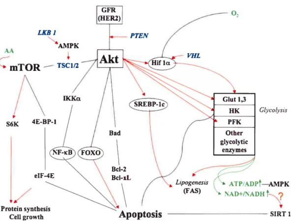
predisposing factors for breast cancer (PTEN and LKB 1) are also linked to nutrient uptake and the regulation of metabolism. Their place in the network of nutrient sensing and signaling for survival is shown inFigure 3.
1.3.3 Non-geneticjàctors involved in breast cancer deveÏopment- estrogens
Breast cancer is a heterogeneous disease in its clinical, genetic and biochemical profile. It arises from the epithelium of the mammary gland (milk producing lobules and ducts, whicb transport rnilk to the nipple). Malignant transformation of the stromal components (fibroblasts, endothelial ceils forrning blood vessels and adipocytes) is very rare and it is flot included in this category [20]. There is evidence that estrogens, the steroid hormones essential for development and function of a normal mammary gland, play an important roTe in the development of breast cancer. Thus, the risk ofthe malignancy is related to the cumulative exposure to endogenous and exogenous estrogens and includes: early menarche, tate age of menopause and honiional therapy after menopause.
Estrogens are steroid hormones produced primarily by ovaries, with some contribution from the placenta, adipocytes and adrenal glands. They act mainly through two nuclear receptors ERa and ER3, which are ligand-inducible transcription factors and are both expressed in breast tissue [30]. Two thirds of ail breast cancers express ERa, which is believed to be responsible for tumor celis proliferation [31]. Curiously, however, the proliferating cells in a normal marnmary gland rarely express steroid hormone receptors t31 ,32]. This apparent paradox is stili the subject of debatc [33, 34]. Nevertheless, antiestrogen therapies are being developed and have been found to be very effective for treatment of this mahgnancy as well as successful in chemoprevntion for high-risk patients, such as those showing abnormnal breast histology or carriers of a genetic predisposition [21, 35]. Cunent antiestrogene strategies include: (a) antiestrogens, such as tamoxifen, that inhibit estrogen binding to its main target, ERa, (b) aromatase inhibitors, that prevent synthesis of endogenous estrogens, and (c) pure antiestrogens, such as flulvestrant, that block the action ofestrogens [36].
10 Although very successftil, endocrine therapies are still the subject of intensive research for at least two reasons:
(1) They have to be designed in such a way so that they do flot interfere with other aspects of women’s health. This is especially true for long-term chemopreventive therapies. Estrogens influence physiology of reproductive and cardiovascular systems, metabolism of bones and the integrity of the central nervous system. The new category of therapeutic agents currently being devetoped for breast cancer treatment, selective estrogen receptor modulators (SERMs), may be suitable for endocrine therapy [36, 37]. These are nonsteroidal agonists/antagonists of estrogens action. It is anticipated that the effects of these drugs may vary depending on the target tissue. Indeed, tamoxifen, a widely used breast cancer therapeutic drug, is the first of this type. It acts as an antagonist in the breast to prevent breast cancer progression and as an agonist in the bone to preserve bone density.
(2) The second challenge facing endocrine therapies is the development of resistance. Some ER-positive breast cancers do not respond to antiestrogen therapy (intrinsic resistance), whil e others stop responding after long-terrn therapy (acquired resistance). Interestingly, tumors that acquire resistance (30 to 50 % of treated ERa positive tumors) retain the expression of the receptor. Resistance to endocrine therapies is a complex phenomenon, which may involve many different mechanisms [38]. For example, the direct interaction between signaling through ERa and several other transduction pathways can convert the inhibitoiy effects of the tamoxifen-ERa complex into a stimuÏatoiy effect. Cross-talk between signaling from ERa and HER2/neu (EGFR type receptor and the common oncogene in breast cancer), rnight be involved in dnig resistance of tumors overexpressing this ceil surface receptor [36, 39] . In addition, ERa activity could be reguÏated by phosphorylation mediated
directÏy by Akt [40]. The signaling from HER2/neu involves the PI3K/Akt pathway activation as wel[. As discussed later in this chapter, this pathway is implicated in the upregulation of glucose uptake and the stimulation of lipogensis in many tumor ceils. Interestingly, there are indications that interfering with lipogensis by inhibiting fatty acid synthase (FAS), the final enzyme responsible for the synthesis of fafty acids (FA), can alter upstream signaling form ERa. This suggests that lipid metabolisrn
itself may be an important factor involved in the estrogen response of breast cancer celi [41].
1.3.4. Breast cancer as a climnic disease—cancer dormancy
Ihe risk ofrecurrence for cancer patients is the highest within five years after diagnosis and more then haif of the patients develop metastasis during this time. Breast cancer belongs to the srnall group of cancers that have a relatively unusual risk of late recurrence, even 20 years after diagnosis. The other cancers belonging to this group are melanoma, non-Hodgkin’s lymphoma and renal carcinoma [42]. The early relapse and late relapse seem to occur through different mechanisms. The characteristic feature of a late relapse is that it occurs in two stages, an early stage, when there is no expansion of tumor ceils and the late stage characterize by exponential tumor growth. The earty phase of relative tumor quiescence followed by late recurrence bas been called cancer or tumor dorrnancy[42]. Cancer dormancy is flot well understood and it is extremely difficuit to study due to the veiy low numbers of doniiant cancer ceils. By applying sensitive irnmunocytochemistry, one can detect disseminated tumor ceils (DTC; one cell per bone marrow aspirate) in the bonemarrow of20-40°/ of cancer patients that do flot show signs of metastasis [43]. Other very sensitive methods can identify circulating tumor ceils (CTC) from the blood. They allow the detection of I celi per 20 ml of blood in about 36 % of cancer patients that do not show clinical signs of disease [42]. The diagnostic value of DTC and CTC detection is not yet clear. Considering that about 20 % of patients will relapse after long-term remission, the phenomenon is worth serious consideration. The late relapse has a stochastic characteristic and currently there are no tests that can predict which patients are at risk for recurrence. Persistence of DTC or dc in the body of cancer patients suggests that cancer may be considered as a chronic disease.
We have already mentioned that environmental and life-style factors may be involved in breast cancer development. Considering the possible chronic character of the disease, prevention may be an important therapeutic option. Consistent with this
12 view, the life-style factors, such as obesity. diet and exercise, were shown to markedly affect breast cancer risk and survival after diagnosis.
1.3.5 Obesity and the risk of devetoping cancer
EpidemiotogicaÏ studies suggest that obesity is a metabolic disorder that affects the development of many different types of tumors. including colon, breast (postmenopausal), endometrium, kidney (renal ce!!), oesophagus (adeno-carcinoma), gastric cancer, pancreati c, gallbladder, I iver, non-Hodgkin’ s iymphoma, leukemia, multiple myeloma, rectum, ovary and prostate [reviewed in 44, 45]
Although the molecular mechanisms linking cancer promotion with obesity are sti!! not understood, the association is strong. Thus, obese individuaÎs with a body mass index above 30 kg/m2 (BMI, defined as weight in kiiograms divided by height in meters squared) have an overali increased risk of developing manytypes of cancer ofapproximately 1.5 to 2 fold and an over 3 fold increased risk ofdeveloping cancers of the endometrium and oesophagus. Overweight postmenopausal women have a 1.66 fold increased risk of developing breast cancer [45]. The parameters indicating metabolic dysfunction, such as insulin resistance, visceral adiposity, hypertriglyceridemia and hyperglycemia, were ail shown to correlate with an increased risk of developing breast cancer [46-49]. Overweight women have not only an elevated risk of developing breast cancer, but also an increased chance of cancer recurrence (1.8-1.9 fold) and increased mortality (1.4-1.6 fold) [50 reviewed in 51]. Obesity appears to be strongly related to mortality in women with estrogen receptor-negative breast cancers, for which there exist fewer therapeutic options [52].
1.3.6 Excessive adipositv and its contribution to cancer
Fat deposits in different anatomical sites of obese individuals are unequal. Upper-body fat, including visceral and abdominal subcutaneous deposits, strongly coiielates with increased risk for the development of insulin resistance, diabetes and cancer [47]. The accumulation of fat in the upper body is controlled by various factors, including heredity (about 50%) and sex (more common in males than in
fernales), and is closely associated with glucose intolerance, hyperinsulinemia, hypertriglyceridemia and other features of what is called metabolic syndrome or syndrome X [53]. A causal Iink between visceral obesity and various disorders has been difficult to determine. There is evidence that visceral adipose tissue is more dynamic and has a higher lipolytic activity. The increased release of free fatty acids (fFA) from visceral adipose tissue to the portal vein, which drains directly into the liver, could cause liver dysfunction. However, it is also likely that visceral obesity is not harrnful by itself but is a sign of an underlying metabolic phenotype, which manifests itself in an altered adipose tissue distribution [reviewed in 53].
It bas been recognized that the storage and release of fat is not the only function of adipocytes. These cells also participate in physiological horneostasis through the production of hormones and cytokines, which can act in autocrine, paracrine or endocrine fashion (for examples: leptin, resistin, adiponectin, tumor necrosis factor a (TNFa), interleukin-6 (IL-6) and apolipoproteins [54]. The endocrine function of adipocytes in obese individuals may well be one of the factors contributing to the development of neoplasia [44]. This is particularly true for brcast cancer in postrnenopausal women, since their adipose tissue is also the main source of estrogen [55]. Adipocytes are major components of human breast tissue, making up about 90 % of their volume. It bas been shown that in postmenopausal women, local estrogen Levels in breast tumors could be as much as 10 times higher than in the circulation [56]. Obesity-related breast cancers are also more often ER-positive [57]. It was proposed that various signaling pathways activated by adipocytes may crosstalk with each other and with signaling from the ER and synergistically promote tumorigenesis [55].
Obesity is also independently positively linked to elevated blood ffA levels, which have been irnplicated in the development of pathologies such as insulin resistance and tissue lipotoxicity [58 reviewed in 59, 60, 61].
14 1.3.7 Metabolic inter’entions (diet and exercise) can ilnprove sttn’ival in breasi
cancer patients
Metabolism can affect the progression of cancer. The recent studies showed that dietary interventions such as a decrease in dietary fat or the promotion of energy expenditure by means of an increase in physical exercise can improve survival in postrnenopausal women who have been treated for early-stage breast cancer. Remarkably, only a few hours of exercise a week (3 to 5 hours of walking) can reduce the risk of death from breast cancer by up to 50% [62] and decreased dietary fat (to 20%, from about 40% of total calories from fat, which reflects the typical Western diet), can reduce the risk of tumor recurrence for ail breast cancer patients by 24%, and for patients with estrogen receptor-negative breast cancer by 42% [63]. The efficacy of these interventions is comparable to that of established adjuvant therapies. Thus, the risk reduction of recurrence after hormone therapy is estirnated at about 50% for estrogen receptor-positive cancers [64] and treatrnent with Trastuzumab, the monoclonal antibody inhibiting the activity of HER2/neu tyrosine kinase receptor (often amplified in breast cancers) reduces mortality by 33% [65].
1.4 Metabolism and the contro] of celi survival
1.4.1 Growth factors regulate cell survival be inhihiting apoptosis and by directly controlling cell access to nutrients
The nutritional environment of most celis in the body of heatthy individuais is highiy regulated. The individual celis within the body are usuaiiy not limited for growth by the extracellular concentration of glucose, fatty acids (FA) and other nutrient substrates. ihus, cell survivai, growth and proliferation are mainly controiied by the signals from exogenous growth factors (GF) rather than nutrient substrates [66]. CelIs within multicellular organisms are constantly exposed to numerous signais from their surroundings, including soluble factors, signals from the extracellular matrix or the signais from neighboring cells. GF signaling is responsible for the balance between ccli accumulation and cell death within tissues.
Individual ceils require GF signaling to maintain their surviva! and in the absence of those permissive signais they undergo apoptosis. They also require GF to initiate ce!! division. During tumorigenesis, individual celis acquire mutations in the signaling pathways that a!!ow them to avoid apoptosis and proliferate in the absence of GF [6]. Recently, Thompson et al. showed that Gf also contro! the access to extrace!!uiar nutrients in individuai ceils. They showed that when ceiÏs are withdrawn from GF, the rates of uptake of glucose and amino acids decrease and the transporters for iron (transferrin receptor), as wel! as cholesterol (LDL receptor), are also down regu!ated [67, 68]. Finally, they dernonstrated that when Rab7 (which regulates endocytic membrane traffic and mediates the intemalization and degradation of nutrient transporters) was inhibited, GF-deprived celis displayed protonged, growth factor independent ce!1 surviva!. Therefore, GF reguiate celi survivai by inhibiting apoptosis as we!1 as by direct!y control!ing ce!! access to nutrients [69].
1.4.2 Signating via Akt serine/threonine kinases regutates celi survivat by severaÏ mechctnisnis
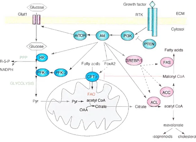
Thompson et al. hypothesized that cancer ceils have to acquire autonomy for uptake of nutrients in order to become ful!y transforrned. They showed that activation of the Akt farniÏy of serine/threonine kinases, often triggered by oncogenic alterations in GF signaiing, promotes increased nutrient uptake [70, 71 reviewed in 72]. Thus, they were the flrst to reveai that Akt can contro! celi survival by contro!!ing ce!!ular metabo!ism.
Akt serine/threonine kinases are activated in many types of cancers and this activation leads to enhanced resistance to apoptosis [73]. Signaling via Akt can induce ce!! growth and division in addition to survival. Sorne of the survival signaling pathways overlaps with the mitogenic pathways, but some are distinct. A recent review on ceil cycle reguiation by Akt was published by Brazi! et al [74] and reviews on regu!ation of ce!l survival by Akt signaling can be found in Harnrnenrian et al. and in Mc Corniick [75, 76]. Figtire 3 briefly summarize the cuiTent know!edge regarding Akt induced regulation of ce!! surviva!, and can be described as fo!lows. Akt can be activated by signais from various tyrosine kinase receptors in breast
16
cancers, sucli as the HER2 oncogene. Activated Akt increases ce!! survival by inhibiting various proapoptotic targets, such as TSC1/2, IKKa, FOXO and Bad [761. In addition, Akt activation promotes ce!! survival by stimulating ce!!ular g!ucose and fatty acid metabolism. Akt activates g!ycolysis, presumably by stabilizing HIF-1 transcription factor [77], which in turn is responsib!e for the upregulation of a!1 the glycolytic enzymes (for detailed discussion see section 1.5.2). In addition to this, Akt may directly activate the expression of several enzymes invotved in metabolism of glucose, including glucose transporters, hexokinase [7$] and phosphofructokinase 1 and 2 [79]. The intracellular metabolism of glucose and FA are tightly intercomiected (for detai!ed discussion sec section 1.6.3). Akt activition of glycolysis will resuit in the production of more substrate for FA synthesis. Thus, Akt was shown to activate enzymes involved in FA synthesis via SREBP-! [$0] and it can direct!y phosphoiylate ACL, the enzyme cata!yzing the first step in conversion of glycolytically-derived citrate to cytosolic acetyl CoA ta precursor of FA, cholesterol and isoprenoid synthesis) [8!]. There is evidence that upregulation of FA synthesis itself could be important for cancer ce!! surviva! but the mechanisms are not yet understood [$2].
.4.3 CeÏls can sense changes in the ÏeveÏs ofinani’ nutrients and cciii adapt their
metaboÏism
As discussed above, the ce!!ular metabo!ism of g!ucose and fatty acids may be directly up regulated by growth factors to promote the survival of pro!iferating ceils. However, it lias long been recognized that individual ce!ls are a!so able to sense their metabolic status independent of GF. Such homeostatic responses are evolutionarily conserved [$3]. For example, a deciine in the ce!lu!ar ATP/ADP ratio resuits in activation of AMPK, which tums on ATP-generating catabolic pathways while turning off ATP-consuming processes (ccl! growth) (Figure 3).
Activated AMPK can adjust the activities of various metabo!ic enzymes by phosphorylation and may also modulate intrace!!u!ar signa!ing for ccli growtb and division [84]. A recent discovery is that celis may also sense their NAD7NADH ratio. It was demonstrated that the yeast !ongevity protein Sir2 (sirtuin) is a NAD
dependent histone deacetylase [$5]. Rernarkably, sirtuins are highly conserved in evolution, and are implicated in the controt of metabolism and lifespan in yeast, wornis and flues, particularty in the effects of calorie restriction (CR) on longevity [86]. Mammalian sirtuin (SIRIY) inhibits apoptosis by direct deacetylation of
FOXO, p53 or Bax-binding partner Ku-70 [87,8$]. The exact mechanism of regutation of sirtuins in humans is not wetl understood and its coimection to NAD/NADH ratio stiil needs to be clarffied.
Another protein involved in sensing and communicating the metabolic status of celis is mTOR (Figure 3), the mammalian kinase which is a target of rapamacin.
mIOR is located downstream ofAkt. h is known to respond to arnino acid starvation
and regulates protein translation. However, mTOR also integrates signais regarding celiular metabolism and energy status by communication with AMPK and by directiy responding to ATP leveis. In addition, mTOR controls aspects of glucose homeostasis and recent studies suggest that it may aÏso control fat metabotism [reviewed in 89]. The physiological function of mTOR is the control of cellular
growth (increase in celi mass). mTOR is also involved in the response to starvation by controlling degradation of cellular content, including organelles, to ensure survival under nutrient-depleted conditions, a process known as macroauthophagy. The yeast TOR protein can forrn two types of complexes, TOR complex 1 (TORd) and TOR complex 2 (TORC2). TORCI is sensitive to rapamycin and it regulates the timing of ccli growth. TORC2 is insensitive to rapamycin and regulates spatial growth (where ceil wilÏ grow). One interesting aspect of mTOR signaling is the recently discovered existence of a negative feedback loop from nutrient sensing to the insulin-responsive Akt signaling pathways. Thus, mTOR is emerging as a very important factor in the control of flot only ceil growth but also ceil survival and metabolism [89].
1$
Glycolvsis
SIRT 1
Figure 3. Akt signaling for celi survival
Akt inactivates by phosphorylating proteins, which are directly or indirectty involved in the induction of apoptosis (Bad, IKKa, FOXO). Akt signais through the mammalian target of rapamycin (mTOR) by inactivating its repressor, tuberous scierosis complex (TSC1/2). TSC1/2 is a tumor suppressor mutated or deleted in several cancers [90]. Interestingly AMPK can activate the complex having an antagonistic effect on mTOR function. Akt activates directly or indirectly glycolysis and lipogenesis. This resuits in changes in the leveis of important metabolites (AMP, NAD+), wbich can signal back via proteins sensitive to their levels (AMPK and SIRT1 respectively) to modulate metaboiism and/or celi survival. Hexokinase (HK) directly prevents apoptosis by binding to the mitochondriai outer membrane. Black unes indicate inhibition and red airows indicate stimulation. Transcription factors are encircled, metabolites are green and tumor suppressors are in blue.
LKB 1 AA AMPK
o,
mTOR
— TSC1/2 VHL p S6K Protein synthesisCeli growth
Apoptosis
h Lzpogczeszs ATPADP4—AMPK
(FAS)
1.4.4 Some inetabolic enz’ines direct!y participate in signaling for ce!! sun’ival The interesting aspect of upregulation of glycolysis in the context of the control of ccli survivai is that glycolytic enzymes themselves can directiy participate in antiapoptotic effects. The best-known exampie of this is hexokinase (HK) (Figure 3), which binds with high affinity to the outer mitochondriai membrane and interacts with other mernbrane-associated proteins, including VDAC to inhibit apoptosis [91, 92]. In fact, rnany enzymes involved in glycolysis have additional, nonglycolytic functions, which include transcriptionai reguÏation, stimulation of ccii motiiity, apoptosis [reviewed in 93], and even DNA repair [94]. Glycolysis is one ofthe rnost ancient metabolic pathways, and these recently discovered nonglycolytic functions of glycolytic enzymes may reflect an evoiutionaiy strategy, designed to coordinate rnetabolism with other ceilular functions.
1.5 New concepts regarding the role of glucose metabolism in tumorigenesis
1.5.1 Glucose metabolisin is ttp regttlatedin tuinor celÏs: the Warbttrg effect
Cancer ceiis show major changes in the rnetabolisrn of glucose. While the majority ofnon-transforrned ceiis use oxidative phosphorylation in mitochondria for energy production and switch to glycolysis oniy upon oxygen deprivation (hypoxia), tumor ceils show high glycoiytic rates and rely on glycolysis for energy production even in the presence of oxygen. Up-reguiation of both glucose uptake and giycoiysis are common features of many cancers and have application in noninvasive cancer imaging used for diagnosis and staging of turnors by positron emission tomography (PET) [95]. This test uses radioiabeled glucose analogs to detect differentiai glucose uptake by fast-growingtumors and metastases.
The reason for the metaboiic switch to giycolysis in the presence of oxygen in tumors, the so-caiied “Warburg effect”, is stili not ciear. Over 70 years ago Warburg proposed that the effect is a resuit of mitochondriai dysfunction [96] and developed a cancer theory based on altered metabolism [97]. However, according to the generally accepted views, aiterations in turnor ccii metaboiisrn are symptoms of
20
transformation or response to the turnor’s microenvironment rather than a cause of cancer. Thus, the Warburg’s theory was neyer accepted.
1.5.2 The transcrttion factor HIf-1 coordinateÏy reguÏates an integrated response
to Ïow oxygen and a switch to high glvcolysis
Growing tumors are often hypoxic due to insufficient blood supply. Thus, up regulation of glycolysis could occur in response to hypoxia and could be an adaptation to hostile tumor environment. The ceilular response to oxygen deprivation is controlled by the transcription factor: hypoxia-inducible factor 1 (HIF-1), which regulates a rnuitiplicity of genes, including those coding for ail of the giycoiytic enzymes, and others encoding paracrine growth factors such as vascular endotheiial growth factor (VEGF). Activation of these genes assures an integrated response to Iow oxygen, including a switch to high giycolysis and recruitment of new biood vessels [9$].
1.5.3 Tumor suppressor genes and oncogenes are invoÏved in the upregiclation of glvcoÏys’is: the Warhttrgeffect re-examined
HIF-1 is degraded in the presence ofoxygen. Disruption ofHIF-l’s oxygen dependent degradation leads to its constitutive activation. Interestingly, recent data shows that HIFI could be activated by factors other then tumor environment and that it may possibly play a crucial role in the process of transformation [99]. First of ail, it was demonstrated that genes involved in the control of HIF- 1 degradation function as ciassicai tumor suppressors ta germiine mutation in one aiiele predisposes carriers to develop tumors in specific organs). Three genes of this type were identified: the von-Hippel-Lindau (VHL) gene, which predisposes carriers of gerniline mutations to kidney, blood vessel and adrenal tumors, and two genes encoding enzymes of the tricarboxylic acid cycle: succinate dehydrogenase (SCD) and fumarate hydratase (Fil), which predispose calTiers of germtine mutations to hereditary paraganglioma and Ieiomyomato sis/renal ccli cancer syndromes, respectively [reviewed in 100, 101]. Moreover, HIF-l could be detected in non
growth factor, such as insulin-like growth factor (IGF), or in ceils transformed by oncogenes including v-Src, c-Src, ras or HER 2 [reviewed in 103].
Stabilization of HIF-1 is flot the only way in which ceils can acquire higli
glycolytic rates. Some oncogenes can directly activate glycolytic enzymes. So far two examples are known: MYC and Akt oncogenes. In the case of MYC, it was shown that several key glycolytic genes have highÏy conserved Myc binding sites. Myc directly binds to their promoters and transactivates them in non-hypoxic conditions [104]. The exact mechanisrn involved in activation of glycolysis by Akt is flot known, but it appears to be independent of HIf-1 [70, 105]. Akt is involved in redirecting cellular metabolism in response to growth factors stimulation or oncogenic alterations to support ce!! growth and proliferation [reviewed in 72].
The present views on up-regulation of glycolysis in cancer ceils are surnmarized in Figure 4 [103]. Thus, it seems clear that the up-regulation of glycolysis that is so often observed in cancer celis may flot be just an adaptive phenomenon, since it could be autonomously acquired during tumor progression. Its contribution to tumorigenesis, however, is stiil controversial and flot widely accepted [106].
22 Glucose uxterriail GLUT ,—GCUT Glucose inlrsa) ‘ /7 GlUcoseHK
D;t£5,CSYE,vioday D:scaze?Schr,vts
Figure 4. CeIl autonomous oncogenic alterations and adaptation to hypoxia contribute to tumor aerobic gIyco1ysi
The figure denotes a tumor mass that could have sustained genetic alterations that activate either AKT or MYC, resulting in ceil autonomous activation of aerobic glycolysis. In the case of MYC, which encodes a transcription factor, glycolytic genes are directly activated. As the tumor mass continues to enlarge, diffusion limitation causes local hypoxia that induces the HIF- 1. HIF- 1, in turn, activates the glycolytic genes as well as factors such as VEGF, which induces angiogenesis. The glycolytic pathway is shown on the right. Genes affected by either HIF-l or MYC are indicated by unes. For MYC, the thickness represents the level of direct binding ofMyc to glycolytic genes.
Source: Kim, J.W., Gardner, L.B. and Dang, C.V. (2005) Dnig Discovery Today: Disease Mechanisms 2 (2): 233-238. Celi aut000moos r MYC r AKT -S --J / / GP5 / Frr’to,p 6-phnsphate -__— PFrÀ’ HIE-1 : trtjcijse -: -, Â sLDA’ y flihVOr)xyacctoccphcph1 t°i MYC Adptee I 3-phospho’e — — SAPDH — — O, r Ji rI _lycea’s ECK —--. I ii 1 ert VEGF PGM ,‘ ru I rstr /‘ /
•
\
CNO1 °hosphoe ilpvrutatc’/ Pyruvate . LDHA/
, Anqiogenesis1.6 Overview of FFA metabolism and ïts regulation
1.6. 1 Control offfA Ïevels in the bÏood
The level of FFA (those which are flot components of circulating triacylglycerols, Figure 5) in the blood is highly controÏÏed between 0.1 - 0.2 mM
[107]. After a meal, FFA released from dietary fat are absorbed by enterocytes that une the smali intestine [102]. Inside the enterocytes, they are esterified to form triacylglycerols (TG) and then are exported into the circulation as chylomicrons. They are released from chylomicrons after hydrolysis mediated by lipoprotein lipases (LPL), which are produced by adipose tissue and muscles, and then secreted to the surface of capillary endotheliat celis nearby. In fed state, the majority of FfA released from chylomicrons will be taken up by adipocytes and immediately re esterified to form storage TG inside the ceil. Thus, the multistep process of trapping dietary FFA inside adipocytes consists of sequential cycles of esterification and lipolysis. In normal individuals, this complex process is almost 100% efficient 1 h afier a meal and the efficiency then decreases to 10-30% by 6 h [109]. This allows for a graduai release of FFA into the blood at longer times after the mea!. Thus, the
FfA levels in the blood are highest in a fasted state (over-night fast) when they are
needed as a fuel in various tissues. Lipolysis in adipocytes is calTied out by the highly regulated enzyme hormone sensitive lipase (HSL) and other associated lipases [110, 111].
Obesity is often linked to insulin resistance, which is flot well understood, and manifests itself in an inability of various tissues to respond to insulin signaling [reviewed in 112]. In the obesity-induced insulin resistant state, trapping of dietary FFA in adipocytes becomes inefficient, causing elevated blood FFA levels even in the fed state. In this situation, higher than normal amounts of FfA are available to rnany tissues and organs, even though they do flot need them for energy production [54]. Little is known how these tissues deal with the surplus ofFFA fuel. For human tissues other than adipocytes, liver, skeletal muscles, heart and pancreatic 3-ceHs. there is littie or no data describing the metabolism of FFA, their rate of oxidation,
24
esterification, formation of lipid stores or lipolysis in the healthy or obese states. Considering the profound influence of obesity, diet and life-style on the developrnent of breast and other types of cancer, as welI as development of metabolic syndrome and diabetes, more knowledge is needed regarding Ff A metabolism in different normal and tumor tissues.
A C o A/AA/V’V\. -oc. C ______ At,/J\.t\/\/\ 1_oc C CH c I4pho,, td . fi Hdrophilk h.d IOi Ni PIIOSPHOLIPID IPL)
Figure 5. Structure of selected fatty acids and common complex lipids
A, Model molecules and chemical structure of common FA: palmitate (C 16:0) and
oeate (C18:1) B, Structure of most common polyunsamrated fatty acids: linoleic
acid (C1$:2) and linolenic acid (C1$:3) C, Structure of triacylgtycerol (TG) D, Structure of phosphatidylcholine, a common membrane glycerophopholipid.
Source: Lodish H, Berk A, Zipursky L, Matsudaira P, Baltimore D, Darneil J: Molecular Celi Biology. Fourth edition. W.H. Freeman and Company (2000)
•_\ ,—,—,—
:E*
1*,
sc c C ç c -c c C- C—C C C-c C-C C C
COMMON EATTY &CIDS TRI AClGLY CEROL (TG)
B D
f.iflyacvI çt.ail,
12 9
/%.\/\\/==\/==\\/\\J/\//\/COOH
CI)
Linoleic AciLi (onieçja-6)
Aipha-Linolenic Acidtomeua-])
1 .6.2 Intracellular metaboÏism ojexogenous fFA: introduction
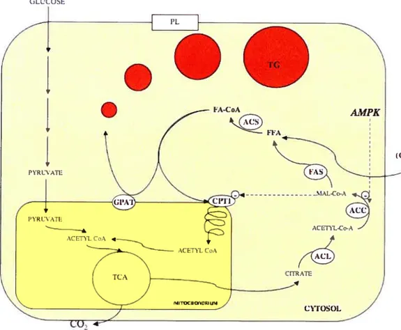
FA are the simplest lipids, consisting of long aikyl chains with a terminal carboxyl group. Most FA in human body exist in complex form, as storage TG as wetl as structural phospholipids (PL) (figure 5). Circulating FFA (those which are flot components of complex lipids) are bound to serum albumin and their level in the blood is tightly controlled [107]. The transport ofFfA into celis is believed to occur via passive diffusion as well as by protein mediated transmembrane transport [113, 114]. Once inside the ce!!, FFA are activated to the corresponding fatty acyl-CoA (FA-CoA) by acyl-CoA synthetase (ACS). The fate of FA-CoA subsequently varies between tissues and depends on the overait metabolic state of the body as wel[ as on the individual needs of different specialized ceils [115]. When esterified with glycerol-3-phosphate (G3P), FA-CoA are preserved in the form of storage TG or can be chaimeled to form phospholipids. Enzymes, which initiate the esterification process, are called G3P acyl transferases (GPAT) [116]. Alternatively, FA-CoA can be transported to mitochondria by carnitine palmitoyl transferase 1 (CPT 1) and oxidized, generating ATP and CC2 [117]. Thus, FA, like glucose, are fuel substrates.
1.6.3 Coordinate regutation of glucose and FFA metaboflsm in the bodv and within an individual ce!!
The metabolic pathways of the fuel substrates: FFA and glucose are interdependent and reciprocally regulated. Utilization of glucose and FfA by different tissues within the body is largely coordinated by insulin but the fine-tuning is brought about by various intracellular mechanisms. Hence, elevated glucose concentration stimulates pancreatic f3-celts to secrete insulin, which suppresses lipolysis in adipocytes, preventing release of FFA. Ibis eliminates competition for fuel substrates in peripheral tissues (muscles). Thus, in healthy individuals, FFA becorne the major fuel substrate only when glucose and insulin concentrations are low [107, 11$, 119]. Fine-tuning mechanisms, which allow individual cells to sense the availability of fuels and adjust their metabolic pathways accordingly, include the inhibitory effect of etevated FFA concentrations on glucose metabotism described in
26 skeletal muscles by Randie et al [120], and the inhibitory effect of high glucose concentration on fatty acid oxidation discovered by McGarry and Foster [121]. The McGarry’s effect described in hepatocytes is briefly illustrated in Figure 6. When glucose is abundant, ceils in various tissues, in particular liver celis, have an elevated glycolytic rate and produce large amounts ofpyruvate, which enters the tricarboxylic acid (TCA) cycle in mitochondria. TCA cycle intermediates are replenished and the surplus is transported from the mitochondrial matrix back to the cytoplasm in the form of citrate. Citrate is then metabolized by a sequence of cytoplasmic enzymes: ATP citrate lyase (ACL), acetyl CoA carboxylase (ACC) and finally fatty acid synthase (FAS) to produce endogenous fatty acids. MalonyÏ-CoA, which is an intennediate in this process, acts as an allosteric inhibitor of CPT I. Therefore, the presence of malonyl-CoA signals the abundance of glucose in liver cells that begin to produce endogenous FA from glucose and shuts off oxidation of the newly forrned FFA, prornoting their esterification and storage. Inhibition of CPI I by malonyl-CoA will also prevent oxidation of exogenous FFA if they are available.
An additional level of complexity to this intracellular metabolic regulation is added by signaling from AMP-activated protein kinase (AMPK). AMPK is an evolutionarily conserved sensor and regulator of energy balance in celis. It becomes activated by phosphorylation when levels of AMP increase, indicating a reduced ATP/ADP ratio. Actïvated AMPK turns on ATP-generating catabolic pathways while tuming off ATP-consuming processes (in particular celi growth) in response to an energy crisis. Thus, it will induce oxidation ofFFA when glucose is not providing enough energy or in the situation of high energy consumption (muscle contraction) [122, 123]. lnterestingly, the upstream kinase responsible for AMPK activation is a turnor suppressor LKB1, responsible for the developmcnt ofbenign intestinal tumors (Peutz-Jeghers syndrome) and predisposing carriers to malignant cancers in other tissues (including a breast) [124]. The deregulation of the AMPKImalonyl-CoA fuel sensing and signaling network bas been proposed to be involved in the developrnent of the metabolic syndrome, which predisposes to several chronic disorders, including obesity, diabetes, hypertension and premature atherosclerosis
t
125].Exogenous FFA become activated inside the celi by ACS to form FA-CoA, which can be further metabolized in mitochondria to produce ATP. CPT-1 catalyzes the first rate-limiting step of its transport to mitochondria. Atternatively, fA-CoA can be esterified to glycerol-3 phosphate to fonri storage TG or PL. When glucose is available, it is metabolized via glycolysis and the TCA cycle to produce ATP. At high glycolytic rates, the TCA cycle will be replenished and the surplus will be transported from the mitochondrial matrix back to the cytoplasrn in the form of citrate. Citrate is a precursor for de novo FFA synthesis. MaÏony-CoA, an intermediary metabolite between citrate and FFA, is an allosteric inhibitor of FFA oxidation. Its levels can also be controlled by AMPK, which inactivates ACC by phosphorylation.
f.I.’ t OLLAlE)