-1-
BCL-3 degradation involves its polyubiquitination through a FBW7-independent pathway and
its binding to the proteasome subunit PSMB1
Aurore Keutgens1,2,3, Xin Zhang1,2,3, Kateryna Shostak1,2,3, Isabelle Robert1,2,3, Sabine Olivier1,2,3, Alain Vanderplasschen4, Jean-Paul Chapelle1,2,3, Patrick Viatour1,2,3, Marie-Paule Merville1,2,3, Françoise Bex5, André
Gothot1,6 and Alain Chariot1,2,3* 1
Interdisciplinary Cluster for Applied Genoproteomics (GIGA-Research), 2Unit of Medical Chemistry and 3
GIGA-Signal Transduction, 4Immunology-Vaccinology, Department of Infectious and Parasitic Diseases, Faculty of Veterinary Medicine, University of Liege, Sart-Tilman, 4000 Liège, 5Institute for Microbiological Research J-M Wiame and Laboratory of Microbiology, University of Brussels and 6Department of Medicine
/Hematology, University of Liege, Liege, Belgium.
*To whom correspondence should be addressed
Dr. Alain Chariot, Laboratory of Medical Chemistry, GIGA-R, Tour GIGA, +2 B34, Sart-Tilman, University of Liège, 4000 Liège, Belgium; Tel : 32 (0) 4 366 24 72; FAX : 32 (0) 4 366 45 34
e-mail : Alain.chariot@ulg.ac.be
Running title: « PSMB1 as a BCL-3 interacting protein»
The oncogenic protein BCL-3 activates or represses gene transcription through binding
with the NF-κκκκB proteins p50 and p52 and is
degraded through a phospho- and GSK3-dependent pathway. However, the mechanisms underlying its degradation remain poorly understood. Yeast-two-hybrid analysis led to the identification of the proteasome subunit PSMB1 as a BCL-3-associated protein. The binding of BCL-3 to PSMB1 is required for its degradation through the proteasome. Indeed, PSMB1-depleted cells are defective in degrading polyubiquitinated BCL-3. The N-terminal part of BCL-3 includes lysines 13 and 26 required for the K48-linked polyubiquitination of BCL-3. Moreover, the E3 ligase FBW7 known to
polyubiquitinate a variety of substrates
phosphorylated by GSK3 is dispensable for BCL-3 degradation. Thus, our data defined an unique motif of BCL-3 that is needed for its recruitment to the proteasome and identified PSMB1 as a key protein required for the proteasome-mediated degradation of a nuclear
and oncogenic IκκκκB protein.
BCL-3 is an nuclear protein that belongs to the IκB family and regulates gene transcription when bound to the NF-κB proteins p50 or p52 (1-5). BCL-3 was originally identified through molecular cloning of the breakpoint of the t(14 ;19) chromosomal translocation found in a subset of human B-cell chronic lymphotic leukemias (6). This translocation triggers BCL-3 overexpression and consequently the deregulation of many target genes that may be involved in survival and cell
proliferation (7). Since then, it has been shown that over-expressed BCL-3 contributes to the constitutive NF-κB activation seen in most solid and haematological malignancies (8). Indeed, BCL-3 overexpression has been reported in multiple myelomas and subtypes of lymphomas, even in the absence of any t(14 ;19) chromosomal translocation (9-12). Interestingly, a deregulated BCL-3 expression has also been reported in solid tumours such as breast cancers, nasopharyngeal and hepatocarcinomas, although the underlying mechanisms remain unclear (13-15). Familial cylindromatosis, a genetic disease characterized by benign tumours of hair-follicle keratinocytes (called cylindromas) results from loss-of-function mutations of CYLD, a deubiquitin ligase that specifically removes K63-linked polyubiquitin chains from various substrates, including BCL-3 (16,17). As a result, the nuclear import of K63-linked polyubiquitinated BCL-3 is enhanced and cell proliferation occurs through cyclin D1 gene induction (17). Therefore, multiple mechanisms that include chromosomal rearrangements or mutations of BCL-3-associated proteins ultimately trigger BCL-3 overexpression in the nucleus, a key event in BCL-3-mediated oncogenesis. This IκB protein has been formally defined as an oncoprotein based on the ability of BCL-3 expressing NIH3T3 cells to form foci and induce tumour growth in nude mice and also because bcl-3 transgenic mice developed lymphoproliferative disorders (18,19).
To gain more insight on BCL-3 functions, we conducted yeast-two-hybrid analysis and defined the proteasome subunit PSMB1 as a BCL-3-interacting protein that binds an unique sequence within the N-terminal domain of this oncoprotein.
at UNIV DE LIEGE-MME F PASLE, on August 2, 2010
www.jbc.org
This interaction is required for BCL-3 degradation, as evidenced by the stabilization of BCL-3 in PSMB1-depleted cells. Therefore, our data defined PSMB1 as a key protein required for the proteasome-dependent degradation of a nuclear IκB protein.
Material and Methods
Cell culture, biological reagents and treatments 293, HeLa and Karpas cells were cultured as previously described (18).
LiCl, TNFα and MG132 were purchased from Sigma (St-Louis, MO), Peprotech (Rocky Hill, NJ) and A&E Scientific (Marcq, Belgium), respectively. Polyclonal anti-HA, -Hsp90, -Myc, -BCL-3, -IκBα and monoclonal -αtubulin, Myc, PML, and -phospho-IκBα antibodies were purchased from Santa Cruz Biotechnologies (Santa Cruz, CA). Monoclonal anti-FLAG antibodies and beads were purchased from Sigma. Monoclonal anti-NBS1 antibody was from BD Biosciences (Franklin Lakes, NJ). The polyclonal anti-PSMB1 antibody was from Biomol (Plymouth Meeting, PA). Polyclonal anti-p50 and monoclonal anti-p52 were from Millipore (Temecula, CA). GFP and PSMB1 siRNAs were purchased from Dharmacon (Lafayette, CO). pMT2T p50, pMT2T p52, pMT2T BCL-3, pMT2T BCL-3 K13-26R, pMT2T BCL-3 MTS, pMT2T BCL-3 ∆N (which lacks the first 112 amino acids), FLAG-BCL-3, -∆N, -∆C, -∆N∆C and MTS were previously described, as were FLAG-IκBα and FLAG-IκBα NLS MT (1,18,20). FLAG-BCL-3 ∆N10, -∆N30, -∆N43, -∆N67, -∆N92, and -∆N120 constructs were generated by PCR whereas FLAG-BCL-3 ANK M1, -M12, -M123 and BCL-3 5KR mutants were generated by mutagenesis. The BCL-3 ∆67-92 mutant that corresponds to the BCL-3 ∆PSMB1 mutant was generated by subcloning the cDNA sequence corresponding to aa 1 to 66 and subsequently the domain corresposnding to aa 93 to the stop codon of BCL-3 into the pcDNA3-FLAG construct. FLAG-BCL-3 S394/398/419A was generated by mutagenesis using the FLAG-BCL-3 MTS mutant as template. Myc-PSMB1 was generated by PCR amplification and subcloning into the pCMV-Myc vector (Invitrogen, Carlsbad, CA), using the PSMB1 cDNA clone pulled out from yeast-two-hybrid analysis as template. HA-Ub, Ub K63R Myc were previously described (21). Myc-FBW1 was a gift from Dr. Nakayama (Department of Molecular and Cellular Pathology, Medical Institute of Bioregulation, Kyushu University, Japan) whereas FBW7 and
Myc-FBXW8 were generated by PCR using FLAG-FBW7 (a gift from Dr. Clurman, Department of Medicine University of Washington School, Seattle, USA) and HA-FBXW8 (a gift from Dr. Jung, Institute für Pathologie, Bochum, Germany) as template, respectively.
Yeast-two-hybrid, Immunoprecipitations and Immunofluorescences
The bait was generated by subcloning the cDNA sequence of BCL-3 coding for the first 112 N-terminal amino acids into the pGBKT7 vector (Clontech, Mountain View, CA) and yeast-two-hybrid experiments were conducted as previously described (21), as were immunoprecipitations involving ectopically expressed proteins (18) as well as immunofluorescences (22).
Results
The N-terminal domain of BCL-3 harbours a nuclear localization signal dispensable for the
interaction with the NF-κκκκB proteins p50 and p52
Both the N-terminal and the C-terminal domains of BCL-3 were previously defined as key regions for the regulation of the transactivation potential of this oncoprotein, yet the underlying mechanisms remain unclear (1). To learn more on the functions of these domains of BCL-3, we generated a variety of mutants lacking sequences either in the N- or C-terminal domain and first addressed their subcellular localization by immunofluorescence (Fig. 1A and 1B). We identified a nuclear localisation signal within the first 30 N-terminal amino acids. Indeed, whereas wild type (WT) BCL-3 was mainly but not exclusively located in the nucleus (Fig. 1B), removing the entire domain upstream of the ankyrin (“BCL-3 ∆N”) or the first 30 N-terminal amino acids of BCL-3 (“BCL-3 ∆N30”) localizes BCL-3 mainly in the cytoplasm (Fig. 1B). While the N-terminal domain was required for the nuclear localization of BCL-3, its C-terminal domain was dispensable as a mutant that lacks the entire C-terminal domain downstream of the ankyrin repeats (“BCL-3 ∆C”) remained largely nuclear (Fig. 1B). Finally, removing both the N- and C-terminal domains of BCL-3 (“BCL-3 ∆N∆C”) led to an equal distribution of the resulting mutant in the cytoplasm and in the nucleus (Fig. 1B). Whereas the first 30 N-terminal amino acids were required for the nuclear localization of BCL-3, they were dispensable for the interaction with p50 or with p52 as the BCL-3 ∆N30 mutant still bound both NF-κB
at UNIV DE LIEGE-MME F PASLE, on August 2, 2010
www.jbc.org
proteins, as judged by co-immunoprecipitation analysis (Fig. 1C, top panels, lanes 5). Taken together, these data defined the N-terminal domain of BCL-3 as a key region for the nuclear localization of this oncoprotein.
The binding of BCL-3 with p50 and p52 is required for its constitutive phosphorylation
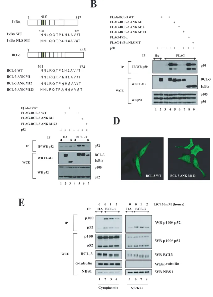
Whereas the first 30 N-terminal domain of BCL-3, which harbours the nuclear localization signal, is dispensable for the interaction with the NF-κB proteins p50 and p52, the so-called ankyrin repeats are required for binding with those proteins (1).
The sequence of BCL-3 spanning from amino acids 161 to 174 is highly similar to the one of IκBα spanning from amino acids 108 to 121 (Fig. 2A). Interestingly, this second ankyrin repeat harbours the nuclear localization sequence (NLS) of IκBα and three point mutations within this domain indeed impaired IκBα nuclear localization as well as its interaction with p65 (23). To more precisely define the role of this highly conserved domain for the interaction with p50 and p52, we next generated BCL3 mutants (referred to as “BCL3 ANK M1”, -M12, and -M123”) that harbour one, two, or three point mutations identical to those generated on the corresponding residues of the IκBα sequence (23) (Fig. 2A). We first determined whether these point mutations interfered with BCL-3 binding to p50 or p52 by co-immunoprecipitations in 293 cells. Whereas wild type (WT) BCL-3, and to a less extent BCL-3 ANK M1, associated with endogenous and ectopically expressed p50, both BCL-3 ANK M12 and -M123 failed to do so (Fig. 2B, top panel, compare lanes 4 and 5 with lanes 6 and 7). The same conclusion also applied for the ability of WT BCL-3 or BCL-3 mutants to interact with p52 (Fig. 2C, top panel, compare lane 5 with lanes 6 and 7). Of note, an IκBα mutant harbouring three mutations (“IκBα NLS MT”) did not bind as efficiently as wild type IκBα to p50 (Fig. 2B, top panel, compare lanes 8 and 9). Importantly, both BCL-3 ANK M12 and -M123 mutants were no longer constitutively phosphorylated, as judged by their migrating profile on an SDS PAGE gel (Fig. 2B, middle panel, compare lane 4 with lanes 6 and 7). Thus, our data suggest that binding of BCL-3 to p50 or p52 is required for the constitutive BCL-3 phosphorylation. Point mutations within this ankyrin repeat also had consequences on BCL-3 subcellular localization, as the BCL-3 ANK M123 mutant equally distributed in the cytoplasm and in
the nucleus while WT BCL-3 was mainly located in the nucleus (Fig. 2D). To further support the notion that the binding of BCL-3 to p50 or p52 is required for the GSK3-mediated phosphorylation of this oncogenic protein, we looked for these modified forms of BCL-3 in the cytoplasm and in the nucleus of Karpas cells left untreated or stimulated with LiCl, a GSK3 inhibitor. We noticed that BCL-3 was constitutively phosphorylated in a GSK3-dependent manner in both the nucleus and in the cytoplasm and was also bound to p52 in both cell compartments (Fig. 2E, top panels, lanes 2 to 4 and 6 to 8). Indeed, the phosphorylated forms of BCL-3 progressively disappeared upon LiCl treatment in the nucleus but also in the cytoplasm (Fig. 2E, third panel from the top, compare lanes 7 and 8 with lane 5 as well as lanes 3 and 4 with lane 1, respectively). Therefore, these data suggest that the binding of BCL-3 to p50 or p52 is the key issue for its constitutive GSK3-mediated phosphorylation, rather than its nuclear localization.
The degradative K48-linked polyubiquitination of BCL-3 requires the N-terminal lysine 13 and 26 residues
The critical role of the N-terminal domain of BCL-3 does not only result from the presence of this nuclear localization signal. Indeed, this domain is also targeted by post-translational modifications such as polyubiquitination (18). In fact, BCL-3 is subjected to both degradative (K48-linked) and non degradative (K63-linked) polyubiquitination but the signalling pathways involved in those modifications are poorly characterized (17,18). To prove that BCL-3 is indeed polyubiquitinated in vivo, we performed immunoprecipitation experiments in denaturing conditions in order to exclusively detect the polyubiquitinated forms of BCL-3 but not of the associated molecules. Polyubiquitinated adducts of BCL-3 were still detectable even in denaturing conditions that prevented its binding to the NF-κB protein p50 (Fig. 3A, compare lanes 2 and 6). As expected, MG132 treatment facilitated the detection of polyubiquitinated adducts of BCL-3, even in denaturing conditions, further supporting the notion that the proteasome is required for BCL-3 degradation (Fig. 3A and 3B, respectively, compare lanes 2 and 4). To learn more on the residues required for BCL-3 degradation, we next tested the K48-linked polyubiquitination of WT BCL-3 or the BCL-3 ∆N mutant in presence of MG132 and with an ubiquitin mutant that only makes K48 ubiquitin chains (“Ub K63R”). Removing the N-terminal domain of BCL-3 severely compromised the formation of K48-linked polyubiquitin chains on
at UNIV DE LIEGE-MME F PASLE, on August 2, 2010
www.jbc.org
this protein (Fig. 3C, top panel, compare lanes 2 and 3). As this domain harbours two lysines (residues 13 and 26), we mutated both of them (“BCL-3 K13-26R”) and addressed the consequences on the K48-linked polyubiquitination of BCL-3 in presence of MG132. The detection of polyubiquitinated adducts on BCL-3 was severely impaired upon mutation of both lysine 13 and 26 residues (Fig. 3C, top panel, compare lanes 2 and 4). As expected, the mutations of all 5 lysine residues of BCL-3 (“BCL-3 5KR”) also impaired its K48-linked polyubiquitination (Fig. 3C, top panel, compare lanes 2 and 5). Interestingly, while MG132 did not modify BCL-3 localization, the mutant that lacks all lysine residues or the GSK3 phosphorylation sites (“BCL-3 MTS”) (18) was mostly nuclear with bigger dots, as judged by immunofluorescence analysis, presumably because of an impaired degradation (Fig. 3D). Taken together, our data defined lysines 13 and 26 as key residues for the degradative K48-polyubiquitination of BCL-3.
The N-terminal domain is required for the association of BCL-3 with the proteasome subunit PSMB1
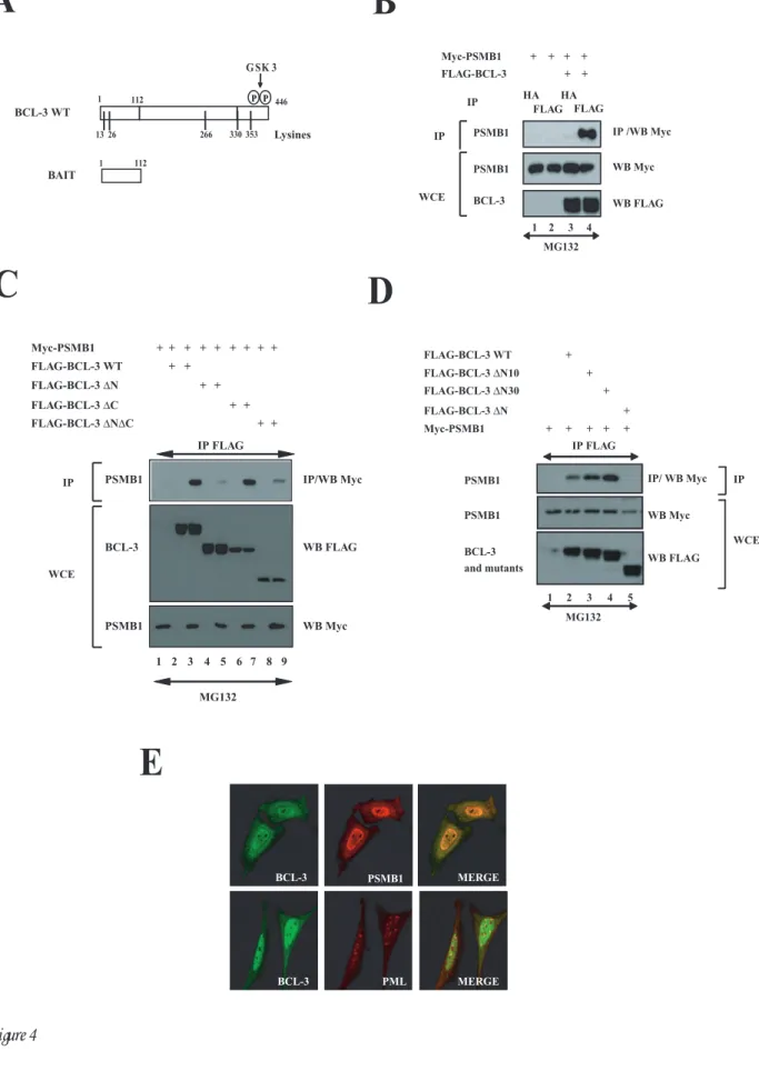
To further identify BCL-3-interacting partners involved in its K48-degradative polyubiquitination, yeast-two-hybrid experiments were conducted using the N-terminal domain of BCL-3 (amino acids 1 to 112) as bait (Fig. 4A). Positive clones included the co-repressor N-CoR, SUMO 1 (data not shown) as well as the entire coding sequence of PSMB1, a subunit of the 20S proteasome. This latter interaction also occurred in mammalian cells as overexpressed FLAG-BCL-3 associated with Myc-PSMB1, as judged by co-immunoprecipitation experiments performed in 293 cells (Fig. 4B, top panel, lane 4). The N-terminal domain of BCL-3 was confirmed to be required for binding to PSMB1 as the BCL-3 ∆N mutant failed to bind this proteasome subunit (Fig. 4C, top panel, lane 5). Interestingly, the BCL-3 products lacking the first 10 or 30 N-terminal amino acids (“BCL-3 ∆N10 and BCL-3 ∆N30”) still interacted with PSMB1 (Fig. 4D, top panel, lanes 3 and 4, respectively). Therefore, these results suggest that BCL-3 interacts with the proteasome via the N-terminal domain between amino acids 31 and 112. We next performed immunofluorescence analysis in HeLa cells and observed that ectopically expressed BCL-3 colocalized with PSMB1 mainly but not exclusively in the nucleus (Fig. 4E, top panel on the right). Importantly, BCL-3 did not colocalize with the PML bodies in the nucleus (Fig. 4E, bottom
panel on the right). Thus, the association of BCL-3 with the proteasome subunit requires its N-terminal domain, mainly occurs in the nucleus and does not involve any shuttling into the PML bodies.
PSMB1 is required for BCL-3 degradation We next assessed the role of this proteasome subunit in the degradative polyubiquitination of BCL-3 through PSMB1-depleted 293 cells. Because IκBα is also degraded through the proteasome pathway upon TNFα stimulation (24), we first determined whether PSMB1 depletion impaired TNFα-mediated IκBα degradation. Whereas PSMB1 depletion did not interfere with TNFα -mediated IκBα phosphorylation (Fig. 5A, second panel from the top, compare lanes 2 to 5 with lanes 7 to 10), IκBα degradation triggered by this pro-inflammatory cytokine was attenuated in PSMB1-depleted 293 cells (Fig. 5A, top panel, compare lanes 3 and 4 with lanes 8 and 9). Thus, PSMB1 is required for the signal-induced IκBα degradation. The half-life of BCL-3 is also extended in PSMB1-depleted Karpas cells (Fig. 5B, top panel, compare lanes 1 to 5 with lanes 6 to 10). In conclusion, PSMB1 is also required for BCL-3 degradation. A role for PSMB1 in BCL-3 degradation was further supported by the accumulation of BCL-3 polyubiquitinated adducts upon PSMB1 depletion. Indeed, we transfected FLAG-BCL-3 and HA-Ub in siRNA GFP (negative control) or -PSMB1 293 cells, isolated the nuclear proteins and looked for the BCL-3 polyubiquitinated forms in the anti-FLAG immunoprecipitates. As a positive control, we pretreated the siRNA GFP cells with the proteasome inhibitor MG132. As expected, MG132 treatment dramatically increased the amount of BCL-3 polyubiquitinated forms (Fig. 5C, top panel, compare lanes 2 and 4). Interestingly, an accumulation of BCL-3 polyubiquitinated forms was also detected upon PSMB1 depletion (Fig. 5C, top panel, compare lanes 2 and 6). Of note, this accumulation was not as dramatic as the one seen upon MG132 treatment (Fig. 5C, top panel, compare lanes 4 and 6). This may most likely be due to the partial PSMB1 loss of function as residual amounts of this proteasome subunit were still detectable in the siRNA PSMB1 cells (Fig. 5C, bottom panel, compare lanes 5 and 6 with lanes 1 to 4). In conclusion, our data provide experimental evidence for a role of PSMB1 in BCL-3 proteasomal degradation.
As the degradation of BCL-3 through the proteasome is regulated by GSK3-mediated phosphorylation (18), we next explored whether
at UNIV DE LIEGE-MME F PASLE, on August 2, 2010
www.jbc.org
GSK3 inhibition by pharmacological means impaired the association of BCL-3 with PSMB1. To address this issue, 293 cells were transfected with FLAG-BCL-3 and Myc-PSMB1 and subsequently treated with LiCl, a GSK3 inhibitor. This inhibitor indeed impaired constitutive BCL-3 phosphorylation (Fig. 5D, bottom panel, compare lanes 3 and 4) and also interfered with the binding of Myc-PSMB1 to FLAG-BCL-3 (Fig. 5D, top panel, compare lanes 3 and 4). This conclusion was also true when Myc-PSMB1 instead of FLAG-BCL-3 was immunoprecipitated in unstimulated or LiCl-treated cells (Fig. 5E, top panel, compare lanes 2 and 3, respectively). Thus, our results suggest that GSK3 phosphorylation triggers the recruitment of BCL-3 to the proteasome via binding to PSMB1. It is however important to note that the BCL-3 MTS mutant that lacks both GSK3 phosphorylation sites still bound PSMB1, although less efficiently than the wild type protein (Fig. 5E, top panel, compare lanes 4 and 2, respectively). Thus, BCL-3 may also be recruited to the proteasome through GSK3-independent pathways.
To further confirm that PSMB1 is required for BCL-3 degradation, we next generated a BCL-3 mutant that specifically lacks the PSMB1-interacting domain (“BCL-3 ∆PSMB1”) and subsequently addressed its half-life. We first deleted the first 43, 67, 92 or 120 N-terminal amino acids of BCL-3 and noticed that the BCL-3 ∆N67 but not the ∆N92 mutant bound PSMB1 in co-immunoprecipitation experiments in 293 cells (Fig. 6A, top panel on the left, compare lanes 4 and 6, respectively). Of note all these BCL-3 mutants still bound the NF-κB protein p50 (Fig. 6A, top panel on the right, lanes 4 and 6). The sequence of BCL-3 between amino acids 68 and 92 harbours the PSMB1-interacting domain as the BCL-3 ∆67-92 mutant (also named “BCL-3 ∆PSMB1”) failed to bind this proteasome subunit but still bound p50 (Fig. 6A, top panels, lanes 5). Having generated the BCL-3 ∆PSMB1 mutant, we next addressed its half-life when expressed in 293 cells treated or not with cycloheximide. Whereas WT BCL-3 almost totally disappeared after 6 hours of CHX treatment, the BCL-3 ∆67-92 remained easily detectable at that time and its level of expression was very moderately decreased after 8 hours of CHX treatment, when compared to the levels of WT BCL-3 (Fig. 6B, compare lanes 6 to 10 with lanes 1 to 5, respectively). A quantification of WT BCL-3 or BCL-3 ∆67-92 levels confirmed this conclusion (Fig. 6B at the bottom). Therefore, our data strongly suggest that this BCL-3 ∆67-92 mutant has a
prolonged half-life due to its inability to bind PSMB1. This last result thus defines PSMB1 as a key protein required for BCL-3 degradation.
The E3 ligases FBW1, FBW7 and FBXW8 are
dispensable for GSK3-mediated BCL-3
degradation
BCL-3 is degraded once phosphorylated by GSK3 on serines 394 and 398 located on its C-terminal domain (18). Importantly, we identified a phosphodegron within this C-terminal domain of BCL-3 that is similar to the ones seen in other proteins known to be phosphorylated by GSK3 and polyubiquitinated by the E3 ligase FBW7 (Fig. 7A) (25-28). Therefore, we investigated whether BCL-3 binds to this E3 ligase by co-immunoprecipitation analysis. As expected, FBW1, an E3 ligase required for TNFα-mediated IκBα degradation (29), efficiently bound this IκB protein in MG132 and TNFα-treated cells but failed to associate with BCL-3 (Fig. 7B, top panel, lanes 12 and 6, respectively). Moreover, FBW7 also failed to bind BCL-3, which indicates that this E3 ligase is dispensable for the GSK3-dependent pathway that leads to BCL-3 degradation. Of note, FBXW8, another E3 ligase, efficiently bound BCL-3 (Fig. 7B, top panel, lane 8). This binding required the N-terminal domain of BCL-3 as the BCL-3 ∆N mutant failed to bind this E3 ligase while the BCL-3 ∆C did (Fig. 7C, top panel, lanes 5 and 7, respectively). However, FBXW8 is also dispensable for BCL-3 degradation through the GSK3-dependent pathway as a BCL-3 mutant that lacks both GSK3 phosphorylation sites as well as serine 419 still bound this E3 ligase similarly to WT BCL-3 (Fig. 7D, top panel, compare lanes 3 and 5). Taken together, our data indicate that BCL-3 binds to FBXW8, yet this binding is not required for the GSK3, -phospho-dependent degradation of this nuclear oncoprotein.
Discussion
We report here the identification of the proteasome subunit PSMB1 as a BCL-3-associated protein that is critical for its degradation. This interaction requires the N-terminal domain of BCL-3, which also harbours a NLS sequence as well as the lysine residues required for the K48-linked polyubiquitination of this nuclear IκB protein. Our data therefore defined the N-terminal domain of BCL-3 as a key element for its activity.
The mechanisms underlying BCL-3 degradation remained poorly characterized. We
at UNIV DE LIEGE-MME F PASLE, on August 2, 2010
www.jbc.org
previously identified a GSK3-dependent pathway that involves the phosphorylation of two C-terminal residues (18). We now show that BCL-3 is associated to the proteasome and interacts with the proteasome subunit PSMB1 once phosphorylated by GSK3. Interestingly, the GSK3-dependent pathway is negatively regulated by Akt (18), RAS as well as by PI3K (data not shown). Functional links, that appear to be cell-specific, were previously established between NF-κB and RAS. Although the NF-κB proteins p65 and c-Rel are dispensable for RAS-induced cellular transformation in NIH3T3 cells, they nevertheless potentiate RAS oncogenic potential (30). Indeed, some NF-κB-dependent genes such as Serpin B2 and TSLP require p65 and/or c-Rel for their induction of expression by RAS (30). Of note, these genes are not induced by cytokines known to activate NF-κB (TNFα, IL-1β, …), which supports the idea that RAS induces the expression of some NF-κB-dependent genes while suppressing the ability of pro-inflammatory cytokines to activate NF-κB in fibroblasts (30,31). Another study reported that oncogenic RAS induces IKK activation and therefore IκBα degradation in liver epithelial cells (32). These studies, combined with our report, therefore suggest that multiple cross-talks exist between oncoprotein RAS and NF-κB. The ability of RAS to stabilize BCL-3 defines a mechanism by which BCL-3 expression may be enhanced even in cases where the t(14 ;19) chromosomal translocation does not occur. Elevated levels of BCL-3 in the nucleus can also be the result of loss-of-function mutations targeting the deubiquitine ligase CYLD (17). Similarly, human papillomavirus (HPV)-positive cancers such as cervical as well as head and neck malignancies also have elevated levels of nuclear BCL-3 because the HPV-encoded E6 protein inactivates CYLD under hypoxic conditions (33). It is also tempting to speculate that loss-of-mutations of E3 ligases that target BCL-3 may ultimately causes the accumulation of this protein. These results thus considerably extent the pathological contexts that ultimately lead to BCL-3 over-expression in solid and haematological malignancies.
Multiple proteins whose GSK3-dependent phosphorylation triggers their subsequent degradation can be stabilized by RAS. Among those candidates is cyclin E whose degradation requires the E3 ligase FBW7 (34). The FBW7 substrates such as c-Myc, c-Jun, Notch1, cyclin D1 and cyclin E share a so-called Cdc4 phospho-degron (CPD)
(35) that we actually found on BCL-3 (Fig. 7A). Yet, FBW7 does not bind phosphorylated BCL-3 and the half-life of this IκB oncogenic protein is not enhanced in FBW7-depleted cells (data not shown). Importantly, BCL-3 binds to FBXW8, another E3 ligase, through its N-terminal domain, yet this binding is not modulated by GSK3 phosphorylation. Thus, the E3 ligase that polyubiquitinates BCL-3 upon phosphorylation by GSK3 remains unknown. Additional interactomic studies are currently in process in order to isolate the E3 ligase that specifically polyubiquitinates BCL-3 through the GSK3-dependent pathway.
BCL-3 may actually be degraded through more than one pathway. Indeed, we show here that a mutant that lacks the GSK3 phosphorylation sites is still, although more weakly, associated to the proteasome. Although this recruitment may not be exclusively required for BCL-3 degradation and may also be involved for the modulation of the transactivation potential of BCL-3, this indicate that other pathways, potentially phospho-independent, may also ultimately triggers BCL-3 degradation. It is also tempting to speculate that some loss-of-function mutations targeting the E3 ligases required for BCL-3 degradation may also explain why the expression level of this oncoprotein is increased even in cases of malignancies where the translocation involving the bcl-3 gene is not observed. Future experimental strategies, mainly relying on interactomic studies, should shed more lights on those issues.
Acknowledgements
The authors are grateful to Drs. Nakayama, Clurman and Jung for the gift of the expression constructs for FBW1, FBW7 and FBXW8, respectively. A.K, I.R., P.V. and A.C. are Research Assistants, Senior Research Assistant and Senior Research Associate at the Belgian National Funds for Scientific Research ("F.N.R.S."), respectively. This work was supported by grants from the F.N.R.S., TELEVIE, the Belgian Federation against cancer, the Concerted Research Action Program (04/09-323, University of Liege), the Inter-University Attraction Pole 6/12 (Federal Ministery of Science), the “ Centre Anti-Cancéreux “, the “Leon Frédéricq” Foundation (ULg) and the King Baudouin Foundation (Brussels, Belgium).
at UNIV DE LIEGE-MME F PASLE, on August 2, 2010
www.jbc.org
References
1.
Bours, V., Franzoso, G., Azarenko, V., Park, S., Kanno, T., Brown, K., and Siebenlist, U.
(1993) Cell 72(5), 729-739
2.
Palmer, S., and Chen, Y. H. (2008) Immunol Res 42(1-3), 210-218
3.
Franzoso, G., Bours, V., Azarenko, V., Park, S., Tomita-Yamaguchi, M., Kanno, T.,
Brown, K., and Siebenlist, U. (1993) Embo J 12(10), 3893-3901
4.
Kerr, L. D., Duckett, C. S., Wamsley, P., Zhang, Q., Chiao, P., Nabel, G., McKeithan, T.
W., Baeuerle, P. A., and Verma, I. M. (1992) Genes Dev 6(12A), 2352-2363
5.
Nolan, G. P., Fujita, T., Bhatia, K., Huppi, C., Liou, H. C., Scott, M. L., and Baltimore, D.
(1993) Mol Cell Biol 13(6), 3557-3566
6.
McKeithan, T. W., Rowley, J. D., Shows, T. B., and Diaz, M. O. (1987) Proc Natl Acad
Sci U S A 84(24), 9257-9260
7.
Ohno, H., Takimoto, G., and McKeithan, T. W. (1990) Cell 60, 991-997
8.
Karin, M., and Lin, A. (2002) Nat Immunol 3(3), 221-227
9.
Mathas, S., Johrens, K., Joos, S., Lietz, A., Hummel, F., Janz, M., Jundt, F.,
Anagnostopoulos, I., Bommert, K., Lichter, P., Stein, H., Scheidereit, C., and Dorken, B.
(2005) Blood 106(13), 4287-4293
10.
Brenne, A. T., Fagerli, U. M., Shaughnessy, J. D., Jr., Vatsveen, T. K., Ro, T. B., Hella,
H., Zhan, F., Barlogie, B., Sundan, A., Borset, M., and Waage, A. (2009) Eur J Haematol
82(5), 354-363
11.
Canoz, O., Rassidakis, G. Z., Admirand, J. H., and Medeiros, L. J. (2004) Mod Pathol
17(8), 911-917
12.
Nishikori, M., Maesako, Y., Ueda, C., Kurata, M., Uchiyama, T., and Ohno, H. (2003)
Blood 101(7), 2789-2796
13.
Cogswell, P. C., Guttridge, D. C., Funkhouser, W. K., and Baldwin, A. S., Jr. (2000)
Oncogene 19(9), 1123-1131
14.
O'Neil, B. H., Buzkova, P., Farrah, H., Kashatus, D., Sanoff, H., Goldberg, R. M.,
Baldwin, A. S., and Funkhouser, W. K. (2007) Oncology 72(1-2), 97-104
15.
Thornburg, N. J., Pathmanathan, R., and Raab-Traub, N. (2003) Cancer Res 63(23),
8293-8301
16.
Bignell, G. R., Warren, W., Seal, S., Takahashi, M., Rapley, E., Barfoot, R., Green, H.,
Brown, C., Biggs, P. J., Lakhani, S. R., Jones, C., Hansen, J., Blair, E., Hofmann, B.,
Siebert, R., Turner, G., Evans, D. G., Schrander-Stumpel, C., Beemer, F. A., van Den
Ouweland, A., Halley, D., Delpech, B., Cleveland, M. G., Leigh, I., Leisti, J., and
Rasmussen, S. (2000) Nat Genet 25(2), 160-165
17.
Massoumi, R., Chmielarska, K., Hennecke, K., Pfeifer, A., and Fassler, R. (2006) Cell
125(4), 665-677
18.
Viatour, P., Dejardin, E., Warnier, M., Lair, F., Claudio, E., Bureau, F., Marine, J. C.,
Merville, M. P., Maurer, U., Green, D., Piette, J., Siebenlist, U., Bours, V., and Chariot, A.
(2004) Mol Cell 16(1), 35-45
19.
Ong, S. T., Hackbarth, M. L., Degenstein, L. C., Baunoch, D. A., Anastasi, J., and
McKeithan, T. W. (1998) Oncogene 16, 2333-2343
20.
Viatour, P., Legrand-Poels, S., van Lint, C., Warnier, M., Merville, M. P., Gielen, J.,
Piette, J., Bours, V., and Chariot, A. (2003) J Biol Chem 278(47), 46541-46548
21.
Gatot, J. S., Gioia, R., Chau, T. L., Patrascu, F., Warnier, M., Close, P., Chapelle, J. P.,
Muraille, E., Brown, K., Siebenlist, U., Piette, J., Dejardin, E., and Chariot, A. (2007) J
Biol Chem 282(43), 31131-31146
at UNIV DE LIEGE-MME F PASLE, on August 2, 2010
www.jbc.org
22.
Robert, I., Aussems, M., Keutgens, A., Zhang, X., Hennuy, B., Viatour, P., Vanstraelen,
G., Merville, M. P., Chapelle, J. P., de Leval, L., Lambert, F., Dejardin, E., Gothot, A., and
Chariot, A. (2009) Oncogene 28(13), 1626-1638
23.
Sachdev, S., Hoffmann, A., and Hannink, M. (1998) Mol Cell Biol 18(5), 2524-2534
24.
Brown, K., Gerstberger, S., Carlson, L., Franzoso, G., and Siebenlist, U. (1995) Science
267, 1485-1488
25.
Wei, W., Jin, J., Schlisio, S., Harper, J. W., and Kaelin, W. G., Jr. (2005) Cancer Cell 8(1),
25-33
26.
Welcker, M., Singer, J., Loeb, K. R., Grim, J., Bloecher, A., Gurien-West, M., Clurman, B.
E., and Roberts, J. M. (2003) Mol Cell 12(2), 381-392
27.
Tetzlaff, M. T., Yu, W., Li, M., Zhang, P., Finegold, M., Mahon, K., Harper, J. W.,
Schwartz, R. J., and Elledge, S. J. (2004) Proc Natl Acad Sci U S A 101(10), 3338-3345
28.
Grim, J. E., Gustafson, M. P., Hirata, R. K., Hagar, A. C., Swanger, J., Welcker, M.,
Hwang, H. C., Ericsson, J., Russell, D. W., and Clurman, B. E. (2008) J Cell Biol 181(6),
913-920
29.
Yaron, A., Hatzubai, A., Davis, M., Lavon, I., Amit, S., Manning, A. M., Andersen, J. S.,
Mann, M., Mercurio, F., and Ben-Neriah, Y. (1998) Nature 396(6711), 590-594
30.
Hanson, J. L., Hawke, N. A., Kashatus, D., and Baldwin, A. S. (2004) Cancer Res 64(20),
7248-7255
31.
Hanson, J. L., Anest, V., Reuther-Madrid, J., and Baldwin, A. S. (2003) J Biol Chem
278(37), 34910-34917
32.
Arsura, M., Mercurio, F., Oliver, A. L., Thorgeirsson, S. S., and Sonenshein, G. E. (2000)
Mol Cell Biol 20(15), 5381-5391
33.
An, J., Mo, D., Liu, H., Veena, M. S., Srivatsan, E. S., Massoumi, R., and Rettig, M. B.
(2008) Cancer Cell 14(5), 394-407
34.
Minella, A. C., Welcker, M., and Clurman, B. E. (2005) Proc Natl Acad Sci U S A
102(27), 9649-9654
35.
Welcker, M., and Clurman, B. E. (2008) Nat Rev Cancer 8(2), 83-93
at UNIV DE LIEGE-MME F PASLE, on August 2, 2010
www.jbc.org
Figure legends
Figure 1: The N-terminal domain of BCL-3 harbours a nuclear localisation signal. A. On the left, schematic representation of the BCL-3 constructs used in immunofluorescence analysis. The GSK3 phosphorylation sites (S394 and S398) as well as the lysine residues (13, 26, 266, 330 and 353) on BCL-3 are illustrated. On the right, anti-FLAG and α-tubulin (loading control) western blots using extracts from 293 cells transfected with the indicated expression plasmids. B. The N-terminal domain of BCL-3 is critical for its nuclear localization. HeLa cells were transfected with the indicated FLAG-tagged expression plasmids and immunofluorescence studies were carried out using the anti-FLAG antibody. C. The 30 N-terminal amino acids of BCL-3 are dispensable for the interaction with p50 and p52. 293 cells were transfected with the indicated expression plasmids and cell extracts were subjected to anti-HA (negative control), -FLAG or -BCL-3 immunoprecipitations (IP) followed by antip50 or -p100/p52 western blots (left and right top panels, respectively). Anti-p100/p52, -p50 and -BCL-3 western blots were carried out with crude cell extracts (WCE) as well (bottom panels).
Figure 2: BCL-3 binding to p50 or p52 is required for constitutive BCL-3 phosphorylation. A. Mutation of residues within the second ankyrin repeats of BCL-3. Both IκBα and BCL-3 are schematically illustrated. The NLS sequence within the second ankyrin repeat of IκBα is shown. The FLAG-IκBα NLS MT construct encodes a FLAG-tagged IκBα protein harbouring three mutations within the NLS sequence (23). Single, double or triple mutants where the corresponding leucine and isoleucine residues within the second ankyrin repeats of BCL-3 were mutated into alanines, as indicated (“BCL-3 ANK M1, -M12 or M123”, respectively). B and C. The integrity of the second ankyrin repeat of BCL-3 is required for binding to p50 (B) or p52 (C). 293 cells were transfected with the indicated expression plasmids and antiHA (negative control) -FLAG or -BCL-3 immunoprecipitations followed by anti-p50 (B) or p52 (C) western blot analysis were carried out (top panels). Cell extracts were also subjected to anti-FLAG, -p50 (B) and -p52 (C) western blot analysis (bottom panels). D. Mutations within the second ankyrin repeat alter the nuclear localization of BCL-3. HeLa cells were transfected with the indicated expression plasmids and the resulting cells were subjected to immunofluorescence analysis using the anti-FLAG antibody. E.
Constitutive GSK3-mediated BCL-3 phosphorylation in the cytoplasm and in the nucleus of Karpas cells. Lymphoma-derived Karpas cells were left untreated (lanes 1, 2, 5 and 6) or stimulated with LiCl for the indicated periods of times (lanes 3, 4, 7 and 8) and cell extracts were subjected to anti-HA (negative control, lanes 1 and 5) or -BCL-3 immunoprecipitations (lanes 2 to 4 and 6 to 8) followed by anti-p52/p100 western blot analysis (top panel). Crude cell extracts were subjected to anti-p52/p100, -BCL-3, α-tubulin and -NBS1 western blots as well (bottom panels).
Figure 3: The degradation of BCL-3 through the proteasome requires lysines 13 and 26. A and B. The proteasome is required for BCL-3 degradation. 293 cells were transfected with the indicated expression plasmids and subsequently left untreated or stimulated for 2 hours with MG132 (20µM) the next day. Cell extracts (non-SDS lysis buffer (lanes 1 to 4 (A)) or SDS lysis buffer (lanes 5 to 6 (A), and B)) were subjected to anti-FLAG immunoprecipitations followed by anti-HA (A and B) and -p50 (A) western blot analysis (top panels). Cell extracts were also subjected to anti-FLAG (A and B), -p50 (A) and -HA (A and B) western blot analysis (bottom panels). C. Critical roles of lysines 13 and 26 for the K48-linked polyubiquitination of BCL-3. 293 cells were transfected with the indicated expression plasmids and subsequently treated with MG132 (20µM) for 4 hours. Cell extracts were subjected to anti-BCL-3 immunoprecipitations followed by anti-Myc western blot analysis (top panel). Anti-BCL-3 and -Myc western blots were also carried out on the crude cell extracts (bottom panels). D. Subcellular localization of wild type and BCL-3 mutants. HeLa cells were transfected with the indicated FLAG-tagged expression plasmid and immunofluorescence studies were carried out using the anti-FLAG antibody
Figure 4: PSMB1 is a BCL-3-interacting protein. A. A schematic representation of both BCL-3 and the bait used for yeast-two-hybrid analyses. The GSK3 phosphorylation sites (S394 and S398) as well as the lysine residues (13, 26, 266, 330 and 353) on BCL-3 are illustrated. B to D. Ectopically expressed BCL-3 and PSMB1 interact in mammalian cells through the N-terminal domain of BCL-3. 293 cells were transfected with the indicated expression plasmids. Cells were treated with MG132 (20µM) for 4 hours and anti-HA (B) (negative control) or -FLAG (B to D) immunoprecipitations followed by an anti-Myc western blot performed on the immunoprecipitates were carried out (top panels). Crude cell extracts
at UNIV DE LIEGE-MME F PASLE, on August 2, 2010
www.jbc.org
were subjected to anti-Myc and -FLAG western blots as well. E. PMSB1 and BCL-3 mainly colocalize in the nucleus. HeLa cells were transfected with FLAG-BCL-3 and with Myc-PMSB1 and their localization were revealed through anti-FLAG and -Myc immunofluorescences, respectively. PML bodies were also visualized using the corresponding anti-PML antibody.
Figure 5: PSMB1-deficient cells show impaired BCL-3 degradation. A. PSMB1 depletion interferes with TNFα-mediated IκBα degradation but not its phosphorylation. 293 cells were transfected with a siRNA targeting either GFP (negative control, lanes 1 to 5) or PSMB1 (lanes 6 to 10). Cells were subsequently left untreated (lanes 1 and 6) or stimulated with TNFα (100U/ml) for the indicated periods of time. Cell extracts were subjected to anti-IκBα, -phospho-IκBα, -PSMB1 and α-tubulin, as indicated. B. BCL-3 half-life is increased upon PSMB1 depletion. Karpas cells were transfected with the indicated siRNA and subsequently left untreated (lanes 1 and 6) or stimulated with CHX (50µg/ml) (lanes 2 to 5 and 7 to 10) for the indicated periods of time. Anti-BCL-3, -PSMB1 and -Hsp90 (used for normalization purposes) western blots were carried out on the crude cell extracts, as indicated. C. K48-linked polyubiquitinated forms of BCL-3 are accumulating upon MG132 treatment or PSMB1 depletion. SiRNA GFP or PSMB1 293 cells were transfected with the indicated expression plasmids and subsequently left untreated (lanes 1, 2, 5 and 6) or stimulated with MG132 (20 µM) (lanes 3 and 4) for 4 hours. Cell extracts were subjected to anti-FLAG immunoprecipitates followed by anti-HA western blot analysis (top panel). AntiHA, FLAG, -NBS1 and -PSMB1 western blots were also carried out with the crude cell extracts (bottom panels). D and E. GSK3 enhances the association of BCL-3 to the proteasome. 293 cells were transfected with the indicated expression plasmids and subsequently left untreated (lanes 1 and 3 (D) and lanes 1, 2 and 4 (E)) or stimulated with LiCl (50mM) for 2 hours (lanes 2 and 4 (D) and lane 3 (E)). Anti-FLAG (D) or -Myc (E) immunoprecipitates were subjected to anti-PSMB1 (D) or BCL-3 (E) western blot analysis (top panels). Crude cell extracts were also subjected to anti-PSMB1 and -FLAG (D) and -BCL-3 (E) western blots (bottom panels).
Figure 6: PSMB1 is required for BCL-3 degradation. A. Identification of the PSMB1-binding domain on BCL-3. 293 cells were transfected with the indicated expression plasmids an anti-HA (negative control) or -FLAG immunoprecipitations
followed by anti-Myc or -p50 western blots were carried out (top panels on the left and on the right, respectively). Crude cell extracts were subjected to anti-Myc, -FLAG and -p50 western blots, as indicated (bottom panels). B. Increased half-life of the BCL-3 mutant that fails to bind PSMB1 (“BCL-3 ∆67-92” also named “BCL-3 ∆PSMB1”). WT or the BCL-3 ∆PSMB1 mutants were transfected in 293 cells and the resulting cells were left untreated or stimulated with cycloheximide (CHX) for the indicated periods of time. Anti-BCL-3 and Hsp90 (loading control) were performed on the crude cell extracts. At the bottom, a quantification of WT BCL-3 or BCL-BCL-3 ∆PSMB1 levels is shown in transfected 293 cells. The signal intensity in unstimulated 293 cells is set to 100% for both experimental conditions.
Figure 7: FBW1, FBW7 and FBXW8 are dispensable for BCL-3 degradation through the GSK3-dependent pathway. A. Listing of the phosphodegrons seen in multiple FBW7 substrates and in BCL-3. The residues phosphorylated by GSK3 are underlined. B to D. FBXW8 but not FBW1 and FBW7 bind BCL-3. 293 cells were transfected with the indicated expression constructs and subsequently left untreated (C and D) or stimulated with MG132 and/or TNFα as indicated (B). Anti-FLAG immunoprecipitations followed by anti-Myc western blot analysis were performed (top panels). Crude cell extracts were subjected to anti-FLAG and -Myc western blots as well (bottom panels).
at UNIV DE LIEGE-MME F PASLE, on August 2, 2010
www.jbc.org
B
Figure 1 BCL-3 WT BCL-3 ?N?C BCL-3 ?C BCL-3 ?N30 BCL-3 ?NC
BCL-3 WT 13 26 266 330353Lysines 31 446 126 446 330 1 126 330 BCL-3 ∆N30 BCL-3 ∆N BCL-3 ∆N∆C BCL-3 ∆C 11 446 BCL-3 ∆N10 BCL-3 ?N10 FLAG-BCL-3 WT FLAG-BCL-3 ∆N30 p50 p52 HA FLAG HA BCL-3 IP/WB p50 WB p50 WB BCL-3 IP/WB p52 WB p100/p52 WB BCL-3 1 2 3 4 5 1 2 3 4 5 IP WCE + + + + + + + + + + + + + + + + BCL-3 and mutants αtubuline BCL-3 ∆N10 BCL-3 ∆N30 BCL-3 ∆N BCL-3 ∆N∆C BCL-3 ∆C+
+
+
+
+
+
1 2 3 4 5 6 7 WB α tubuline WB FLAGat UNIV DE LIEGE-MME F PASLE, on August 2, 2010
www.jbc.org
Figure 2 BCL-3 WT BCL-3 ANK M123
D
121 BCL-3 1 317 N N L Q Q T P L H L A V I T 1 446 N N L R Q T P L H L A V I T 161 108 NLS BCL-3 ANK M1 174 N N L R Q T P A H L A V I T N N L R Q T P A H A A V I T N N L R Q T P A H A A V A T 1 2 3 4 5 6 7 8 9 WB FLAG WB p50 HA FLAG HA BCL -3 1 2 3 4 5 6 7 p52 + + + + + + + + + + + + p50 FLAG-BCL-3 WT -+ + + + + + + + + + + + + + + BCL-3 BCL-3 p52 p100 p50 p105 p50 p52 BCL-3 ANK M12 BCL-3 ANK M123 BCL-3 WTC
IκBα + IP WCE IP WCE IκBα IP/ WB p52 WB FLAG WB p52 IP/WB p50 FLAG-BCL-3 ANK M1 FLAG-BCL-3 ANK M12 FLAG-BCL-3 ANK M123 FLAG-IκBα FLAG-IκBα NLS MT FLAG-I- κBα FLAG-BCL-3 WT FLAG-BCL-3 ANK M1 FLAG-BCL-3 ANK M123 IP IP IκBα WT IκBα NLS MT N N L Q Q T P AH A A V AT WB p100/ p52 WB BCL-3 WB -tubulin WB NBS1 p52 p100 p100 p52 BCL-3 α-tubulin NBS1 LiCl 50mM (hours) 1 2 3 4 5 6 7 8 IP HA BCL-3 HA BCL-3 Cytoplasmic Nuclear WB p100/ p52 1 2 0 0 1 2 0 0E
α IP WCE IκBαat UNIV DE LIEGE-MME F PASLE, on August 2, 2010
www.jbc.org
BCL-3 WT BCL-3 WT + MG132 BCL-3 5KR BCL-3 MTS
C
Figure 3 p50 IP WCE + + + + + + + + + + MG132 FLAG-BCL-3 WT HA-Ub IP FLAG IP/WB HA IP /WB p50 WB FLAG WB HA WB p50 poly-Ub BCL-3 p50 poly-Ub 1 2 3 4 5 6 - -+ + + + + + IP FLAG IP/WB HA poly-Ub poly-Ub BCL-3 WB HA WB FLAG HA-Ub FLAG-BCL-3 MG132 1 2 3 4 + + + + + + + + BCL-3 WT BCL-3 ∆N BCL-3 K13-26R BCL-3 5KR Ub K63R Myc IP BCL-3 poly-Ub BCL-3 and mutants poly-Ub IP/WB Myc WB BCL-3 WB Myc MG132 1 2 3 4 5 +D
IP WCE IP WCEat UNIV DE LIEGE-MME F PASLE, on August 2, 2010
www.jbc.org
Figure 4 Myc-PSMB1 FLAG-BCL-3 WT FLAG-BCL-3 ∆N FLAG-BCL-3 ∆C FLAG-BCL-3 ∆N∆C PSMB1 PSMB1 BCL-3 IP/WB Myc WB FLAG WB Myc IP FLAG 1 2 3 4 5 6 7 8 9 + + + + + + + + + + + + + + + + + MG132 1 112 G SK 3 13 26 266 330 P P PP 353 1 446 BCL-3 WT BAIT Lysines 112 Myc-PSMB1 FLAG-BCL-3 PSMB1 PSMB1 BCL-3 MG132 IP /WB Myc WB Myc WB FLAG + + + + + + 1 2 3 4 FLAG FLAG HA HA IP IP WCE WB FLAG WB Myc IP/ WB Myc PSMB1 PSMB1 1 2 3 4 5 MG132 FLAG-BCL-3 ∆N30 FLAG-BCL-3 ∆N10 FLAG-BCL-3 WT + + + + + Myc-PSMB1 + + + + FLAG-BCL-3 ∆N BCL-3 and mutants IP FLAG BCL-3 BCL-3 PSMB1 PML MERGE MERGE
C
D
E
IP WCE IP WCEat UNIV DE LIEGE-MME F PASLE, on August 2, 2010
www.jbc.org
+ + + + + + + +
E
D
0 2 4 6 8 0 2 4 6 8 + + + Figure 5 0 5 15 30 60 0 5 15 30 60 LiCl Myc-PSMB1 FLAG-BCL-3 PSMB1 WB PSMB1 WB FLAG IP/WB PSMB1 BCL-3 1 2 3 4 PSMB1 WB IκBα WB p-IκBα WB PSMB1 IκBα p-IκBα PSMB1 α-tubulin TNFα (min) siRNA GFP siRNA PSMB1 CHX (hours) BCL-3 PSMB1 Hsp90 WB BCL-3 WB PSMB1 WB Hsp90 IP FLAG IP/WB HA poly-Ub poly-Ub BCL-3 NBS1 PSMB1 WB PSMB1 WB NBS1 WB HA WB FLAG WCE FLAG-BCL-3 siRNA GFP siRNA PSMB1 1 2 3 4 5 6 1 2 3 4 5 6 7 8 9 10 1 2 3 4 5 6 7 8 9 10 IP FLAG + + + + + + LiCl Myc-PSMB1 BCL-3 WT PSMB1 WB PSMB1 WB BCL-3 IP/WB BCL-3 BCL-3 1 2 3 4 BCL-3 IP Myc BCL-3 MTS + +B
WB α-tubulin IP WCE IP WCE IPat UNIV DE LIEGE-MME F PASLE, on August 2, 2010
www.jbc.org
Figure 6
B
IP IP/WB Myc WB Myc WB FLAG FLAG-BCL-3 WT FLAG-BCL-3 ∆N43 Myc-PSMB1 + + + + + + + + + + + + + FLAG-BCL-3 ∆N67 FLAG-BCL-3 ∆N92 FLAG-BCL-3 ∆N120 HA FLAG Myc-PSMB1 Myc-PSMB1 FLAG-BCL-3 and mutants 1 2 3 4 5 6 7 IP IP/WB p50 WB p50 WB FLAG p50 HA FLAG p50 p50 FLAG-BCL-3 and mutants 1 2 3 4 5 6 7 FLAG-BCL-3 ∆67-92 + FLAG-BCL-3 WT FLAG-BCL-3 ∆N43 FLAG-BCL-3 ∆N67 FLAG-BCL-3 ∆N92 FLAG-BCL-3 ∆N120 FLAG-BCL-3 ∆67-92 + + + + + + + + + + + + + + HSP90 WT BCL-3 and BCL-3 ∆67-92 CHX (hours) 0 2 4 6 8 0 2 4 6 8 1 2 3 4 5 6 7 8 9 10 WB HSP90 WB BCL-3 20 40 60 80 100 120 WT BCL-3 BCL-3 ∆PSMB1 R e lat iv e I n te n si ty WT BCL-3 BCL-3 ∆PSMB1at UNIV DE LIEGE-MME F PASLE, on August 2, 2010
WB FLAG WB Myc FBXW8 WB FLAG WB Myc FBXW8 FBXW8 1 2 3 4 5 6 7 8 9 FBXW8 IP FLAG WB Myc Immunoprecipitates WB Myc 1 2 3 4 5 Immunoprecipitates IP FLAG Myc-FBXW8 FLAG-BCL-3 S394/398/419A FLAG-BCL-3 + + + + + + + + +
D
BCL-3 MG 132 T NFα 1 2 3 4 5 6 7 8 9 10 11 12 Immunoprecipitates + + + + + + + + + + + + + + + + + Figure 7 Cyclin E LLT
PPQS
G SREBP1 TLT
PPPS
D Notch1 GGT
PTLS
P c-Myc LPT
PPLS
P c-Jun GET
PPLS
P BCL-3 SSS
PSQS
PC
Myc-FBXW8 FLAG-BCL-3 WT FLAG-BCL-3 ∆N FLAG-BCL-3 ∆C FLAG-BCL-3 ∆N∆C + + + + + + + + + + + + + + + + + FLAG-BCL-3 WT FLAG-BCL-3 ∆N FLAG-BCL-3 ∆CFLAG-BCL-3 ∆N∆C Crude cell extracts
BCL-3 WT and mutant
Crude cell extracts FLAG-IκBα
Myc-FBW1 Myc-FBW7 Myc-FBXW8 FLAG-BCL-3
Crude cell extracts FBW7 FBXW8 IκBα WB Myc WB Myc WB FLAG IP FLAG
at UNIV DE LIEGE-MME F PASLE, on August 2, 2010
www.jbc.org