Caractérisation des infections à Cittainydia trachomatis persistantes induites par l’action des antibiotiques
Par
Philomène Mpiga
Département de Microbiologie et Immunologie Faculté de Médecine
Thèse présentée à la Faculté des études supérieures en vue de l’obtention du grade de
Philosophie Doctor (Ph.D.) en microbiologie et immunologie Septembre, 2005 fL , C.71 4D/ © Philomene Mpiga, 2005
w
o
(lb
de Montréal
Direction des bibliothèques
AVIS
L’auteur a autorisé l’Université de Montréal à reproduire et diffuser, en totalité ou en partie, par quelque moyen que ce soit et sur quelque support que ce soit, et exclusivement à des fins non lucratives d’enseignement et de
recherche, des copies de ce mémoire ou de cette thèse.
L’auteur et les coauteurs le cas échéant conservent la propriété du droit
d’auteur et des droits moraux qui protègent ce document. Ni la thèse ou le mémoire, ni des extraits substantiels de ce document, ne doivent étre imprimés ou autrement reproduits sans l’autorisation de l’auteur.
Afin de se conformer à la Loi canadienne sur la protection des renseignements personnels, quelques formulaires secondaires, coordonnées
ou signatures intégrées au texte ont pu être enlevés de ce document. Bien
que cela ait pu affecter la pagination, il n’y a aucun contenu manquant.
NOTICE
The author of this thesis or dissertation has granted a nonexciusive license allowing Université de Montréal to reproduce and publish the document, in part or in whole, and in any format, solely for noncommercial educational and
research purposes.
The author and co-authors if applicable retain copyright ownership and moral
rights in this document. Neither the whole thesis or dissertation, nor
substantial extracts from it, may be printed or otherwise reproduced without the author’s permission.
In compliance with the Canadian Privacy Act some supporting forms, contact information or signatures may have been removed from the document. While this may affect the document page count, it does flot represent any Ioss of
Cette thèse intitulée:
«Caractérisation des infections à C. trachomatis persistantes
induites par l’action des antibiotiques»
Présentée par: Philomène Mpiga
A été évaluée par un jury composé des personnes suivantes:
Dr Georges Stmar (Président-rapporteur)
Dr Madeleine Ravaoarinor (Directrice de recherche)
de jury) Dr
RÉSUMÉ
ChÏainvdia trachomatis est la bactérie intracellulaire obligatoire de cellules épithéliales humaines responsables des maladies susceptibles de devenir chroniques. Lesdites maladies occasionnent les lésions tissulaires importantes. La cicatrisation caractéristique résultante conduit au dysfonctionnement tissulaire, d’où les séquelles permanentes engendrées: cécité, infertilité tubaire, grossesses ectopiques. Le développement de telles séquelles est imputé à la réponse immunitaire auto-pathologique chronique induite et nécessite que les individus soient plusieurs fois réinfectés ou que l’infection initiale souvent asyrnptomatique demeurent non résolue. Chez certains patients, malgré le traitement avec des antibiotiques appropriés, l’infection même aiguè, est demeurée non résolue. Le fait que C. trachomatis soit susceptible de développer une forme de latence, dite de persistance, permet de suggérer que cette forme puisse être impliquée dans le développement de maladies chroniques avec de réponses immunitaires délétères.
Ainsi, utilisant un modèle d’infection in vitro, des cellules cervicales épithéliales HeLa, l’efficacité de trois antibiotiques habituellement utilisées dans la thérapie des infections chlamydiennes a été testée. Il en résulte que la doxycycline, la tétracycline et l’érythrornycine, bien qu’utilisées aux concentrations minimales bactéricides pendant 24 jours, ne résolvent pas l’infection à C. trachomatis serovar L2. L’infection semble résolue en apparence seulement: ni les inclusions typiques, ni les particules infectieuses ne sont détectées après 10 jours de traitement. Cependant le test de viabilité par RT-PCR, ciblant les trancripts d’ARNr 16$ mature ou non mature, puis les ARNm du gène Ompi spécifique à ChÏamvdia, révèle la présence continue de la bactérie viable après 24 jours de traitement.
À
la différence de ce qui est observé dans les cellules HeLa, l’infection de cellules inflammatoires, les lignées de cellules monocytiques THP-l et U-937, est résolue par le traitement à la doxycycline. Ceci suggère que l’action combinée de la doxycycline et des réactions de défense probablement plus intenses dans les cellules monocytiques (THP-1 et U 937) que dans les cellules épithéliales (HeLa), est requise pour résoudre l’infection chlamydienne. Ces résultats suggèrent que les cellules épithéliales pourraient constituer un réservoir de particules chlamydiennes persistantes, malgré l’antibiothérapie appropriée.Les infections à C. trachomatis sont souvent accompagnées d’une inflammation chronique délétère. Comme les cytokines pro-inflammatoires sont requises dans la promotion de l’inflammation, les profils d’expression différentielle de certaines d’entre elles, l’IL-lfl, IL-6, IL-8, IL-10, IL-12p70 et le TNF-o selon que l’infection est productive ou persistante, ont été étudiés. Pour ce faire, 3 modèles d’infection in vitro ont été utilisés : les cellules HeLa, les cellules THP-1, les cellules HeLa et THP-1 en co-culture. Les différentes cytokines libérées dans le surnageant cellulaire ont été mesurées par le test de cytométrie en flux multiplexe. Les résultats montrent qu’en général toutes ces cytokines exhibent une expression accrue à la suite de l’infection chlamydienne, cependant avec le temps, cette synthèse accrue s’estompe. Quelques exceptions demeurent: l’IL-6 et l’IL-8 ont exhibé une expression accrue et continue dans le modèle de co-culture HeLaJTHP-1 durant les 18 jours d’infection. Par contre, lorsque l’infection persistante est induite à la suite du traitement à la doxycycline, seulement l’IL-8 demeure exprimée de manière accrue et continue. Vu son expression accrue et soutenue durant l’infection persistante, l’IL-8 pourrait jouer un rôle, à déterminer, dans la pathogenèse des infections chlamydiennes chroniques.
MOTS-CLÉS:
Chlamydia trachornatis, Cytokines pro-inflammatoires, Doxycycline, Échec thérapeutique, Érythromycine, Infection chronique, Infection persistante, Inflammation chronique, Tétracycline.
SUMMARY
C7ilamvdia trachomatis is an obligate intracellular bacterium infecting humai; epithelial celis and leading to diseases that are hable to become chronic. The aforesaid diseases cause important tissue lesions. The resulting typical scar ieads to tissue dysfunction that give rise to permanent sequeiae such as preventable blindness, tubai infertility and ectopic pregnancy. Developrnent of these sequelae is attributed to an induced chronic auto-pathologicai inimune response and requires that individuals would be re-infected several times or that the asymptomatic initiai infection remains unresoived. Some peopie with acute chlamydiai infection remain infected despite treatment with suitable antibiotics. Since C. trachomatis is hable to acquire latency or persistence, we have suggested that this persistent form may play a part in the deveÏopment of chronic diseases with deieterious immune responses.
Thtis, to test the effectiveness of 3 antibiotics usuaily used in treatment of chiamydiai infections, cervicai epitheioid HeLa celis have been used as in vitro model of infection. resuit of this study shows that doxycyciine, tetracyciine and erythromycin are unable to resolve C. trachomtis serovar L2 infection, although they are used at the minimal bactericidal concentration for 24 days. Infection seems resolved outwardly: neither chlamydial inciusions, nor infectious particles are found 10 days afler treatment. However, viability test by RT-PCR targeting unprocessed and processed h 6S rRNA franscripts, and Ompi gene niRNA revealed that viable C. trachomatis continuously remained in culture, even after 24-days treatment. Uniike resuits found in HeLa ceils, C. trachornatis serovar L2 infections in monocytic THP- 1 and U-937 ceils fines are resolved by treatment with doxycycline. This suggests that the combined action of doxycycline and defence reactions that are probably higher in inflammatory celis (THP-1 and U-937) than in epitheloid celis (HeLa) is required to resolve chlamydial infection. These resuits showed that epithelial ceils could be a reservoir for persistent chiamydiai particle despite suitable therapy.
Diseases due to C. trachornatis are often accompanied by deleterious chronic inflammation and cytokines can play a part in the promotion of this inflammation. Thus, [L-1/3, Ii-6, IL-8, IL-10, IL-12p70 and TNF-Πexpression profiies in chlamydial productive infection have been
compared with those in persistent infection. To do this, 3 in vitro infection modeis have been used: HeLa ceils, THP-1 ceils and co-cuitured HeLa/THP-1 celis. Cytokines released in celi media have been measured using a cytometry bead array multiplexed assay. Resuits showed that ail cytokines tested have increased expression following chlamydial infection. However, this increased expression fails with time. 2 exceptions are observed in the co-cultured
HeLaJTHP-1 mode! of infection: IL-6 and IL-8 exhibited continuous increased expression over 1$ days of infection. On the other hand, when persistent infection was induced fo!!owing treatment with doxycyciine, only IL-8 remained continuousiy expressed with high intensity. This resuit suggests that IL-8 can play a foie, to be determined, in the pathogenesis of chlamydia! persistent infection.
KEY WORDS:
Chlamydia tracliornatis, Chronic infection, Chronic inflammation, Doxycycline, Erythromycin, Persistent infection, Pro-inflammatory cytokines, Therapeutic failure, Tetracyc!in.
TABLE DES MATIÈRES
PAGE TITRE ï
IDENTIFICATION DES MEMBRES DE JURY ii
RÉSUMÉ iii
SUMMARY y
TABLE DES MATIÈRES vii
LISTE DES TABLEAUX XV
LISTE DES FIGURES xvi
LISTE DES ABBRÉVIATIONS xviii
DÉDICACE xix
RÉMERCIEMENTS xx
PREMIÈRE PARTIE:
INTRODUCTION GÉNÉRALE ET REVUE DE LA LITTÉRATURE 1
INTRODUCTION GÉNÉRALE 2
CHAPITRE I: ARTICLE 1
CHLAMYDIA TRACHOMATIS: FROM ATTACIIMENT TO INFECTION 5
1.1 SUMMARY 7
1.2 RÉSUMÉ 8
1.3 INTRODUCTION 9
1.4EBSVERSUSRBs 11
1.4 REPRODUCTWE CYCLE 13
1.4.1 Attachrnent and penetration 13
1.4.2 Chlarnydial inclusion and lysosomal fusion escape 16 1.4.3 Differentiation and de-differentiation: EB .-* RB 1$
1.4.4 Chlamvdia-hostceil interaction 19
1.4.6EBexit.24
1.5 GENE EXPRESSION 25
1.6 CONCLUSION 26
1.7 ACKNOWLEDGEMENTS 26
l.8REFERENCES 27
CHAPITRE II: ARTICLE 2
CHLAMYDIA TRACHOMATIS PERSISTENCE: AN UPDATE 32
2.1 SUMMARY 34
2.2 RÉSUMÉ 35
2.3 INTRODUCTION 36
2.4 THERAPY AND THERAPEUTIC FAILURE 39
2.5 CHLAMYDIAL PERSISTENCE STUDIES IN VITRO 40
2.5.1 Cytokine effects on C. trachomatis 40
2.5.2 Growth in non-permissive ceils 43
2.5.3 Nutritional deficiency effects 43
2.5.4 Antibiotic effects 43
2.6 IN VIVO STUDIES 0F CHLAMYDIAL PERSISTENCE 44
2.7 MOLECULAR MECHANISMS 0F PERSISTENCE 47
2.8 ESCAPE MECHANISMS TO AVOID THE HOST DEFENCE SYSTEM 48
2.8.1 Insufficient antichiamydial inmrnnity 48
2.8.2 Low inftammatory response 49
2.8.3 Apoptosis modulation 50
2.8.5 Privileged tissue localization 51
2.9 CONCLUSION 51
2.10 ACKNOWLEDGEMENTS 52
CHAPITRE III:
LES MALADIES CAUSÉES PAR C. TRA CHOYA TIS .60
3.1 INTRODUCTION 61
3.2 LES CHLAMYDIOSES 62
3.2.1 Le trachome 62
3.2.2 Les infections oculo-génitales 63
3.2.2.1 Cheziafemme 63
3.2.2.2 Chez le nouveau-né et le nourrisson 66
3.2.2.3 Chez l’homme 67
3.2.3 La Ïymphogranulome vénérienne 68
3.3 IMMUNITÉ ANTI-CHLAMYDIENNE 69
3.3 L’E\4MUNOPATHOGÉNÈ$E 71
3.5 APPROCHE VACCINALE 74
3.6 LES TRAITEMENTS THÉRAPEUTIQUES 77
3.6.1 Les cyclines 77
3.6.2 Les macrolides 77
3.6.3 Les fluoroquinolones 79
3.6.4 Les bêta-lactamines 79
3.7 LA RÉSISTANCE AUX ANTI-CHLAMYDIENS 80
3.8 LES CYTOKINES PRO-INFLAMMATOIRES $0
3.8.1 IL-12 80 3.8.2 IL-6 82 3.8.3TNF-Œ $3 3.8.4 IL-113 84 3.8.5 IL-8 85 3.8.6L’IL-lO $6 3.9 CONCLUSION 87 3.10 PROBLÉMATIQUE ET OBJECTIFS $8
DEUXIÈME PARTIE:
MATÉRIELS, MÉTHODES ET RÉSULTATS .90
CHAPITRE IV: ARTICLE 3
EFFECTS 0F SUSTMNED BACTERICIDAL TREATMENT ON CHLAMYDIA TRACHOMATIS-INFECTED EPITHELIAL-LIKE CELLS (HeLa) AND
MONOCYTE-LIKE CELLS 91
4.1 ABSTRACT 93
4.2 RÉSUIvIÉ 95
4.3 TNTRODUCTION 97
4.4 MATERIALS AND METHODS 99
4.4.1 Ccli culture 99
4.4.2 Chlarnydial organism 99
4.4.3 Antibiotics, minimal inhibitory concentrations (MICs) and minimal
bactericidal concentrations (MBCs) determination 100
4.4.4 C. trachomatis-infected ceils treated with antibiotics 100
4.4.5 Non-molecular viability test 100
4.4.6 Chlamydial persistence in THP-1 and U-937 ceils and their treatment with
Dox 101
4.4.7 ViabilitytestbyRT-PCR 101
4.5 RESULTS 104
4.5.1 Determination ofMICs and MBCs 104
4.5.2 Effects ofDox, Tet and Ery on C. trachornatis growth in HeLa cells 104 4.5.3 Death or viability of C. trachomatis afier antibiotic treatment 104
4.5.4 Viability test by RT-PCR 105
4.5.5 C. trachomatis L2 persistence in THP-1 and U-937 ceils 105 4.5.6 C. trachornatis persistence in permissive THP-1 ceils and non-permissive
U-937 celis 106
4.5.7 Infectious EB release from THP-1 and U-937 ceils harboring persistent
C. trachomatis 107
4.7 AUTHORS’ CONTRIBUTIONS .112 4.8 ACKNOWLEDGEMENT 113 4.9REFERENCES 114 4.1OTABLES 120 4.11 FIGURES 123 CHAPITRE V: ARTICLE 4
SUSTAINED INTERLEUMN-6 AND INTERLEUMN-8 EXPRESSION
FOLLOWING INFECTION WITH CHLAMYDIA TRACHOMATIS SEROVAR L2 IN
HeLa/THP-l CELL CO-CULTURE MODEL 135
5.1 ABSTRACT 137
5.2 RÉSUMÉ 13$
5.3 1NTRODUCTION 139
5.4 MATERIALS ANI METHODS 141
5.4.1 CelI culture 141
5.4.2 Bacterial strain, purification and titration 141
5.4.3 Sample preparation for cytokine quantification 142
5.4.5 THP-1 growth curves 142
5.4.6 Effects ofDoxycycline on cytokine secretion 143
5.4.7 Chtamydia growth 143
5.4.8 Cytometric bead array analysis 143
5.4.9 Statistical analysis 143
5.5RESULTS 145
5.5.1 ftifected THP-1 celi growth 145
5.5.2 C. trachomatis serovar L2 growth in HeLa ceils 145
5.5.3 Cytokine synthesis in C. trachornatis serovar L- infected HeLa celis 146 5.5.4 Cytokine synthesis in C. trachomatis serovar L2- infected THP-1 ceils 146 5.5.5 Cytokine synthesis in C. trachonzatis serovar L2- infected co-cultured HeLa/THP-l
ceils 147
5.6 DISCUSSION. 150
5.7 ACKNOWLEDGEMENTS 153
5.8 REFERENCES 154
5.9 FIGURES 159
5.1OTABLES 167
PARTIE III: DISCUSSION GÉNÉRALE, CONCLUSION ET PERSPECTIVES 169
CHAPITRE VI: DISCUSSION ET CONCLUSION 170
6.1 DISCUSSION 171
6.2 CONCLUSION ET PERSPECTiVES 179
LISTE DES TABLEAUX
CHAPITRE V
Table 1. Resumption of C. trachomatis growth afier antibiotic removal from culture
media 120
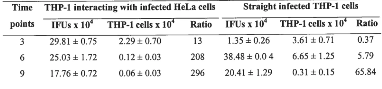
Table 2. C. trachomatis serovar L2 infectious particles (IFUs) compared to THP-1 cdl
number in culture media 121
Table 3. Transmission of infection to HeLa ceils monolayer by THP-1 and U-937
persistently infected with C. trachomatis serovar L2 122
CHAPITRE VI
Table 1. C. trachomatis serovar L2 growth 167
Table 2. Effect ofDox on cytokine secretion by co-cultured HeLaJTHP-1 cells infected
LISTE DES FIGURES CHAPITRE I
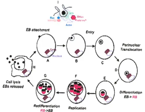
Figure 1. ChlainvcÏia trachomatis life cycle 10
Figure 2. Optical micrography (100x) of Chlarnydia trachomatis serovar L2 inclusion
in HeLa celis 12
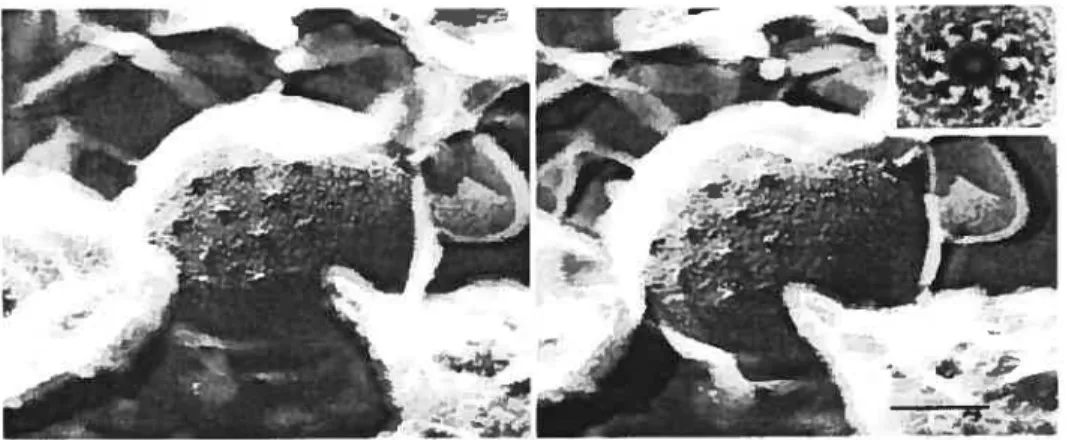
Figure 3. An example of early events during Chlamydia caviae entry 15 Figure 4. Chlamydial growth inside a membrane-bound inclusion 17 Figure 5. Surface projections of Chlamydia psittaci elementary body 22
CHAPITRE II
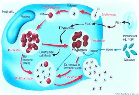
Figure 1. Chlarnydia trachomatis life cycle 38
Figure 2. Chlamydial active life cycle versus persistent cycle 42
CHAPITRE III
Figure 1. Infection of female genital tract with C7ztanzydia trachomatis 65
CHAPITRE IV
Figure 1. Effects of Dox, let and Ery on C. trachomatis inclusions inHeLa cells 124
Figure 2. C. trachomatis L2 viability after treatment with Dox assessed by RT-PCR 126
Figure 3. C. trachomatis L2 viability after treatment with let assessed by Rl-PCR 12$
Figure 5. Growth curves of C. trachomatis-infected THP-1 (A) and U-937 (3) cells 132 Figure 6. C. trachomatis serovar L2 viability in HeLa, THP-1 and U-937 celis after
treatment with Dox assessed by RT-PCR 134
CHAPITRE V
Figure 1: Influence ofC. trachornatis serovar L2 infection on THP-1 ceils alone
or in co-culture with HeLa celis 160
Figure 2: IL-lfl (A), IL-6 (B), IL-8 (C), TNF-a (D), IL-10 (E) and IL-12p70 (F)
synthesis by C. trachornatis serovar L2-infected HeLa celis 162
Figure 3: IL-lfl (A), IL-6 (B), IL-8 (C), TNF-a(D), IL-10 (E) and IL-12p70 (F) synthesis
by C. trachomatis serovar L2-infected THP-1 ceils 164
Figure 4: IL-13 (A), IL-6 (B), IL-8 (C), TNF-a(D), IL-10 (E) and IL-12p70 (F) synthesis
LISTE DES ABBRÉVIATIONS
ADCC: Cytotoxycité dependent des anticorps ADN ou DNA: Acide désoxyribonucléique
APC: “Antigen presenting ce!!”
ARN ou RNA: Acide ribonucléique
ATCC: “American Type Culture Collection”
ATP: Adenosine triphosphate
CADD: “ChÏamydia protein associated with death domains”
CDC: “Center for diseases contro!”
CHANCE: Chirurgie, antibiotique, nettoyage facial, environnement
CHO: “Chineese hamster ovary”
CHUM: Centre hospitalier de l’université de Montréal
CPAF: “Chlamydia protease/proteasome-like activity factor”
CRP: “Cysteine-rich protein”
Ctc: “Chlamydia 2-component”
CTGF: “Connective tissue growth factor”
DAG: Diacylglycérol
DC: “Dentritic ccl!”
DNase: Désoxynucléase
dNTP: Désoxynuc!éoside triphosphate
Dox: Doxycycline
DTH: “Delayed type hypersentivity”
DTNB: “5,5’ -dithiobis(2-nitrobenzoic acid)” EB ou CE: “E!emetary body” ou corps élémentaire
EBV: Virus Epstein-Barr
Ery: Érythromycine
GAG: Glucosaminoglycane
GM-CSF: “granulocyte macrophages- colony stimu!ating factor”
Hsp: “Heat sliock protein”
DO: Jndoléamine dioxygénase
IFN-’y Interféron-’y
LEU ou UFI: “Inclusion forming-units”
IL-: Interleukin
mc: Inclusion
LCR: “Ligase chain reaction”
LPS: Lipopolysaccharide
LVG: lymphogranulome vénérienne
MOI: Multiplicity of infection
MBC: “Minimal bactericidal concentration” MEC: 2,4-méthylérythritol 2,4-cyclodiphosphate MEM/H: Minimal essential medium avec les sels de Hank
MEP: Méthylérythritol phosphate
MHC: “Major Histocompability complex”
MIC: “Minimal inhibitory concentration”
MOMP: “Major outer membrane protein”
NE-KB : “Nuclear factor-KB”
NK: “natural killer ceil”
NO: “nitric oxide”
NOS : “Nitric oxide synthase”
OMS: Organisation mondiale de la santé
ORF: “Open reading frame”
PBS: “phosphate-buffered saline”
PDGF: “Platelet derived growth factor”
PG: Peptidoglycane
PD: “Pelvic inflammatory diseases”
PIP2: PhosphatidyÏinositol4,5 biphosphate
PKC: Protein kinase C
Pmp or Pomp: “Polymorphic outer membrane protein” PrmC: “Protein release factor methylation C” RB ou CR: “Reticulate body” ou corps reticulé
RNase: Ribonucléase
rpm: révolution par minute
RT-PCR: “Reverse transcription-polymerase chain reaction”
SAFE: “Surgery, antibiotic, face washing, environmental
improvement”
SEP: Septum
5FB: Senim foetal bovin
SOCS: “Suppressor of cytokine signalling” SPG: “Sucrose-phosphate-glutamic acid buffer”
TACE: “TNF converting enzyme”
Tarp: “Translocated actin-recntting protein”
Tet: Tétracycline
TGf-a “Transfoniiing growth factor-o”
Th: “T helper”
TLR: “Toli like receptor”
TNFR: “Tumor necrosis factor receptor”
TNF-a: “Tumor necrosis factor-a”
TTSS: “Type III secretion system”
DÉDICACE
REMERCIEMENTS
Je désire tout d’abord exprimer ma gratitude à l’égard du Dr Madeleine Ravaorinoro, ma directrice de thèse qui a bien voulu diriger cette étude avec beaucoup de tact. Durant ces 4 années, Madeleine n’a pas fait que me guider, elle a aussi su susciter en moi différentes qualités ayant accrue mes capacités à mener à bien un projet de recherche. Je suis désormais dotée d’une rigueur et d’une curiosité scientifiques, mes connaissances et mes compétences sont maintenant plus accrues. Madeleine, je te suis énormément reconnaissante pour ta disponibilité et ton enseignement de qualité.
À
chaque fois que j’ai été confrontée à un problème, tu as toujours été présente pour répondre à mes questions et à mes inquiétudes. Merci!Je tiens également à remercier le Programme Canadien des bourses de la francophonie (PCBF) qui a assumé tous mes frais de subsistance et d’études doctorales. Je précise mes remerciements à Mr Yves Galipeau, Coordonnateur du PCBf, et à madame Colette Gagnon, conseillère pédagogique, qui en plus d’assurer convenablement leurs taches au sein du PCBf, ont su donner de leur temps pour m’écouter.
Mes remerciements s’adressent également à mes amis (es) ci-dessous à qui j’ai assailli de questions: Mohamadi kaouass, Mohamed Laakel (laboratoire du Dr Danuta Balicki), Sonia Deschênes et Hélène Brodeur (Laboratoire du Dr Pangala Bhat), Annie Chamberland et Geneviève Bélanger (laboratoire du Dr Cécile Tremblay) Jill Héneault (laboratoire du Dr Yves Raymond), Subadjini Selliah, Omar El Ared et Cathérine Carrier (laboratoire du Dr Madeleine Ravaoarinoro). Merci aussi à mon amie Sandra Akouamba (laboratoire du Dr Hugos Sudens) avec laquelle
j
‘ai passé les nuits à discuter de mon examen pré-doctoral.Je remercie les membres de mon comité de suivi et de ce présent jury, Dr Ah Ahmad, Dr Michel Desjardins, Dr Éric Frost et Dr Georges Szatmari, pour s’être intéressés à mon projet de recherche et d’avoir pris le temps de lire ma thèse.
Je remercie ma soeur Owanga Wilfried, ma fille Kounessoussa Carme et mon fils Epenit Banas Wayne-P qui, ici à Montréal, ont vécu avec moi mes joies et mes peines.
Je profite de l’occasion pour renouveler mon affection pour mes deux frères, Ambroise Banas Germain et Jean-Pierre Kenké. Leurs encouragements constants ont été pour moi une source de force et d’énergie m’ayant permis d’affronter les difficultés quotidiennes et de mener jusqu’à la fin mes études.
Je remercie Mr Mondjo Epenit Mesmin de m’avoir encourager à entreprendre les études doctorales. Merci également à mon ami de coeur Mehdi Hermann Gaètan Mvou-Kouma de m’avoir soutenu lors de la rédaction de cette thèse.
A ma mère, Véronique Kampalari, et à mon père, Camille Étougui, je réitère mon merci. Sans eux, je ne serais pas.
Je nonm-ie les différents membres de ma famille ci-dessous pour le bien qu’ils ont pu m’apporter: Adjandji Marie-Pierrette, Atimbou Blanche, Ayaya Philomène, Benigui Monique, Boumas Marcel, Kassanga Josephine, Kayeghe Yvonne, Mme Kenké née Minko Judith, Lamboumi Élise, Mpiga Germaine, Ndjobolo Hortense, Ndjouba Rigobert, Ngabina Jacques, Ngouadzia Yvette, Obili Steeward, Olayi Sylvie, Ondjani Ariette, Ontoula Antoine, Otaly Émilie, Ontaglia Bruno Bemard, Ntsiégué Claudine, Worogoyo Françoise, Yemayira Antoinette.
PREMIÈRE PARTIE:
Les infections causées par la bactérie intracellulaire obligatoire Chtamydia trachornatis sont en général asymptomatiques et peuvent être résolues spontanément, uniquement grâce à la réponse de défense générée (Morre et al., 2002). Cependant, chez certaines personnes, les infections multiples et les infections initiales non résolues peuvent entraîner la réponse immunitaire spécifique et une inflammation chronique occasionnant les dommages tissulaires sévères (Joyner et al., 2002). Les grossesses ectopiques, l’infertilité tubaire, la cécité curable sont quelques exemples de séquelles résultantes. Les mécanismes par lesquels les infections initiales aigus deviennent chroniques sont peut-être multiples, mais sont encore indéterminés. Chez certains individus chroniquement atteints, C. trachomatis demeure difficile à cultiver alors que son ADN est révélé par PCR, suggérant la présence de l’organisme sous une forme non infectieuse (Grayston et Wang, 1975; Brunham et al., 1985). De plus, malgré la thérapie avec les antibiotiques appropriés (doxycycline, tétracycline, érythromycine, azithromycine), dans certains cas, l’infection chlamydienne demeure toujours présente, quand les réinfections sont exclues par l’adoption de rapports sexuels protégés (Whittington et al., 2001). Différents travaux réalisés in vitro ont permis de montrer qu’en présence de conditions non propices, C. trachomatis pouvait développer une forme latente ou persistante difficile à résoudre par les antibiotiques (Beatty et al., 1994). Ces différents constats ont permis de poser l’hypothèse selon les antibiotiques habituellement utilisés dans la thérapie des infections
à C. trachomatis pourraient induire la forme persistante à la place de résoudre l’infection.
Le but de ce projet de recherche est d’associer les échecs thérapeutiques rapportés dans la littérature à la persistance chlamydienne et de déterminer les conséquences inflammatoires résultantes
Les objectifs sont alors les suivants
1- Démontrer si le traitement des infections chlamydiennes in vitro par des antibiotiques habituellement utilisés dans la thérapie des infections chlamydiennes conduit à l’infection persistante à la place de la résoudre
2- Caractériser les réponses de cytokines pro-inflammatoires induites selon que l’infection est productive ou persistante, dans de modèles d’infection in vitro
Ce travail est subdivisé en 3 parties. La première partie est une revue de littérature décrivant la biologie chlamydienne, les pathologies occasionnées et les réponses immunitaires induites. La deuxième partie décrit: les effets de la doxycycline, la tétracycline et l’érythromycine sur l’infection chlamydiennes dans deux modèles de cultures in vitro; les cytokines inflammatoires induites selon que l’agent pathogène soit métaboliquement actif ou latent. Finalement, la troisième partie inclut la discussion générale pour une synthèse de l’étude.
CHAPITRE I: ARTICLE 1
Chtamydia trachomatis: from attachment to infection Mpiga Philomène, Ravaoarinoro Madeleine
Department of Clinical Microbiology, Centre hospitalier de l’Université de Montréal (CHUM)-Hôtel-Dieu, Montréal, Québec, Canada and Department of Microbiology and linrnunology, Université de Montréal, C.P. 6128, Montréal, QC, Canada
Correspondence and reprint requests:
Madeleine Ravaoarinoro, Department of Clinical Microbiology, CRUM-Hôtel-Dieu, 3840 rue St-Urbain, Montréal, QC, H2W 1T8, Canada, Telephone 514-890-8175, Fax 514-412-7240, E-mail: madeleine.ravaoarinoro.chumssss.gouv.qc.ca
Submitted to
1.1 SUMMARY
Chlamydia trachoinatis is an obligate intracellular bacterium with a growth cycle that occurs in several stages. The infectious chlamydial particle, or elementary body (EB), enters a susceptible celi and remains in a vacuole without fusing to endocytic organelles. It then converts to a growth form, or reticulate body (RB), whose role is to multiply by binary fission. The generated RBs undergo maturation to EBs, which are released from the cell to initiate new rounds of infection in neighbouring ceils. Most stages of its developmental cycle have been established, but corresponding molecular details need to be deterrnined. In this review, we report and discuss recent knowledge in moÏecular biology on the growth cycle of C. trachomatis whose cycle complexity is already noted at the celi entry level, which involves enzymatic processes and requires several molecules or molecular domains as acceptors and receptors. It is suspected that Inc inclusion proteins can contribute to escape from phagolysosomal fusion, and specific isomerases and proteases can play a part in differentiation process. Finally, 3 transcription factors and a 2-component regulation system have been identified, but their role in transcription regulation ofthe chlamydial cycle remains to be elucidated.
Key words: Chlamydia trachomatis biology; elementary body; reticulate body; developmental cycle; inclusion.
1.2 RÉSUMÉ
Chtamvdia trachomatis est une bactérie intracellulaire obligatoire dont le cycle de reproduction se produit en plusieurs étapes. La particule infectieuse chlarnydienne, le corps élémentaire, pénètre dans la cellule-hôte et y demeure dans une vacuole ne fusionnant pas avec les organelles endocytiques. Il s’y différencie en forme de reproduction, le corps réticulé, dont le rôle est de se reproduire par fission binaire. Les corps réticulés engendrés se dédifférencieront en corps élémentaires qui seront expulsés de la cellule-hôte afin d’initier de nouveaux cycles d’infection. Si l’ensemble des étapes de ce cycle de reproduction est établi, il reste cependant à déterminer les détails moléculaires correspondants. Dans cette revue, nous relatons et discutons des dernières connaissances moléculaires de la biologie chlamydienne. La complexité du cycle s’observe déjà au niveau de la pénétration qui, en plus d’être un processus enzymatique, requiert plusieurs molécules ou domaines moléculaires accepteurs et récepteurs. On soupçonne que les protéines des inclusions Inc puissent contribuer à l’échappement à la fusion lysosomale, et les isomérases et protéases spécifiques à la différenciation. finalement, trois facteurs de transcription et un système de régulation à deux composantes ont été identifiés, mais leur rôle dans la régulation transcriptionnelle du cycle chlamydien demeure à déterminer.
Mots-clés: Biologie chlamydienne, Chlamydia trachomatis, corps élémentaire; corps réticulé; cycle de développement; inclusion.
1.3 INTRODUCTION
Chtamydia trachomatis is a bacterium responsible for a broad spectrum of pathologies. Even though C. trachomatis is susceptible to many antibiotics [1-3], it remains the leading cause of sexually-transmitted diseases and the second leading cause of blindness worldwide [4, 5]. This Gram-negative prokaryote, belonging to the chlamydiaceae family, is described as an atypical obligate intracellular parasite with no peptidoglycan (PG) in waÏÏ. Although the eubacteriaÏ origin of C. trachoinatis has been established, its precise phylum stili remains to be determined. It is possible that the chlamydiaceae have a common ancestor with chlorop!asts and cyanobacteria [6]. It is also suggested that C. trachomatis has undergone reductive evolution. According to this assumption, protochiamydia must have lost genes during its evolution as it adapted to different intracellular niches, which would explain why the C. trachomatis species is made up of many serovars, A to L3, differing in pathogenicity and tropism [7].
Several decades after its discovery, C. trachomatis stiil remains a mystery. Nevertheless, recent sequencing of the complete chlamydial genome has allowed us to leam a littie more about this organism. C. trachomatis is a bacterium with one of the smallest genomes, roughly 1,042, 519 ph, that is, approximately 894 genes, of which 28% stili remain to be assigned [8]. This genome sequence also reveals various chlamydial metabolic pathways, but several among them are inteinipted, explaining why C. trachoinatis has Iimited anabolic capacities, and, hence, its energy- and metabolite dependence on the host. This dependence would partly justify strict chlamydial parasitism. However, despite chlamydial genome revelations, littie is known about various aspects of chlamydial biology, specifically gene regulation and interactions with the host.
C. trachornatis is characterized by a unique developmental cycle during which the
extracellular stage altemates with the intracellular stage (Figure 1). The first stage is achieved by particles known as elementary bodies (EBs) that are of small size (0.3 jim of diameter), metabolically inactive and specialized in ce!! infection. To initiate the
developmental cycle, EBs must penetrate cclls without specialized endocytosis. As the resulting phagosome does flot fuse with the lysosome, the EBs can survive and differentiate into rcticulate bodies (RBs) of 0.5-1 .3 j.im, which ensure growth and are less rigid, osmoticafly fragile and metabolically active. The RBs undergo multiple divisions by binary fission, leading to enlarged phagosomes called inclusions (Figure 2). At the end of grow-th, RBs are differentiated back into EBs for release from the ceil to perpetuate the developmental cycle [9]. Reproductive cycle events are globally established, but the precise mechanisms of each cycle step remain to be determined. The objective of this paper is to update C. trachomatisbiology, especially its structure and reproductive cycle.
Figure 1. Chlantydia trachomatis life cycle. Interaction of elementaiy body with host celis plasma membrane lead to Rho GTPase member family Rac recruitment and the component of Arp2/3 complex, WAVE2. With phosphorylated recruited Tarp, these proteins allow actin recruitment at the site of attachment. Actin remodeling resuks in distinct microvillar reorganization tbroughout the host celi surface and the formation ofpedestal-like structure and entry. EB, elernentary body; RB, reticulated body; Tarp, translocated actin recruting protein. Adapted from J. Engel. 2004. Proc. Nati. Acad. Sci. U S A. 101, 9947-8 [10j. \ _ •fl! WM ‘ul flS’ ESatuichrnent Entry C2Jt Iysi E&rCi)SCd Ppdifferptfl r.lfiOfl Replicatior? RB>EB
1.4 EBs versus RBs
EBs are distinguished from RBs flot only by their compacted nucleoid, but also by their rigid wall, conferring osmotic stability. As EBs are specialized in cell infection, it is important to study their wall ultrastructure to understand interactions that allow attacliment and penetration. Despite the presence of ail genes required for peptidoglycan (PG) synthesis in the chlamydial genome, the C. trachomatis wall does flot have a fine PG layer that is specific for Gram-negative bacteria [8]. Instead of PG, there is a supramolecular structure made up of a trimeric major outer membrane protein (MOMP, 39.5 kDa), a large cystein-rich protein (CRP, 60/62 kDa) and a small CRP (12 kDa). This structure, within MOMP and CRPs, is highly disulfide cross-linked and confers EB wall rigidity [10]. In RBs, the wall possesses monomeric MOMP, but loses large and small CRPs; thus, RBs are osmotically fragile [12].
Several studies have attempted to clarify the ultrastructure of these parietal proteins. A topological model of the C. trachomatis MoPn MOMP has been built. According to this model, MOMP is made up of 5 constant domains separated by 4 variable antigenic domains. MOMP folds up 16 times so that its constant domains form transmembrane t3-strands and short periplasmic loops, while its variable domains form long loops exposed at the celi surface. According to the authors, this model is in agreement with the porin function of MOMP [13, 14]. When MOMP is in the outer membrane, CRPs are at the iimer surface of the outer membrane. A small CRP is proposed to be a periplasmic lipoprotein that, like Braun lipoprotein, connects the inner large CRP molecules to the outer membrane through disulfide bonds [15]. C. trachomatis, like other Gram-negative bacteria, lias lipopolysaccliarides (LPS) in its wall, but these LPS are truncated, terminating by 3-deoxy-D-maimose-octulosnic acid [16]. In addition to these wall compounds, examination of the completed chlamydial genome has led to prediction of the family of genes coding for polymorphic outer membrane proteins, called pomps or prnps, of 90-180 kDa. In C. trachomatis species, 9 genes have been identified (prnpA to
pmpI, whereas C. psittaci has 6 (2 familles: pomp9o and pomp98) and C. pneumoniae,
21 (pompi to pomp2l). The corresponding coding proteins differ in their amino acid
V, I) and FXXN) which are repeated several times in an altemating fashion. Henderson and Lam [17] suggest that these proteins belong to the autotransporter family. Pmps B, F and H have so far been detected only in EBs, while Pmps C, D E, G and I have been
found in both EBs and RBs. In contrast, PmpA has been detected only in RBs [18, 19]. Much work is needed to elucidate Pmps function.
Figure 2. Optical micrography (1 OOx) of CÏiÏarnvcfia trachornatis serovar
L2 inclusion in HeLa ccii. Infected HeLa ceils were stained by the May GrLinwald-Giemsa method. Each point in inclusison is either elementary body, or reticulated body.
1.4 REPRODUCTIVE CYCLE
1.4.1 Attachment and penetration
Several chlamydial adhesins, such as MOMP, large CRP and 70 kDa heat shock protein (Hsp7O), are considered to be ligands. However, only a cellular compound, heparan sulfated-like glucosaminoglycans (GAG), was studied as a cellular receptor during chlamydial entry. It is now known that these different compounds are flot high affinity ligands or receptors. Possibly, a given serotype has several ways of penetrating, since inhibition of specific interactions between the celi and the EB surface confers only partial protection.
Carabeo and Hackstad [20] showed that attachment is a specific process that occurs in 2 steps. The initial step consists of electrostatic and reversible interactions that would allow intimate proximity of the EB and host cell. This proximity would promote the second step during which high-affinity ligand and receptor form specific and irreversible bonds. Actually, it is found that Chinese hamster ovary celis (CHO) deficient in GAG expression (mutant pgsA-745) remain resistant to infection by L2 serovar as well as MoPn serovar, whereas CHO expressing GAGs, but deficient in other receptors (D4. 1-3 mutant) remain resistant to the L2 serovar, but flot to MoPn serovar. This means that both serovars use GAGs as the primary receptor as well as 2 distinct but unknown secondary receptors.
The high affinity chlamydial ligand may not be exposed on the EB surface and must become accessible only after reduction of the cysteine-rich outer membrane protein lattice. This possibility was suggested as it was noted that EB penetration but not attachment was compromised by 5,5’-dithiobis(2-nitrobenzoic acid)(DTNB) [21]. DTNB is an impermeable reagent that covalently modifies surface suifflydryls to prevent disulfide bond cleavage. Thus, penetration could be an enzymatic process requiring isomerases, enzymes taking part in reduction of the disulfide bonds. The importance of isomerases in chlamydial penetration has been proved with anti-cellular disulfide isomerase antibodies to reduce HEC-1B celi infection by C. trachornatis serovar E. This
55-kDa cellular isomerase was normally found to act in liaison with cellular Hsp7O and other additional proteins of 45, 48 and 90 kDa, activating estrogen receptors. Thus, Davis et ai. [22] suggested that during the association of EB on the surface of endometrial epithelial celis, this isomerase would act as a universal trigger for reduction of the supramolecular cross-linked EB outer membrane complex, thereby exposing the high affinity MOMP or large CRP adhesin domains. Moreover, 2 chlamydial disulfide isomerases, whose existence was predicted by chÏamydial genome anaÏysis, could be activated for total dissolution ofthe disulfide bonds during this process.
EBs entry does flot occur randomly, but in defined areas of plasma membranes called lipid rafts, characterized by high cholesterol and glycosphingolipid content. These lipid microdomains are enriched in molecules involved in signal transduction events. Coalescence of lipid rafis may trigger EB intemalization. Disturbance of lipid rafis in the plasma membrane inhibits C. trachomatis serovar L2 entry into cells [23-25]. Although most studies argue for the involvement of cholesterol in Chlamydia entry, there is nevertheless one that questions this possibility [26].
Upon ineversible C. trachomatis EB attachment to the host cell, various events trigger EB intemalization. In addition to lipid rafi coalescence, actin remodelling is required to facilitate infection. Carabeo et al. [27] noted that the presence of cytochalasin D, an inhibitor of actin formation, prevented celi infection. Microscopy has revealed actin recrujtment at the attachruent site of C. trachomatis serovar L2 with HeLa cells. Accumulated actin would lead to the formation of a pedestal-like structure carrying EB and, finally, the formation of microvillar extensions, which would coat the EB. hi this entry process, the Rho GTPase family ofproteins, which controls actin filament assembly and organization, plays an important role. Rac, but not Rho or Cdc42, is specifically actjvated afler C. trachornatis serovarL2 and D attachment. Clostridial toxin B, which is a known enzyrnatic inhibitor of Rac, Rho and Cdc42, significantly reduces C. trachomatis invasion ofHeLa ceils [28, 29] (figures 1 and 3).
The ChÏarnydia signal required to trigger a cascade of events for entry remains unknown. A recent smdy has shown that immediately upon attachment, EB secreted, by a type III system, a protein named translocated actin-recruiting protein (Tarp) of about 103 kDa, encoded by cifiamydial ORF ct456. This protein is tyrosine-phosphorylated at the cytopisamic face of plasma membrane at the site of EB attachment. Tarp appears to initiate or participate in signalling events that regulate actin recruitment which, ultimately, Ïeads to EB intemalization [30, 31] (Figures 1 and 3). Despite this knowledge, much remains to be done to clarify the Chlamydia entry process.
Ï
Ij .• l;n%I i ElO I”. ii ii ‘t. :ç_rr.iwt//
/1/1/
/:
t,4t’
I s t*
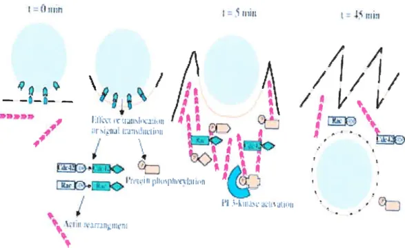
Figure 3. An example of early events during Chlarnydia caviae entry. Interaction of bacterium (grey circle) with the host plasma membrane induces the clustering of cholesterol rich membrane domain (orange une). This interaction initiates intracellular signalling that is also mediated by activated recruited small GTPase Cdc42 and Rac. Phospohoiylated proteins (orange shapes) accumulate at the site of entry where they interact with PI 3 kinase (blue) and participate in the signalling cascade. The activation of these different proteins is followed by actin recruitement and depolarization and bacterium entry. Reproduced from A. Subtil et aI. 2004. J. ceil. Sci. 117, 3923-3933 [291.
I= I = mli -f 41 1 ltl—l
1.4.2 Chlamydial inclusion and lysosomal fusion escape
Afler penetration, the EB finds itself in the ccli, intemalized by a phagosomal or endosomal membrane. b survive in the ccli, it must avoid being lysed by lysosomal enzymes contained in endophytic vesicles. Whereas many bacteria, such as Coxiella burnetii, survive in ceils by resisting lytic enzymes, C. trachomatis EB must limit its contact with these enzymes by preventing phagolysosomal fusion. Actually, early endosomal markers such as Rab5 and FF1, are not found in the inclusion membrane. Markers of late endosomes and lysosomes, including LAMP 1, LAMP2 and the hydrolase cathepsin, are also excluded. The proton ptimp ATPase is equally not found, explaining why the lumen of the inclusion is not acidified, and an inhibitor of vacuole acidification, such as bafilomycin Ai, does not affect C. trachomatis replication. Only the early endosome marker transferrin is seen near, but not in the inclusion [32, 33].
Several hypotheses have been offered to explain how Chlamydia escapes lysosomal fusion. Heinzen et al. [34] demonstrated that afier formation, chlamydial inclusions underwent retrograde transport, moving then to the exocytic pathway of the Golgi apparatus where they fused with exocytic vesicles containing sphingomyelin (Figure 4).
Ibis interaction allows chlamydial inclusions to be hidden in the exocytic network, which
would expiain how they escape from phagolysosomal fusion [35]. However, recent studies showing that even the initial phagosome does flot contain any early endosomai markers raised the possibility that chlamydial components transform the inclusion membrane so that it is not recognized as being destined to fuse with the lysosome [34, 36]. This attractive possibiÏity was reinforced by Taraska et aÏ. [37] who reported that if an inclusion membrane seems derived from the plasma membrane, it does flot necessarily contain host proteins, but could be rather decorated with various chlamydial Inc proteins [38].
Jnitially discovered in C. psittaci [39], Inc proteins as a group of proteins differ in their sequence, but resemble each other by their unique bilobed hydrophobic domain of 50 to
$0 amino acids. In C. trachornatis, even if 40 open reading frames (ORFs) coding for these special proteins are present in the genome, only some of them have been identified (IncA to IncG). IncA, Incf and IncG are exposed at the cytoplasmic side of the inclusion [40]. The various functions of these proteins are flot known. It is only clear that IncA is involved in homotypic fusion between C. trachomahs inclusions [41]. The inclusion membrane would be covered with these proteins, each playing a unique ftinction, conferring multiple properties. One of their possible functions would be, notably, participation in avoidance of phagolysosomal fusion. Other functions must be considered, such as participation in nutrient acquisition and in EB differentiation into RBsand of RBs into EBs. Given the importance of inclusion for the establishment of conditions favourable to chlamydial growth and in its direct interactions with the host cytosol, the identification of various Inc proteins as well as their functions will provide much more information on chlamydial biology.
ATI:f,JIJ) _.,i.-Ur lipici — f1ow
••
•
e
e
.J,,.
Figure 4. Chlamydial growth inside a membrane-bound inclusion. The nascent inclusion intercepts epithelial exotic vesicles carrying sphingomyelin (SM), phosphatidylinositol (PI), phosphatidylcholine (PC) and cholesterol (CL) for inclusion membrane expansion. ER, endoplasmic reticulum; mc, inclusion proteins. Reproduceil from P. Wyrick. 2000. CelI. Microbiol. 2, 275-2$ [9J.
1.4.3 Differentiation and de-differentiation: EB÷-+ RB
Two hours afler penetration, EBs must differentiate into RBs. Differentiation can be defined as ail events taking place after intemalisation, leading to breakdown of the supramolecular structure responsible for wall rigidity, and to nucleoid decondensation. The resuit is an increased membrane penTieability with cytoplasmic decondensation and chromatin dispersion. These changes ultimately elicit metabolic activation. As EBs derive from the de-differentiation of RBs afier multiple binary fissions at the reproductive cycle end, various authors have attempted to find late proteins expressed at the time of dedifferentiation of RBs into EBs, to understand the distinctive characteristics of EBs. $ome of them succeeded in cloning and sequencing genes whose expression in Escherichia cou led to DNA compaction. It was suggested that Chlamydia histone Hi like protein, Hcl, could be involved in the compaction of chlamydial DNA, and that its expression during the reproductive cycle could trigger RB de-differentiation into EB [42]. Pedersen et al. [43] showed that the C-terminal domain of Hcl is able ta bind ta DNA and RNA in limited proteolysis, SouthWestem blotting and gel retardation assays, whereas the N-terminal extremity is a dimerization domain. This could explain how Hcl at high molarity allowed DNA and RNA compaction, as revealed by microscopic observations [44]. In addition, previous studies also disclosed that Hcl binding ta DNA strongly repressed its transcription and translation in vitro [45]. These different facts indicated that late Hcl expression could bring some change of DNA topology, allowing differential late protein expression. High production of Hcl and other proteins would likely cause, finally, the total compaction of DNA and RNA, inhibiting transcription and replication. These events would coincide with the de-differentiation ofRBs into EBs.
The dissociation or degradation of proteins compacting chromatin, such as Hc 1, is necessary at the time of differentiation of EBs into RBs. One report has suggested proteolytic degradation as a possible mechanism of Hcl dispersai. A chlamydial gene, euo, coding for an early protease of 121 amino acids and 20.9 kDa, has been identified [46], and whose expression has lcd ta selective degradation of the C-terminal extremity of Hc 1 bound to DNA. This would elicit Hc 1 dissociation, and hence, the decompaction of DNA necessary for the resumption of transcription and metabolism. However, very
recently, another report suggested a simple dissociation of Hcl as a rnechanism of chlamydial chromatin relaxation. Chlamydial 2-C-methylerythritol 2,4-cyclodiphosphate (MEC) lias been found to dissociate Hcl from chlamydial chromatin. MEC is a
metabolite of the non-mevalonate methylerythritol phosphate (MEP) isoprenoid
biosynthetic pathway. The precise mechanism is flot known but MEC may be a competitive inhibitor or an allosteric effector of Hcl. In addition, a chlamydial IspE (CT$04), an intermediate enzyme of the MEP pathway, is required in this process. Actually, chlamydial ispE expression in Escherichial cou removes the lethality resulting from chromatin compaction after Hcl expression [47].
As mentioned above, disulfide bonds form at the time of de-differentiation, and it appears that cellular and chlamydial isomerases are required for their rupture. However, differentiation being a complex process, several other events likely occur. EBs couid have ail enzymes capable of mediating early events immediately afler intemalization, such as lysosomal fusion avoidance and differentiation of EBs into RBs. It would be interesting to determine the protein differential expression pattem in EBs and RBs to identify all the proteins necessary in these processes. For these reasons, RBs should be collected 1$ h afler infection, before de-differentiation, to ensure that the RB population is flot contarninated by EBs.
1.4.4 Chtamydia-host cett interaction
Once within the host celI, C. trachomatis does not completely control host metabolism, but must modulate host reactions to develop suitable conditions for its growth. Thus, the intracellular pathogen must secrete compounds in the cytoplasm that have the ability to change host behaviour. C. trachomatis are found to express 2 eukaryote-like Ser/Thr kinases, Pknl (CT145) and PknD (CT3OÏ). The activity of these kinases may confer signais to regulate ceiiular functions [48]. In addition, the presence ofthe gene encoding PP2C-type protein phosphatase (CT259) in the chlamydiai genome argues for a functional kinase-based signalling cascade in Chlamydia for interaction with host signalling pathways.
Among the chlamydial compounds released in the ceIl cytosol that interact with the host for maintenance of the infection process, Chlamydia protease/proteasome-like activity factor (CPAF) has been identified. This protein is synthesized as 70-kDa pro-CPAF that is gradually processed for a mature dimeric CPAF with 35- and 29-kDa fragments [49]. Active CPAF is found to degrade host transcription factors (RFX5) required for major histocompatibility complex gene activation. CPAY also degrades keratin-8, a key component of the intermediate filaments involved in celi integrity. This keratin degradation may allow increased cytosol fluidity, facilitating chlamydial inclusion expansion [50]. The way that CPAF is secreted from chlamydial inclusions to the host cytosol remains unknown.
To favour its growth, C. trachomatis also interacts with the host to inhibit programmed celi death [51]. Very recently, it has been discovered that C. trachomatis inhibits apoptosis by broad cleavage of proapoptotic proteins with Bd-2 homology domain 3 (BH3-only proteins), among them Bad, Bmf’ active Bid (tBid), Puma, Noxa, and Bim [52-54]. BH3-only proteins are required for the release of cytochrome c from mitochondria. Since proteasomal activity is needed in the degradation of some of these BH3-only proteins, CPAF may be involved in degradation ofthese compounds. Tse et al. [55] showed also that C. trachomatis inhibited apoptosis by sequestering protein kinase ô
(PKC8) in inclusions. By binding PKC6 through its Cl domain in the context of
ancillary protein moieties, diacylglycerol directs the PKCô in or near the chlamydial vacuole, away from the mitochondria. In fact, PKCô, a lipid-dependent serine/threonine kinase, is a proapoptotic regulator. Once cleaved, this kinase releases a catalytically active fragment that transiocates to the mitochondria where it promotes the release of cytochrome c with subsequent activation of the apoptotic pathway. In contrast, Ïate at the end of growth, C. trachomatis must induce apoptosis to favour EB propagation, probably through apoptotic bodies. Chlamydia protein associating with death domains (CADD) that is synthesized late in the developmental cycle has been found to modulate host cell apoptosis [56]. CADD has a death domain-like region that can interact with tumour necrosis factor family proteins and induce cell apoptosis. Since CADD seems to be an oxydoreductase, it can also induce ceÏÏ necrosis [57]. Thus, Chlainydia interacts with the
host machinery through chlamydial-secreted compounds. About 130 cellular genes are induced after C. trachomatis infection [58].
The question is to deterniine the mechanisrn utiiised by C. trachomatis to release some cornpounds modulating the host response when the inclusion membrane is impermeabie, even to small molecules whose molecular weight is at least 0.52 kDa [59]. The answer to this question has corne from chlamydiaÏ genorne analysis, which lias made it possible to predict the existence ofthe type III secretion system (TTS$). In generai, the TTSS is used by Gram-negative bacteria for secreting and injecting pathogenicity proteins into the eukaryote ccli cytosol. Whereas the genes coding for this system are typically linked in small pathogenicity islands containing an A+T-rich signature among enterobacteriaceae, the chlamydial orthologs are divided into 3 different loci in the genome and iack the A+T-rich signature [8].
While there is no doubt about the existence of the chlamydial TTSS, the identification of proteins forming this apparatus is stiil in progress. Some authors could provide experirnental evidence for a flinctional TTSS in C. trachomatis by referring to the homology of structural proteins constituting this system among Gram-negative bacteria. Thus, by combining RT-PCR, immunoblotting and matrix-assisted laser desorption ionization time-of-flight (Maldi-Tof), Fieids et al.[60, 61] successfuiiy identified some of these proteins, including CdsJ (contact-dependent secretion J) and CopN (Chlamydia outer membrane protein N). CdsJ, a predicted lipoprotein homologous to Yersinae spp YscJ, is essential in the TTSS because of its inter-connections with the internai and external components of this system. By homology with Yersinae YopN, CopN is a reguiating protein that could be localized on chiamydiai surfaces where it would block TTSS pores, preventing protein secretion in the absence of a suitable signai. Recently, a chaperone of CopN, the chiamydiai LcrH-2, has been proposed [62]. As mentioned eisewhere, Tarp is also secreted by the TTSS [30].
The anti-host proteins released have been identified according to the fact that proteins secreted by a type III machine of one pathogen can aiso be secreted by the heterologous
machine of another pathogen. Subtil et al. [63] have reported that proteins expressed by the hybrid genes inc3/cya and incC/cya in wild type Shigella fiexneri were excreted in the supematant, while in the mutant mxiD, in which the TTSS is completely compromised, there was no excretion of these proteins in the supematant. Cya is Bordetella pertussis gene encoding calmodulin-dependent adenylate cyclase used in this experiment as a reporter gene. This made it possible to state that inclusion proteins, such as IncA, IncB and IncC, pKn5 and CopD, were secreted by the TTSS in ChÏamydia. Pkn5 may be a serine/threonine kinase or an aminoglycoside 3 ‘phosphotransferase [48]. CopD, similar to Yersina YopD, may associate with another protein to form a porin-like structure in the membrane, facilitating the translocation of other type III secreted substrates [64].
In the genus Chlarnydia, the TTSS appears to correspond physically to pili-like structures
(figure 5), discovered for the first time by Matsumoto [65] more than 20 years ago.
Recently, Chang et al. [66] combined electron microscopy and computer image analysis, generating more information on the organization of these appendages. They noted that the piÏi-like structures are made up of helical arrangements of protein subunits having a periodicity of 50
À.
These appendices, 60-$0À
in diameter and 500À
in length, leave the muer membrane, traverse the periplasmic space and pass in the outer membrane through a hexagonal ring. The proteins constituting the appendages have not yet been identified, but the possibility is flot excluded those the proteins secreted, such as IncB, JncG or IncA, could be some ofthem.Figure 5. Surface projections of Chlamydia psittaci elementary body.
1.4.5 Growth
Since several chlamydiai metabolic pathways are incomplete, C. trachomatis must acquire different nutrients from host ceils. Although chÏamydiaï nutritive needs are known, there is no rich medium for its growth in the absence of host celis. Studies must be undertaken to detennine if tlie inabiÏity C. trachornatis to grow in the absence of host ceils resuits from the need for cellular signais allowing EB differentiation into RB, or from the need to transform nutrients at the level of the inclusion membrane to make them easier to assimilate by the pathogen. Since C. trachomatis does not control host metabolism, both host and parasite take up the same available nutrients. To date, no compound allowing C. trachomatis to compete efficiently with the host celi lias been identffied. This is why it is important to add cycioheximide to culture media to strengthen chiamydial growth rather than that of the host ceil. C. trachornatis may have developed mechanisms allowing it to survive during food shortages. Indeed, it was noted that C. trachomatis developed an aberrant and persistent form when intracellular tryptophan pools were reduced in response to gamma interferon [68]. In addition, chlamydial genome sequence analysis supports the hypothesis of an ATP-iimited biosynthesis pathway, probably an emergency one [8].
In regard to cellular division, it is difficuit to conceive the chiamydial mechanism of septum formation, in the apparent absence of FtsZ protein. Indeed, ftsZ is a GTPase required for ceilular division, because it participates in the ring-like structure formation essential to septal protein assembly. Until now, no chiamydial protein sharing significant homology with FtsZ lias been identified. However, Brown and Rockey [70], using ftuorescent-antibody labeling, detected a chlamydial antigen caiied SEP (for septum) in dividing RBs. SEP was localized to a ring-like structure at or near the plane of chlamydial division. According to these authors, this iocalization pattem corresponds to that ofFtsZ in other bacterial species.
Moreover, even if the C. trachomatis genome contains ail the genes necessary to encode proteins of the PG biosynthesis pathway, attempts to identify PG in RBs have been
unsuccessful. Just like FtsZ, PG plays a part in cellular division by allowing the formation of invaginations between 2 daughter celis during cytokinesis. Recent studies showed that the chlamydial murA gene was expressed, and the resulting enzyme was functional. Jndeed, MurA is a uridine diphosphate N-acetylglucosamine enolpyruvyl transferase, an enzyme essential to the PG biosynthesis pathway [70]. The authors also noted that C. trachomatis serovar L2 MurA contains a cysteine-to-aspartate change at amino acid 1 19. This active site substitution alters the pH optimum for the enzyme. Chlamydial MurA enzyme has high activity at pH values less than 7.0, and loses activity at pH values greater than 7, whereas wild typeMurA activity remains constant over a pH of 5.0 to 9.0. Considering the fact that the pH of inclusion is higher than 7.0, 20 h after chlamydial infection, McCoy et aÏ. [70] suggested that PG could be synthesized only during the early stages of the C. trachomatis life cycle. According to them, the PG syntl;esized precociously could be recycled later to take part in cellular division. However, UDP-N-acetylmuramoylalanylglutamyl DAP ligase has been found in RBs. This enzyme is involved in the synthesis of the muramyl-peptide unit, the first stage in PG assembly. This finding suggests that PG biosynthesis occurs during RB growth and ceil division [19]. Nevertheless, this PG and its precurseurs remain to be identified in chlamydial RBs.
1.4.6 EB exit
EBs coming from the de-differentiation of RBs must, at the cycle end, leave the infected host ceil to infect new surrounding cells. The exit mechanisms deployed by EBs are stiil unknown. Electron microscopy studies showed that some serotypes from A to K can leave without causing host ceil lysis. The overall inclusion would exit the ceil by a process similar to exocytosis [71]. Other serotypes from Li to L3 could be released afier host cell lysis. It is not know if this lysis requires specific chlamydial lytic enzymes or if it resuits from the pressure exerted by the inclusion. Recently, Perfettini et al. [72] noted that C. trachomatis MoPn could use celi apoptosis to spread. Jndeed, apoptotic bodies containing chlamydial particles, could be intemalized by surrounding cells in which the particles, instead ofbeing digested, perpetrate infection.
1.5 GENE EXPRESSION
Shaw et al. [73] have identified 3 classes ofchlamydial developmental genes: early genes detected 2 h afler intemalization, that are necessary to complete the differentiation of EBs into RBs; mid-cycle genes appearing 6-12 h post-infection and required in RB metabolism and growth; and, finally, late genes that are transcribed 12-20 h post infection and are important in the de-differentiation of RBs into EBs. Using the microarray approach to identify development stage-specific gene sets, Nicholson et al. [74] demonstrated that only 22% of the chlamydial genome was differentially expressed during the developmental cycle. The majority of other genes had constitutive expression in the basic functions of the chlamydial celi. Gene expression, according to chlamydial growth stages, means transcriptional regulation of development. However, until now, the signais and mechanisms ailowing global gene expression regulation are unknown. Aithougli 3 transcription factors (u66, u54 and u28) have been identified in Chlamydia, nothing to date indicates that chlamydial gene expression control is similar to that allowing Bacillus spp. sporulation, and implies a cascade of sigma factors [75]. u66 could be involved in some late gene expression such as hctA and hctB. The u54 promoter has been identified for 2 chlamydial late genes of unknown function. u28 encoded by rspD gene recognizes the promoter of hctB, that is, the gene encoding Hc2, a histone-like protein implicated in DNA compaction during back differentiation of RBs into EBs [76,77]. Recently, Koo and Stephens [7$] discovered a unique 2-component regutation system which can play an important part in late gene expression. This single system is made up of a sensor kinase, CtcB (Chlamydia 2-component B), and a response regulator, CtcC, having homology to the u54 activator. It appears that in the presence of Mg2, Mn2 and Fe2, the CtcB sensor can autophosphorylate and then transfers its phosphoryl group to the activator CtcC. Phosphorylated CtcC becomes oligomerized and then activates u54 holoenzyme. Since CtcB and CtcC are expressed late, it is possible that they have roles in the de-differentiation ofRBs to EBs.
Methylation may also play a role in chlamydial gene regulation. Recently C. trachomatis serovar D protein release factor methylation (PrmC) has been identified. PrmC (CT024)
contains a hallmark sequence of an N6-adenine-specific DNA methyl transferase. PrmC is constitutively expressed during the chlamydial intracellular developmental cycle and is able to methylate chlamydial release factor within the tryptic fragment containing the universally conserved motif glycine-glycine-glutamine (gly-gly-gln) [79]. The role of methylation in the regulation of gene expression and protein activity of Chlamydia remains to 5e revealed.
When the EB enters the host ceil, its DNA is decompacted, allowing gene expression. One may wonder if DNA decompaction ends before transcription restarts. Indeed, it is possible that DNA relaxation happens gradually so that the genes to be transcribed are in the portion ofrelaxed DNA, whereas those ofthe condensed portion cannot 5e expressed. The decondensation origin point ami the direction of this decondensation can influence transcription factor activity and thus determine the order in which the genes are transcribed. Further studies are required to explore the global regulation of chlamydial genes. This will constitute an important advance in understanding the biology of the pathogen.
1.6 CONCLUSION
The sequelae of chlamydial diseases resuit from a long process requiring multiple recurrences. New strategies besides antibiotics to prevent recurrences must be considered. Chlamydial biology studies can hasten the design of new anti-chlamydial approaches. hiileed, many aspects of C. trachomatis biology stili remain obscure. Current or future research should focus on the following stages: the mechanisms of EB penetration and differentiation, the means of division used by RBs in the absence of ftsZ, the transcriptional regulation of chlamydial genes, the identification of anti-host proteins modulating host metabolism, the inclusions role in nutrition and chlamydial growth.
1.7 ACKNOWLEDGEMENTS
We thank Ovid MichaeÏ Da $iÏva for editing this manuscript. M.P. is the recipient of theTrogramme Canadien des Bourses de la Francophonie” scholarship.
1.8 REFERENCES
[1] Sangare, L., Morisset, R, and Ravaoarinoro, M. 1999, 1. Med. Microbiol., 4$, 689.
[2] Sangare, L., Morisset, R., Gaboury, L., and Ravaoarinoro, M. 2001, J.
Antimicrob. Chemother., 47, 323.
[3] Sangare, L., Morisset, R., and Ravaoarinoro, M. 2001, Pathol. Biol. (Paris), 49, 53.
[4] Thylefors, B., Negrel, A.D., Pararajasegaram, R., and Dadzie, K.Y. 1995, Buli. WIIO, 73, 115.
[5] Sciarra, J.J. 1997, Int. J. Gynaecol. Obstet., 58, 107.
[6] Brinkman, F.S., Blanchard, J.L., Cherkasov, A., Av-Gay, Y., Brunham, R.C.,
Femandez, R.C., Finlay, B.B., Otto, $.P., Ouellette, B.F., Keeling, P.J., Rose, A.M., Hancock, R.E., Joncs, S.J., and Greberg, H. 2002, Genome Res., 12, 1159.
[7] Read, T.D., Brunham, RC., Shen, C., Gui, S.R., Heidelberg, J.F., White, O.,
Hickey, E.K.,Peterson, J., Utterback, T., Berry, K., Bass, S., Linher, K., Weidman, J., Khouri, H., Craven, B., Bowman, C., Dodson, R., Gwinn, M., Nelson, W., DeBoy, R., Kolonay, J., McClarty, G., Salzberg, S.L., Eisen, J.,
and Fraser, C.M. 2000, NucI. Acid. Res., 28, 1397.
[8] Stephens, R.S., Kalman, S., Lammel, C., fan, J., Marathe, R., Aravind, L.,
Mitcheli, W.,Olinger, L., Tatusov, R.L., Zhao,
Q.,
Koonin, E.V., and Davis, R.W. 1998, Science, 282, 754.[9] Wyrick, P.B. 2000, Cell. Microbiol., 2, 275.
[10] Engel, J. 2004, Proc. Nat. Acad. Sci. U S A., 101, 9947.
[11] Yen, T.Y., Pal, S., and de la Maza, L.M. 2005, Biochemistry, 44, 6250-6256. [12] Hatch, T.P. 1996, J. Bacteriol., 178, 1.
[13] Rodriguez-Maranon, M.J., Bush, R.M., Peterson, E.M., Schirmer, T., and de la Maza, L.M. 2002, Prot. Sci., 11, 1854.
[14] Findlay, H.E., McClafferty, H., and Ashley, R.H. 2005, BMC Microbiol., 5, 5. [15] Mygind, P., Christiansen, G., and Birkelund, S. 199$, J. Bacteriol., 120, 5784.
[161 Rund, S., Lindner, B., Brade, H., and Holst, 0. 1999, J. Biol. Chem., 274, 16819.
[17] Henderson, I.R., and Lam, A.C. 2001, Trends Microbiol., 9, 573. [18] Tanzer, RI., and Hatch, T.P. 2001, J. Bacteriol. 183, 2686.
[191 Skipp, P., Robinson, J., O’Connor, C.D., and Clarke, I.N. 2005, Proteomics, 5, 1558.
[20] Carabeo, R.A., and Hackstad, T. 2001, Infect. liumun., 69, 5899.
[21] Raulston, I.E., Davis, C.H., Paul, T.R., Hobbs, J.D., and Wyrick, P.B. 2002, Infect. Immun., 70, 535.
[22] Davis, C.H., Raulston, J.E., and Wyrick, P.B. 2002, Infect. Immun., 70, 3413. [23] Jutras, I., Abrami, L., and Dautry-Versat, A. 2003, Infect. Immun., 71, 260. [24] Stuart, E.S., Webley, W.C., and Norkin, L.C. 2003, Exp. Celi. Res., 287, 67. t25] Webley, W.C., Norkin, L.C., and Stuart, E.$. 2004, BMC Infect. Dis., 4, 23. [26] Gabel, B.R, EIwell, C., van Ijzendoom, S.C., and Engel, J.N. 2004, Infect.
Immun., 72, 7367.
[27] Carabeo, R.A., Grieshaber, S.S., fisher, E., and Hackstadt, T. 2002, Infect. Immun., 70, 3793.
[28] Carabeo, R.A., Grieshaber, S.S., Hasenkrug, A., Dooley, C., and Hackstadt, T. 2004, Traffic, 5, 418.
[29] Subtil, A., Wyplosz, B., Balana, M.E., and Dautry-Varsat, A. 2004, J. Cell. Sci., 117, 3923.
[30] Clifion, D.R., fields, K.A., Grieshaber, S.S., Dooley, C.A., fischer, E.R., Mead, D.J., Carabeo, RA., and Hackstadt, T. 2004, Proc. Natl. Acad. Sci. USA, 101, 10166.
[31] Dautry-Varsat, A., Balana, M.E., and Wyplosz, B. 2004, Traffic, 5, 561. t32] A1-Younes, H.M., Rudel, T., and Meyer, T.f. 1999, Celi. Microbiol., 1, 37. [33] Scidmore, M.A., Fisher, E.R., and Hackstadt, T. 2003, Infect. Immun., 71,
973.
[34] Heinzen, R.A., Scidmore, M.A., Rockey, D.D., and Hackstadt, T. 1996, Infect. Immun., 64, 796.