Role of the homeodomain transcription factor Hoxa13 in
embryonic development and formation of
extra-embryonic structures.
par Martina Scotti
Programmes de Biologie Moléculaire Faculté de Médecine
Thèse présentée à la Faculté de Médecine
en vue de l’obtention du grade de Philosophiae Doctor (Ph.D) en Biologie Moléculaire
Décembre, 2011
Université de Montréal Faculté de Médecine
Cette thèse intitulée:
Role of the homeodomain transcription factor Hoxa13 in embryonic development and formation of extra-embryonic structures
présentée par: Martina Scotti
a été évaluée par un jury composé des personnes suivantes :
Dr Jean-Philippe Gratton, président-rapporteur Dre Marie Kmita, directrice de recherche
Dr Hans Larsson, membre du jury
Dre Jacqueline Deschamps, examinatrice externe Dr François Dubé, représentant de la Doyenne
Résumé
La famille des gènes Hox code pour des facteurs de transcription connus pour leur contribution essentielle à l’élaboration de l’architecture du corps et ce, au sein de tout le règne animal. Au cours de l’évolution chez les vertébrés, les gènes Hox ont été redéfinis pour générer toute une variété de nouveaux tissus/organes. Souvent, cette diversification s’est effectuée via des changements quant au contrôle transcriptionnel des gènes Hox. Chez les mammifères, la fonction de Hoxa13 n’est pas restreinte qu’à l’embryon même, mais s’avère également essentielle pour le développement de la vascularisation fœtale au sein du labyrinthe placentaire, suggérant ainsi que sa fonction au sein de cette structure aurait accompagné l’émergence des espèces placentaires.
Au chapitre 2, nous mettons en lumière le recrutement de deux autres gènes Hoxa, soient Hoxa10 et Hoxa11, au compartiment extra-embryonnaire. Nous démontrons que l’expression de Hoxa10, Hoxa11 et Hoxa13 est requise au sein de l’allantoïde, précurseur du cordon ombilical et du système vasculaire fœtal au sein du labyrinthe placentaire. De façon intéressante, nous avons découvert que l’expression des gènes Hoxa10-13 dans l’allantoïde n’est pas restreinte qu’aux mammifères placentaires, mais est également présente chez un vertébré non-placentaire, indiquant que le recrutement des ces gènes dans l’allantoïde précède fort probablement l’émergence des espèces placentaires. Nous avons généré des réarrangements génétiques et utilisé des essais transgéniques pour étudier les mécanismes régulant l’expression des gènes Hoxa dans l’allantoïde. Nous avons identifié
un fragment intergénique de 50 kb capable d’induire l’expression d’un gène rapporteur dans l’allantoïde. Cependant, nous avons trouvé que le mécanisme de régulation contrôlant l’expression du gène Hoxa au sein du compartiment extra-embryonnaire est fort complexe et repose sur plus qu’un seul élément cis-régulateur.
Au chapitre 3, nous avons utilisé la cartographie génétique du destin cellulaire pour évaluer la contribution globale des cellules exprimant Hoxa13 aux différentes structures embryonnaires. Plus particulièrement, nous avons examiné plus en détail l’analyse de la cartographie du destin cellulaire de Hoxa13 dans les pattes antérieures en développement. Nous avons pu déterminer que, dans le squelette du membre, tous les éléments squelettiques de l’autopode (main), à l’exception de quelques cellules dans les éléments carpiens les plus proximaux, proviennent des cellules exprimant Hoxa13. En contraste, nous avons découvert que, au sein du compartiment musculaire, les cellules exprimant Hoxa13 et leurs descendantes (Hoxa13lin+) s’étendent à des domaines plus proximaux du membre, où ils contribuent à générer la plupart des masses musculaires de l’avant-bras et, en partie, du triceps. De façon intéressante, nous avons découvert que les cellules exprimant Hoxa13 et leurs descendantes ne sont pas distribuées uniformément parmi les différents muscles. Au sein d’une même masse musculaire, les fibres avec une contribution Hoxa13lin+ différente peuvent être identifiées et les fibres avec une contribution semblable sont souvent regroupées ensemble. Ce résultat évoque la possibilité que Hoxa13 soit impliqué dans la mise en place de caractéristiques spécifiques des groupes musculaires, ou la mise en place de connections nerf-muscle.
Prises dans leur ensemble, les données ici présentées permettent de mieux comprendre le rôle de Hoxa13 au sein des compartiments embryonnaires et extra-embryonnaires. Par ailleurs, nos résultats seront d’une importance primordiale pour soutenir les futures études visant à expliquer les mécanismes transcriptionnels soutenant la régulation des gènes Hoxa dans les tissus extra-embryonnaires.
Abstract
The Hox family of transcription factors is well known for its key contribution in the establishment of the body architecture in all the animal kingdom. During vertebrate evolution, Hox genes have been co-opted to pattern a variety of novel tissues/organs. Often, this diversification has been achieved by changes in Hox transcriptional control.
In mammals, Hoxa13 function is not restricted to the embryo proper, but is also essential for the proper development of the fetal vasculature within the placental labyrinth, suggesting that its function in this structure accompanied the emergence of placental species.
In chapter 2, we report on the recruitment of two other Hoxa genes, namely Hoxa10 and Hoxa11, in the extra embryonic compartment. We show that Hoxa10, Hoxa11 and Hoxa13 expression is required in the allantois, the precursor of the umbilical cord and fetal vasculature within the placental labyrinth. Interestingly, we found that Hoxa10-13 gene expression in the allantois is not restricted to placental mammals, but is also present in a non-placental vertebrate, indicating that the recruitment of these genes in the allantois most likely predates the emergence of placental species. We generated genetic rearrangements and used transgenic assays to investigate the regulatory mechanisms underlying Hoxa gene expression in the allantois. We identified a 50 kb intergenic fragment able to drive reporter gene expression in the allantois. However, we found that the regulatory mechanism
controlling Hoxa gene expression in the extra-embryonic compartment is very complex and relies on more than one cis-regulatory element.
In chapter 3, we used genetic fate mapping to assess the overall contribution of Hoxa13 expressing cells to the different embryonic structures. In particular, we focused on Hoxa13 fate-mapping analysis in the developing forelimbs. We could determine that, in the limb skeleton, all autopod (hand) skeletal elements, with the exception of a few cells in the most proximal carpal elements, originate from Hoxa13 expressing cells. In contrast, we found that, in the muscle compartment, Hoxa13 expressing cells and their descendants extend to more proximal limb domains, where they contribute to most of the muscle masses of the forearm and, in part, to the triceps. Interestingly we found that Hoxa13 expressing cells and their descendants are not identically distributed among different muscles. Within the same muscular mass, fibres with different Hoxa13lin+ contribution can be identified, and fibers with similar contribution are often clustered together. This result raises the possibility that Hoxa13 might be involved in establishing specific features of muscle groups, or in establishing nerve-muscle connectivity.
Altogether, the data presented herein provide a better understanding of the role of Hoxa13 in both the embryonic and extra-embryonic compartment. Moreover, our results will be of key importance for further investigations aimed at unravelling transcriptional mechanisms underlying Hoxa gene regulation in extra embryonic tissues.
Table of contents
Résumé ... iii
Abstract ...vi
Table of contents ... viii
List of figures and tables...xvi
Chapter 1: Introduction... 1
1.1 The Hox genes... 2
1.1.1 Key features of Hox genes. ...2
1.1.2 Evolution of Hox gene clusters. ...5
1.1.3 Collinearity. ...8
1.2 Hox gene function. ... 11
1.2.1 Homeotic transformations... 11
1.2.2 Hox gene function in the primary vertebrate body axis. ... 12
“Hox code” and posterior prevalence: ...12
Loss-of-function phenotypes: ...14
Gain-of-function phenotypes: ...15
Unexpected phenotypes:...16
1.2.3 Hox genes and evolutionary novelties. ... 21
Hox gene functions in the limbs: ...21
Hox genes functions in external genitalia and reproductive tract:...23
1.2.4 Hoxa13 functions in the embryo proper... 25
Hoxa13 functions in the limb...26
Hoxa13 function in the urogenital system and gastrointestinal tract...29
Hoxa13 functions in the extra-embryonic compartment. ...31
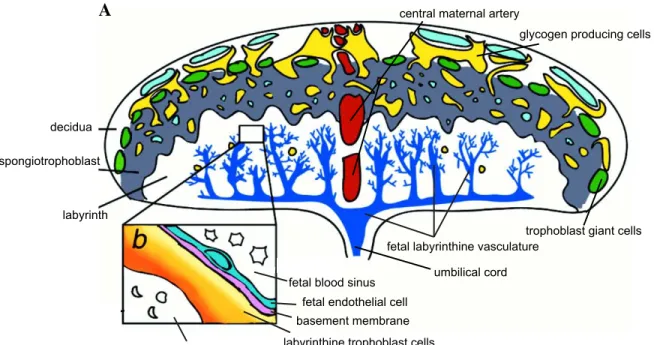
1.3 The murine chorioallantoic placenta... 32
1.3.1 The placenta: overview. ... 32
1.3.2 Early development and the origin of embryonic and extra-embryonic lineages in the mouse. ... 35
1.3.3 Labyrinth development... 38
Gastrulation and emergence of the allantois...38
Elongation and vascularization of the allantois. ...40
Chorio-allantoic union. ...41
Chorion vascularization...42
1.4 Hox gene regulation. ... 46
1.4.1 Hox gene regulation in the primary body axis... 46
Initiation of Hox gene expression. ...46
Establishment of expression domains...47
Maintenance of gene expression...51
Aim of the thesis. ... 60 Chapter 2... 62 2.1 Author contribution:... 64 2.2 Abstract: ... 65 2.3 Introduction :... 66 2.4 Results:... 67
2.4.1 Hoxa10 and Hoxa11 together with Hoxa13 contribute to the development of the labyrinthine vasculature... 67
2.4.2 5’Hoxa genes are expressed in progenitors of the labyrinthine vasculature... 69
2.4.3 Expression of 5’Hoxa genes in the allantois is required for embryonic survival. ... 72
2.4.4 Extra-embryonic recruitment of 5’Hoxa genes is specific to the allantois and is not restricted to placental mammals... 73
2.4.5 The transcriptional control of 5’Hoxa genes in the allantois involves an enhancer-sharing mechanism. ... 74
2.5 Discussion. ... 77
2.6 Materials and Methods... 83
2.6.1 Mouse strains... 83
2.6.2 In Situ Hybridization, Immunohystochemistry and X-gal staining. ... 84
2.8 Legends to figures... 86 Figure 2.1 Deletion of the HoxA cluster leads to impaired vasculature in the placental labyrinth... 86 Figure 2.2 Hoxa10, Hoxa11 and Hoxa13 are the only members of the HoxA cluster
expressed in the allantois... 87 Figure 2.3: Initial expression of Hoxa13 does not occur in endothelial cells of the allantoic vasculature... 87 Figure 2.4: Hoxa13lin+ cells become progressively endothelial only in the labyrinth... 88 Figure 2.5: Delay in the induction of Hoxa13 inactivation is sufficient to ensure proper development of the labyrinth and survival of the embryo. ... 89 Figure 2.6: Expression of 5’Hoxa genes in chick allantois, and evidence for a shared
allantois enhancer in mice. ... 89 Figure 2.7: Deletion of the Hoxa13-Evx1 intergenic region does not prevent Hoxa10,
Hoxa11 and Hoxa13 expression in the allantois. ... 90 2.9 Legends to supplementary figures. ... 98
Figure S 2.1 Expression of Hoxa10 and Hoxa11 is detected at early stages in the allantois, but is not maintained in the allantois-derived labyrinthine vasculature. ... 98 Figure S 2.2 Inactivation of Hoxa genes does not interfere with the formation of the
primary vascular plexus within the allantois... 99 Figure S 2.3 The fetal vasculature of the mature labyrinth is formed of Hoxa13lin+ and Hoxa13lin- endothelial cells. ... 99 Figure S 2.4: Hoxa13 function is dispensable for endothelial cell differentiation. ...100
Figure S 2.5: The yolk sac from HoxAdel/del mutant is indistinguishable from wild-type
yolk sac. ...100
Figure S 2.6: Loss of allantois expression upon subdivision of the Hoxa13-Evx1 intergenic region. ...101 Chapter 3...109 3.1 Author contribution:...111 3.2 Abstract: ...112 3.3 Introduction:...113 3.4 Results:...116
3.4.1 Generation and validation of the Hoxa13Cre mice. ...116
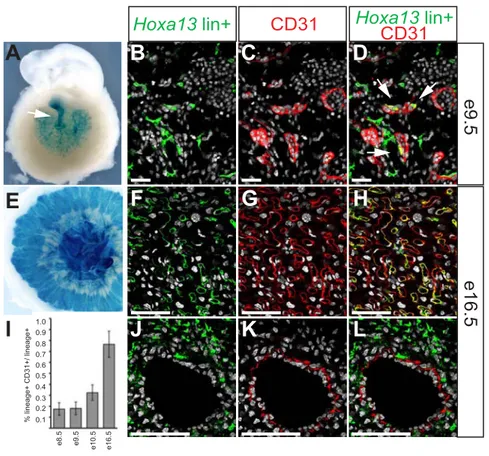
3.4.2 Hoxa13 fate-mapping analysis in the developing embryo. ...117
3.4.3 Hoxa13-expressing cells and their descendants mark a subpopulation of myogenic progenitors in the developing forelimb pre-muscular masses...119
3.4.4 Hoxa13lin+ cells form muscular fibers of a subset of limb muscles...121
3.4.5 Hoxa13lin+ cells contribution to the limb skeleton is restricted to the autopod. ...123
3.5 Discussion. ...124
3.5.1 Hoxa13 is a distal marker for the limb skeleton...124
3.5.2 Hoxa13lin+ cells are part of the limb musculature...125
3.5.3 Hoxa13 function in the limb musculature...127
3.6 Materials and Methods...130
3.6.1 Targeting and generation of the Hoxa13Cre mice. ...130
3.6.2 Genotyping and mating schemes...131
3.6.3 Whole mount in situ hybridization, X-gal staining and imaging. ...132
3.6.4 Immunostaining...132
3.6.5 Muscle nomenclature...133
3.7 Legends to figures...133
Figure 3.1: Generation of the Hoxa13Cre mouse line. ...133
Figure 3.2: Comparison between Hoxa13 expression and Hoxa13lin+ cells distribution at different stages of embryonic development...134
Figure 3.3: Hoxa13lin+ cells in the forelimb bud and early forelimb are not restricted to the presumptive autopod domain...135
Figure 3.4: Myogenic progenitors within the developing ventral and dorsal muscular masses of the forelimb are also Hoxa13lin+. ...135
Figure 3.5: At later stages of development, the distribution of Hoxa13lin+ cells in the forelimb has a pattern reminiscent of the forming musculature...136
Figure 3.6: The distribution of Hoxa13lin+ cells in the limb skeleton marks the transition between autopod and zeugopod. ...136
Figure 3.7: Hoxa13lin+ contribution to muscular masses of the zeugopod and stylopod at e14.5...137
Figure 3.8: Hoxa13lin+ contribution to muscular masses of the zeugopod and stylopod at e18.5...137
Chapter 4: Discussion...148
4.1 Hoxa gene regulation in the extra-embryonic compartment. ...149
4.1.1 Identification of cis-regulatory elements for Hoxa genes active in the allantois... 150
4.1.2 Loss of enhancer activity upon fragmentation of the IR50 transgene... 154
4.1.3 Hoxa gene expression in the allantois is restricted to 5’Hoxa genes... 156
4.1.4 Conclusion... 158
4.2 Hoxa gene function in the muscles...159
4.2.1 The Hoxa13Cre line is a tool to explore molecular identities of limb muscles. ... 160
4.2.2 The Hoxa13Cre line is a tool to explore Hoxa13 function in limb muscles at later stages of development. ... 162
4.2.3 Hoxa gene function in muscle development... 166
4.2.4 Conclusion... 168
List of figures and tables
Figure 1. 1 Hox gene clusters in the mouse. ___________________________________________________ 4 Figure 1. 2 Homeotic transformations and functional redundancy among paralogous Hox genes. ________ 20 Figure 1. 3 The murine placenta. ___________________________________________________________ 34 Figure 1. 4 Stages of mouse preimplantation development. ______________________________________ 37 Figure 1. 5 Labyrinth development in the mouse. ______________________________________________ 45 Figure 1. 6 Global regulatory mechanisms of the murine HoxD locus. _____________________________ 59 Figure 2 .1 Deletion of the HoxA cluster leads to impaired vasculature in the placental labyrinth. _______ 91 Figure 2. 2 Hoxa10, Hoxa11 and Hoxa13 are the only members of the HoxA cluster expressed in the
allantois. ______________________________________________________________________________ 92 Figure 2. 3 Initial expression of Hoxa13 does not occur in endothelial cells of the allantoic vasculature. __ 93 Figure 2. 4 Hoxa13lin+ cells become progressively endothelial only in the labyrinth. _________________ 94 Figure 2 5 Delay in the induction of Hoxa13 inactivation is sufficient to ensure proper development of the labyrinth and survival of the embryo. _______________________________________________________ 95 Figure 2. 6 Expression of 5’Hoxa genes in chick allantois, and evidence for a shared allantois enhancer in mice. _________________________________________________________________________________ 96 Figure 2. 7 Deletion of the Hoxa13-Evx1 intergenic region does not prevent Hoxa10, Hoxa11 and Hoxa13 expression in the allantois.________________________________________________________________ 97 Figure S 2. 1 Expression of Hoxa10 and Hoxa11 is detected at early stages in the allantois, but is not maintained in the allantois-derived labyrinthine vasculature. ___________________________________ 102 Figure S 2. 2 Inactivation of Hoxa genes does not interfere with the formation of the primary vascular plexus within the allantois. ____________________________________________________________________ 103 Figure S 2. 3 The fetal vasculature of the mature labyrinth is formed of Hoxa13lin+ and Hoxa13lin-
Figure S 2. 4 Hoxa13 function is dispensable for endothelial cell differentiation. ____________________ 105 Figure S 2. 5 The yolk sac from HoxAdel/del mutant is indistinguishable from wild-type yolk sac._______ 106 Figure S 2. 6 Loss of allantois expression upon subdivision of the Hoxa13-Evx1 intergenic region.______ 107 Table S 2. 1 Transgenic analysis of the intergenic region between Hoxa13 and Evx1. ________________ 108 Figure 3. 1 Generation of the Hoxa13Cre mouse line. _________________________________________ 139 Figure 3. 2 Comparison between Hoxa13 expression and Hoxa13lin+ cells distribution at different stages of embryonic development._________________________________________________________________ 140 Figure 3. 3 Hoxa13lin+ cells in the forelimb bud and early forelimb are not restricted to the presumptive autopod domain._______________________________________________________________________ 141 Figure 3. 4 Myogenic progenitors within the developing ventral and dorsal muscular masses of the forelimb are also Hoxa13lin+. ___________________________________________________________________ 142 Figure 3. 5 At later stages of development, the distribution of Hoxa13lin+ cells in the forelimb has a pattern reminiscent of the forming musculature. ____________________________________________________ 143 Figure 3. 6 The distribution of Hoxa13lin+ cells in the limb skeleton marks the transition between autopod and zeugopod._________________________________________________________________________ 144 Figure 3. 7 Hoxa13lin+ contribution to muscular masses of the zeugopod and stylopod at e14.5. _______ 145 Figure 3. 8 Hoxa13lin+ contribution to muscular masses of the zeugopod and stylopod at e18.5. _______ 146 Table 3. 1 Limb muscle nomenclature.______________________________________________________ 147
Acknowledgments
I wish to thank my supervisor, Dr. Marie Kmita, for giving me the opportunity to work in her lab. During all these six years, I greatly appreciated her support, help and encouragement, both from the professional and personal point of view. I also wish to thank Prof. Denis Duboule of the University of Geneva and the Swiss Ph.D program NCCR “Frontiers in Genetics” for giving me the privilege of joining their doctoral school and for their financial support at the beginning of my Ph.D in Geneva and during the first year here in Canada.
I am also honored and pleased that Dr. Jacqueline Deschamps, Dr. Hans Larrson and Dr. Jean-Philippe Gratton accepted to be members of my thesis jury.
I also want to thank the members of my Ph.D committee for their help and advice over these years: Dr. Jean-Francois Coté, Dr. Jean Vacher, Dr. Jean-Philippe Gratton, Dr. Maxime Bouchard and Dr. Michel Cayouette.
All these years wouldn’t have been the same without the Kmita lab members. It has been a privilege to work with such great people. Many of you are and have been my family and friends over the last six years. A special thank you to Damien Gregoire, Gemma de Martino, Mark Cwajna and Tong Yu Wang. I miss you very much and it was very hard to finish this Ph.D without your daily presence. Thank you also to the current members, especially Rushikesh Sheth, for his advice and discussion, which I really appreciated in this
last phase of my studies, and Jessica Pham, for her good mood and energy. Moreover, I greatly appreciated Jessica’s help with the French translation of the abstract of this thesis.
I want also to thank many scientist and friends here at the IRCM, for exchanging ideas and coffees: Elena, Wendy, Manishha, Amel, Chris, Mat, Christine, Vas, Jimmy, Dom&Shuofei.
This thesis wouldn’t exist without Luisa. Thanks for your daily encouragement and help during these last months and for being such a good friend! Thanks also to Elena, Luisa’s mother, for adopting me and for being so generous and kind.
Finally I wish to thank my father Maurizio and his wife Tiziana for their constant support, and Nasr, whose presence over the last year has been extremely precious for my equilibrium.
1.1 The Hox genes.
1.1.1 Key features of Hox genes.
Members of the Hox gene family play a pivotal role in axial patterning of animals with bilateral symmetry, among which they are surprisingly conserved (Carroll, 1995; Duboule, 1992; Krumlauf, 1994). Hox genes were first identified in the fruit fly Drosophila melanogaster, in which they confer segment identity along the primary anterior-posterior (AP) body axis (Lewis, 1978). When mutated in the fly, loss of Hox genes cause dramatic homeotic phenotypes, conditions in which one body segment is transformed into the identity of another one. It was subsequently discovered that Hox genes code for transcription factors sharing a sequence of 60 amino acids DNA-binding domain, referred to as the homeodomain (Scott and Weiner, 1984), related to the helix-turn-helix motif of prokaryotic DNA-binding proteins (Kissinger et al., 1990; Otting et al., 1990). Despite their key function during embryogenesis, relatively little is known about the molecular events that these transcription factors trigger. Hox proteins bind DNA with little specificity, recognizing a core sequence composed by only four nucleotides. Moreover, due to the high conservation of the homeodomain, most Hox proteins bind in vitro to simple sequences with the same affinity (Ekker et al., 1994; Hoey and Levine, 1988). This suggests that the specificity achieved by these transcription factors in vivo is most likely due to the additional presence of cofactors on target genes promoters. Different Hox co-factors have been identified, including members of the Pbx and Meis TALE homoedomain transcription
factors (reviewed for example in (Mann and Affolter, 1998; Moens and Selleri, 2006)). Multiprotein complexes composed by Hox and cofactors bind DNA sequences, acting both as activators or repressors of transcription.
In most vertebrates 39 Hox genes have been identified. These genes are clustered on four genomic loci spanning over 100 to 150 kilobase pairs (kb), referred to as HoxA, B, C and D clusters. Each Hox cluster contains a series of nine to eleven contiguous genes transcribed from the same DNA strand, thus defining a 5’ to 3’ polarity to the cluster. Genes are referred to paralogous groups 1 to 13 based on their sequence similarity with genes located on the other clusters. Even if some paralogous group are missing in each cluster, the gene order is always maintained, such that group 1 genes are always located at the 3’ end of the complex, and group 13 at the 5’ (Krumlauf, 1992; Scott, 1992) (Fig. 1.1).
3’ 5’ HoxA HoxB HoxC HoxD anterior posterior
Figure 1.1 Hox gene clusters in the mouse.
In the mouse, as in most vertebrates, 39 Hox genes have been identified. These genes are clustered on four genomic loci, referred to as HoxA, B, C and D clusters. Each Hox cluster contains nine to eleven contigous genes transcribed from the same DNA strand, conferring a 5’ to 3’ polarity of the cluster. Hox genes are expressed in overlapping domains during embryogenesis, such that their anterior limit of expression is collinear with the gene positions along the complex. Accordingly, genes located at the 3’ of each Hox clusters are expressed in more anterior structures of the developing embryo, while more 5’ Hox gene transcripts are restricted to more posterior domains of the body.
1.1.2 Evolution of Hox gene clusters.
Importantly, the function of Hox genes in patterning the AP axis is conserved among all species, and changes in the body plan organization among different species are generally associated with changes in Hox gene number, pattern or boundary of expression (Burke et al., 1995; Cohn and Tickle, 1999). Hox genes have been identified in all bilaterian animals investigated so far (de Rosa et al., 1999), and, in most cases, they show a clustered organization, with the exception of the platyhelminth Schistosoma mansoni and the urochordate Oikopleura dioica, in which Hox genes are found scattered in the genome, with little if any linkage (Pierce et al., 2005; Seo et al., 2004). Despite its evolutionary and developmental significance, the origin of the Hox gene clusters remains obscure. However, the highly conserved organization described in most bilaterian Hox clusters indicates that the common bilaterian ancestor had a set of clustered Hox genes. It has been proposed that during early evolution, an ancestral ProtoHox cluster has emerged by tandem gene duplication in cis of a single ancestral ProtoHox gene (Garcia-Fernandez, 2005). This early gene amplification would also explain the clustered organization of Hox genes that is found already in cnidarians (animals with a radial symmetry) (Chourrout et al., 2006; Ryan et al., 2007). Subsequently, whole cluster duplication and split of the resulting sister clusters generated the Hox and ParaHox clusters (Brooke et al., 1998). From this putative ground state, a wide variety of Hox gene organizations have evolved. In all invertebrates investigated so far, including the insect Drosophila melanogaster, only one single Hox
cluster has been identified. The Drosophila Hox cluster (HOM-C) contains eight genes: Labial (lb), Proboscipedia (Pb), Deformed (Dfd), Sex comb reduced (Scr), Antennapedia (Antp), Ultrabihtorax (Ubx), Abdominal-A (Abd-A) and Abdominal-B (Abd-B). This single cluster was split into two sub-clusters, the Antennapedia (ANT-C) containing the first five genes, and the Bithorax (BX-C) containing the last three (Kaufman et al., 1980). The consensus view is that a single Hox cluster was at the origin of vertebrate evolution, a situation potentially reflected today by the single cluster of the cephalochordate Amphioxus (Garcia-Fernandez and Holland, 1994). The number of Hox genes within the cluster increased during evolution, as exemplified by the expansion of the Abd-B-related gene repertoire in Amphioxus, leading to a cluster of 14 Hox genes in this organism (Ferrier et al., 2000). This single ancestral Hox cluster was subsequently amplified in the vertebrate lineage, possibly following successive whole genome duplications near the origin of vertebrates. For example, the lamprey, a jawless primitive vertebrate, has three (possibly four Hox clusters) (Force et al., 2002; Irvine et al., 2002). In higher vertebrates, like mammals, two rounds of whole genome duplications most likely originated a total of four paralogous clusters, referred to as HoxA to HoxD. Members of each cluster can be classified in paralogy groups, from 1 to 13. Paralogy groups 1 to 8 are homologous to the Drosophila genes Labial, Proboscipedia, Deformed, Sex comb reduced, Antennapedia, Ultrabitorax, and Abdominal-A, while the paralogy group 9 to 13 are related to the Abdominal-B gene (Krumlauf, 1994). In teleost fishes, an additional round of genome
duplication occurred, generating seven Hox clusters in zebrafish, one of the HoxD clusters being lost (Amores et al., 1998; Woltering and Durston, 2006). Interestingly however, the total number of genes in zebrafish is not much higher than in other species with only four clusters, most of the duplicated genes being lost.
It was proposed that the expansion of Hox clusters resulted in looser evolutionary constraints on Hox genes, allowing them to acquire novel functions (Holland and Garcia-Fernandez, 1996). Accordingly, the duplication of Hox clusters in higher vertebrates was accompanied by gene loss: as a result, none of the four Hox clusters displays all 13 paralogy groups. Comparing vertebrate Hox gene clusters with their invertebrate counterparts reveals that the firsts show the higher degree of organization (reviewed in (Duboule, 2007)). In fact, in vertebrates, Hox clusters are considerably more compact than in other species, and no non-Hox genes are found interspersed in the complexes, as it is the case for Drosophila and the sea urchin. Furthermore, in most vertebrates, repetitive sequences are excluded from the Hox clusters, arguing for a strong selective pressure to exclude the invasion by mobile genetic elements at the base of vertebrate evolution (Amemiya et al., 2008; Fried et al., 2004). An exception to repetitive elements exclusion within Hox complexes in vertebrates has been recently reported in both squamates (like lizards and snakes) and caecilieans (snake-like amphibians) (Di-Poi et al., 2009; Mannaert et al., 2010). Interestingly these organisms display a divergent body plan organization from the other vertebrates. Changes in Hox genes expression, possibly deriving from this altered
Hox cluster organization, have been proposed as a possible explanation for this striking morphological diversity (Di-Poi et al., 2010b). Two scenarios can account for the increased organization of vertebrate Hox genes. Either the tightly structured organization found nowadays in vertebrates represents the evolutionary ground state that eventually deteriorated during the evolution of other lineages, or, alternatively, a disorganized Hox complex underwent a “consolidation” and compaction process in the case of vertebrates (Duboule, 2007). To support this latter hypothesis, an increased regulatory complexity at vertebrate loci has been proposed as a potential evolutionary constraint for Hox cluster compaction, including both newly acquired expression specificity, as well as ancient collinear transcription mechanisms.
1.1.3 Collinearity.
One of the most fascinating features of Hox genes is the phenomenon of collinearity, which stands for the correspondence between gene order within each Hox cluster and the distribution of gene transcripts along the main body axis (reviewed in (Kmita and Duboule, 2003)). This property was first described in Drosophila, where the distributions of Hox transcripts, as well as their domain of action along the AP axis of the embryo, are collinear to the Hox gene location along the chromosome (Lewis, 1978). Subsequently, the same kind of “spatial collinearity” was found to occur also in vertebrates, where genes of all four clusters show expression territories along the AP axis according to
the relative position they occupy within their respective complexes (Duboule and Dolle, 1989; Gaunt, 1988; Graham et al., 1989). Hox genes in vertebrates are expressed in overlapping domains during embryogenesis, such that their anterior limit of expression is collinear with the gene positions along the complex. Accordingly, genes located at the 3’ of each Hox clusters are expressed in more anterior structures of the developing embryo, while more 5’ Hox gene transcripts are restricted to more posterior domains of the body (Duboule and Dolle, 1989; Graham et al., 1989). This “spatial collinearity”, analogous to the one described in Drosophila, is observed in a variety of tissues along the main body axis, such as the neural tube and the paraxial mesoderm, but also in the gastrointestinal tract and urogenital system (Gaunt, 1988; Yokouchi et al., 1995) (Graham et al., 1989). Additionally, similar links between gene order and nested expression domains are found in secondary body axis, such as the external genitalia and limbs (Dolle and Duboule, 1989; Dolle et al., 1991; Haack and Gruss, 1993; Nelson et al., 1996).
“Spatial collinearity” has been reported in all bilaterian organisms investigated so far, independently from the strict clustered organization of the Hox complex (as reported in C. elegans (Wang et al., 1993)). Strikingly, even organisms with a completely fragmented Hox gene organization present such a “trans-collinearity”, and Hox genes are still expressed in the body axis accordingly to groups of paralogy (Duboule, 2007; Seo et al., 2004). This apparent lack of interdependence between clustering and “spatial collinearity” challenges the claim that this phenomenon might represent a major constraint on maintaining Hox
genes clustered together. Furthermore, in the mouse, randomly integrated transgenes carrying single Hox genes with relatively reduced neighboring sequence could reproduce major aspects of the endogenous gene’s spatial transcript distribution (Puschel et al., 1991; Whiting et al., 1991).
However, a Hox transgene randomly inserted into the genome shows some differences in the temporal dynamics of activation as compared to the endogenous gene, raising the possibility that the clustering of Hox genes is required for the fine-tuning during Hox gene activation process. In fact, unlike Drosophila genes, vertebrate Hox genes display an additional degree of complexity, resulting in the link between the onset of their expression during development and their position inside the complexes. This phenomenon is referred to as “temporal collinearity” and is observed in all vertebrates, in both the primary and secondary axes of the developing embryo ((Dolle and Duboule, 1989); reviewed in (Deschamps and van Nes, 2005)). In the vertebrate primary body axis, Hox genes start to be expressed during early gastrulation, following a subsequent activation from the 3’ end to 5’ end of the clusters. This process is completed by late tail bud stage, around embryonic day 9 (e9) (Dolle et al., 1989; Izpisua-Belmonte et al., 1991). Genes activated early on are expressed in more anterior structures of the embryo, while genes activated later on are progressively restricted to more posterior embryonic compartments.
1.2 Hox gene function.
1.2.1 Homeotic transformations.
Genetic analysis in Drosophila has shown that mutations in Hox genes give rise to transformation of a specific part of the body of the fly into a completely different one: this phenomenon is called homeotic transformation. For example, in the Antennapedia mutation, the fly develops an extra pair of legs in place of the antenna, while the Bithorax mutation leads to the growth of an extra pair of wings (Lewis, 1978). It was shown that loss of function mutation in Hox genes usually lead to anteriorizing homeotic transformations, i.e., the transformation of a body segment into a more anterior one. Vice-versa, gain of function of a specific Hox gene anterior to the position in which it is normally expressed imposes a posterior identity to the segment where the Hox gene is ectopically expressed (Akam, 1987; Lewis, 1978; McGinnis and Krumlauf, 1992). Results obtained over the last three decades have demonstrated that the function of Drosophila Hox genes in specifying the identity of the different body segments along the AP axis is largely conserved in vertebrates. Each of the 39 Hox genes, as well as entire Hox clusters, has been genetically inactivated in the mouse. In parallel, classical transgenesis in the mouse was used to investigate the phenotypic outcomes of misexpressing a specific Hox gene in an embryonic domain in which it is not normally expressed.
Altogether, these studies have provided much support to the idea that vertebrate Hox genes, as well as their Drosophila counterparts, can specify morphological identity of metameric structures in the body plan. This view is also supported by evidence in other animal model organisms, like Xenopus and Zebrafish, in which mRNA injections or antisense approach experiments have been performed (Cho et al., 1991; McClintock et al., 2002).
1.2.2 Hox gene function in the primary vertebrate body axis.
During vertebrate embryogenesis the extension of the body along the AP axis is coupled with the process of somitogenesis. The segmented structure of the vertebrate body plan is mostly apparent at the level of somatic mesoderm derivatives, such as the vertebrae, but also at the level of the central nervous system, where, for example, the hindbrain is subdivided into eight rhombomeres. Consistently, alteration of Hox expression mainly affects the morphology of these segmented structures (reviewed in (Burke, 2000; Lumsden and Krumlauf, 1996; Trainor and Krumlauf, 2001)).“Hox code” and posterior prevalence:
In vertebrates, Hox expression patterns in the trunk, unlike in Drosophila, is characterized by large domains of expression, partially overlapping in the posterior region
of the main body axis. This led to the hypothesis that the identity of segments at different axial levels of the embryo relies on a different combination of Hox proteins, referred to as the “Hox code” (Kessel and Gruss, 1991). However, loss-of-function experiments in the mouse usually lead to morphological changes confined to the most rostral segments in which a given Hox gene is normally expressed. These observations indicate that a functional hierarchy exists among Hox genes, and that the most posterior Hox gene that is expressed in a determined segment imposes its function over more anterior Hox genes. This model is referred to as “posterior prevalence” (Duboule and Morata, 1994). In this view, nested and overlapping expression domains are only a way to confer discrete identities to the undifferentiated extending body axis. Notably, the phenomenon of “posterior prevalence” is not limited to the main body axis, but occurs also in secondary axis, like the limbs (see e.g. (Herault et al., 1997; Kmita et al., 2002a; Peichel et al., 1997; van der Hoeven et al., 1996)). “Posterior prevalence” is related to the phenomenon of “phenotypic suppression” described in Drosophila. Unlike in vertebrates, the distribution of Hox transcripts along the body axis in Drosophila is characterized by largely non-overlapping patterns. Posterior Hox proteins were shown to down-regulate the expression of more anterior Hox genes. Ectopic expression of Hox genes in Drosophila, however, demonstrated that this effect does not involve transcriptional repression, but rather acts at the post-transcriptional level (Duboule and Morata, 1994).
Loss-of-function phenotypes:
As mentioned earlier, loss-of-function mutations induce anteriorization mainly confined to the region corresponding to the anterior limit of expression of a given Hox gene. For example, the inactivation of Hoxa1, which is normally expressed up to rombobere 4 (r4) in the hindbrain, produces abnormalities only in r4 to r8 and their derivatives, leaving more anterior or posterior structures of the body unaffected (Carpenter et al., 1993; Chisaka et al., 1992; Lufkin et al., 1991). The inactivation of Hoxa2 leads to anterior homeotic transformation of the cranial neural crest derivatives from the second branchial arch to a first branchial arch identity (Gendron-Maguire et al., 1993; Rijli et al., 1993).
In the axial skeleton, inactivation of Hox genes leads to anterior transformation of vertebrae. For example, Hoxa4 mutants show partial transformation of the third cervical vertebra (C3) towards a C2 identity (Kostic and Capecchi, 1994). Inactivation of Hoxb4 or Hoxd4 leads to partial transformation of C2 into C1 (Horan et al., 1995a; Ramirez-Solis et al., 1993). In more posterior domains of the trunk, homeosis of thoracic vertebrae is observed in many mutants. For example, Hoxc4 inactivation leads to anterior transformation of the third thoracic vertebra (T3) (Saegusa et al., 1996). Hoxc8 mutants show anterior transformation of T8 to T7 and of the fist lumbar vertebrae (L1) to T13 (Le Mouellic et al., 1992). Finally, several examples of inactivations of Abd-B- related genes illustrate the requirement for these genes in conferring proper lumbo-sacral vertebral
identity. Following inactivation of Hoxa9, anterior homeosis of the first five lumbar vertebrae is observed (Fromental-Ramain et al., 1996a). Inactivation of the paralogous Hoxd9 leads to anterior transformations from L3 to the first caudal vertebra, which assumes a sacral identity (Fromental-Ramain et al., 1996a). Similarly, upon Hoxd11 inactivation, the sacral region displays anterior transformation, resulting in a posterior shift of the sacrum (Davis and Capecchi, 1994; Favier et al., 1995).
Gain-of-function phenotypes:
In many cases, ectopic Hox expression leads to posterior transformations, mainly restricted to regions of the body that are rostral to the normal limit of expression of the gene. For example, ectopic widespread expression of Hoxa1 leads to rhombomere transformation, resulting in change of r2 and r3 towards r4 identity (Zhang et al., 1994). Ectopic expression of Hoxa7 under the control of the ubiquitous β-actin promoter leads to the appearance of an additional first cervical vertebra and other severe cranio-facial defects (Kessel et al., 1990). The expression of Hoxd4 under the control of the Hoxa1 promoter leads to transformation of the occipital bones into structures resembling cervical vertebrae, since neural arches are induced (Lufkin et al., 1992). Hoxb8 expression under the retinoic acid receptor (RAR) β2 promoter leads to cervical vertebrae adopting a more posterior identity (Charite et al., 1995).
Unexpected phenotypes:
In some cases, Hox gene inactivation results in posterior, rather than anterior transformations, or in a complex combination of both. These results may, however, reflect technical limitations of gene-targeting technology as it was originally developed. For instance, mice lacking Hoxb2 present partial transformation of C2 in C1 and splitting of the sternum into two longitudinal structures (Barrow and Capecchi, 1996). This sternal defect is reminiscent of Hoxb4 inactivation, and consistently, Hoxb4 expression is decreased or suppressed in these mutants. In parallel, Hoxb2 mutant mice display facial paralysis, as do mice with Hoxb1 inactivation (Goddard et al., 1996). Accordingly, Hoxb1 expression in neural crest cells is abrogated in these mutants (Barrow and Capecchi, 1996). Hoxa11 inactivation leads to posterior homeotic transformation probably linked to Hoxa10 misexpression (Branford et al., 2000; Small and Potter, 1993).
Targeting constructs employed to generate gene inactivation typically include a selection cassette, to allow selection of integration events in ES cell clones. The presence of strong promoters in the selection cassettes, such as the phosphoglycerate kinase promoter (PGK), can perturb the regulation of neighboring genes, competing for enhancer activity, or, alternatively, the targeting of the cassette can disrupt regulatory elements, ultimately leading to loss or gain-of-expression of one or several genes (Beckers and Duboule, 1998; Rijli et al., 1994; Zakany et al., 1997b). More recently, the Cre-loxP recombinase approach has been used to ultimately remove the selection cassette. However, most Hox gene
inactivations were produced before the advent of this technique, and still include the selection cassette. Thus, the resulting phenotypes should be interpreted with caution.
Conversely, Hox gene gain of function can also induce unexpected anterior vertebral transformations. For example, transgenic mice carrying over 40 copies of a human Hoxc6 transgene present anterior transformation of thoracic and lumbar vertebrae. However, this doesn’t happen when a lower copy number of the transgene is integrated, suggesting that a quantitative effect is responsible for the former phenotype (Jegalian and De Robertis, 1992). More recently, to circumvent this problem, single-copy integration of a transgene can be achieved using retroviruses and transposons (Ding et al., 2005; Lois et al., 2002; Mates et al., 2009).
Functional redundancy:
In some cases, the inactivation of a single Hox gene does not result in any obvious phenotype. This is the case for instance with Hoxa7 and Hoxd8 mutation (Chen et al., 1998; van den Akker et al., 2001). In general, compared to the drastic effect of homeotic transformation in Drosophila, loss-of-function mutations in mice result in relatively mild effects.
For example, inactivation of the Hox4 paralogs (Hoxa4, Hoxb4 and Hoxd4) results in dose dependent increase in the severity of the resulting phenotype, leading to an increment in the number of cervical vertebrae adopting a C1 identity, compared to what is
observed in single mutants (Horan et al., 1995a; Horan et al., 1995b). Inactivation of Hox9 paralogs (Hoxa9, Hoxb9, Hoxc9 and Hoxd9) leads to transformation of posterior thoracic and anterior lumbar vertebrae into a more anterior thoracic morphology (McIntyre et al., 2007). Surprisingly, the presence of a single wild-type allele out of eight was sufficient to drastically reduce the severity of all these observed phenotypes. The inactivation of Hox10 paralogs (Hoxa10, Hoxc10 and Hoxd10) results in mice with transformation of all lumbar vertebrae into rib-bearing structures, similar to thoracic vertebrae. Moreover, inactivation of Hox11 paralogs (Hoxa11, Hoxc11 and Hoxd11) causes transformation of sacral vertebrae to a lumbar phenotype (Wellik and Capecchi, 2003). Also in these cases, the presence of a single wild-type allele out of eight could drastically reduce the severity of the phenotypes observed in Hox triple mutants (Fig. 1.2).
Thus, the duplication of the ancestral Hox cluster in the vertebrate lineage, and the resulting generation of different members for the same paralogy group, is likely to account for this effect. This suggests that Hox genes of the same paralogy group, if not others, can have partially redundant functions.
In other cases, genetic interactions among paralogs are more complex. For example, Hoxa3 and Hoxd3 single mutants do not display overlapping phenotypes, even if they are expressed in the same structures. However, double gene inactivation causes exacerbation of both individual phenotypes revealing an unexpected synergism (Chisaka and Capecchi, 1991; Condie and Capecchi, 1993). Another interesting result is obtained by combined
gene inactivation of Hoxb8, Hoxc8 and Hoxd8. Single mutants display some distinct phenotypes (Le Mouellic et al., 1992; Tiret et al., 1993; van den Akker et al., 1999). Nevertheless, compound inactivation reveal synergistic interaction in the patterning along the AP axis, yet some phenotypes observed in Hoxc8/Hoxd8 double mutants are rescued by Hoxb8 inactivation, suggesting that qualitative differences among Hox8 paralogous proteins may exist (van den Akker et al., 2001).
Even though Hox paralogous genes show closer sequence similarity than neighboring genes of the same cluster, there are several examples of functional redundancy among non-paralogous genes located in trans or even in cis. If we look at Hoxa10 and Hoxd11 compound mutants, animals display up to eight lumbar vertebrae with concomitant shift of the sacrum, a phenotype reminiscent of Hoxa11/Hoxd11 compound mutants, but distinct from single mutants involving Hoxa10, Hoxa11 or Hoxd11 inactivation (Favier et al., 1996). In some other cases, functional redundancy was even reported between neighboring genes in cis. Hoxb5/Hoxb6 trans-heterozygous mutants show the same phenotype as both homozygous, carrying C6 to C5 and T1 in C7 anterior transformations (Rancourt et al., 1995).
Many other examples of functional redundancy have been reported in other tissues, such as the limbs (see for example (Fromental-Ramain et al., 1996a; Fromental-Ramain et al., 1996b; Wellik and Capecchi, 2003)) or the kidney (Davis et al., 1995).
Figure 1.2 Homeotic transformations and functional redundancy among paralogous Hox genes.
Representation of a wild-type skeleton (B).The inactivation of Hox10 paralogs (Hoxa10, Hoxc10 and Hoxd10) results in mice with transformation of all lumbar vertebrae (yellow) into rib-bearing structures, similar to thoracic vertebrae (A). Inactivation of Hox11 paralogs (Hoxa11, Hoxc11 and Hoxd11) causes transformation of sacral vertebrae (red) to a lumbar phenotype (C). Surprisingly, the presence of a single wild-type allele out of eight is sufficient to reduce drastically the
observed phenotype (D, E).
(modified from Wellik and Capecchi, 2003)
B
C
1.2.3 Hox genes and evolutionary novelties.
In the course of vertebrate evolution, besides their ancestral function in patterning the main body axis, Hox genes have been recurrently co-opted to pattern novel structures of the body. Due to two rounds of whole-genome duplications, higher vertebrates present four Hox clusters and thus up to four paralogous genes for each paralogy group. The presence of up to four paralogous genes allowed the allocation of different functions to different genes, often through the acquisition of new expression specificities. In this section, I will review some examples relevant for this thesis and subsequently discuss the specific functions of Hoxa13 in embryonic and extra-embryonic structures.
Hox gene functions in the limbs:
The vertebrate limb is one of the most important morphological adaptations that enabled the transition from aquatic to the terrestrial environment in the course of vertebrate evolution. Work from different laboratories has demonstrated that Hox genes have been recruited to pattern this secondary body axis. Gain and loss-of-function experiments have demonstrated that 5’ members of the HoxA and HoxD clusters are essential for the proper patterning and growth of limb skeleton. Genes of these two clusters are expressed in complex and dynamic patterns during forelimb and hindlimb development, characterized by two different waves of expression (reviewed in (Zakany and Duboule, 2007)). In the early limb bud, 5’ Hoxa and Hoxd genes are sequentially activated in time and space,
reminiscent of the spatial and temporal collinearity occurring in the trunk and their expression patterns are progressively restricted towards the posterior margin of the bud (Dolle et al., 1989; Haack and Gruss, 1993). Later in development, during the second wave of expression, genes of the HoxA and HoxD clusters show a quite distinct and more complex expression profile. In fact, while Hoxd gene expression still show an anterior-posterior polarity, Hoxa genes rapidly loose this anterior-posterior restriction, but are progressively confined to the distal limb domain (proximo-distal polarity). This observation suggests that the regulatory mechanisms underlying Hoxa and Hoxd gene expression in this second phase of expression may have evolved separately, after cluster duplication occurred (Zakany and Duboule, 2007).
It was proposed that the appearance of the second wave of Hox genes expression in the developing limb accompanied the evolution of the most distal limb structures, the digits, from an ancestral fin-like appendage (described as the “fin to limb” transition) (Sordino et al., 1995). However, this view was recently challenged by the discovery that two waves of Hox gene expression are also present in different fishes (Ahn and Ho, 2008; Davis et al., 2007; Freitas et al., 2007; Johanson et al., 2007). Only the analysis of regulatory elements in fishes, and the comparison with the ones already identified in the mouse, will help to understand whether tetrapod digits are an adaptation of pre-existing structures or are a functional novelty that has evolved together with specific new regulatory elements (Woltering and Duboule, 2010).
Patterning defects along the proximo-distal (PD) axis of the limb, caused by Hox gene inactivation, are collinear with the gene position in the complex. Mutation in Hoxa9 and Hoxd9 paralogs leads to mild growth defects in the most proximal domain of the limb, the stylopod, group 11 gene inactivation affects the intermediate region of the limb, the zeugopod, Hoxa13 and Hoxd13 mutants display defects in the digits and carpal/tarsal bones (Davis and Capecchi, 1994; Fromental-Ramain et al., 1996b; Small and Potter, 1993). Compound Hoxa and Hoxd paralogous gene inactivation usually results in much more severe phenotypes than single gene inactivation (Davis et al., 1995; Fromental-Ramain et al., 1996b). Finally, the complete deletions of the HoxA and HoxD clusters in the limbs result in early developmental arrest, leading to a truncated limb at the proximal level of the humerus (Kmita et al., 2005).
Hox genes functions in external genitalia and reproductive tract:
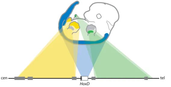
Interestingly, group 9 to group 13 genes of the HoxA and HoxD clusters are also expressed in the genital bud, the structure that will give rise to external genitalia. Deletion of group 13 paralogous genes results in a similar dose-dependent reduction of both penian bone and digits (Kondo et al., 1997; Zakany et al., 1997a). Both external genitals and digits develop in a similar way and share a number of common signalling pathways (Yamada et al., 2006). Most recently, similar regulatory mechanisms have been identified for both structures. Enhancer sequences for both limb and genitals reside on the centromeric side of the HoxD cluster (Spitz et al., 2001; Spitz et al., 2005). Moreover, the Ulnaless inversion,
in which Hoxd genes are moved away from regulatory sequences located on the centromeric side of the cluster, results in down-regulation of Hoxd genes in both digits and genitals (Spitz et al., 2003). Transgenic assays also demonstrated that enhancer elements able to drive reporter gene expressing in the limbs are also active in the genitals (Gonzalez et al., 2007). Finally, Hoxd genes are expressed in a quantitative collinear manner in both structures (Montavon et al., 2008). Altogether, this suggests that the acquisition of new Hox expression specificities has been instrumental for the emergence of both digits and external genitalia during evolution, these structures being a combined adaptation to terrestrial life, allowing for both effective locomotion and internal fertilization.
Placental mammals present essential innovations compared to other animals, such as the differentiation of the oviducts into uterus and upper vagina, the presence of a highly specialized epithelium, referred to as the endometrium, and the placenta, a specialized organ that mediates the feto-maternal exchanges (Wagner and Lynch, 2005). 5’ Hoxa genes are expressed in a collinear manner in the female reproductive tract, such that Hoxa9 transcript is detected in the oviduct, Hoxa10 in the developing uterus, Hoxa11 in the uterus and cervix and Hoxa13 in the upper vagina (Taylor et al., 1997; Warot et al., 1997). Hoxa10 is expressed in the uterine epithelium and is up-regulated during implantation, when the uterine stroma is transformed into decidua. Hoxa10 inactivation leads to anterior homeotic transformation of the uterus in oviduct and homozygous mutant females are partially infertile, as a result of implantation defects (Benson et al., 1996; Satokata et al.,
1995). Similarly, females lacking Hoxa11 present with a small uterus and implantation defects (Gendron et al., 1997; Hsieh-Li et al., 1995). The combined inactivation of Hoxa11 and Hoxd11 results in anterior homeotic transformation of the male reproductive tract, while Hoxd11 loss-of-function confers male sterility (Davis et al., 1995).
1.2.4 Hoxa13 functions in the embryo proper.
Preliminary insights into Hoxa13 gene function came from the discovery that the mouse spontaneous mutant “Hypodactyly” (Hd) carries a deletion of 50 base pairs (bp) in the Hoxa13 coding sequence (Mortlock et al., 1996). The Hd mutation was first described in 1970 (Hummel, 1970) as a semidominant mutation leading to digit defects and shortening of digit I in heterozygous animals, while the majority of Hd/Hd embryos die in utero. Rare homozygous survivors are infertile and display severe autopod defects, characterized by the presence of only one digit and defects in carpal and tarsal elements (Hummel, 1970; Mortlock et al., 1996). Subsequently, two loss-of-function alleles have been generated in mice. The first engineered inactivation of Hoxa13 (referred to as Hoxa13-/-) disrupts the homeobox in the second exon (Fromental-Ramain et al., 1996b) and the second Hoxa13 loss-of-function allele was generated by targeting of a green fluorescent protein (GFP) into exon 2 (hereafter referred to as Hoxa13GFP) (Stadler et al., 2001). Interestingly, the limb phenotype of these Hoxa13 mutants is much less severe than
the Hd/Hd one. In fact, it was found that the Hd mutation leads to the production of a dominant negative Hoxa13 protein, in which the first wild-type 25 amino acids are followed by 275 new residues, resulting from the frame-shift caused by the 50 bp deletion in the gene sequence (Post et al., 2000). In the limb bud of these mutants the expression of other Hox genes is impaired and massive cell-death is detected (Post and Innis, 1999).
Many spontaneous mutations in different regions of the Hoxa13 gene have been described in humans, causing the Hand-Foot-Genital syndrome (HFGS) (Frisen et al., 2003; Goodman et al., 2000; Jorgensen et al., 2010; Mortlock and Innis, 1997; Utsch et al., 2007). HFGS is a rare, dominantly inherited condition in which affected individuals present a phenotype very similar to the one of heterozygous Hd mutants, including digit 1 hypoplasia (short digit 1), brachydactyly (shortening) of the other digits and brachydactyly and clinodactyly (curvature) of digit 5. HFGS is also accompanied by lower genito-urinary tract malformations, such as hypospadias in males and Mullerian duct fusion defects in females, leading in the most severe cases to the presence of a “duplicated uterus” together with ectopic ureteric orifices (Poznanski et al., 1975; Stern et al., 1970)(Donnenfeld et al., 1992; Halal, 1988).
Hoxa13 functions in the limb.
In the mouse, Hoxa13 starts to be expressed in the posterior and distal part of the limb bud around e10. At e12.5 its expression is detected in the entire presumptive autopod,
both in digit condensations and interdigital tissue and later, at e13.5-e14.5, Hoxa13 transcript is restricted to peridigital tissue and interarticular condensations (Haack and Gruss, 1993; Stadler et al., 2001).
Hoxa13-/- adults are not viable due to mid-gestation embryonic lethality. Hoxa13 homozygous mutant embryos can be analyzed up to e15 and display limb abnormalities in the autopod of both forelimb and hindlimbs, characterized by lack of digit 1 chondrogenic condensation, variable syndactyly and barachydactyly of other digits, loss of the second phalangeal cartilage, and delayed or absent pre-cartilaginous condensation for carpal and tarsal elements. Rare homozygous mutants have been recently recovered after birth, allowing for the analysis of skeletal defects in adult mutants that confirmed previous conclusions (Perez et al., 2010). Heterozygous animals are fully viable and fertile and display only mild limb abnormalities, such as fusion of digits 2 and 3 at the level of the soft tissues and alteration of the claw of digit 1 (Fromental-Ramain et al., 1996b). Hoxa13/Hoxd13 compound mutants display a much more severe phenotype compared to the one observed in single loss-of-function inactivation and the severity increases progressively with the number of alleles inactivated (Dolle et al., 1993; Fromental-Ramain et al., 1996b). In fact, the most compromised autopod is observed in Hoxa13-/-; Hoxd13-/- embryos, which display almost complete digit loss and absence of carpal/tarsal condensations, demonstrating the fundamental importance of these paralogous genes in digit formation. Hoxa13 mutants show a more severe phenotype in limb domains where
Hoxa13 function cannot be completely compensated by its paralog Hoxd13, including the forming digit 1 and carpal/tarsal elements of the limb (Haack and Gruss, 1993)
Subsequent studies have aimed to understand the molecular pathways regulated by Hoxa13. Studies using avian embryos suggested that Hoxa13 expression is important to confer cell-cell adhesiveness properties and that Hoxa13 expressing cells are able to selectively associate and form aggregates in vitro (Yokouchi et al., 1995). Further analysis demonstrated that Hoxa13 directly activates the expression of the tyrosine kinase receptor ephrin A7 (EphA7) in limb mesenchymal condensations, and that this activation is required for proper mesenchymal condensation in the limb autopod (Salsi and Zappavigna, 2006; Stadler et al., 2001). Moreover, Hoxa13 activates the expression of bone morphogenetic protein 2 and 7 (Bmp2 and Bmp7) in the autopod by binding to upstream sequences of these genes. In the developing limb, Bmp2 and Bmp7 are key regulators of chondrogenesis as well as interdigital programmed cell death (IPCD) required for proper digit separation (Macias et al., 1997; Merino et al., 1998; Yokouchi et al., 1996; Zou et al., 1997; Zou and Niswander, 1996; Zuzarte-Luis and Hurle, 2002). Decreased expression of Bmp2 and Bmp7 thus underlies loss of IPCD in Hoxa13 homozygous mutants, leading to fused digits (Knosp et al., 2004).
In addition to its function in skeletal pattering, Hoxa13 is also activated in myogenic progenitors of the limb, derived from the hypaxial dermomyotome of the somites. After these cells have entered the limb bud territory, some of them start expressing Hoxa13, and
Hoxa13 expression is maintained in limb the musculature at least until e13.5 (Yamamoto et al., 1998; Yamamoto and Kuroiwa, 2003). However, the functional relevance of Hoxa13 expression in limb muscles remains unclear, and the phenotype of Hoxa13-/- mouse embryos has not been characterized for the presence of patterning or functional defects in the muscular tissues. Studies in chick showed that Hoxa13 expression in myogenic progenitors of the limb is not under the control of signals derived from the apical ectodermal ridge (AER), unlike Hoxa13 expression in mesenchymal cells, but is controlled by signals from the zone of polarizing activity (ZPA), via Bmp2 regulation (Hashimoto et al., 1999). One study has linked Hoxa13 function to the regulation of the transcription factor MyoD, which is involved in the process of muscular differentiation (reviewed in (Tajbakhsh and Buckingham, 2000)). Electroporation experiments in the chick and in vitro studies using myoblast cells showed that forced expression of Hoxa13 inhibits expression of MyoD and the expression of MyoD is consistently enhanced in Hoxa13-/- mutants (Yamamoto and Kuroiwa, 2003). However, the functional relevance of this regulation is not clear and further studies in this direction have not yet been performed, leaving the potential function of Hoxa13 expression in the limb musculature still largely unexplored.
Hoxa13 function in the urogenital system and gastrointestinal tract.
Hoxa13 and Hoxd13 are co-expressed in the mesenchyme of the genital bud and urogenital sinus and, at lower levels, in the urogenital sinus epithelium (Oefelein et al., 1996; Podlasek et al., 1997; Warot et al., 1997). In particular, Hoxa13 is expressed in the