HAL Id: tel-01191476
https://tel.archives-ouvertes.fr/tel-01191476
Submitted on 4 Sep 2015
HAL is a multi-disciplinary open access
archive for the deposit and dissemination of sci-entific research documents, whether they are pub-lished or not. The documents may come from teaching and research institutions in France or abroad, or from public or private research centers.
L’archive ouverte pluridisciplinaire HAL, est destinée au dépôt et à la diffusion de documents scientifiques de niveau recherche, publiés ou non, émanant des établissements d’enseignement et de recherche français ou étrangers, des laboratoires publics ou privés.
Role of vascular plasticity in muscle remodeling in the
child
Cyril Gitiaux
To cite this version:
Cyril Gitiaux. Role of vascular plasticity in muscle remodeling in the child. Molecular biology. Université Sorbonne Paris Cité, 2015. English. �NNT : 2015USPCB029�. �tel-01191476�
1
Université Paris-René Descartes
Ecole Doctorale bioSPC
THESE
En vue d’obtenir le grade de
Docteur de l’université Paris-Descartes
Spécialité: Biologie Cellulaire et PhysiopathologieRole of vascular plasticity in muscle remodeling in the child
Présentée et soutenue publiquement le 27 mars 2015
Cyril Gitiaux
Jury
Dr Alain-Pierre Gadeau, Rapporteur
Pr François Rivier, Rapporteur
Pr Romain Gherardi, Examinateur
Pr Pierre Quartier-dit-Maire, Examinateur
Dr Bénédicte Chazaud, Directeur de Thèse
Pr Isabelle Desguerre, Directeur de Thèse
2 Je remercie,
Isabelle Desguerre de m’avoir convaincu de débuter le long parcours, parfois épineux, de la thèse de science et de m’avoir fait découvrir le monde passionnant des maladies neuromusculaires.
Bénedicte Chazaud d’avoir accepté la codirection de ma thèse et de m’avoir initié à la recherche fondamentale.
Romain Gherardi de m’avoir soutenu depuis de nombreuses années et pour les nombreuses heures passées ensemble lors de discussions scientifiques.
Fabrice Chrétien pour m’avoir accueilli dans son laboratoire à l’institut Pasteur.
Tous les fameux (ex) membres de la « muscle team » de l’Institut Cochin : Rémi Mounier, Mélanie Magnan, Sylvain Cuvellier, Marine Théret, Marielle Saclier, Emmeran Le Moal et Claire Latroche, ma comparse de thèse qui ont chaleureusement accueilli un pédiatre en leur sein.
Francois Rivier et Pierre-Alain Gadeau d’avoir accepté d’être les rapporteurs de ma thèse. Pierre Quartier d’avoir accepté d’être examinateur de ma thèse.
Mes parents pour leur présence et leur soutien.
3
Summary
Introduction ... 8
1. Adult skeletal muscle anatomy ... 8
2. Microvasculature structure in skeletal muscle ... 10
2-1. The concept of microvascular unit (MVU) ... 11
2-2. Muscle capillarization ... 13
2-3. Cellular mechanisms of angiogenesis ... 16
2-4. Development of the muscle microvasculature ... 19
3. Physiological adaptation of the skeletal muscle microvascular bed ... 22
3-1. Exercise ... 23
3-2. Hypoxia ... 25
3-3. Aging ... 28
4. Vascular niche (cellular and molecular aspects) ... 30
4-1. Hematopoietic stem cell vascular niche ... 30
4-2. Neural stem cell vascular niche ... 32
4-3. Satellite cell vascular niche ... 34
5. Skeletal muscle microvasculature under pathological conditions ... 38
5-1. Juvenile Dermatomyositis ... 38
5-2. Duchenne muscular dystrophy ... 50
Specific aims of the study ... 58
Results ... 61
Article 1: Vasculopathy as a major marker of severity in Juvenile Dermatomyositis. ... 61
Article 2: Myogenic precursor cells differentially participate to vascular remodeling in Juvenile Dermatomyositis and Duchenne Muscular Dystrophy. ... 88
Conclusions and perspectives ... 141
4
Abbreviations:
ALP: Alkaline Phosphatase Ang1/Ang2: Angiopoietin 1/2
bFGF/FGF-2: basic Fibroblast Growth Factor / Fibroblast Growth Factor 2 CCL: CC Chemokine Ligand
CCR: CC Chemokine Receptor CD: Capillary Density
C/F: Capillary/Fiber
CD31/PECAM: Cluster of Differentiation 31 / Platelet Endothelial Cell Adhesion Molecule CD56/NCAM: Cluster of Differentiation 56 / Neural Cell Adhesion Molecule
CM: Conditioned Media
CMAS: Childhood Myositis Assessment Scale CPK: Creatine Phosphokinase
CXCL: Chemokine Ligand CXCR: Chemokine Receptor
(J)DM: (Juvenile) Dermatomyositis DMD: Duchenne Muscular Dystrophy DGC: Dystrophin Glycoprotein Complex DNA: Deoxyribonucleic acid
DWI: Diffusion Weighted Imaging ECs: Endothelial Cells
ECM: Extracellular Matrix
EDL: Extensor Digitorum Longus EGF: Epidermal Growth Factor
eNOS: endothelial Nitric Oxide Synthase ERK: Extracellular signal-regulated kinases ERRα: Estrogen-related receptor alpha FACS: Fluorescence-activated cell sorting FGF: Fibroblast Growth Factor
GFP: Green Fluorescent Protein GO: Gene Ontology
GRMD: Golden Retriever Muscular Dystrophy HE: Hematoxylin and Eosin
HGF: Hepatocyte Growth Factor Hh: Hedgehog
HIF: Hypoxia Inducible Factor HLA: Human Leukocyte Antigen HSC: Hematopoietic Stem Cell
ICAM: InterCellular Adhesion Molecule IFN: Interferon
IGF-1: Insulin Growth Factor 1 IL: Interleukin
iNOS: inducible Nitric Oxide Synthase MAC: Membrane Attack Complex
MAPK: Mitogen-activated Protein Kinase MCP-1: Macrophage Chemoattractant Protein 1 M-CSF: Macrophage Colony Stimulating Factor
5 Mdx: X chromosome-linked muscular dystrophy
MMP: Metalloproteinase MMT: Manual Muscle Testing MO/MP : Monocytes/Macrophages MPCs: Myogenic Precursor Cells MRI: Magnetic Resonance Imaging MRP: Myeloid Related Protein
MSAs: Myositis Specific Autoantibodies MVU: Microvascular unit
NF-κB: Nuclear Factor kappa B NG2: Nerve/Glial antigen 2 NGF: Nerve Growth Factor NO: Nitric Oxide
NSC: Neural Stem Cells
Anti-NXP2: Anti-Nuclear Matrix Protein 2 Pax: Paired box gene
PDCs: Plasmocytoid Dendric Cells PDGF: Platelet-derived Growth Factor PDGFR: PDGF Receptor
PECAM : Platelet Endothelial Cell Adhesion Molecule
PGC-1α: Peroxisome proliferator-activated receptor gamma coactivator 1-alpha RNA: Ribonucleic acid
SPP1: Secreted Phosphoprotein 1 SV2: Synaptic Vesicle glycoprotein 2 TA: Tibialis Anterior
TGF: Transforming Growth Factor β
TIF1ɣ: anti-transcriptional intermediary factor 1ɣ TIMP: Tissue Inhibitors of Metalloproteinases TLR: Toll Like Receptor
Tie: Tyrosine kinase with immunoglobulin and EGF homology domains TNF: Tumor Necrosis Factor
TNF: Tumor Necrosis Factor
VECAM: Vascular Cell Adhesion Molecule VEGF: Vascular Endothelial Growth Factor VEGFR: VEGF Receptor
6
List of figures:
Figure 1:Schematic diagram of the general organization of muscle tissue. P9 Figure 2: Microanatomic organization of the muscle arterioles. P12
Figure 3: Microvascular unit of the normal deltoid muscle vasculature. P13
Figure 4: Early morphogenic events during sprouting and splitting (intussusception) angiogenesis. P18 Figure 5: Model for the effect of the vasculature on muscle development. P21
Figure 6: Proposed model for PGC-1α-mediated orchestration of functional angiogenesis in skeletal muscle.P26 Figure 7: The model of HSCs niche. P33
Figure 8: Interactions between vessels and satellite cells during muscle regeneration. P39 Figure 9: Clinical characteristic feature of juvenile dermatomyositis. P45
Figure 10: Type 1 interferon pathway. P50
Figure 11: Components of the pathogenesis of juvenile dermatomyositis. P52 Figure 12: Vascular changes in Duchenne Muscular Dystrophy (DMD). P56 Figure 13. Alteration of microvascular network in 12 month-old mdx mice. P146 Figure 14: Participation of non myogenic cell types in muscle regeneration. P147 Figure 15: Proposed model for the JDM pathophysiology. P149
7
8
Introduction
In vertebrates, there are three types of muscles: the striated skeletal muscle, the smooth muscle and the striated cardiac muscle. Skeletal muscle represents 40% of the body mass. Muscles provide motility to an organism through muscle fiber contraction/relaxation events. In order to obtain these contraction/relaxation cycles, muscles need to be vascularized and innervated. Skeletal muscle is one of the most vascularized tissues. The importance of vessels in providing oxygen and nutriments has been known for a long time, illustrated by the highly adaptive behavior of microvessels depending on the muscle demand (rest vs. activity, acute vs. endurance exercise…). However, more recent studies have shown that vessels, and particularly microvessels, fully participate to skeletal muscle homeostasis through the development of specific interactions with neighboring cells including myofibers and satellite cells. Moreover, peri-endothelial cells and perivascular cells have been shown to possess myogenic stem cell properties. The present introduction aims at giving an overview of the interactions developed between vessels and muscle cells, at the tissue, cell and molecular levels. It also provides a description of two myopathies in the child, which may be used as opposite paradigmatic models of vessel plasticity during human muscle regeneration: Juvenile Dermatomyositis (JDM) and Duchenne muscular dystrophy (DMD).
1. Adult skeletal muscle anatomy
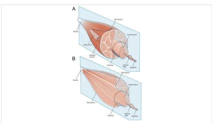
The contractile unit of human skeletal muscle is the muscle fiber, which is a long cylindrical cell measuring up to several cm long and 10 to 100 μm of diameter. The muscle fibers are multinucleated cells, containing several hundreds of nuclei in peripheral position. Each muscle fiber is surrounded by a basement membrane, while a thin layer of connective tissue filling the intercellular space is the endomysium (Figure 1). Approximately 20–100 of these
9 muscle fibers are grouped together in a parallel arrangement called a muscle fascicle that is encapsulated by a perimysium connective tissue. Finally, several muscle fascicles form the muscle, surrounded by connective tissue called epimysium wrapping the whole muscle (Engel and Franzini-Armstrong, 2004; Gillies and Lieber, 2011).
Figure 1:Schematic diagram of the general organization of muscle tissue and muscle extracellular matrix (ECM)-tendon organization. (A) Muscle ECM can be categorized as epimysium (surrounding the muscle),
perimysium (surrounding muscle fascicles), and endomysium (surrounding muscle fibers). (B) Cross-section of muscle tissue indicating that the perimysium may be continuous with the tendon, whereas endomysium is contained within muscle fascicles (adapted from Gillies et al., 2011).
Skeletal muscle is composed of an heterogeneous collection of muscle fiber type classified using by their histochemical (myosin ATPase immunolabeling), biochemical (aerobic/oxidative or anaerobic/glycolytic enzymes) and morphological (size) characteristics. As compared with type fast contractile II fibers, slow-twitch or type I fibers are generally thinner, invested by a denser capillary network, and appear red owing to the presence of a large amount of the oxygen-binding protein myoglobin. These type I fibers are resistant to
10 fatigue, relying on oxidative metabolism for energy, and thus exhibit high mitochondrial and oxidative enzyme content, and low glycogen levels and glycolytic enzyme activity (Engel and Franzini-Armstrong, 2004).
The muscle fiber contraction, and consequently the entire muscle contraction, is provided by myofibrils. The myofibrils consist of chaining sarcomeres. The sarcomere is the smallest contractile entity measuring 1.5-2.5 μm long. It is made up of regions presenting different structures, which explains the striated look of myofibrils in optical microscopy. This aspect is due to the combination of two types of myofilaments: actin thin myofilaments (5-7 nm diameter) and myosin thick myofilaments (14-16 nm diameter). During muscle contraction, the sarcomere decreases its length. This is due to a bigger overlap between thin and thick filaments. During the muscle relaxation, this overlap decreases and the sarcomere increases its length (Engel and Franzini-Armstrong, 2004).
2. Microvasculature structure in skeletal muscle
Skeletal muscle is one of the most vascularized tissues. Beyond oxygen and nutriment supply, new functions for vessels have been recently identified. Vessels, and particularly microvessels, fully participate to skeletal muscle homeostasis through the development of specific interactions with neighboring cells, particularly with muscle stem cells, the satellite cells. Satellite cells are found in close proximity and interact with endothelial cells for their expansion and differentiation. Thus, vessels participate to the control of muscle homeostasis. Thanks to these interactions, vessel cells play a central role in the tissue remodeling after an injury.
11 2-1. The concept of microvascular unit (MVU)
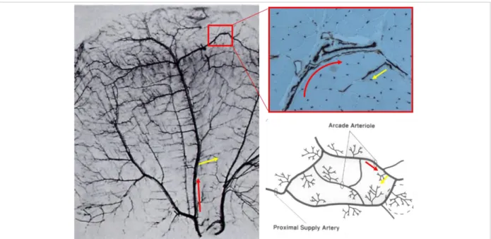
Mammal skeletal muscle is highly vascularized. The role of microcirculation is to support muscle contractile activity that depends on an active consumption of energy substrates and requires a continuous replenishment of their pools and the removal of end products of energetic metabolism. These functional characteristics are based on intimate anatomical interrelations between muscle cells and vessels. Knowledge on skeletal muscle microvascularization exclusively comes from historic studies performed in small animals (e.g. rats, hamsters or cats). Most studies were performed on muscles which could be transilluminated in vivo such as cremaster and spinotrapezius in rat, tenuissimus in cat and rabbit, or gluteus in mice (Myrhage and Eriksson, 1980; Skalak and Schmid-Schönbein, 1986). Muscle vessels are embedded in the extracellular matrix (ECM). Muscle arterioles are classified according to both their location in the ECM and their diameter (~20 to ~50 µm). In most muscles, microcirculation branches from one or more feed arteries in the epimysium into a network of interconnected arcade arterioles in the perimysium (Engelson et al., 1985; Snyder, 1988) (Figure 2). These interconnections allow for compensation if blood flow is compromised by occlusion of one arcade arteriole. At regular intervals, arcades give rise to transverse arterioles which penetrate into the endomysium and divide asymmetrically to yield terminal arterioles which, in turn, give rise to capillary networks. Blood returns to collecting venules which merge to form larger venules, arranged in a similar manner to arterioles and veins (Engelson et al., 1985, 1986; Eriksson and Myrhage, 1972; Hudlicka, 2011; Saltzman et al., 1992; Schmid-Schönbein et al., 1987).
12
Figure 2: Microanatomic organization of the muscle arterioles: Arcade (red arrow) and transverse arterioles
(yellow arrow) are illustrated in rodent transilluminated vascular bed (left panel), in human deltoid biopsy immunostained for CD31 and in a schematic drawing recapitulating the different type of arterioles (right panel).
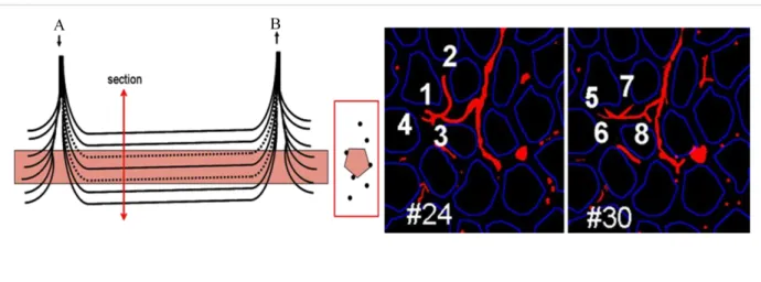
Capillaries embedded in the endomysium run parallel to the muscle fibers to collect into venules, which merge to form larger venules. All capillaries perfused by one terminal arteriole and collected by one venule define the muscle microvascular unit (MVU), which represents the smallest functional unit for blood flow regulation in skeletal muscle. One MVU irrigates a cylinder of muscle tissue of 500-1000 µm in length in all animals studied. In rat and in other small mammals, it comprises 5-10 capillaries located in-between 3-4 adjacent myofibers (Bloch and Iberall, 1982; Cheung, 1996; Lo et al., 2003; Skalak and Schmid-Schönbein, 1986).
Techniques used to study muscle microvasculature in small animals (based on intravascular ink injection) cannot apply to human muscle biopsy explaining that the literature is scarce in Human. A human post-mortem study of the temporal muscle microvascularization has shown an arteriole-to-venule distance of about 900 µm but did not provide data on the MVU size (Cheung, 1996). We confirmed, using optic stacks and serial sectioning of alkaline
13 phosphatase-treated human muscle biopsies, that the overall vascular organization is strikingly similar to those observed in small animals (Gitiaux et al., 2013) (Figure 3). Interspecies conservation of the microcirculation structure stresses a major role of capillary-muscle interactions in physiological and pathological conditions.
Figure 3: Microvascular unit of the normal deltoid muscle vasculature: On the left, schematic representation
of the normal human muscle MVU and its spatial relationship with a myofiber (A: terminal arteriole; V: postcapillary venule), on the right, two optic sections allowing visualization of the 8 capillaries of the MVU issued from one of the two endings of the terminal arteriole (Alkaline phosphatase histoenzymatic reaction with DAPI, x 160) (adapted from Gitiaux et al., 2013).
2-2. Muscle capillarization
Muscle capillarization is related to aerobic functions because it represents the exchange of respiratory gases and metabolites between the muscle fiber and its vascular supply. Historically, the microscopic anatomy of capillaries in muscle tissue has been investigated in relation to their functional role in oxygen transport. Krogh et al. originally proposed that capillaries are the primary site of oxygen exchange between blood and muscle and developed a mathematical model of oxygen transport (Krogh, 1919). Since these pioneering publications, a number of variations on the theme of this model have been proposed but it is
14 still considered as a unifying principle in the study of oxygen transport (Lo et al., 2003; Pittman, 2011). Capillaries are not only uniform static semi-permeable tubes that act as a passive barrier delineating the blood/tissue interface but are now considered as dynamic structures involved in gas exchanges. It was demonstrated that these gas exchanges and the corresponding difference in oxygen partial pressure gradient from capillary to tissue required to maintain adequate oxygen supply are correlated with the capillarity (Hepple et al., 1997; McCall et al., 1996). As a result, the capillary–muscle interface is viewed as a critical determinant of maximal O2 flux capacity in skeletal muscle function. To determine the capillary supply, morphometric indexes are measured from histochemical or immunocytochemical stainings of transverse muscle sections. The indexes used to quantify capillarity include “global indexes”: capillary density (CD), capillary/fiber ratio (C/F). Although CD is one of the most commonly reported global indexes of capillarity, it has drawbacks related to tissue swelling or shrinking and does not account for fiber size or fiber type. The C/F attempts to account for some of these factors and therefore provides a better mean to compare results obtained from different studies. Both parameters are easily used particularly for human muscle capillarity description and provide a global aspect of muscle blood supply (McCall et al., 1996; Qu et al., 1997). However, the use of CD and C/F indices: i) assumes a regular distribution of capillaries in the whole muscle and gives no information on the intramuscular heterogeneity in capillary distribution, ii) does not take into account the fiber size and the fiber type. Thus, “local indexes” allowing the evaluation of capillarity at the level of the individual muscle fiber were introduced: capillaries contact to fiber (CC), fiber area per capillary (FA/C), fiber perimeter per capillary (FP/C) and capillary-to-fiber perimeter exchange index (the CFPE index) derived as the quotient of the individual capillary-to-fiber ratio (i.e., the capillary-to-fiber ratio calculated individually for each fiber) on the fiber
15 perimeter (Harris, 2005; Hepple and Mathieu-Costello, 2001; Mathieu-Costello et al., 1991; Porter et al., 2002a).
Table1: Morphometric indexes used to describe the capillarization of skeletal muscle (adapted from Harris, 2005 and Porter et al., 2002a)
Global indexes
Capillary density (CD) Number of capillaries per unit area
Little information on capillary supply to individual fibers. Dependent on fiber size and sensitive to fiber size changes.
Fiber density (FD) Number of fibers per unit area Capillaries per fiber ratio
(C/F)
Ratio of capillary density to
fiber density Little information on capillary supply to individual fibers. Not effected by fiber size changes, but dependent on capillary : muscle fiber geometry.
Stereology
(three-dimensional modelling) Information on longitudinal capillary geometry. Little information on capillary supply to individual fibers.
Individual fibers indexes
Capillary contacts (CC) Number of capillaries in contact
with each fiber No information on the effects of fiber type size. Fiber area per capillary
(FA/C) and Fiber perimeter per capillary (FP/C)
Fiber area and perimeter supplied by each capillary
Reflects changes in fiber dimensions and proportions, and potential effects of diffusion.
Capillary to Fiber Perimeter Exchange (CFPE) Index
Capillary to individual fiber ratio
divided by fiber perimeter Does not account for capillary geometry.
These “local indexes” provide a more direct depiction of capillarity specific to fiber size and fiber type. However, results obtained from different studies are difficult to compare due to the wide range of type of muscles studied and the different capillary indices chosen. Skeletal muscle is composed of an heterogeneous collection of muscle fiber type and the proportion of each fiber type varies among the different muscles and among the different species (Brooke and Kaiser, 1970; Schroeder et al., 2014). It has been shown that type II fibers have generally lower level of capillary supply than type I fibers (Buckley and Bossen, 2013; Sjøgaard, 1982). For example, in human tibialis anterior muscle, although type II fibers have higher numbers of capillary in contact (CC) compared with type I fibers, the fact that type II fibers are larger than type I fibers for both men and women results in a higher FA/C and FP/C for type II fibers as compared with type I fibers. This indicates that larger area is being supplied by each
16 capillary, and this reflects the lower level of capillarization of type II fibers (Porter et al., 2002a, 2002b). Capillary supply of skeletal muscle indicates the potential exchange capacity between the vascular system and the muscle fibers and therefore reflects the muscle fiber composition in steady state conditions. Although the evaluation of capillary supply remains a useful tool for the understanding of muscle-vessel interactions, the comparison between different pathological and physiological conditions must be interpreted with cautious depending on which morphometric parameter is used. Furthermore, due to physiological variations of capillarization during post-natal muscle development, patient age is an important parameter to consider when assessing capillarity in children (Kottlors and Kirschner, 2010; Sallum et al., 2013).
2-3. Cellular mechanisms of angiogenesis
Angiogenesis, the growth of blood vessels, is a major biological process that controls embryonic development and is also involved in numerous human diseases. New blood vessels are formed by various mechanisms, including assembly by mesodermal endothelial progenitor cells or angioblasts (vasculogenesis, i.e. differentiation of hemangioblast-derived cells into endothelial cells), longitudinal splitting of existing vessels (intussusceptions, i.e. splitting angiogenesis) and enlargement of the vasculature through sprouting, proliferation and remodeling processes (sprouting angiogenesis) (Figure 4). Splitting or sprouting angiogenesis occurs depending on the mechanical stimulus operating from either the luminal or the extravascular side of the vessels. Splitting angiogenesis is defined by the division of the lumen by endothelial protrusion and vessel splitting without breakdown of the basement membrane. In contrast, sprouting angiogenesis is characterized by the migration and proliferation endothelial cells towards an extravascular angiogenic stimulus (Egginton, 2011; Egginton et al., 2001; Hansen-Smith et al., 1996; Hudlicka, 1998; Hudlicka and Brown,
17 2009). Splitting and sprouting angiogenesis differently involve pericytes, which are the
perivascular cells located in close proximity to endothelial cells in capillaries and in post-capillaries venules beneath a common basement membrane (Egginton et al., 1996). Pericytes are generally considered to stabilize the vessel wall, controlling endothelial cell proliferation and thereby the growth of new capillaries. In addition, they are believed to participate in the regulation of microvascular blood flow via a contractile mechanism (Ribatti et al., 2011). Splitting angiogenesis is a mostly pericyte independent process whereas sprouting angiogenesis requires the release of growth factors into the local environment and migration and activation of pericytes (Bloor, 2005; Lindahl et al., 1997). Under certain controlled experimental conditions, such as chronic muscle stretching and vasodilatation, angiogenesis has been shown to proceed in the two distinct modes: by abluninal lateral sprouting and intraluminal splitting (Egginton, 2011; Egginton et al., 2001). Under other conditions both types of angiogenesis may occur. Whereas sprouting angiogenesis appears to be a major process involved in the formation of the majority of blood vessels during tissue repair or diseases (Flamme et al., 1997; Herbert and Stainier, 2011) physiological angiogenesis during exercise in skeletal muscle likely does not occur by sprouting angiogenesis (i.e. tip-cell formation) but rather via intussusception, a process that remains poorly understood due to the paucity of appropriate experimental model (Brown et al., 2003; Gianni-Barrera et al., 2013, 2014).
Figure 4: Early morphogenic events during sprouting and splitting (intussusception): In sprouting,
specialized endothelial tip cells (white) are first selected (A), which then migrate and eventually connect with each other, whereas trailing stalk cells proliferate to form the new vascular trunk (B). In intussusception, at first,
18 pre-existing vessels enlarge by circumferential growth (C) and subsequently endothelial processes invaginate from opposing vascular walls to form transluminal pillars, leading to the splitting into two new vessels (D) (adapted from Gianni-Barrera ., et al 2011).
Blood vessel formation by sprouting angiogenesis is a multistep process that requires the tight control and coordination of endothelial cell behavior. It is generally assumed that common sequence of events is initiated by the proteolytic breakage of the basement membrane surrounding capillaries. In quiescent vasculature, endothelial cells form a cobblestone monolayer of quiescent cells that covers the luminal surface of the vessels. Upon angiogenic cues, endothelial cells lose their cell to cell junctions, activate matrix metalloproteases that degrade the surrounding basement membrane and, in turn, become mobile, invasive and initiate sprouting from the basal surface of the blood vessels. Several types of specialized endothelial cells required to build a new functional vessel have been identified. Tip cells located at the distal end of each sprout extend numerous filopodia, respond to attractive or repulsive guidance signals (e.g. vascular endothelial growth factor (VEGF) gradient) within their immediate environment and proliferate minimally. Tip cells are trailed by endothelial stalk cells which maintain the connectivity with the parental vessels, are less mobile and more proliferative and initiate the formation of the new vessel lumen (De Smet et al., 2009). Sprouting angiogenesis is mainly controlled by the secreted VEGF tyrosine kinase receptor-2 (also known as Flk1). Tie-2 and Notch signalings are also key pathways in sprouting angiogenesis. Ang-Tie signaling controls vessel quiescence in adults and also regulates the later steps of the angiogenic cascade that are related to vessel maturation (Augustin et al., 2009; Carmeliet and Jain, 2011). Although the understanding of the anastomotic process remains limited, it is clear that other cell types may also influence vessel fusion. In particular, the recruitment of other cells is a critical factor in the subsequent maturation of the nascent vasculature. Factors such as platelet-derived growth factor B (PDGFB) secreted by
19 endothelial cells recruit mural cells (pericytes and vascular smooth muscle cells) to the developing vasculature, which stabilizes vessel walls (Naylor et al., 2014).
2-4.
Development of the muscle microvasculatureMuscle vasculogenesis is mainly confined to the formation of the first primitive vasculature in the embryo. Both myogenic and endothelial cells share a common embryonic origin. Myogenic progenitor cells derive from the dorsal somite, the dermomyotome, which also gives rise to derma, to endothelial and smooth muscle cells of blood vessels (Buckingham and Vincent, 2009). Multipotent Pax3-positive (Pax3+) cells in the somites give rise to skeletal muscle and to cells of the vasculature. FoxC2, a member of the Forkhead box transcription factors family is also expressed in the somite, and mutual repression between FoxC2 and Pax3/7 has been shown to determine myogenic (high Pax3/7:FoxC2 ratio) or vascular (low Pax3/7:Foxc2 ratio) fates in the murine dermomyotome (Lagha et al., 2009). The Notch pathway affects the Pax3:FoxC2 balance and promotes the endothelial versus myogenic cell fate in the somite, before migration to the limb (Mayeuf-Louchart et al., 2014).
At the tissue level, the muscle development of the chick wing consists of an initial migration phase of somatic cells and organization into dorsal and ventral premuscle masses. During the second phase, theses masses undergo a series of splits thus giving rise to all the muscles of the wing. The timing of vascular invasion of the limb occurs at a later stage, just prior the establishment of the final muscle pattern, suggesting that myogenic differentiation is not oxygen or nutrient dependent (Murray and Wilson, 1997). Furthermore, in mouse Extensor Digitorum Longus, at early stage (12 days in utero) the developing muscle consists of primary myotubes surrounded by a pleomorphic population of mononucleated cells devoid of myofilaments. At this stage, blood vessels and nerves are found in the periphery but not within the developing muscle mass. By 14 days in utero, small blood vessels and
20 unmyelinated nerve bundles are found throughout the developing muscle in parallel with maturation of myotubes leading from 18 days in utero to the polygonal shape fascicular arrangement and ultrastructure characteristic of more mature myofibers (Ontell and Kozeka, 1984). Vessels and nerves progressively penetrate into the developing muscle with a clear temporal pattern and progressively separate the various muscle masses producing the individual muscles (Schroeter and Tosney, 1991).
At the cell level, during chick and mouse limb development, angioblasts colonize the limb regions before muscle precursors cells and organize correctly in space, even in the absence of muscle progenitors (in Pax3-/- mice). In addition, anarchic ectopic blood vessels, induced by overexpression of VEGF, inhibit muscle formation while the absence of vessel formation, induced by soluble form of VEGF receptor 1 (VEGF1/Flt1), leads to muscle fusion, preventing the splitting into distinct limb muscles. At the molecular level, PDGBB secreted by endothelial cells crossing the muscle mass may act as a paracrine signal. PDGFB may increase locally the production of connective tissue by promoting connective tissue cell migration and accumulation at the future sites of cleavage, excluding myogenic cells and forcing them to split at these sites, and finally allowing the formation of separate muscles of the limb (Tozer et al., 2007) (Figure 5).
Figure 5: Model for the effect of the vasculature on muscle development. (A) The endothelial cells (blue)
delineate the future cleavage site in the muscle mass, which is composed of myogenic cells (red) and muscle connective tissue cells (green). (B) At a later stage, the muscle masses are separated. (C) The PDGFB secreted by the endothelial cells acts in a paracrine manner on muscle connective tissue cells, which express PDGF receptor. In response to PDGFB, connective tissue cells increase the secretion of extracellular matrix by producing collagen I allowing muscle mass separation. (adapted from Tozer et al., 2007).
21 After birth, an intensive construction takes place, which is correlated with the gradual morphological and functional maturation of the muscle tissue (Oertel, 1988; Ontell and Dunn, 1978). In mouse, while the number of myofibers remains constant after birth, post-natal growth is characterized by an extensive hypertrophy of the muscle fibers. This hypertrophy is initially supported until day 21 (P21) by a rapid increase in the number of myonuclei, as satellite cells proliferate to generate fusion-competent myoblasts for muscle growth (White et al., 2010). It was shown in human deltoid muscle a 5-fold increase in fiber diameter (25 fold increase in cross sectional area) from 10 µm at birth up to 50 µm at 15-20 years (Oertel, 1988) associated with a progressive increase of satellite cells per myofiber and a decrease of satellite cells per square millimeter (Sallum et al., 2013; Verdijk et al., 2014; White et al., 2010). At birth, muscle vascularization is rudimentary, most growing myofibers being unconnected to capillaries during early post-natal development. The postnatal maturation of muscle fibers is supported by an architectural rearrangement of the microvascular bed (Stingl and Rhodin, 1994). The architecture of the terminal vessels develops gradually from an immature shape to its final feature. In rat, the average length of terminal arterioles increases progressively and the capillary network consisting initially of numerous small meshes, very irregular in size and shape, gradually develops into a characteristic pattern of long meshes, parallel to the course of muscle fibers (Snyder and Coelho, 1989; Stingl and Rhodin, 1994). Meanwhile, similarly in rat/mouse model and in Human, capillarization progressively declines (decreased CD) and capillary to fiber ratio (C/F) increases in all fiber types consistent with post-natal muscle capillary growth and myofiber enlargement, respectively (Sallum et al., 2013; Smith et al., 1989).
Signaling mechanisms involved in embryonic development and more particularly in vasculogenesis and myogenesis have been deeply studied but mechanisms that may orchestrate and interegulate the two processes remain poorly understood (Buckingham and
22 Vincent, 2009). In particular, the Notch signaling pathway has been involved in satellite cell activation and cell fate determination in postnatal myogenesis (Conboy and Rando, 2002) and also appears to play myriad roles during vascular development (Gridley, 2010). The role of Notch signaling in regulating vascular development is intertwined with another major regulator of vascular development and physiology, the VEGF pathway. Myofibers targeted-VEGF deficient mice (mtargeted-VEGF-/-) present a significant decrease of muscle capillary to fiber ratio (-48%) and in capillary density (-39%) in gastrocnemius, without changes in muscle fiber type composition, leading to a major intolerance to aerobic exercise. These results indicate that since muscle-VEGF-deficient mice survive to adulthood, VEGF is essential to the physiologic regulation of postnatal muscle capillarity and therefore to the maintenance of adult skeletal muscle microvasculature (Olfert et al., 2009). Thus, the development of muscle vasculature before and after birth needs coordinated angiogenesis and myogenesis. Interactions between vessels and muscle are also required upon various physiological conditions (hypoxia, exercise, and aging) in which both muscle fibers and vessels adapt to changing demands by altering myofiber size or type composition and capillary density, respectively. All the pathways studied during development may be also recruited postnatally in response to muscle injury. For example, the Hedgehog (Hh) signaling was demonstrated to be further reactived in adult ischemic tissues and to be impaired in aging mice (Pola et al., 2003; Renault et al., 2013a, 2013b).
3. Physiological adaptation of the skeletal muscle microvascular bed
Skeletal muscle is the organ containing the highest microvascular mass in Human and constitutes a highly adaptable tissue, responding to environmental and physiological demands.
23 The vasculogenesis/angiogenesis processes described above are initiated during fetal development. By contrast, postnatal physiological (i.e. non pathological) angiogenesis is relatively rare, limited to uterine changes during the estrous cycle, and to exercise and altitude-induced angiogenesis in skeletal muscle (Egginton, 2011; Olfert and Birot, 2011; Shimizu-Motohashi and Asakura, 2014). The following paragraph aims at focusing on current knowledge about molecular aspects in some physiological conditions (exercise, hypoxia and aging) to highlight specific interactions between muscle cells and vessels.
3-1. Exercise
Exercise is a potent angiogenic stimulus and is one of the few situations of non-pathological angiogenesis that occurs in mammals after development (Egginton, 2009; Malek et al., 2009). Because the vascular supply to muscles is a major contributor to endurance capacity, physiological angiogenesis has been studied in trained and untrained subjects as well as after training or detraining (Hoier and Hellsten, 2014). The increase of capillarity that occurs in skeletal muscle in response to endurance exercise training is a major example of physiological capillary growth in a mature differentiated tissue (Malek et al., 2009). Indeed, the number of capillaries per fiber is higher in endurance athletes than in sedentary subjects and it increases with endurance training in parallel with the changes in oxidative metabolism (Olenich et al., 2013). Under exercise, skeletal muscle presents increased blood flow. Capillaries are exposed to repeated shortening and elongation due to changes in sarcomere length during contractions (Egginton, 2011). However, how exercise regulates the complex process of physiological angiogenesis remains poorly described. One prevailing notion is that the metabolic needs during exercise lead to a local hypoxia with induction of VEGF secretion (Breen et al., 1996; Richardson et al., 1999). Among possible cellular sources, myocyte is a critical source for paracrine VEGF production during exercise. In myocyte-VEGF-/- mouse model, the
24 physiological angiogenesis (i.e. increase in capillarity) does not occur and metabolic enzyme activity levels after training are not increased. This may indicate that other cellular sources of VEGF such as endothelial cells, fibroblasts, pericytes and macrophages are unable to compensate for the loss of myocyte-derived VEGF (Delavar et al., 2014; Olfert et al., 2010). The mechanism by which myocyte-derived VEGF controls skeletal muscle angiogenesis is not well understood. Confocal microscopy has revealed an increase of the density of immunostained VEGF vesicles in the subsarcolemmal region of teased human muscle fibers after exercise suggesting that VEGF is pre-stored in vesicles and secreted to the extracellular space (Hoier et al., 2013). This release seems to be partially mediated by adenosine via mitogen activated protein kinase (MAPK) (Høier et al., 2010a).
Factors capable of coordinating the complex process leading to VEGF dependent angiogenesis remain elusive. Several studies have investigated regulatory factors proposed to be related to VEGF expression in skeletal muscle. Matrix remodeling via Matrix Metalloproteases (MMP) as well as angiopoietin-2 (Ang-2) are important for the destabilization of the capillary and the division of capillary lumen (Høier et al., 2010b). They have been shown to both induce VEGF secretion and regulate skeletal muscle angiogenesis (Bobadilla et al., 2014; Hoier and Hellsten, 2014). VEGF is a downstream target of hypoxia-inducible factor (HIF). HIF is a heterodimeric complex composed of a constitutively expressed HIF β-subunit and an oxygen sensitive HIF subunit (Semenza, 2003). Three α-subunits are known to date (HIF-1α, -2α, and -3α). In skeletal-muscle-specific-HIF-1α-KO mice, the increase of expression of HIF-1α target genes, including VEGF, normally observed in response to exercise is lacking, suggesting a HIF dependent adaptation of skeletal muscle to exercise (Mason et al., 2004). The peroxisome proliferator-activated receptor gamma (PPARɤ) coactivator-1 alpha (PGC)-1α, a potent metabolic sensor involved in multiple aspects of skeletal muscle physiology, has been also described as a key regulator of skeletal
25 muscle angiogenesis. PGC-1α is robustly induced in the skeletal muscle in response to exercise (Baar et al., 2002; Gouspillou et al., 2014) resulting in mitochondrial biogenesis, a switch from glycolytic fibers to mitochondrial-rich oxidative fibers (Handschin and Spiegelman, 2011; Miura et al., 2008; Wende et al., 2007). PGC-1α induces VEGF expression by co-activating the transcription factor estrogen related receptor alpha (ERRα) on an enhancer located in the first intron of the VEGF gene (Arany et al., 2008; Leick et al., 2009). Furthermore, deletion of PGC-1α in skeletal muscle prevents exercise-mediated angiogenesis (Chinsomboon et al., 2009; Rowe et al., 2014). Several studies have pointed out the role of non-myocyte cell types in exercise-induced angiogenesis. Expression microarray analysis using RNA from muscles of an inducible mice transgenic model of PGC-1α overexpression has shown that the vast majority of upregulated genes are known to be strongly expressed in macrophages. It was demonstrated that PGC-1α induces the recruitment of macrophages to skeletal muscle and the secretion of phosphoprotein 1 ([SPP1]; also known as osteopontin) by myocytes. It was suggested that SPP1 could lead to secretion of monocyte chemoattractant protein-1 (MCP-1) from macrophages, which in turn would activate adjacent endothelial cells, pericytes and smooth muscle cells (Rowe et al., 2014) (Figure 6). Thus, proper temporal and spatial interactions between myocytes, the main source of VEGF in muscle and their microenvironment are crucial to ensure that the metabolic demands of the muscle during exercise are met.
Figure 6: Proposed model for PGC-1α-mediated orchestration of different cell types, myokines, and cytokines to mediate functional angiogenesis in skeletal muscle (adapted from Rowe et al.,2014).
26 Similarly, modifications of the muscle vascular bed also occur as an adaptation to exposure to hypoxia defined by the decrease of partial pressure of oxygen. Whatever the origin of the hypoxic stimulus may be (environmental or pathological (chronic obstructive pulmonary disease, stroke, cardiac dysfunction...), the ultimate consequence is an inadequate oxygen delivery at the tissue level leading to ischemia. Exposure to systemic chronic hypoxia in rats stimulates significant arteriolar changes including an increase of the number of terminal arterioles and of arcade arteriole loops (Bailey et al., 2008). Additional branches are also formed along each category of arterioles to maintain an adequate tissue perfusion and oxygenation. This reduction of the branching interval might be attributed to an increase in number of newly formed arterioles from pre-existing capillaries (Price and Skalak, 1998; Smith and Marshall, 1999). These arteriolar remodeling begin within the first few days after the onset of hypoxia but gradually develop over the following 3-4 weeks (Olfert et al., 2001; Ou et al., 1985). However, the question of whether chronic hypoxia induces angiogenesis within skeletal muscle is still controversial. Due to the high variations in experimental protocols (duration, oxygen pressure, sedentary vs. trained subjects…), animal and human studies show discrepant results regarding the effect of hypoxia on VEGF and on growth of skeletal muscle capillaries. Generally, it is accepted that in response to chronic hypoxia, the resting mRNA levels of VEGF, and of its receptors (VEGFR1/Flt1, VEGFR2/Flk1) increase, CD is unaltered or somewhat increased and the C/F remains unchanged (Desplanches et al., 1996; Lundby et al., 2009; Olfert et al., 2001, 2009; Mounier et al., 2009, 2011; Pisani, 2005; Vogt et al., 2001; Tuomisto et al., 2004). While many cellular functions such as overall protein synthesis are down regulated under hypoxia, selected subsets of gene are upregulated. Prominent among them is the family of genes governed by the transcription factor HIF-1. HIF-1 is a heterodimer composed of an alpha and a beta subunit. HIF-1α protein is usually not present at high levels in tissues or cells in a normoxic environment (Brown et al., 2005; Flann
27 et al., 2014; Hudlicka and Brown, 2009; Semenza, 2011; Silvestre et al., 2013). HIF-1α is hydroxylated, ubitiquined and degraded in normoxia but is stable in hypoxia and translocates into the nucleus to form an active complex with HIF-1β, which is constitutively expressed (Jaakkola et al., 2001). HIF-1 induces transcription of numerous target genes involved in
erythropoiesis, energy metabolism and angiogenesis (Forsythe et al., 1996; Hudlicka and Brown, 2009; Jewell et al., 2001; Semenza, 2011; Silvestre et al., 2013). Human and mice studies show also discrepant results regarding the effect of hypoxia on HIF expression according predominantly oxidative or glycolytic muscles (Mounier et al., 2010; Pisani, 2005).
HIF-1α likely plays a more prominent role in pathological angiogenesis (Semenza, 2011) whereas deletion of HIF-1α in skeletal muscle leads to more blood vessels, rather than fewer (Mason and Johnson, 2007). By contrast, PGC1-α expression appears to play a major role in physiological angiogenesis. In human skeletal muscle, PGC1-α expression is coupled to HIF-1α dependent gene expression by increasing mitochondrial consumption, leading to a decrease in intracellular oxygen availability for HIF hydrolases, resulting in activation and stabilization of HIF-1α (O’Hagan et al., 2009).
Furthermore, the hypoxic responsiveness of satellite cells, the main muscle stem cells, in term of their angiogenic capacity remains to be defined. Hypoxia induces modifications in the normal interactions between vessels and satellite cells for maintaining muscle homeostasis and therefore the mechanical properties, to meet the functional demand. Satellite cells seem to prefer hypoxic niches, which is in apparent contradiction with satellite cell proximity to vessels (Latil et al., 2012). VEGF expression in satellite cells is modulated by hypoxia (Germani et al., 2003; Rhoads et al., 2009; Rissanen et al., 2002a). A second potential candidate that may modulate satellite cell angiogenic capacity is Hepatocyte Growth Factor (HGF). HGF possesses a dual role regulating angiogenesis and myogenesis, raising the possibility that it is a critical factor coordinating both aspects during skeletal muscle
28 regeneration. Hypoxic conditions increase satellite cell HIF-1α and VEGF protein content but decrease HGF mRNA content compared to normoxic satellite cells. This reduction of satellite cell HGF expression under hypoxia directly impacts the ability of satellite cells to drive angiogenic process (see below, paragraph 4.3) (Flann et al., 2014).
3-3. Aging
Aging is another physiological condition leading to significant modifications in the angiogenic and regenerative capacity of skeletal muscle. Progressive generalized loss of skeletal muscle mass and function (sarcopenia) occurs as a consequence of aging. The reduced regenerative potential associated with age is correlated with a decline in satellite cell number (Day et al., 2010; Renault et al., 2013a; Verney et al., 2008). Multiple mechanisms are thought to be involved in these age-induced alterations of satellite cell niche and microenvironment, which ultimately have an effect on satellite cell activity. Recent studies have highlighted that both intrinsic and extrinsic factors are responsible for the loss of myogenic capacities of satellite cells with age but the exact etiology of sarcopenia remains unknown (Bernet et al., 2014; Biressi et al., 2014; Brack et al., 2007; Cosgrove et al., 2014; Fry et al., 2015; Price et al., 2014; Sousa-Victor et al., 2014; Tierney et al., 2014). In particular Notch ligand Delta is down regulated in old satellite cells resulting in defective satellite cell proliferation and an inability to generate myoblasts during regeneration (Conboy et al., 2003) whereas Wnt signaling is hyperactivated (Brack et al., 2007). Furthermore, with increasing age, activation of JAK-STAT signaling in satellite cells substantially contributes to myogenic commitment and results in their regenerative deficiency by exhausting the pool of satellite cells (Doles and Olwin, 2014; Price et al., 2014; Tierney et al., 2014). Aging is associated with higher expression of known activators of JAK-STAT signaling, including basic fibroblast growth factor (bFGF, also called FGF2), inflammatory cytokines including interleukin-6 (IL-6), PDGF, and Epidermal Growth Factor (EGF) (Shuai and Liu, 2003).
29 Changes in the microenvironment in aging muscle could impede the physiological angiogenesis and myogenesis and thus alter muscle capacities to regenerate. In parallel, the expression of VEGF has been shown to be down regulated in aged animals (Rivard et al., 1999). Moreover downregulation of VEGFR2/Flk1 observed in old mice has been suggested to be responsible for impaired VEGF-induced angiogenesis in the ischemic limb of old mice (Pola et al., 2001; Qian et al., 2006). From a histological perspective, muscle aging is characterized by a decrease in myofiber size and number, with a preferential loss of fast type II myofibers (type II atrophy) (Piec et al., 2005). Data on the impact of aging in skeletal muscle capillarization are variable, depending on species and muscles studied as well as the methodology used (Degens et al., 1994). They show either unchanged or increased C/F ratio (Mathieu-Costello, 2005). Impaired microvascular functions associated with aging have been poorly investigated in skeletal muscle. They involve several processes likely to interfere with the homeostasis of myofibers and myogenic cells, such as a decreased NO activity, increased production of vasoconstrictor factors, increased oxidative stress associated with increased expression of pro-inflammatory cytokines/compounds (Baraibar et al., 2013; Herrera et al., 2010). In addition, aged satellite cells exhibit decreased ability to promote angiogenesis) (Rhoads et al., 2013) (see below part 4). Hedgegog (Hh) signaling and Hh dependent regulation of angiogenesis and myogenesis (cf below paragraph 4.3) is impaired in aged mice (Renault et al., 2013a). Evidence indicates that apoptosis is involved in mediating the progression of sarcopenia in old mice. Apoptosis is not only confined to myofibers but is preponderant (over 75%) in capillary endothelial cells (Wang et al., 2014). Altogether, these findings indicate that age related changes affecting both myogenic cells and endothelial cells may unbalance muscle homeostasis and alter the normal muscle angiogenesis abilities.
In summary, the adaptation to physiological conditions (exercise, hypoxia and aging) needs specific and proper interactions between muscle and vessels at the tissue level. These
30 interactions are substantiated at the cellular level by a close proximity between endothelial and myogenic cells.
4. Vascular niche (cellular and molecular aspects)
A stem cell niche can be defined as a spatial structure in which stem cells are housed and maintained by allowing survival and slow self-renewal in the absence of differentiation. The niche comprises cellular structures, extracellular matrix proteins, and soluble factors (Rezza et al., 2014). Muscle stem cells, i.e. satellite cells play a key role in muscle regeneration process
(Wang et al., 2014). They lie along the myofiber in a quiescent state until a damage signal stimulates their entry into the cell cycle. These myogenic precursor cells or transit-amplifying myogenic cells proliferate and further commit into terminal myogenic differentiation to replace the damaged myofibers while a subset of these cells self-renew to maintain the satellite cell pool (Wang et al., 2014). To achieve these processes, satellite cells interact with several cell partners in the muscle tissue, among them endothelial cells, of which they are in close proximity. The vascular stem cells niche concept has been characterized in several tissues: hematopoietic stem cells (HSCs) and neural stem cells (NSCs) while it remains poorly characterized in skeletal muscle, particularly at the molecular level.
4-1. Hematopoietic stem cell vascular niche
HSCs give rise to all hematopoietic lineages and provide appropriate numbers of mature blood cells throughout the lifetime. A plethora of studies has investigated HSC niche. Due to the particular organization of bone marrow, HSCs may find several cell types as a niche support including osteoblasts, macrophages, platelets, specific interstitial cells (Mendelson
31 and Frenette, 2014). We will focus here on the interactions HSCs develop with endothelial cells. It seems established that a hypoxic HSC microenvironment is crucial for HSC maintenance and survival (Rehn et al., 2011). HSCs reside in a hypoxic microenvironment as pharmacologic increase in HIF-1α enhances HSC homing (Speth et al., 2014; Suda et al., 2011). During several years, the concept of two main HSC niches prevailed, with the “endosteal or osteoblastic niche” serving as a quiescent storage niche for dormant HSCs and the “vascular or sinusoidal niche” for HSC differentiation and mobilization (Colmone and Sipkins, 2008; Wilson and Trumpp, 2006) (Figure 7). However, recent studies broke this paradigm and demonstrated that vascular niche is indispensable for maintaining HSC quiescence (Scadden, 2014; Spencer et al., 2014). HSCs in close proximity to sinusoids would allow the constant screening of the status of hematopoietic system in the vascular network (Wilson and Trumpp, 2006). A detailed analysis of the location of HSCs and progenitors along the entire femur at short time points after transplantation revealed that they preferentially and consistently migrate through endothelium and reside in an endosteal niche in close association with blood vessels, which do not form a separate niche (Ellis et al., 2011). Similarly, 3D analysis of bone marrow vascular structures demonstrated that quiescent HSCs specifically associate with small arterioles that are preferentially found in the endosteum, indicating that arteriolar niches are indispensable for maintaining HSC quiescence (Kunisaki et al., 2013; Morrison and Scadden, 2014; Scadden, 2014) A large number of molecular signaling delivered by the various components of the HSC niche have been shown to regulate and control HSC homeostasis (Mendelson and Frenette, 2014). Some are specifically delivered by endothelial cells, such as Stem Cell Factor (SCF) (Ding et al., 2012). HSCs are located adjacent to perivascular cells (reticular cells) highly expressing chemokine (C-X-C motif) ligand 12. CXCL12 is involved in the regulation of HSC trafficking between the bone marrow niche and the peripheral circulation (Guerrouahen et al., 2011). Platelet endothelial
32 cell adhesion molecule (PECAM-1) also known as cluster of differentiation 31 (CD31) expressed by vascular cells and HSCs regulate their ability to respond to a CXCL12 gradient (Ross et al., 2008). VEGF, which expression in HSCs is upregulated by a hypoxic environment, is involved in HSC survival (Rehn et al., 2011), as well as in the regeneration of the bone marrow vascular niche after myelosuppression (Kopp et al., 2009).
Figure 7: The model of HSCs niche. HSCs niche is composed of complex components including HSCs and
other functional elements such as vessels, stromal cells, ECM proteins, neural inputs, and endothelial cells.
HSCs are found mainly adjacent to sinusoids throughout the bone marrow, where endothelial cells and mesenchymal stromal cells promote HSC maintenance by producing SCF, CXCL12 and probably other factors (adapted from Morrison and Scaden, 2014).
4-2. Neural stem cell vascular niche
Neurogenesis in the adult brain has been demonstrated within the subventricular zone (SVZ) and hippocampus (Kuhn et al., 1996). Neurogenesis and angiogenesis are closely linked in the germinal zones of the adult brain where neural stem cells (NSCs) reside associated with the vasculature (Capela and Temple, 2002; Palmer et al., 2000). Bursts of angiogenesis occur at the same time as neurogenesis and endothelial cells secrete soluble factors that regulate neuronal differentiation in vitro (Alvarez-Buylla and Lim, 2004; Shen, 2004). Adult neurogenesis supported by proliferative cells that generate neurons are found in dense clusters associated with the vasculature (Palmer et al., 2000). Anatomically, NSCs are closely apposed to the laminin-containing ECM surrounding endothelial cells (Shen et al., 2008). Moreover, transplanted human NSCs were found in the proximity of cerebral microvessels upon induction of angiogenesis by cerebral infusion of VEGF into the murine brain (Schmidt et al., 2009; Shen et al., 2008). Blocking the laminin receptor α6β1 integrin, which is expressed by NSCs, inhibits their adhesion to endothelial cells and alters their proliferation, indicating that direct contact with vessel plays a functional role in regulating NSC properties (Shen et al.,
33 2008). Indeed, co-transplantation of endothelial cells with NSCs increases their survival and proliferation and accelerates neuronal differentiation (Nakagomi et al., 2009). Unique neurovascular niche was defined in which angiogenesis and neurogenesis are linked through specific vascular growth factors and chemokines. A focal cortical stroke in the cortex induces the long-distance migration of neuroblasts originate from NSCs in the SVZ, which form close associations with peri-infarct blood vessels. These interactions are, at least, in part mediated by the vascular ligands CXCL12 and Angiopoetin 1 (Ang1) acting on neuroblast receptors CXCR4 and Tie-2, respectively, which exert a tropic effect on migrating neuroblasts (Ohab et al., 2006). Ang1 has a unique role in neurogenesis independent of its role in angiogenesis. Ectopic expression of Ang1 promotes neuronal differentiation and neurite outgrowth in neuronal progenitors, while this effect was blocked by the presence of anti-Tie-2 receptor antibody (Bai et al., 2009). Of note, Ang1 stimulates in vitro neurogenesis in neural progenitor cells (NPCs) through the Akt pathway (Bai et al., 2009). VEGF also plays a role in the proliferation, homing and recruitment of NSCs to the site of injury. In vivo, microvasculature modulates the local guidance of NSCs through endothelial cell-derived chemo-attractants such as PDGF-BB, chemokine (C-C motif) ligand 5 (CCL5) also known as RANTES, chemokine (C-X-C motif) ligand 11 (CXCL11) also known as I-TAC, chemokine (C-X-C motif) ligand 1 (CXCL1) also known as GROα, Neurophil-activating peptide 2 (NAP-2), Angiopoietin 2 (Ang2), and macrophages-colony stimulating factor (M-CSF). Moreover, in vitro chemotactic migration of human NSCs is enhanced in the presence of conditioned media from human endothelial cells stimulated with VEGF (Schmidt et al., 2009). Reconstitution of NSC-vascular niche in 3D with vascular cells and extra cellular matrix provides precise control of NSC self-renewal, proliferation and differentiation into astrocytes and oligodendrocytes through Notch effectors. Astrocyte differentiation is more active when NSCs are in close proximity to brain vasculature (Shin et al., 2014). Indeed,
34 endothelial cells participate in the regulation of NSC homeostasis by secreting soluble factors that activate Notch and Hes 1 to promote self-renewal (Shen, 2004).
4-3. Satellite cell vascular niche
Recent studies identified vessel cells as privileged partners of satellite cells and myogenic precursor cells (MPCs) and showed that vessels contribute to the regulation of satellite cell fate (Mounier et al., 2011). Skeletal muscle is laced with a dense microvasculature, and most quiescent satellite cells are found strikingly close to capillaries. At the tissue level, angiogenesis and myogenesis take place concomitantly during muscle regeneration after ischemia (Scholz et al., 2003). Christov et al. indicated that satellite cells and endothelial cells were tightly juxtaposed at steady state in normal adult muscle. Satellite cell number was significantly correlated with capillarization of myofibers, regardless to their type, in normal muscle. They also varied in paradigmatic physiological and pathological situations associated with variations of capillary density. Satellite cell number was decreased in amyopathic dermatomyositis, a condition in which muscle capillary loss occurs without myofiber damage, while it was increased in athlete muscles presenting a higher number of capillaries (Christov et al., 2007). In vitro analysis using human cells in coculture showed that myogenic cells and endothelial cells reciprocally interact to promote both angiogenesis and myogenesis. Endothelial cells support myogenesis through the secretion of a series of growth factors such as Insulin Growth Factor (IGF-1), HGF, bFGF, PDGF-BB and VEGF (Christov et al., 2007). Reciprocally, myogenic cells promote angiogenesis, a process that mainly depends on VEGF, which expression was increased in myogenic cells while they differentiate along the myogenic program (Chazaud, 2003; Christov et al., 2007). This was confirmed in a 3D co-culture model composed of rat satellite cells and microvascular fragments, a multicellular structure consisting of endothelial cells, pericytes, and smooth muscle cells. Active myogenic
35 cells possess a potent pro-angiogenic program that may participate in revascularization of damaged muscle through the secretion of soluble factors, such as VEGF, HGF, IGF and FGF (Rhoads et al., 2009). VEGF is involved in myogenesis and angiogenesis regulation as satellite cells express Flk1 and Flt1 receptors for VEGF that modulate their migration and survival (Germani et al., 2003). Several studies have demonstrated that VEGF is necessary for muscle repair: intramuscular AAV-VEGF treatment or recombinant proteins VEGF associated with IGF1 enhanced muscle regeneration, innervation and revascularization after ischemia (Arsic et al., 2004; Borselli et al., 2010). It has been shown that in vitro VEGF stimulates myogenic differentiation of MPCs and that in vivo it mediates skeletal myogenesis (Bryan et al., 2008; Chazaud, 2003). Increase of VEGF expression in myogenic cells is mediated by β-catenin signaling and induces both angiogenesis through endothelial cell proliferation, and muscle regeneration (Kim et al., 2006). Controlled overexpression of VEGF in muscle induces vessel splitting whereas uncontrolled expression causes aberrant vascular structures (Gianni-Barrera et al., 2013; Karvinen et al., 2011).
Analysis of Hh signaling showed the tight coregulation of myogenesis and angiogenesis in the context of post-ischemia muscle regeneration (Renault et al., 2013a). Specific depletion of Gli3, a transcription factor mediating Hh signaling in myofibers induces both impaired myogenesis and angiogenesis. Gli3 regulates myogenic differentiation as well as the delivery of proangiogenic factors by myogenic cells, including thymidine phosphorylase and Ang1. This study demonstrates that impaired myogenesis affects angiogenesis in the context of skeletal muscle regeneration (Renault et al., 2013b). Other factors such as Ang1, Ang2, FGF, Nerve Growth Factor (NGF) and chemokine (C-C motif) ligand (CCL-2/MCP-1) are implicated in angiogenesis and their expression is increased after an injury at early stages during muscle regeneration (Wagatsuma, 2007). Ang2 stimulates angiogenesis during muscle regeneration (Bellamy et al., 2010). NGF plays a functional role in neovascularization after
36 treatment of ischemic muscle, through stimulation of angiogenesis and protecting myofiber from necrosis (Emanueli et al., 2002). At steady-state, vessels are surrounded by peri-endothelial cells including smooth muscle cells, pericytes and interstitial fibroblastic cells. These cells are known to secrete Ang1 that binds to its Tie-2 receptor on endothelial cells to stabilize vessels. This Ang1/Tie-2 signaling has also been shown to act on muscle stem cell behavior (Abou-Khalil et al., 2009; Bentzinger et al., 2013). In an autocrine manner, myogenic cell-derived Ang1 signals on Tie-2, through ERK1/2 pathway, to decrease both proliferation and differentiation and increase their own self-renewal. Peri-endothelial cells, by secreting Ang1, reinforce and maintain the quiescent state in satellite cells in a paracrine manner (Abou-Khalil et al., 2009). Thus endothelial cells and peri-endothelial cells have opposite effects on satellite/myogenic cells. During muscle regeneration, endothelial cells, free from peri-endothelial cells due to vascular remodeling, may interact with myogenic cells to promote angiogenesis and myogenesis. Then peri-endothelial cells promote quiescence of both satellite cells and vessels once homeostasis of the skeletal muscle is recovered (Abou-Khalil et al., 2010). While few studies, described above, have investigated the concomitant regulation of angiogenesis and myogenesis during skeletal muscle regeneration, a number of molecules have been separately described to be involved in these two processes. The "musculo-vascular niche" should be envisaged in a broader aspect than strictly defines the place where stem cells reside in a quiescent state, since interactions with endothelial cells have been demonstrated for both quiescent, cycling and differentiating myogenic cells (Christov et al., 2007). The characterization of this niche will benefit from further identification of molecular regulators of these dynamic interactions, as well as the potential identification of satellite cell subsets exhibiting different interactions with vessels. Nevertheless, vessels fully participate to the coordination of the acute satellite cells response as well as the sequential steps of muscle regeneration until the returns to homeostasis