HAL Id: tel-02495351
https://tel.archives-ouvertes.fr/tel-02495351
Submitted on 2 Mar 2020
HAL is a multi-disciplinary open access archive for the deposit and dissemination of sci-entific research documents, whether they are pub-lished or not. The documents may come from teaching and research institutions in France or abroad, or from public or private research centers.
L’archive ouverte pluridisciplinaire HAL, est destinée au dépôt et à la diffusion de documents scientifiques de niveau recherche, publiés ou non, émanant des établissements d’enseignement et de recherche français ou étrangers, des laboratoires publics ou privés.
Inhibition of Transcription by Dactinomycin Reveals a
New Characteristic of Immunogenic Cell Stress
Juliette Humeau
To cite this version:
Juliette Humeau. Inhibition of Transcription by Dactinomycin Reveals a New Characteristic of Im-munogenic Cell Stress. Immunology. Université Paris-Saclay, 2019. English. �NNT : 2019SACLS492�. �tel-02495351�
Inhibition of transcription by
dactinomycin reveals
a new characteristic
of immunogenic cell stress
Thèse de doctorat de l'Université Paris-Saclay Préparée à Gustave Roussy
École doctorale n°582 CBMS Cancérologie Spécialité de doctorat : Aspects moléculaires et cellulaires de la biologie
Thèse présentée et soutenue à Paris, le 03 décembre 2019, par
Juliette Humeau
Composition du Jury :
François Ghiringhelli
Pr, Centre Georges Françcois Leclerc, Dijon (– UMRS866) Rapporteur et président Lubka Roumenina
Dr, Centre de recherche des Cordeliers, Paris (– UMRS1138) Rapporteur Michaela Semeraro
Dr, Hôpital Necker, Paris Examinateur
Sébastien Apcher
Dr, Gustave Roussy, Villejuif (– UMRS1015) Examinateur Guido Kroemer
Pr, Centre de recherche des Cordeliers, Paris (– UMRS1138) Directeur de thèse Oliver Kepp
Dr, Gustave Roussy, Villejuif (– UMRS1138) Invité
NNT : 2 0 1 9 S A CL S 4 9 2
Université Paris-Saclay Espace Technologique / Immeuble Discovery
Route de l’Orme aux Merisiers RD 128 / 91190 Saint-Aubin, France
Titre : L’inhibition de la transcription par la dactinomycine révèle une nouvelle caractéristique du stress cellulaire immunogène
Mots clés : cancer, mort cellulaire immunogène, dactinomycine, eIF2 transcription, traduction
Résumé : La chimiothérapie constitue encore le traitement de référence pour la majorité des
cancers. Or certains agents
chimiothérapeutiques sont capables de déclencher des signaux de stress pre-mortem permettant d’activer une réponse immunitaire antitumorale et confèrent ainsi une protection à long terme. A l'aide d'un modèle construit par intelligence artificielle, nous avons identifié, parmi une librairie comprenant 50 000 composés, des agents anti-cancéreux qui, d'après leurs propriétés physico-chimiques, pourraient induire une mort cellulaire immunogène (ICD, de l'anglais "immunogenic cell death"). Cet algorithme nous a permis d'identifier la dactinomycine, qui, en effet, active les mécanismes sous-jacents à l'activation des cellules dendritiques in vitro et a un effet anti-cancéreux dépendant du système
immunitaire in vivo. La dactinomycine, utilisée en clinique pour le traitement de sarcomes pédiatriques, est connue pour sa capacité à inhiber la transcription. Nous nous sommes donc demandé si d'autres inducteurs de l'ICD partageaient cette propriété. Différentes chimiothérapies immunogènes induisent en effet une inhibition de la synthèse d’ARN, qui est suivie d'une inhibition de la traduction et s’accompagne de l’activation des différentes voies de l’ICD. De plus, une étude rétrospective in silico révèle que les agents classés comme inhibiteurs de la synthèse d’ARN ou de protéines sont prédits comme étant immunogènes. Ces résultats montrent que l’inhibition de la transcription est un évènement précurseur essentiel à l’activation d’une mort cellulaire immunogène.
Title: Inhibition of transcription by dactinomycin reveals a new characteristic of immunogenic cell stress
Keywords: cancer, immunogenic cell death, dactinomycin, eIF2, transcription, translation
Abstract: Chemotherapy still constitutes the standard treatment for most cancers. Yet, some chemotherapeutics are able to trigger pre-mortem stress signals which activate an antitumor immune response and thereby confer long term protection. We used an established model built on artificial intelligence to identify, among a library of 50,000 compounds, anticancer agents that, based on their physicochemical characteristics, were predicted to induce immunogenic cell death (ICD). This algorithm led us to the identification of dactinomycin, which indeed activates the mechanisms preceding dendritic cell activation in vitro and demonstrates immune-dependent
anticancer effects in vivo. Dactinomycin, mainly used to treat pediatric sarcomas, is known as able to inhibit transcription. We therefore investigated whether other ICD inducers would share this characteristic. Different immunogenic chemotherapeutics indeed inhibited RNA synthesis and secondarily translation, accompanied by an activation of ICD-related signaling. A retrospective in silico study revealed that agents annotated as inhibitors of RNA or protein synthesis are predicted as immunogenic. These results establish the inhibition of RNA synthesis as a major initial event for ICD induction.
3
Acknowledgements
To Lubka Roumenina and François Ghiringhelli, for being rapporteur of my thesis. To Sebastien Apcher and Michaela Semeraro, for accepting to examine this work.
To Guido, for welcoming me into your lab during these four and a half years and for all the precious scientific inputs. Doing my PhD in this team have been a truly enriching experience, both scientifically and personally.
To Oliver for letting me into the team at Gustave Roussy and for your precious help for figures and manuscript preparation and submission.
To Laura S. for introducing me to the lab and teaching me fundamental biology techniques, always with patience.
To Allan for so many things: your contribution to this work, R teaching, your help with data analysis, your ideas, your trust, our passionate discussions regarding science and life in general and the happy mood you carry and spread into the team.
To Giulia for your calm, your tolerance, your kindness and your friendship, as well as for the trusted working partner you have become.
To Marion, for your kindness and your help with platform use and data analysis.
To Sabrina, for sharing the office, always in a pleasant and confident atmosphere, as well as for your energy in maintaining good lab organization.
To Lucillia, for initiating the different projects I have been working on and your encouragements.
To Ligia for your infinite goodwill, your scientific and personal advice, your support and trust; I’m keeping at the back of my mind a potential experience in Coimbra.
To Wei for the very nice and curious persons you are, for all scientific and personal exchanges. To Sylvère, for contributing to improve our general knowledge every lunchtime and for all the interesting discussions.
To Jo, for the very kind person you are, always ready to help. For your advice in mice experimentation and immunology.
To Friedo, for your help with gene engineering and for your continuous good mood. To Aitzi for patiently teaching me how to work with mice, always in a good atmosphere. To Norma, for teaching me different techniques, for your very kind and wise advice on how to deal with my PhD, but also for your sincerity and friendship.
To Sarah and Flo, for your continuous presence and personal as well as scientific support, but especially for all the great moments spent together and our friendship that continues beyond the lab.
4 To Franci for the crazy and so engaging person you are, as well as for our scientific discussions. To Lelly, for all the events you organized and the good moments spent together.
To Maria P. and Adriana for our ever so rewarding scientific questionings and discussions. To Sylvie, Lynda, Gauthier, for your kindness, your fun, for all the positive energy you put into keeping this lab a cohesive place to work and for all the “apéro” you initiate. To you, as well as Mehdi, Norma, Sarah and Juliette P., for our political debates and the memorable weekends spent together in Switzerland, in Copenhagen and in Italy.
To the engineers at CRC who helped me with my work: Didier, for your help with cell sorting and your jokes, but also for the judicious advice; Yohann and Isabelle for your help with flow cytometry; Gauthier for your inputs in statistics.
To Maria C. for your remarkable goodwill and tolerance, as well as for your advice at the appropriate time.
To Chiara, for your energy and involvement in making the lab work well and helping anyone in need.
To Carlos for your infinite kindness and for supporting me at a time I needed it most.
To all the other current and past members of the team, Naoufal, Mojgan, Thierry, Aména, Lorenzo, Eri, Fede, Chema, Vale S., Vale I., Shaoyi, Flora, Fatima, Julie, Gerasimos, Adrien, Antoine, Pan, Hui, Margerie, Lorella, Fanny, Deborah, David, Alexis, Noélie, Peng, Liwei, Guo, Yan, Wu Qi, Ai-Ling, Shuai, Hui, Ale, Georgio, Laura M., Barbara, Heng, Pauline and Francesca I. that have contributed to make these years so special.
To Brigitte Cyrille and Léa Poisot for your patience and help with all the administrative tasks. To Flo, Eri and Oliver, for proofreading this manuscript.
To Susanne Brix Pedersen, for your fascinating lecture in immunology which gave me an insight in oncoimmunology and inspired me to continue on this path.
To my grand-parents, uncles, aunts, cousins, friends as well as to Quentin’s family for your curiosity and support.
To Quentin for the day-to-day support, for your happy mood and jokes that make the difficulties fly out the window every evening.
To Eva and Marius for your support and all the great family time. To my parents for passing on to me the passion for learning and understanding, for providing guidance in crucial choices of my life and for your constant trust, support and love.
5
Preamble
Cancer is a leading cause of death worldwide, requiring to find out more efficient strategies of treatment. The objective of this project was initially to discover agents that induce an immunogenic cell death among clinically used chemotherapeutics. We found that the chemotherapeutic agent dactinomycin, well known to inhibit transcription, induces such mechanism. This finding revealed that the inhibition of RNA synthesis constitutes a new characteristic of immunogenic cell stress. In consequence, after a short general presentation of cancer, the second part of this introduction deals with anticancer immunity, including a detailed presentation of immunogenic cell death. The third part focuses on the mechanisms of transcription and translation in eukaryotic cells, followed by the anticancer mechanisms, the pharmacokinetics and clinical use of the chemotherapeutic dactinomycin. In agreement with the doctoral school, the chapters “Material and methods” and “Results” are extracted from the article “Immunogenic cancer cell stress involves inhibition of transcription, Humeau J., Sauvat A., Cerrato G., Loos F., Iannantuoni1 F., Bezu L., Lévesque S., Paillet J., Pol J., Leduc M., Zitvogel L., De Thé H., Kepp O., Kroemer G.”, which is in the reviewing process for the scientific journal EMBO Molecular Medicine, with minor modifications and supplements.
6
Content
ACKNOWLEDGEMENTS ... 3 PREAMBLE ... 5 CONTENT ... 6 ABBREVIATIONS ... 8 INTRODUCTION ... 12 1. Cancer ... 12 1.1. Overview ... 12 1.2. Mechanisms of oncogenesis ... 122. Role of the immune system in antitumor therapy ... 15
2.1. From the discovery of tumor antigens to the development of immunotherapies .. 15
2.2. Mechanisms of antitumor immunity ... 17
2.3. Immunoediting accompanies cancer progression ... 18
2.4. Adjuvanticity through immunogenic cell death ... 20
2.4.1. Mechanisms of chemotherapeutics-driven immunogenic cell death ... 22
2.4.1.1. eIF2 phosphorylation-dependent calreticulin exposure ... 23
2.4.1.2. Autophagy-mediated ATP secretion ... 28
2.4.1.3. HMGB1 release and TLR4 mimicry ... 31
2.4.1.4. Autocrine signaling of type I interferon ... 32
2.4.1.5. The ANXA1 FPR1 axis ... 33
2.4.2. Methods to assess immunogenic cell death ... 34
2.4.1. Immunogenic cell death inducers ... 36
3. The anticancer agents dactinomycin inhibits transcription ... 43
3.1. Transcription and translation in eukaryotic cells ... 44
3.1.1. Mechanisms of transcription ... 44
3.1.2. Mechanisms of translation ... 47
3.1.2.1. From mRNA to protein ... 47
3.1.2.2. ER stress inhibits cap-dependent translation ... 49
3.1.3. The inhibition of transcription and translation to prevent neoplastic cells proliferation ... 50
3.2. Dactinomycin intercalates into the DNA and inhibits transcription ... 51
3.3. Other anticancer mechanisms of dactinomycin ... 53
3.3.1. Dactinomycin is a topoisomerase inhibitor ... 53
3.3.2. Dactinomycin inhibits protein synthesis ... 54
7
3.3.4. Dactinomycin induces photosensitization ... 56
3.3.5. Dactinomycin inhibits respiration and glycolysis ... 56
3.3.6. The effect of dactinomycin on the immune system ... 57
3.4. Pharmacokinetics of dactinomycin ... 57
3.5. Dactinomycin in the clinic ... 60
3.5.1. Cancers treated with dactinomycin-based chemotherapy ... 60
3.5.2. Clinical trials involving dactinomycin ... 63
AIM OF THE WORK ... 66
MATERIAL AND METHODS ... 68
RESULTS ... 80
Identification of dactinomycin as a bona fide ICD inducer. ... 80
Immune-dependent anticancer effects of dactinomycin. ... 85
Inhibition of transcription by a panel of ICD inducers. ... 88
Inhibition of transcription as an ICD hallmark. ... 93
SUPPLEMENTARY FIGURES AND TABLES ... 96
DISCUSSION ... 110
PERSPECTIVES ... 118
COLLABORATIONS ... 121
BIBLIOGRAPHY ... 123
8
Abbreviations
5’UTR 5′ untranslated region
ABC Adenosine triphosphate binding cassette
ADCC antibody-dependent cell mediated toxicity
ADP adenosine triphosphate
ALK anaplastic lymphoma kinase
AML acute myeloid leukemia
AMP adenosine monophosphate
ANXA1 annexin A1
ATF activating transcription factor
ATG autophagy related genes
ATP adenosine triphosphate
ATRA all-trans-retinoid acid
b-ZIP basic leucine zipper
BAK bcl-2-associated K
BAP31 B-cell receptor-associated protein 31
BAX bcl-2-associated X
BDP B-double-prime
BiP binding immunoglobulin protein
BMDC bone marrow derived cells
bp base pair
BRE transcription factor IIB recognition element BRF transcription factor IIB-related factor
CALR calreticulin
CAR chimeric antigen receptors
CCL C-C motif chemokine ligand
CD cluster of differentiation
CDDP cis-diamminedichloridoplatinum(II) (also known as cisplatin)
CF core factor
CPSF cleavage and polyadenylation specificity factor
CSTF cleavage stimulation factor
CTD carboxy terminal domain
CTL cytotoxic T cell
CTLA-4 cytotoxic T-lymphocyte-associated protein 4
CXCL C-X-C motif chemokine ligand
CXCR C-X-C chemokine receptor
DACT dactinomycin
DAMP damage-associated molecular pattern
DDIT3 DNA damage inducible transcript 3
DMBA 7,12-Dimethylbenz[a]anthracene
DNA deoxyribonucleic acid
DC dendritic cell
DPE downstream core promoter elements
dsDNA double-stranded deoxyribonucleic acid
eEF1a1 elongation factor 1-alpha 1
9
eIF eukaryotic initiation factor
eIF eukaryotic translation initiation factor 2-alpha kinase
ER endoplasmic reticulum
ERAD endoplasmic reticulum-associated protein degradation
EF-Tu elongation factor thermo unstable
ERG erythroblast transformation-specific related gene
ETV ETS translocation variant
EWSR Ewing’s sarcoma breakpoint region
FDA Food and Drug Administration
FLI1 friend leukemia integration 1
FPR formyl peptide receptor
GDP guanosine diphosphate
GFP green fluorescent protein
GTP guanosine triphosphate
HER-2 human epidermal growth factor receptor 2
HHV-8 human gammaherpesvirus 8
HIF hypoxia inducible factor
HIV human immunodeficiency virus
HMGB1 high-mobility group box 1
HPV human papilloma virus
HSP heat shock protein
IC50 half maximal inhibitory concentration
ICD immunogenic cell death
ICB immune checkpoint blockade
IFN interferon
IFNAR interferon-α/β receptor
IL interleukin
Inr initiator
i.p. intraperitoneally
IRE1 inositol-requiring enzyme 1
ISRIB integrated stress response inhibitor
i.v. intravenously
kbp kilobase pair
KRAS V-Ki-ras2 Kirsten rat sarcoma viral oncogene kinase domain like
LAMP lysosomal-associated membrane protein
LC3 (or MAP1LC3B) microtubule-associated proteins 1A/1B light chain 3B LRP1 low density lipoprotein receptor-related protein 1
MAMP microbe-associated molecular pattern
MDSC myeloid-derived suppressor cells
MetRNAiMet methionine charged initiator transfer ribonucleic acid
MHC major histocompatibility complex
MIC major histocompatibility class I polypeptide related sequence
miRNA micro ribonucleic acid
MLKL mixed lineage kinase domain like
mRNA messenger ribonucleic acid
MTX mitoxantrone
MX1 myxovirus 1
10 MYD88 myeloid differentiation primary response protein 88
NCI National Cancer Institute
NK natural killer
NKG2D natural killer group 2 member D
NLRP3 NOD-like receptor family pyrin domain containing-3
NOD nucleotide-binding oligomerization domain
NOR nucleolar organizer region
NSCLC non-small-cell lung carcinoma
OXA oxaliplatin
(p)eIF2 (phosphorylated) eukaryotic initiation factor 2 P-TFEB positive transcription elongation factor
P2RX7 purinergic receptor P2X7
P2RY2 purinergic receptor P2Y2
PABP poly(A)-binding protein
PANX1 pannexin 1
PERK PKR-like ER protein kinase
PIC pre-initiation complex
PKR protein kinase R
PD-1 programmed cell death 1
PD-L1 programmed cell death ligand 1
PDIA3 protein disulfide isomerase family A member 3 (also known as ERp57)
PDT photodynamic therapy
PML-RAR promyelocytic leukemia gene-retinoic acid receptor alpha
PP1 protein phosphatase 1
PPP1R15A protein phosphatase 1 regulatory subunit 15A (also known as GADD3 for growth arrest and DNA damage-inducible protein 4 protein phosphatase 1)
PRR pattern recognition receptor
RB retinoblastoma
rCALR recombinant calreticulin
RAGE receptor for advanced glycation end products RIPK receptor interacting serine threonine kinase
ROS reactive oxygen species
RFP red fluorescent protein
rRNA ribosomal ribonucleic acid
RUSH retention using selective hook
s.c. subcutaneous
SERCA sarco/endoplasmic reticulum Ca2+ ATPase
siRNA small interfering ribonucleic acid
shRNA short hairpin ribonucleic acid
SIOP International Society of Pediatric Oncology
SMAC synthetic second mitochondria derived activator of caspase SNAP23 synaptosomal-associated protein 23
SNARE soluble N-ethylmaleimide-sensitive-factor attachment protein receptor ssDNA single-stranded deoxyribonucleic acid
STAT1 signal transducer and activator of transcription 1
TAA tumor associated antigen
TAM tumor associated macrophage
11 P-TEFb positive transcription elongation factor
TGF tumor gowth factor
Th T helper
TILs tumor infiltrating lymphocytes
TLR toll-like receptor
TNF tumor necrosis factor
TP53 tumor protein 53
TRAIL TNF related apoptosis inducing ligand
Treg regulatory T cell
tRNA transfer ribonucleic acid
(u)ORF (upstream) open reading frame
UAF upstream activating factor
UPR unfolded protein response
UVC ultraviolet C
VAC vincristine, dactinomycin, cyclophosphamide
VAD vincristine, dactinomycin, doxorubicin
wt WT
wild-type Wilms tumor
12
Introduction
1. Cancer
1.1. Overview
Cancer (or neoplasm or malignant tumor) is a generic term applied to a large group of diseases which can affect any part of the organism. One characteristic defining cancer is the apparition of abnormal cells growing out of control and above their physiological limits. The invasion of other organs by cancer cells is called metastasis.
In 2018, 18 million new cases of cancers occurred worldwide with 9.5 million in men and 8.5 million in women. This same year, 9.6 million persons succumbed to cancer, which makes it the second cause of death after cardiovascular disease and accounts for one over six deaths in the world. In France, it has even become the first cause of death since 2017. The four most common types of neoplasms are lung cancer, female breast cancer, colorectal cancer and prostate cancer, while lung, liver, stomach and colorectal cancers account for the highest number of deaths in the world (CancerResearchUK, Retrieved 2019-08-01; WHO, Retrieved 2019-08-01). The number of cancers is expected to reach 23.6 million new cases in 2030 (NCI, Retrieved 2019-08-01).
Tumorigenesis is due to close interaction between genetic factors and exposition to external agents that can be classified into three categories: physical agents (e.g. ultraviolet, ionizing radiations), chemical agents (e.g. abestos, compounds of tobacco smoke, arsenic) and biological agents (e.g. viral, bacterial or parasite infections, food). Aging is another essential element in cancer apparition: the number of cancers increases a lot throughout life, which may be due to accumulation of exposition to risk factors together with repair mechanisms that lose efficiency with aging (WHO, Retrieved 2019-08-01).
1.2. Mechanisms of oncogenesis
Tumorigenesis is a multistep process which initiates in normal cells undergoing DNA damage that in turn is not (or improperly) repaired, thus affecting gene functions. Certain mutations are particularly involved in tumorigenesis such as loss-of-function of tumor suppressor genes, like
TP53 or RB and gain of function of proto-oncogenes, such as KRAS or MYC.
During tumor promotion, selective clonal expansion of initiated cells results in tissue hyperplasia. Eventually, some of these pre-neoplastic cells may enter the progression stage
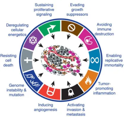
13 where they undergo malignant transformation by accumulating further genetic modifications. This can lead to invasion of neighboring tissues and further migration to other organs where it forms metastasis (Abel & DiGiovanni, 2011). Each process of sequential accumulation of mutations is unique, which explains (part of) the diversity of this disease and the complexity in identifying reliable biomarkers and efficient treatment options. However, certain characteristics essential to cancer development are shared by most tumors, such as intrinsic physiological alterations including growth autonomy, insensitivity to inhibitory growth signals, ability to escape apoptosis, unlimited replicative potential, strong ability to induce angiogenesis and the metastatic invasion of distant tissues (Figure 1) (Hanahan & Weinberg, 2000).
Consequently, in addition to surgical removal of malignant tissues, the first anticancer therapies that were developed in the past century and are still widely used in clinics aim at destroying malignant cells by targeting their rapid uncontrolled proliferation. For this, several, mechanistically diverse, anticancer strategies were designed that can be broadly subdivided based on their main mode of action into: (i) alkylating agents which induce the formation of inter- or intra- DNA crosslinks interfering with DNA replication (e.g. cyclophosphamide, oxaliplatin), (ii) topoisomerase inhibitors which impair DNA unwinding during replication and/or transcription (e.g. anthracyclines, topotecan), (iii) antimetabolites which prevents from DNA or RNA synthesis (e.g. 5-fluorouracil), (iv) spindle poisons affecting the (de)polymerization of tubulin and the formation of mitotic spindle (e.g. taxols, vinca-alkaloids), (v) radiation therapies which induce DNA damages and consequent apoptotic cell death, (vi) cytotoxic antibiotics which display anticancer effects through various mechanisms, such as reactive oxygen species production (e.g. bleomycin) or DNA intercalation (e.g. dactinomycin). Of note, most chemotherapeutics belong to several categories. Targeted therapy, yet another strategy that emerged during the past 20 years, focuses on cancer specific alterations such as oncogenic signaling pathways (e.g. anaplastic lymphoma kinase (ALK) inhibition with crizotinib).
More recently, ample evidence showed that cancer is not a merely cell autonomous genetic disease, but also a process depending on the tumor micro-environment and thus new characteristics were added to the list of cancer hallmarks: modifications in the cell metabolism and the ability to escape from immunosurveillance, together with genome instability that further drives mutational alterations and the capacity to subvert proinflammatory signals for cancer development (Figure 1) (Hanahan & Weinberg, 2011). These discoveries offer additional opportunities for the development of treatment options, including the immunotherapies, which aim at the reactivation of intrinsic anticancer immunosurveillance.
14
Figure 1. The hallmarks of cancer (Hanahan & Weinberg, 2011). Malignant cells share a set of common cell
15
2. Role of the immune system in antitumor therapy
2.1. From the discovery of tumor antigens to the development of immunotherapies In 1890, William B. Coley demonstrated that injection of dying bacterial cultures called Coley toxins could be beneficial for patients with soft tissue sarcoma, which was the first manifestation of the existence of an anticancer immune response. Fifty years later, it was shown that injection of irradiated sarcoma in syngeneic mice promoted immunity against a rechallenge with live sarcoma cells of the same kind (Burnet, 1957; Foley, 1953; Klein et al, 1960).
The specific recognition of tumors by the immune system suggests that cancer cells express characteristic antigens, usually proteins, peptides or polysaccharides, but also nucleic acids combined with proteins or polysaccharides, which are different from “self” antigens. Tumor associated antigens (TAAs) can indeed result from mutations that drive tumorigenesis, like alteration in the p53 transcription factor, or be viral antigens, such as E6 and E7 expressed by human papilloma virus (HPV), both giving rise to new peptides, therefore also called neoantigens, which constitute particularly interesting targets for immunotherapy due to their high tumor specificity. Nevertheless, “self” antigens presented by cancer cells can also be immunogenic if they are (i) over-expressed, with the example of human epidermal growth factor (HER2) in breast cancer, (ii) associated with differentiation, like melanocyte differentiated antigens, or (iii) expressed in an abnormal location, with the example of cancer/testis antigens, a group of proteins expressed by tumor cells, but not by normal tissues except germinal cells and trophoblasts (Scanlan et al, 2004; Schreiber et al, 2011). These TAAs are presented by the major histocompatibility complex (MHC) class I at the surface of tumor cells and can be recognized by the immune system. The first human TAA, the melanoma antigen 1 (MAGE-A1), was identified in 1991 (van der Bruggen et al, 1991). Since then, more than 400 antigens have been discovered (Wurz et al, 2016).
In 1994, it was shown that the immune system not only recognizes TAAs but also danger signals emitted by cells undergoing stress or abnormal differentiation (Matzinger, 2002). In spite of these elements, the role of the immune system against cancer has been long debated, in part due to the evidence that tumor cells could benefit from a proinflammatory environment, including proliferative and proangiogenic signaling (Balkwill & Mantovani, 2001; Karin et al, 2002).
In the early 2000’s, the group of Robert Schreiber demonstrated that interferon (IFN) plays a crucial role in cancer immunosurveillance and that tumors coming from
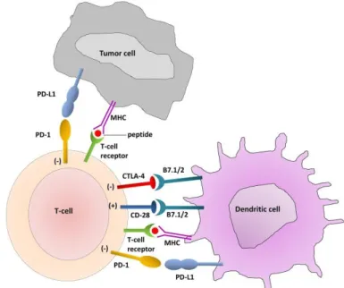
16 immunodeficient mice are more immunogenic than tumors arising on immunocompetent mice, giving birth to the concept of immunoediting (Shankaran et al, 2001). These two fundamental findings led to a regain of interest for cancer immunotherapy. Today, immunotherapy comes in four main flavors including (i) monoclonal antibodies which target specific cancer antigens promoting complement fixation and antibody-dependent cell-mediated toxicity (ADCC), (ii) anticancer vaccines to boost adaptive anticancer immune responses with tumor specific antigens, (iii) chimeric antigen receptor (CAR) T cells which consists of autologous T cells of patients activated in vitro and (iv) immune checkpoint blockade (ICB) to target specific immune inhibitory signaling with monoclonal antibodies. Indeed, T lymphocytes express inhibitory receptors, such as programmed cell death 1 (PD-1) and cytotoxic T-lymphocyte-associated protein 4 (CTLA-4), and cancer cells may express ligands (like programmed cell death ligand 1, PD-L1) as a strategy to escape from immunosurveillance (Figure 2). This approach has largely demonstrated its antineoplastic efficacy first in melanoma and then in different other kinds of tumors (Lesokhin et al, 2015), with several ICBs currently on the market: a CTLA-4 targeting antibody ipilimumab (Yervoy), three PD-1 targeting antibodies, pembrolizumab (Keytruda), nivolumab (Opdivo) and cemiplimab (Libtayo), as well as three PD-L1 targeting antibodies, atezolizumab (Tecentriq), durvalumab (Imfinzi) and avelumab (Bavencio) (Cancer.org, Retrieved 2019-08-17; FDA, Retrieved 2019-08-17).
Figure 2. Activator and inhibitory T cell receptors and their ligands (Zaravinos, 2014). CTLA-4, cytotoxic
T-lymphocyte-associated protein 4; MHC, major histocompatibility complex; PD-1, programmed cell death 1; PD-L1, programmed cell death ligand 1.
17 2.2. Mechanisms of antitumor immunity
The immune system is a biological defense system which protects the organism against infection. It consists of innate immune effectors, which elicit an immediate local response to pathogens in a rather nonspecific fashion, and the adaptive immune system, which takes longer to be activated but confers a specific reaction and durable protection against the initial insult. In the context of cancer, immunosurveillance is an extrinsic mechanism engaged after cell intrinsic mechanisms of control and elimination have failed.
During tumorigenesis, tumor cells require more important blood influx and stromal reorganization to pursue their expansion. This environment promotes proinflammatory cytokine release by tumor and stromal cells. Such cytokines include tumor necrosis factor (TNF)-, transforming growth factor (TGF)-, interleukin (IL)-1, IL-6 and IL-10, altogether leading to the recruitment of natural killer (NK) cells, NKT cells, T lymphocytes, macrophages and dendritic cells (DCs) on the site of the tumor (Matzinger, 2002; Smyth et al, 2001; Yamamoto et al, 2003). Recruited immune cells in turn secrete proinflammatory cytokines, like IL-12 and IFN- (Yamamoto et al, 2003). Tumor infiltrating NK cells bind to tumor cells via interaction between natural killer group 2 member D (NKG2D) and its ligands, MHC class I polypeptide-related sequence (MIC)-A and -B and mediate cytotoxic effect on neoplastic cells promoting tumor antigens release (Gajewski et al, 2013). Dying tumor cells and released antigens are ingested by DCs, which in addition sense damage-associated molecular patterns (DAMPs) via their pattern recognition receptor (PRRs). Altogether, these signals promote DCs maturation with the presentation of tumor antigens by MHC class II, cross-presentation by MHC class I, expression of the co-stimulatory molecules CD40, CD80 and CD86 and secretion of proinflammatory cytokines including IL-12, IL-6, TNF, type I IFN (Albert et al, 1998; Banchereau & Steinman, 1998; Gardner & Ruffell, 2016). At the same time, DCs migrate to the draining lymph node where they activate naïve CD8+ T cells, promoting the
clonal expansion of cytotoxic T lymphocytes (CTLs) as well naïve CD4+ T cells driving their
differentiation in T helper (Th) 1 cells secreting IFN, IL-12, IL-6 and eventually in Th17 cells secreting IL-17 and IL-22 (Bailey et al, 2014) in the context of anticancer immune response. CTLs are recruited to the tumor site where they elicit cytotoxicity against cancer.
Altogether, CTLs, NK and NKT lymphocytes are considered as the main anticancer effectors as they induce apoptosis of their target cells through perforin, granzyme and granulysin secretion, as well as via the expression of TNF related apoptosis inducing ligand (TRAIL) (Mori et al, 1997; Takeda et al, 2001). Thanks to their ability to present antigens to
18 CD4+ and CD8+ T cells, DCs constitute the key mediator between innate and adaptive immune responses. In addition, T cells seem to have a rather antitumor effect even though their role and exact underlying mechanisms need to be further investigated (Fridman et al, 2017; Gajewski et al, 2013). In the context of cancer, macrophages can exert different roles. When activated by IFN, they display an M1 phenotype including the secretion of proinflammatory cytokines such as IL-2, IL-6, IL-1, IL-12, IL-23 and TNF, altogether driving the anticancer response through a Th1 polarization. However, tumor-associated macrophages (TAMs), observed in most tumor infiltrates, rather display an M2 phenotype characterized by low MHCII expression and secretion of immunosuppressive cytokines such as IL-10 and TGF. M2 macrophages also secrete cytokines and chemokines which promote angiogenesis and metastasis (Hao et al, 2012). In addition, this subtype of macrophages can recruit CD4+ FoxP3+
regulatory T cells (Treg) through the secretion of the C-C motif chemokine ligand 2 (CCL22) (Curiel et al, 2004). Tregs in turn promote tolerance by the secretion of inhibitory cytokines (e.g. TGF-, IL-10) and the expression of the inhibitory ectoenzymes CD39 and CD73 among other suppressive mechanisms (Curiel et al, 2004; Fridman et al, 2017; Knochelmann et al, 2018). Like macrophages, neutrophils can have a dual role, proinflammatory under the N1 phenotype while protumoral when displaying as N2 phenotype.
2.3. Immunoediting accompanies cancer progression
Tumors develop despite the ability of the immune system to recognize and eliminate tumor cells. This paradox can be explained by tumor immunoediting, a process associated to the pressure exerted by the immune system during tumorigenesis.
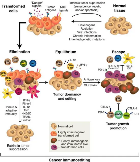
Immunoediting, first described by the team of Robert Schreiber, occurs in three sequential steps: elimination, equilibrium and escape. In the elimination phase, long before a tumor gets clinically detectable, emerging cancer cells are under immunosurveillance by the innate and adaptive immune system, including in particular CTLs, NK cells and proinflammatory cytokines, as previously described. This might lead to complete tumor destruction. However, some cancer variants may survive and enter the “equilibrium” phase, in which tumor outgrowth is maintained in a state of dormancy by mediators belonging to the adaptive immune system. NK cells in particular are dispensable in this phase. Under the continuous pressure exerted by the immune system on genetically unstable cancer cells, tumors undergo transformation (or immunoediting), until resistant clones may emerge, which (i) are not recognized by the immune system due to antigen variation or a defect in antigen
19 presentation, (ii) are resistant to mechanisms of immune destruction and/or (iii) promote an immunosuppressive tumor micro-environment. In the escape phase, tumor proliferation cannot anymore be controlled by the immune system and a clinically detectable tumor forms (Dunn et al, 2004a; Dunn et al, 2004b; Schreiber et al, 2011) (Figure 3).
Figure 3. Cancer immunoediting (Schreiber et al, 2011). When tumor cells emerge, they are recognized and
eliminated by the immune system, including in particular natural killer (NK), NKT cells, dendritic cells (DCs) primed CD8+ T cells, T cells, macrophages (Mᶲ) and CD4+ T cells, the pro-inflammatory cytokines interferon
(IFN), IFN, interleukin (IL)-12, tumor necrosis factor (TNF), natural killer group 2 member D (NKG2D) and the cytotoxic enzymes TNF related apoptosis inducing ligand (TRAIL) and perforin; this constitutes the elimination phase. Under the pressure of these immune mediators, tumor will undergo transformation to resist to the immune attacks; this corresponds to the equilibrium phase, a state of functional dormancy in which proliferation is controlled by mediators of the adaptive immune response. This may last for a while, until variants arise which have acquired the capacity not to be recognized and/or eliminated by the immune system; this constitutes the escape phase. CTLA-4, cytotoxic T-lymphocyte-associated protein 4; IDO, indoleamine 2,3-dioxygenase; MDSC, myeloid-derived suppressor cells; MHC, major histocompatibility complex; NKR, natural killer receptor; PD-1, programmed cell death 1; PD-L1, programmed cell death ligand 1; Treg, regulatory T cell.
20 At this stage, the constitution of the tumor immune infiltrate indicates the outcome of the disease, with the presence of CTLs, tertiary lymphoid structures and M1 macrophages as good prognostic factors, whereas Tregs and M2 macrophages being worst prognosis factors (Fridman et al, 2017; Pages et al, 2018). It can be driven by the choice of anticancer therapy: ICB prevents from CTLs exhaustion, vaccination enhances tumor specific CD8+ T cells and immunogenic cell death inducers promote the emission of danger signals.
2.4. Adjuvanticity through immunogenic cell death
For a long time, cell death has been classified based on morphological features in a dichotomic manner. Apoptosis was described as a regulated, physiological and non-immunogenic type of cell death, whereas necrosis was considered as an uncontrolled pathological and proinflammatory mechanism. Since then, we learned that regulated cell death can exert morphological features of necrosis whereas apoptosis can trigger an immune response (Galluzzi et al, 2015a). The immunogenicity of cell death cannot be defined merely based on morphological traits, but need to be addressed with vaccination experiment which consists in the injection of dying cells into syngeneic host on their sequential rechallenge with live cells (Kepp et al, 2014). An absence of (or delayed) tumor growth in this model is an indication for anticancer vaccination efficacy and thus for the immunogenicity of cell death.
Immunogenic cell death (ICD) following pathogens infection constitutes an ancient mechanism of defense. Indeed, when cells are infected by viruses or intracellular bacteria, they sense microbe-associated molecular patterns (MAMPs) via their specific PRRs which trigger an intracellular and micro-environmental danger response and consequent activation of immune cells. In 2005, it has been demonstrated that anthracyclines can also drive a kind of cell death which induces an anticancer adaptive immune response (Casares et al, 2005). In the following years, many other chemotherapeutics, like oxaliplatin (OXA), as well as physical modalities, like irradiation, photodynamic therapy (PDT) or high hydrostatic pressure (Fucikova et al, 2014; Garg et al, 2012a; Obeid et al, 2007a), were shown to be able to trigger ICD. Similarly to their microbial counterparts, these anticancer agents induce pre-mortem stress mechanisms leading to the emission of DAMPs, recognized by PRRs expressed on DCs (Figure 4). Calreticulin (CALR), adenosine triphosphate (ATP) and high-mobility group box 1 (HMGB1) are DAMPs common to almost all instances of ICD discovered until now (Galluzzi et al, 2017). Type I IFN and annexin A1 (ANXA1), discovered in the context of chemotherapeutics-induced ICD, have been further added to the list (Sistigu et al, 2014; Vacchelli et al, 2015a), but their general
21 involvement in ICD remains to be investigated. Even though these five DAMPs may be general features of ICD, the pathways underlying their emission is particular to each form of ICD, as exemplified by autophagy, which is involved in anthracyclines-induced ICD but displays an immunosuppressive action in the context of hypericin-based PDT (Garg et al, 2013).
Figure 4 Activation of immunogenic cell death (Galluzzi et al, 2017). Chemotherapeutics induce calreticulin
(CALR) exposure at the plasma membrane, as well as adenosine triphosphate (ATP), annexin A1 (ANXA1) and high-mobility box 1 (HMGB1) release from stressed and dying cancer cells, which are recognized by low density liproprotein receptor related protein 1 (LRP1), purinergic receptors P2Y2 (P2RY2) and P2X7 (P2RX7), formyl peptide receptor 1 (FPR1) and toll-like receptor 4 (TLR4) expressed by dendritic cells (DCs) respectively. In addition, they induce the autocrine/paracrine secretion of type I interferon (IFN) leading to C-X-C motif chemokine ligand 10 (CXCL10) release. These mechanisms allow DCs recruitment, activation and maturation and elicit an adaptive immune response involving IL-17 producing T cells and type I IFN secreting T cells. In addition to the establishment of an immune memory, this has the potential to eliminate cancer cells through cytotoxic T lymphocytes (CTLs)-mediated cytotoxicity.
In the context of ICD, inflammatory DCs (CD11b+ CD11c+ Ly6Chigh) sense the emission
of DAMPs and in turn secrete proinflammatory cytokines and cross-present antigens to naïve T cells, most of which differentiate in IFN producing CD8+ T cells. IL-17 producing T cells are also activated and sustain the proliferation of CD8+ T cells (Figure 4) (Ma et al, 2013; Ma et al, 2011). In addition, neutrophils are recruited through the release of DAMPs, but also through the secretion of certain chemokines, such as C-X-C motif chemokine ligand (CXCL)1,
22 C-C motif chemokine ligand (CCL)2 and CXCL10, by dying cancer cells (Garg et al, 2017b). Altogether, these mechanisms participate in elimination of residual cancer cells.
Some signaling events in dying cells involving in particular RIPK1 and nuclear factor B (NF-B)-induced transcriptional programs have also been reported to be required for DCs cross-presentation and consequent CD8+ T cells activation, but the precise mechanism by which they activate DCs is not well understood (Giampazolias et al, 2017; Yatim et al, 2015).
Even though ICD was first described as an apoptotic kind of cell death (Casares et al, 2005), it has been recently reported that necroptosis, a form of regulated necrosis mediated by receptor-interacting serine-threonine kinase 1 (RIPK1), RIPK3 and mixed lineage kinase domain-like (MLKL), is also able to drive an anticancer immune response (Aaes et al, 2016; Yang et al, 2016). Necroptosis can be induced in cells engineered for RIP3K3 to be inducible by doxycycline or by the combination of TNF- with a synthetic second mitochondria derived activator of caspase (SMAC) mimetic and the caspase inhibitor z-VAD-FMK. Similarly to apoptotic ICD, it triggers the emission of DAMPs (CALR, ATP, HMGB1) that confer long term protection in mice against subsequent challenge with living cancer cells of the same type. Interestingly, anthracyclines and OXA’s immunogenic properties are attributable to necroptosis in addition to apoptosis (Aaes et al, 2016; Yang et al, 2016).
The success of a specific anticancer immune response relies both on tumor antigenicity, which is related to its mutational load (Samstein et al, 2019), and on the choice of the therapy driving the antigenicity through DAMPs emission.
2.4.1. Mechanisms of chemotherapeutics-driven immunogenic cell death
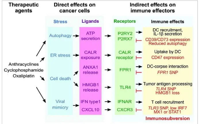
Most ICD mechanisms discovered so far rely on the exposure of CALR, the secretion of ATP and the release of HMGB1, but the mechanisms underlying the emission of these DAMPs depend on the type of ICD. Moreover, more recently discovered hallmarks of ICD, i.e. type I IFN and ANXA1 release have been much less studied and we don’t know yet if they are specific to chemotherapeutics-induced ICD or shared with other modalities. The present work aims at finding new ICD inducers among chemotherapeutics agents. Thus, here, we will present the mechanisms of chemotherapeutics-driven ICD (Figure 5), which have been most described so far.
23
Figure 5. Mechanisms of immunogenic cell death induction by chemotherapeutics (modified from (Vacchelli
et al, 2016b). Immunogenic cell death agents trigger various pre-mortem stress pathways which activate the exposure and release of DAMPs recognized by dendritic cells (DCs) through particular receptors that exert different functions on immune cells. Cancer cells have developed certain strategies to avoid danger signaling. ANXA1, annexin A1; ATP, adenosine triphosphate; CALR, calreticulin; CXCL10, C-X-C motif chemokine ligand 10; CXCR3, C-X-C chemokine receptor 3; ER, endoplasmic reticulum; FPR1, formyl peptide receptor 1; HMGB1, high-mobility box 1; IFN, interferon; IFNAR, interferon-α/β receptor; IRF7, interferon regulatory factor 7, LRP1, low density lipoprotein receptor related protein 1; MX1, myxovirus 1; P2RY2, purinergic receptors P2Y2; P2RX7, purinergic receptors P2X7; STAT1, signal transducer and activator of transcription 1; TLR3/4, toll-like receptor3/ 4; SNP, single nucleotide polymorphism.
2.4.1.1. eIF2 phosphorylation-dependent calreticulin exposure
Effect of CALR on the immune system
CALR, a 60 kDa chaperone, is the most abundant protein in the ER. It regulates calcium homeostasis and protein folding. When treated with ICD inducers, a fraction of CALR together with other ER chaperones, such as heat shock protein (HSP) 70 (also known as HSPA1A), HSP 90 (also known as HSP90AA1) and the protein disulfide isomerase family A member 3 (PDIA3, also known as ERp57), are externalized from the ER lumen to the plasma membrane of stressed and dying tumor cells (Fucikova et al, 2011; Gardai et al, 2005; Garg et al, 2012b; Obeid et al, 2007b; Panaretakis et al, 2008). Theses events occurs before exposure of phosphatidylserine, meaning that it is not related to plasma membrane alterations accompanying apoptosis (Obeid et al, 2007b). At the surface of dying cells, CALR acts as an “eat-me” signal; it is recognized
24 by low density liproprotein receptor related protein 1 (LRP1; also known as CD91) expressed by myeloid cells (mostly macrophages and DCs), unless the dying cells express simultaneously CD47, a “don’t eat-me” signal (Gardai et al, 2005; Garg et al, 2012b). Following, signaling through CD91 in DCs promote the activation of a Th1 or Th17 immune response according to the profile of danger signals (Pawaria & Binder, 2011). Accordingly, vaccination induced by anthracyclines-, OXA- or crizotinib-treated cells is abolished if dying cells are incubated with neutralizing CALR antibodies or if CALR is depleted with siRNAs (Liu et al, 2019b; Obeid et al, 2007b; Tesniere et al, 2010). In addition, PDT-driven ICD is reduced when LRP1 expression is down regulated with shRNAs (Garg et al, 2012b), bortezomib- and capsaicin-induced ICDs are abolished by LRP1 targeted monoclonal antibody or by silencing LRP1 with siRNA (Gilardini Montani et al, 2015) and mouse macrophages lacking LRP1 exhibit a reduced phagocytotic potential (Gardai et al, 2005; Lillis et al, 2008).
EIF2 phosphorylation-dependent mechanism of CALR exposure
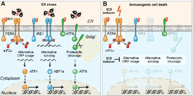
The canonical response to ER stress is called unfolded protein response (UPR) and involves three pathways, which are mediated by the transmembrane proteins eukaryotic translation initiation factor 2-alpha kinase 3 (eIF2AK3, also called PERK for protein kinase R (PKR)-like endoplasmic reticulum kinase), inositol-requiring enzyme 1 (IRE1α) and the activating transcription factor (ATF) 6. In normal conditions, these proteins are maintained in an inactive state by the binding immunoglobulin protein (BiP) chaperone. During ER stress, BiP dissociates from them to bind the misfolded/unfolded proteins, resulting in the activation of the three arms of UPR. EIF2AK3 is a type I ER transmembrane kinase that contains a PEK-like catalytic domain in its cytosolic C-terminal region. When dissociated from BiP, EIF2AK3 oligomerizes, autophosphorylates and promotes phosphorylation of the serine 51 on eukaryotic initiation factor 2 (eIF2α). EIF2α phosphorylation leads to messenger RNA (mRNA) translational attenuation by preventing cap-dependent ribosomal initiation complexes formation. In addition, phosphorylated eIF2α selectively activates the translation of the mRNA encoding for ATF4. ATF4 is a basic leucine zipper (b-ZIP) transcription factor that induces growth arrest and upregulates genes coding for chaperones, antioxidants, XBP1 as well as DNA damage-inducible transcript 3 (DDIT3, also known as CHOP for CCAAT/enhancer-binding homologous protein). IRE1 is also a type I ER transmembrane endoribonuclease/kinase that has a kinase domain and an endoribonuclease domain in its cytosolic N-terminal luminal domain. IRE1 exists as two isoforms: IRE1α and IRE1β. IRE1α is present in all cell types and has been widely studied. Upon ER stress conditions, it becomes active by dimerization and
25 autophosphorylation. Activated IRE1α catalyzes the splicing of a 26 nucleotides intron from the X-box binding protein 1 (XBP1) mRNA. Spliced XBP1 encodes a b-ZIP transcription factor that upregulates UPR genes, including genes involved in ER-associated protein degradation (ERAD) as well as genes involved in protein folding. The third pathway involves ATF6, a type II ER transmembrane protein that has a transcriptional activation domain in its cytosolic region. ATF6 has two isoforms, ATF6α and ATF6β; when dissociated from BiP, ATF6α transits from the ER membrane to the Golgi where it is cleaved by the Golgi resident site 1 and 2 proteases (respectively S1P and S2P), leading to the generation of an activated b-ZIP factor. The N-terminal fragment of ATF6α is transported to the nucleus where it activates UPR genes, like chaperones, XBP1 and BiP overexpression, which promote protein folding (Figure 6-A) (Kato & Nishitoh, 2015; Oslowski & Urano, 2011).
Importantly, eIF2 phosphorylation is induced by eIFAK3 during ER stress, but can also be activated by eIF2AK1 (also called HRI for heme regulated inhibitor) upon heme deprivation, eIF2AK2 (also named PKR for protein kinase R) in response to viral infection or eIF2AK4 (also called GCN2 for general control nonderepressible 2) in the context of nutrient deprivation.
In ICD induced by chemotherapeutics, CALR exposure is preceded by the phosphorylation of eIF2, which has been showed to be mediated by eIF2AK3 in the murine methylcholanthrene-induced fibrosarcoma MCA205, the human osteosarcoma U2OS and colon carcinoma CT26 (Bezu et al, 2018a; Obeid et al, 2007b; Panaretakis et al, 2009). However, it was recently demonstrated that in melanoma human cells, anthracyclines induce eIF2 phosphorylation via the activation of other kinases: eIF2AK2 or eIF2AK4 (Giglio et al, 2018). Importantly, during ER stress elicited by chemotherapeutics, solely eIF2 phosphorylation is involved, without the downstream expression of ATF4 and DDIT3 and without activation of the two other arms of ER stress, i.e. the translocation of ATF6 and the alternative splicing of XBP1 (Figure 6-B) (Bezu et al, 2018a). This split ER stress response was also observed for crizotinib-induced ICD in various cell lines (Liu et al, 2019b) and for anthracyclines-treated human melanoma cells (Giglio et al, 2018). In addition, following administration of the EGFR specific antibody cetuximab, ICD was abolished by overexpression of XBP1 (Pozzi et al, 2016).
26
Figure 6. Split ER stress response involved in chemotherapeutics driven ICD (Bezu et al, 2018b). Canonical
endoplasmic reticulum (ER) stress activates three pathways: (i) protein kinase R-like endoplasmic reticulum kinase (PERK)-mediated eukaryotic initiation factor 2 (eIF2) phosphorylation leading to activating transcription factor (ATF) 4 translation due to particular open reading frame (ORF) arrangement, (ii) inositol-requiring enzyme 1 (IRE1) dimerization leading to X-box protein 1 alternative splicing (XBP1s) and (iii) ATF6 translocation to the Golgi where it undergoes proteolytic cleavage followed by translocation to the nucleus of the N-terminal part; all aiming at eliminating and/or repairing misfolded proteins (A). Immunogenic cell death (ICD) inducers mediate PERK-dependent eIF2 phosphorylation but none of the other protein activation (B).
CALR needs to be engaged in a complex with PDIA3 to be able to translocate from the ER to the membrane (Liu et al, 2019a; Panaretakis et al, 2008). Following eIF2 phosphorylation, B-cell receptor-associated protein 31 (BAP31) is proteolyzed in a caspase 8-dependent manner, leading to the activation of the pro-apoptotic Bcl-2–associated factors X et K (BAX and BAK) and the anterograde translocation of the CALR/PDIA3 complex from the ER lumen to the plasma membrane via the Golgi apparatus. CALR is finally secreted by soluble N-ethylmaleimide-sensitive-factor attachment protein receptor (SNARE)-dependent exocytosis (Panaretakis et al, 2009). Inhibition of any step of this pathway abolished CALR exposure. In addition, the inhibition of (i) eIF2AK3 phosphorylation with a siRNA, (ii) caspase 8 activation with a pharmacologic inhibitor (Z-VAD-fmk) or siRNA, (iii) BAX with a siRNA, (iv) the ER-Golgi traffic with brefeldin A or (iv) SNARE-dependent exocytosis with a synaptosomal-associated protein 23 (SNAP23) siRNA also abolished vaccination capacity of anthracyclines (Panaretakis et al, 2009). Of note, even though eIF2AK3-dependent CALR exposure is also required for the immunogenicity of cells treated with hypericin based PDT, this does not involve eIF2 phosphorylation, caspase 8 and PDIA3 (Garg et al, 2012b).
Additionally, CALR exposure following anthracyclines chemotherapy is not only dictated by cell intrinsic pathways, but also relies on autocrine/paracrine signalization pathways
27 in stressed cancer cells, mediated by the CXCL8 (also known as IL-8) and its receptor CXCL2 (Sukkurwala et al, 2014b).
Eventually, it has been recently shown that CALR exposure also relies on ANXA1. Indeed, following treatment with anthracyclines, CALR exposure and the consequent tumor growth control is abolished in Anxa1-/- cells, effect which is restored by supplementation with rCALR. The underlying mechanism remains to be elucidated, but it may be related to the involvement of ANXA1 in unconventional protein secretion pathways or in intracellular trafficking (Baracco et al, 2019).
Strategies to modulate CALR exposure
Chemotherapeutics like CDDP or mitomycin C, which induce all ICD hallmarks apart from CALR exposure, are not able to induce ICD (Casares et al, 2005; Tesniere et al, 2010), which could be compensated by the co-incubation with CALR recombinant protein (rCALR) prior to vaccination (Obeid et al, 2007b; Tesniere et al, 2010). In line with this, coating with rCALR enhances phagocytosis and vaccination with apoptotic melanoma cells (Dudek-Peric et al, 2015; Qin et al, 2011). CALR trafficking can also be targeted: -integrins and PDIA3 interact with CALR in the ER and coordinate CALR translocation. Incubation of cells with 9EG7, a 1 integrin activating antibody, reduces CALR exposure; conversely, knocking down integrins leads to enhanced CALR exposure, even though this strategy can obviously not be transferred to the clinics (Liu et al, 2019a). The immunogenicity of CDDP or mitomycin C could also be restored in combination with activators of ER stress like the inhibitor of sarco/endoplasmic reticulum Ca2+ ATPase (SERCA) thapsigargin, or the inhibitor of N-glycosylation tunicamycin (Martins et al, 2011). Similarly, pyridoxine, a precursor of the bioactive vitamin B6 as well as zinc supplementations were able to restore immunogenicity to CDDP-induced cell death (Aranda et al, 2014a; Aranda et al, 2015; Cirone et al, 2013).
Another strategy consists in the administration of inhibitors of the eIF2 phosphatase complex formed by protein phosphatase 1 regulatory subunit 15A (PPP1R15A, also called growth arrest and DNA damage-inducible protein, GADD34) and protein phosphatase 1 (PP1), like calyculin A, tautomycin or salubrinal, which mediate eIF2 phosphorylation-dependent CALR exposure (Kepp et al, 2009; Obeid et al, 2007b). All these approaches underline the relevance of eIF2 phosphorylation for CALR exposure and may be promising, but only coating with recombinant CALR could circumvent any kind of deficiency including downregulation of CALR itself.
28
Clinical relevance of CALR exposure
In patients with non-Hodkin’s lymphoma, vaccinated with DCs which were previously activated by co-culture with radiation-treated lymphoma cells, high amounts of CALR and HSP90 were correlated with improved clinical outcome (Zappasodi et al, 2010). Growth of monocytes from melanoma patients were shown to be slower when LPR1 expression was elevated (Stebbing et al, 2004). In lung and ovarian cancers, high CALR expression was associated with better clinical response to therapies with ICD inducers (Garg et al, 2015). In two independent cohorts of patients with non-small cell lung carcinoma (NSCLC), high level of CALR correlated with eIF2 phosphorylation and with improved clinical outcome (Fucikova et al, 2016a). The analysis of acute myeloid leukemia (AML) from patients showed that (i) CALR expression and exposure are associated with increased relapse free survival and overall survival (Fucikova et al, 2016b), (ii) spontaneous CALR exposure correlates with the phosphorylation of eIF2, with a stronger T cell response and with improved disease outcome, regardless of the chemotherapeutics regimen (Wemeau et al, 2010) and (iii) CALR exposure is higher in daunorubicin-treated patients accompanied by elevated CD8+ T cells infiltration and improved disease outcome (Aurelius et al, 2019). Immunohistochemical analysis of NSCLC patients’ tumors revealed that loss of CALR expression was a negative prognostic factor and that CALR expression was correlated to CTLs infiltration (Stoll et al, 2016). These clinical data emphasize the importance of the CALR-LRP1 axis for the outcome of cancer treatment and the reestablishment of immunosurveillance. Another element that emphasize this finding is the negative prognostic value of CD47 expression in AML (Majeti et al, 2009), in ovarian cancer (Wang et al, 2015a) and in esophageal carcinoma (Suzuki et al, 2012).
Interestingly, in a cohort of breast cancer patients treated with neoadjuvant chemotherapy, none of the ER stress induced transcription factors ATF4, ATF6 and XBP1 could reflect therapy outcome, whereas a metagene analysis showed that IFN T cells infiltration was implicated (Stoll et al, 2014). This strengthens recent findings showing a split ER stress response involving exclusively eIF2 phosphorylation in the response to anthracyclines (Bezu et al, 2018a).
2.4.1.2. Autophagy-mediated ATP secretion
Effect of ATP on the immune system
29 biochemical reactions. It is also an important messenger that can act via the ligation of purinergic receptors (Burnstock, 2007). In addition to its involvement in transmission of neurological signals, ATP can be released into the extracellular space by various stimuli including ICD inducers, where it exerts chemotactic and adjuvant effects, serving as a “find-me” signal for myeloid cells (Elliott et al, 2009). Extracellular ATP interacts with purinergic receptors P2Y2 (P2RY2) and P2X7 (P2RX7) expressed by antigen presenting cells (Elliott et al, 2009; Ghiringhelli et al, 2009; Kronlage et al, 2010). Accordingly, injection of ATPS, a non-hydrolysable analog of ATP, induces local recruitment of DCs via P2Y (Muller et al, 2010).
Besides its role as chemoattractant, ATP can influence the function of immune cells by triggering DCs maturation in vivo (Idzko et al, 2007). Moreover, intradermal injection of ATPS stimulates vaccination effects dependent on the expression of MHC class II molecules, CD80, CD86, IL-1 and IL-12 by Langerhans cells (Granstein et al, 2005). Conversely, enzymatic degradation of ATP (by administration of apyrase, an ectonucleotidase) or the deletion of P2Y2 (by incubation with suramin, a P2Y antagonist, or by gene knockout) reduces phagocytosis of thymocytes by macrophages (Elliott et al, 2009).
Accordingly, ICD fails when ATP is neutralized with apyrase or with the membrane bound nucleosidase CD39, or if the receptors P2RY2 or P2RX7 are not expressed at the surface of antigen presenting cells of the host (Elliott et al, 2009; Ghiringhelli et al, 2009; Liu et al, 2019b; Ma et al, 2013; Michaud et al, 2011). ATP-induced purinergic signaling activates the NOD-like receptor family pyrin domain containing-3 (NLRP3) caspase-1 activation complex (the inflammasome) in DCs, leading to IL-1 secretion (Ghiringhelli et al, 2009). IL-1 promotes activation of IL-17 producing T cells and further recruitment of IFN producing by CD8+ T cells (Ma et al, 2011). Accordingly, mice deficient for P2RX7, caspase 1, NLRP3, IL-1, IL-1 receptor, IL-17 or IL-17 receptor fail to elicit an adaptive anticancer immune response after treatment with anthracyclines (Ghiringhelli et al, 2009; Ma et al, 2011).
Autophagy-dependent mechanism of ATP release
ATP released from dying tumor cells in the course of ICD depends on the autophagic machinery. Autophagy is a mechanism which entails the engulfment of cytoplasmic material within doubled membrane cytosolic organelles, called autophagosomes. Subsequently, autolysosomes form through the fusion of autophagosomes with lysosomes, which leads to the degradation of autophagosomes cargo by lysosomal hydrolases (Kroemer et al, 2010). Of note, microtubule-associated proteins 1A/1B light chain 3B called (MAP1LC3B, hereafter referred to as LC3) is
30 the most widely used marker of autophagosomes. Accordingly, in cancer cells treated with anthracyclines, reduced or absent expression of the autophagy related genes (ATG) Atg5, Atg7,
Atg10, Atg12, Beclin 1, Vsp34 or lysosomal-associated membrane protein (LAMP) 1 and 2,
either by shRNAs or by genetic alterations, were unable to induce ATP release, T cell activation and subsequent vaccination (Michaud et al, 2011). Consistently, autophagy-deficient virus-infected fibroblasts were unable to induce cross-presentation (Uhl et al, 2009). During anthracycline-induced autophagy, ATP switches from lysosomes to autolysosome and is further secreted by a mechanism dependent on LAMP1 which translocates to the membrane in a caspase-dependent manner. ATP secretion also requires membrane blebbing and the opening of pannexin 1 (PANX1) channels, both mechanisms depending on caspases activation (Martins et al, 2014). Of note, autophagy not only is dispensable for ICD induced by hypericin-based PDT but its inactivation can enhance immune cells activation (Garg et al, 2013).
Modulation of ICD via the autophagy-ATP axis
CD39 (also known as ENTPD1 for ectonucleosidase triphosphate diphosphohydrolase 1), which converts ATP into adenosine triphosphate (ADP) and adenosine monophosphate (AMP) as well as CD73, which converts AMP into adenosine, can degrade ATP and inhibits the activation of an anticancer immune response mediated by CD8+ T cells and NK cells (Stagg et al, 2012; Sun et al, 2010). CD39 is expressed by Tregs and a subset a Th17 cells, both described as immunosuppressive and tumor promoting (Chalmin et al, 2012; Sun et al, 2010). Accordingly, mice lacking the CD73 encoding genes develop less induced cancers due to enhanced immunosurveillance (Stagg et al, 2012). Consistently, co-administration of ARL67156, a nucleosidase inhibitor can restore the ability of ATP to trigger an immune response (Michaud et al, 2011; Michaud et al, 2012).
Another strategy to elicit ATP release consists in inducing autophagic flux. Caloric restriction mimetics (CRMs) have been recently described as agents able to mimic the effect of starvation, i.e. they promote autophagy without inducing any toxicity, and are therefore particularly interesting for clinical applications (Pietrocola et al, 2016). When combined with the anthracycline mitoxantrone (MTX), CRMs are able to potentiate the anticancer effect by enhancing ATP release and subsequent activation of an autophagy-dependent anticancer immune response (Pietrocola et al, 2016).
Clinical relevance of ATP as a hallmark of ICD
31 variation of P2RX7 led to rapid metastatic disease development (Ghiringhelli et al, 2009). In two other independent cohorts of breast cancer patients treated with anthracyclines, LC3 correlated with autophagic flux and the combination of LC3 and HMGB1 constituted a good prognostic factor for metastasis free survival (Ladoire et al, 2015). Another immunohistochemical study in a cohort of breast cancer patients revealed that the absence of autophagy was correlated with poor disease outcome as well as with reduced CD8+CD68+ T cells and increased CD4+FoxP3+ T cells in the tumor infiltrate (Ladoire et al, 2016).
2.4.1.3. HMGB1 release and TLR4 mimicry
Effect of HMGB1 on the immune system
HMGB1 is the most abundant non-histone chromatin-binding protein, present in the nucleus of all cells. It can be actively secreted by myeloid cells in response to infection or released by cells undergoing necrosis and promotes strong inflammation (Scaffidi et al, 2002; Sims et al, 2010; Wang et al, 1999). In line with this, induction of necroptosis in melanoma leads to HMGB1 release, as well as expression of MHC class II and CD86 by DCs and macrophages, effects which were abolished in Rage-/- and Myd88-/- mice (which respectively lack receptor for advanced glycation end products and myeloid differentiation primary response protein 88).
In response to tissue damage, HMGB1 could also form a complex with CXCL12 leading to the recruitment of immune cells (Schiraldi et al, 2012). However, in the context of ICD, the only receptor of HMGB1 is the toll-like receptor (TLR) 4. Indeed, both Tlr4-/- and Myd88-/- mice cannot mount an efficient anticancer immune response in response to anthracylines or OXA (Apetoh et al, 2007). This is reinforced by the fact that in co-culture experiments, neutralization or depletion of HMGB1 abolishes the cross-presentation, whereas administration of HMGB1 promotes inflammation (Apetoh et al, 2007). These findings reveal that the HMGB1-TLR4-MYD88 axis is required for ICD (Apetoh et al, 2007). However, the molecular mechanisms underlying HMGB1 release from the nucleus of cells undergoing ICD remains to be elucidated.
Modulation of ICD via the HMGB1 pathway
Dendrophilin, a highly potent and exclusive Tlr4 agonist could both reestablish anticancer immune responses when HMGB1 expression is depleted by siRNA, and increase IFN production and consequent tumor growth reduction in several mice models when combined to anthracycline-based chemotherapy (Yamazaki et al, 2014). Besides, lots of ongoing clinical