Full Terms & Conditions of access and use can be found at
ISSN: 1758-4299 (Print) 1758-4302 (Online) Journal homepage: https://www.tandfonline.com/loi/tlip20
Challenges to determining whether DHA can
protect against age-related cognitive decline
Marie Hennebelle, Emilie Harbeby, Sébastien Tremblay, Raphael
Chouinard-Watkins, Fabien Pifferi, Mélanie Plourde, Philippe Guesnet & Stephen C
Cunnane
To cite this article:
Marie Hennebelle, Emilie Harbeby, Sébastien Tremblay, Raphael
Chouinard-Watkins, Fabien Pifferi, Mélanie Plourde, Philippe Guesnet & Stephen C Cunnane (2015)
Challenges to determining whether DHA can protect against age-related cognitive decline, Clinical
Lipidology, 10:1, 91-102
To link to this article: https://doi.org/10.2217/clp.14.61
Copyright 2015 Future Medicine Ltd
Published online: 18 Jan 2017.
Submit your article to this journal
Article views: 360
View Crossmark data
part of Clin. Lipidol.
10.2217/CLP.14.61
Review
Challenges to determining whether DHA can
protect against age-related cognitive decline
10
1
2015
DHA, an omega-3 fatty acid, is an important constituent of brain membranes and has a key role in brain development and function. This review aims to highlight recent research on DHA’s role during age-related cognitive decline and Alzheimer’s disease. Animal and in vitro studies have provided some interesting mechanistic leads, especially on brain glucose metabolism, that may be involved in neuroprotection by DHA. However, results from human studies are more mitigated, perhaps due to changing DHA metabolism during aging. Recent innovative tools such as 13C-DHA
for metabolic studies and 11C-DHA for PET provide interesting opportunities to study
factors that affect DHA homeostasis during aging and to better understand whether and how to use DHA to delay or treat Alzheimer’s disease.
Keywords: aging • Alzheimer’s disease • apoE • brain glucose metabolism • cognitive
decline • docosahexaenoic acid • metabolism • omega-3 fatty acids
Alzheimer’s disease (AD) is the most common
form of age-related cognitive decline in
West-ern countries
[1]and is characterized by a
pro-gressive loss of memory that affects daily living
activities. The main neuropathological features
of AD are the accumulation of senile plaques,
the presence of neurofibrillary tangles, regional
brain atrophy and a regional hypometabolism
of glucose. In the absence of typical features
of AD, individuals with cognitive decline
greater than expected during normal aging
are defined as having mild cognitive
impair-ment (MCI)
[2]. About 50% of MCI progress
to AD within 5 years. With increasing life
expectancy, age-related cognitive decline has
become a major concern for healthcare
poli-cies and research. Age is the most important
risk factor for AD, but the sporadic form of
AD is also associated with genetic risk factors,
especially carrying the APOE4 allele
[3], higher
risk in women, comorbidities, such as diabetes,
hypertension and cardiovascular disease
[1]and
poor lifestyle, including sedentarity, stress and
poor nutrition
[4,5].
No pharmacological treatment is currently
available to cure AD. Modifying lifestyle
habits to delay the onset of AD has attracted
special attention, especially strategies that
improve nutrition and physical activity. Due
to their central position within the central
nervous system, omega-3 polyunsaturated
fatty acids (PUFA) may be good candidates
to promote cognitive health during aging.
Indeed, omega-3 PUFA, especially
docosa-hexaenoic acid (DHA), are important
com-ponents of all cell membranes
[6]and play
a critical role in optimal brain development
and function
[7]. This review aims to update
the potential protective role of dietary DHA
intake in age-related cognitive decline which
has been intensively investigated over the
past decade. First, we will describe a
selec-tion of the evidence from in vitro and animal
studies highlighting the protective role of
omega-3 PUFA against some
neuropatholog-ical features of age-related cognitive decline,
especially glucose hypometabolism. Then we
will discuss the results obtained from human
studies, especially the divergence of results
obtained from epidemiological studies and
randomized clinical trials (RCT). Finally, we
will highlight some metabolic features that
Challenges to determining whether DHA
can protect against age-related cognitive
decline
Marie Hennebelle*,1,
Emilie Harbeby2,
Sébastien Tremblay3,
Raphael Chouinard-Watkins1,4, Fabien Pifferi5,
Mélanie Plourde1,4,6,
Philippe Guesnet7
& Stephen C Cunnane1,4,6 1Research Center on Aging, Université de Sherbrooke, QC, Canada 2INRA, Nutrition et Régulation lipidique des fonctions cérébrales, Jouy-en-Josas, France 3Sherbrooke Molecular Imaging Center, Université de Sherbrooke, QC, Canada 4Department of Physiology & Biophysics, Université de Sherbrooke, QC, Canada 5Mécanismes Adaptatifs et Evolution, UMR 7179 Centre National de la Recherche Scientifique, Muséum National d’Histoire aturelle Brunoy, France 6Department of Medecine, Université de Sherbrooke, Sherbrooke, QC, Canada 7PG Consulting, Versailles, France *Author for correspondence: Tel.: +1 819 780 2220 (ext. 45383) Fax: +1 819 829 7141 [email protected]
may need to be taken into account in clinical trials to
better understand the metabolism of omega-3 PUFA
during aging and age-related cognitive decline and to
adapt dietary recommendations in this population.
Omega-3 fatty acids & the neuropathology
of Alzheimer’s disease: evidence from
in vitro & animal studies
Senile plaques
The most important neuropathological hallmark of
age-related cognitive decline is the formation of senile
plaques through the aggregation of β amyloid (Aβ)
peptides, resulting from the amyloidogenic
cleav-age of amyloid precursor protein (APP)
[1]. In vitro
and animal studies have shown that providing DHA
decreases Aβ accumulation and neurotoxicity
[8–10]in cells implicated in the neurovascular unit, in other
words, neurons, glial cells, endothelial cells and
pericytes
[11]. DHA inhibits β-secretase activity and
promotes α-secretase stability, leading to the
inhibi-tion of amyloidogenic pathway in favor of the
non-amyloidogenic pathway
[12]. DHA also stimulates the
phagocytosis of Aβ42 by microglia
[13]and inhibits the
oligomerization and fibrillation of Aβ peptides
[14,15].
DHA regulation of the amyloidogenic pathway also
implicates membrane lipid rafts. The immediate
envi-ronment of lipid rafts is highly enriched in cholesterol
and favors the interaction between APP and β-secretase
and, hence, Aβ accumulation
[16]. DHA reduces
cho-lesterol biosynthesis and promotes a delocalization of
cholesterol from lipid rafts to nonrafts membrane
frac-tions
[12]. Part of the protective effect of DHA against
Aβ neurotoxicity seems also to be mediated through
its hydroxyl-derivative, as shown in vitro with the
natural derivative, neuroprotectin D1 (NPD1)
[17,18]or
synthetic forms of NPD1
[19].
Neurofibrillary tangles & Tau
hyperphosphorylation
AD neuropathology is also characterized by the
pres-ence of neurofibrillary tangles, composed of abnormally
hyperphophorylated Tau proteins
[1]. Under
physi-ological conditions, Tau proteins bind to microtubules
to ensure their stability. The hyperphosphorylation of
Tau proteins occurring during AD prevents them from
binding to microtubules and so contributes to
micro-tubule disassembly. Studies in mouse models of AD
show that supplementation with DHA
[20],
hydroxyl-derivative of DHA
[19], as well as endogenous
conver-sion of n-6 PUFA to omega-3 PUFA in fat-1 mouse
model
[21]all reduce Tau accumulation and
phos-phorylation. This effect may go through the
inactiva-tion of c-Jun N-terminal kinase, a kinase implicated
in Tau phosphorylation
[20,22]. Interestingly, DHA
also promotes the expression of cytoskeletal proteins
associated with stable and dynamic microtubules in
primary rat hippocampal neurons
[23].
Brain glucose hypometabolism
Regional brain glucose hypometabolism on the order
of 10–15% occurs in healthy aging in the absence
of any measurable cognitive decline
[24–26]. In AD,
brain glucose hypometabolism is more severe reaching
upwards of 35% in some brain areas
[27]. There is a
positive association between glucose hypometabolism
and the degree of cognitive decline in MCI or AD
patients
[28,29]. Brain glucose hypometabolism may be
a consequence of neural degeneration and synaptic loss
associated with AD. However, it is also clearly present
in cognitively normal individuals with risk factors for
AD, in other words, aging, family history of AD or
prediabetes, so may also be contributing to the cause
of AD
[30].
We have recently shown in rats that a low DHA
content in brain induced by feeding a diet deficient in
omega-3 PUFA is associated with lower glucose uptake
by the brain.
18F-fluorodeoxyglucose (
18F-FDG)
posi-tron emission tomography (PET) was used to
deter-mine glucose uptake expressed as standardized uptake
values (SUVs). Two-month-old male Wistar rats
received either an omega-3 PUFA deficient diet (1370
mg LA and 6 mg ALA/100 g of diet) or a control diet
supplying adequate amount of omega-3 PUFA (1307
mg LA and 309 mg ALA/100 g of diet). The omega-3
PUFA deficiency was associated with a 60% reduction
in brain phospholipid DHA and a significant decrease
of glucose utilization in the brain corresponding to
both a reduction of the rate of FDG input during
the early phase (27% lower than in controls) and a
lower plateau level of FDG incorporation (12% lower
SUVmax)
(Figure 1). Brain glucose uptake is highly
dependent on glucose transporter activity, especially
GLUT1, which is localized in both endothelial cells
of the blood–brain barrier and astrocytes
[31].
Interest-ingly, omega-3 PUFA deficiency in rats reduces gene
and protein expression of GLUT1
[32,33], without
changing other glucose transporters such as the
neuro-nal GLUT3 isoform. In vitro studies on rat brain
endo-thelial cells depleted of DHA show that the
incorpora-tion of DHA in cell membranes by its addiincorpora-tion in the
culture medium led to a 35% increase in glucose
trans-port activity associated with an increase in GLUT1
density
[34,35]. Altogether, these results suggest an
important role of omega-3 PUFA in the regulation of
brain glucose metabolism, in part due to the regulation
of the endothelial and astroglial GLUT1. Modulating
DHA dietary intakes may therefore help to prevent or
correct the glucose hypometabolism observed during
age-related cognitive decline
[36]. Nevertheless, a small,
short-term pilot study showed that a 3-week fish oil
supplementation providing daily 680 mg of DHA
and 323 mg of EPA was not sufficient to alter brain
FDG uptake in older persons despite them having mild
glucose intolerance
[37].
Omega-3 fatty acids & prevention of
age-related cognitive decline in humans:
epidemiological studies vs clinical trials
Fish & dietary docosahexaenoic acid
intakes: results from cross-sectional
& prospective studies
The endogenous synthesis of DHA from its dietary
essential precursor, α-linolenic acid, is very low in
humans
[38]. Providing sufficient amount of DHA
through dietary intakes, especially through the
con-sumption of fatty fish, is therefore recommended to
ensure the optimal bioavailability of DHA to the
tissues, notably to the brain. A number of
cross-sectional and prospective studies have highlighted a
positive correlation between fish and/or DHA dietary
consumption and cognitive performances in healthy
elderly
[39–42], as well as with lower accumulation of
Aβ peptides
[43]and lower brain atrophy
[44,45].
Com-pared with individuals with MCI or AD, cognitively
healthy elderly tend to consume higher amount of
fatty acid and/or DHA
[46–49], an observation that
agrees with prospective studies showing an inverse
correlation between fish consumption and the
inci-dence of dementia, especially AD
[50–54]. Consuming
fatty fish at least once a week may therefore help
reduce the risk to develop AD by more than 50%
[52].
Although many epidemiological studies suggest a
protective role of fish and/or DHA consumption on
the incidence of age-related cognitive decline, three
prospective studies do not show any significant
asso-ciation between higher fish consumption and risk of
AD. The first study was based on a subsample of
par-ticipants and may have a lack of statistical power, the
association between fish consumption and a
reduc-tion of the risk of AD bordering on significant
[55]. In
the Rotterdam study, the 2-year follow-up reported a
protective role of fish consumption against the risk of
AD, but this result was no longer found in the 6- and
10- year follow-up of the same cohort
[56,57]. In this
study, the consumption of fish was quite low and
con-sisted principally of lean fish that are not particularly
rich in omega-3 PUFA. Moreover, the APOE
poly-morphisms may need to be considered in the
statisti-cal models since three studies have shown that the
relationship between higher consumption of fish and
lower risk of AD is reduced or absent in carriers of
APOE4
[53–54,58].
Docosahexaenoic acid blood levels: is there a
link with dietary intake?
Fish and DHA dietary intake are usually
evalu-ated through the administration of food-frequency
questionnaires. Measuring dietary DHA intake is a
laborious measurement based on self-reported
con-sumption and is subject to high day-to-day
variabil-ity. Conversely, measuring DHA level in the plasma
is a simple and reliable technique. Hence, it would be
helpful if dietary DHA intake correlated linearly and
significantly with plasma DHA. Most
epidemiologi-cal studies have shown a positive association between
fish consumption and lower risk of AD, yet the results
from blood samples are more mitigated, as reviewed
previously
[39,59]. The discrepancy of the results may
be partly explained by a lack of an established
refer-ence lipid class in the blood, because studies have been
performed either on DHA in erythrocytes, plasma
Figure 1. Omega-3 fatty acid dietary supply and brain glucose uptake. (A)18F-fluorodeoxyglucose (18F-FDG)standardized uptake values (SUVs) were obtained by in
vivo PET imaging of the whole brain of control (▪; n = 5) and omega-3 PUFA deficient rats (○; n = 6). SUVs are expressed as measured activity/normalized dose (mean ± SEM). *Indicates SUVmax at 45 min was significantly different between the two groups. (B) A linearization of 18F-FDG uptake during the first 80 s following 18F-FDG infusion was used to calculate the rate of
brain glucose uptake in the two groups (previously unpublished data). A B * 2 1 0 0 0 40 50 Deficient: y = 0.042x-1.659, R2 = 0.76 Control: y = 0.053x-2.282, R2 = 0.84 70 60 80 Time (s) 10 20 30 40 50 Time (min) SUV 2 3 1 SUV
phospholipid, cholesteryl esters, triglycerides, free
fatty acids or plasma total lipids. In addition, several
factors may modify DHA metabolism and hence
the relationship between dietary and plasma DHA
concentrations.
We speculate that lower DHA intake in AD patients
could still be associated with similar plasma DHA
con-centrations as in the healthy elderly
[46], because
age-related cognitive decline may be accompanied by a shift
in the diet-plasma relationship for DHA. It has recently
been proposed that the relationship between diet and
plasma DHA is disrupted in carriers of APOE4, but
may also vary with age, sex or dietary habits
[60]. These
factors were also identified as modulating DHA
con-centrations in erythrocytes following omega-3 PUFA
supplementation
[61]. In this latter study
[61],
supple-mentation explained 66% of the variance in omega-3
PUFA content in the erythrocytes suggesting that
other factors such as body weight, physical activity,
age or sex contribute to about one third of the variance
in DHA.
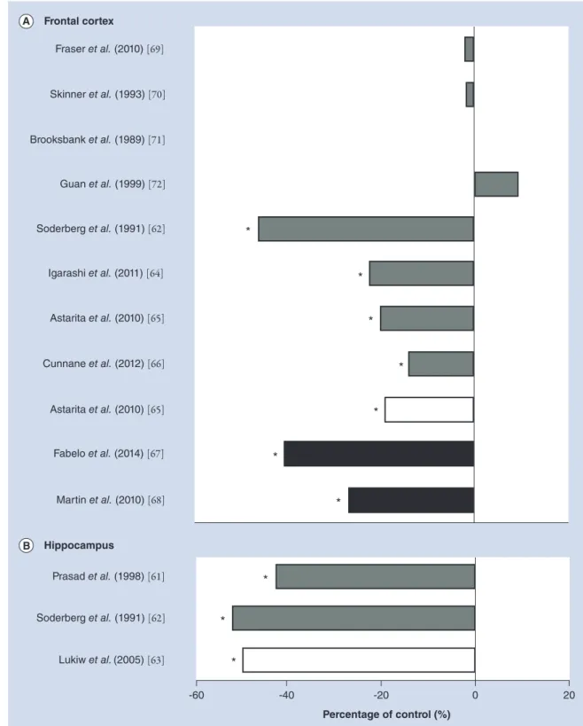
Post-mortem brain studies
Several studies have used postmortem samples of
human brain to measure brain DHA concentration in
AD patients
(Figure 2) [59]. As expected due to lower
DHA dietary intakes observed during age-related
cognitive decline, some studies have shown that AD
patients have lower DHA levels in the hippocampus,
a brain structure implicated in memory and learning
[62–64]
. Similar results were observed in the frontal
cor-tex, which is important for reasoning, attention,
plan-ning and emotions
[63,65–69]. Nevertheless, other
stud-ies did not show any modification of DHA content of
the frontal cortex
[70–73]or in other brain regions that
are also implicated in AD, such as the temporal or
pari-etal lobes and the parahippocampus
[62,66–67,70–71,74].
In accordance with animal studies, it has been recently
proposed that an altered composition of lipid rafts in
human frontal cortex, including a decrease in their
DHA content, may be related to the neuropathology
of AD, especially the formation of senile plaques
[68,69].
Randomized clinical trials
A recent prospective study showed that daily
consump-tion of an omega-3 PUFA supplement prevented
cogni-tive impairment as measured by MMSE in elderly
Chi-nese
[75], but the type or concentration of the omega-3
PUFA supplement were not specified. Several other
RCTs showed that DHA-enriched supplements delay
cognitive decline in elderly with cognitive impairments
(Table 1)[76–78]. Some of them have suggested that DHA
supplementation improves cognitive performances
in elderly with memory complaints
[79,80], MCI
[81,82]and AD
[83]. However, other studies do not show any
cognitive improvement due to DHA supplement in
AD
[84–86].
The discrepancy in the results from RCT may partly
been explained by the highly heterogeneous protocols
used, especially considering the dose, the type and the
duration of the DHA supplementation
(Table 1). For
example, most studies used fish oil as a
supplementa-tion, providing both DHA and eicosapentaenoic acid
(EPA), with the latter also potentially contributing
to the preventive effect of fish oil against cognitive
decline. Moreover, the protective role of DHA against
age-related cognitive decline may depend on APOE
genotype
[84]and on the degree of cognitive decline at
baseline, and may be stronger in those with the mildest
cognitive impairment
[85–86,88–91]. This suggests that
DHA may be more efficient as a preventive
interven-tion rather than as a curative treatment. As we will
discuss in the following section, we hypothesized that
age-relative cognitive decline may be accompanied by a
shift in DHA metabolism in older persons with
mem-ory problems such that they may be less able to benefit
from DHA-enriched supplements as cognitively
healthy individuals.
Docosahexaenoic acid metabolism during
age-related cognitive decline
To attempt to explain the divergent results between
epidemiological studies and RCT and between various
RCT, we hypothesized that DHA metabolism may be
altered during age-related cognitive decline. The
devel-opment of uniformly labelled tracers with stable
iso-topes constitutes a new approach to safely assess DHA
metabolism in human clinical studies. A 1-month
fol-low-up of a single dose of uniformly carbon-13 labelled
DHA (
13C-DHA) showed that
13C-DHA remained
longer in the blood of elderly (mean age 76 years)
com-pared with young adults (mean age 27 years)
[92].
13C-DHA homeostasis also differed between the different
plasma lipid classes: compared with young adults,
plasma enrichment in
13C-DHA in elderly was fourfold
to fivefold higher in triglycerides and free fatty acids
within hours and twice higher in PL and CE within
days to weeks following the ingestion of the tracer.
Carrying APOE4 also disrupts DHA
metabo-lism
[58,93–94]. The protective role of higher fish
con-sumption or higher blood DHA against age-related
cognitive decline may not be observed in carriers of
APOE4
[53–54,95]. Whole body half-life of DHA in the
healthy elderly was 77% shorter in carriers of APOE4
compared with the noncarriers
[93], due to higher
β-oxidation of DHA in the APOE4 carriers as
mea-sured by expired
13C-CO
2
after the ingestion of the
Figure 2. Summary of the published literature on docosahexaenoic acid content in (A) the frontal cortex and (B) the hippocampus during Alzheimer’s disease.Bars show docosahexaenoic acid values as a percentage of age-matched healthy elderly groups for each study shown. Date were obtained from either brain phospholipids (gray bars), brain free fatty acids (white bars) or lipid raft fractions (black bars).
*Studies presenting a significant difference between control and Alzheimer’s disease.
Frontal cortex Hippocampus Prasad et al. (1998) [61] Soderberg et al. (1991) [62] Lukiw et al. (2005) [63] -60 -40
*
*
*
*
*
*
*
*
*
*
-20 0 20 Percentage of control (%) Fraser et al. (2010) [69] Skinner et al. (1993) [70] Brooksbank et al. (1989) [71] Guan et al. (1999) [72] Soderberg et al. (1991) [62] Igarashi et al. (2011) [64] Astarita et al. (2010) [65] Astarita et al. (2010) [65] Fabelo et al. (2014) [67] Martin et al. (2010) [68] Cunnane et al. (2012) [66] A BTable 1. Impact of docosahexaenoic acid supplementation in elderly with cognitive complaints, mild cognitive impairment or Alzheimer’s disease.
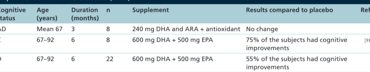
Cognitive status Age (years) Duration (months)
n Supplement Results compared to placebo Ref.
MCI >60 12 18 Placebo [87]
17 1290 mg DHA + 450 mg EPA ↑ Memory improvement
AD >55 12 12 Placebo [83]
11 675 mg DHA + 975 EPA Less ↓ in IADL 11 675 mg DHA + 975 mg EPA + 600
mg LA
Less ↓ in MMSE and IADL
MC 50–90 4 62 Placebo [88]
60 300 mg PS + 79 mg DHA + EPA (DHA:EPA ratio of 3:1)
↑ Immediate recall compared to P; Greater ↑ in subgroup with higher baseline cognitive performances MC 50–90 4/4 60 Placebo / 100 mg PS + 26 mg DHA +
EPA (DHA:EPA ratio of 3:1)
↑ Cognitive performances (sustained attention and memory recognition) [79] 4/4 300 mg PS + 79 mg DHA + EPA/100 mg PS + 26 mg DHA + EPA (DHA:EPA ratio of 3:1) Maintained cognitive performances AD Mean 76 18 112 Placebo [84]
152 2g Algal DHA containing 45–55% of DHA
No change on cognitive decline and brain atrophy
C >65 6 28 Placebo [89]
29 180 mg DHA + 120 mg EPA Less ↓ in AMT scores
MCI >65 6 71 Placebo
71 180 mg DHA + 120 mg EPA No change
MCI 55–90 6 6 Placebo [85]
12 1080 mg EPA + 720 mg DHA Significant ↑ in ADAS-Cog score
AD 55–90 6 9 Placebo
8 1080 mg EPA + 720 mg DHA No change
MC >55 6 218 Placebo [80]
219 900 mg DHA ↑ Cognitive performances in CANTAB
AD Mean 74 6/6 85 Placebo /1.7 g DHA +0.6 g EPA Less ↓ in MMSE score in subgroup with very mild AD
[90]
12 89 1.7 g DHA + 0.6 g EPA Less 6-month ↓ in MMSE score in subgroup with very mild AD
MCI >65 6 11 Placebo [82]
16 1.55 g DHA + 0.4 g EPA ↑ GDS score and verbal fluency 13 1.67 g EPA + 0.16 g DHA ↑ GDS score
MCI Mean 70 3 9 Placebo [86]
Mean 67 3 12 240 mg DHA and ARA + antioxidant ↑ Immediate memory and attention
AMT: Abbreviated Mental Test; AD: Alzheimer’s disease; ADAS-Cog: Alzheimer’s Disease Assessment Scale–Cognitive; C: Control;
CANTAB: Cambridge Neuropsychological Test Automated Battery; D: Dementia; DHA: Docosahexaenoic acid; GDS: Geriatric depression scale; IADL: Instrumental activities of daily living; MC: Memory complaints; MCI: Mild cognitive impairment.
and plasma DHA therefore seems to shift in carriers
of APOE4, who need higher dietary fish intakes to
increase plasma DHA concentration
[60]. In carriers of
APOE4, β-oxidation may preferentially affect omega-3
PUFA compared with monounsaturated or saturated
fatty acids, explaining the modification of DHA
metabolism occurring with APOE4
[96,97].
Daily dietary intake of DHA has been estimated to be
lower in many populations than the 250 mg/day
recom-mended
[98]. At the same time, a higher intake of DHA
may also modify its own metabolism. In a recent study,
the metabolism of
13C-DHA in older adults (mean age
72 years) was followed before and in the last month of
a 5-month supplementation providing 1.4 g of DHA
and 1.8 g of EPA daily
[99]. While on the supplement,
DHA clearance from plasma was faster and β-oxidation
of
13C-DHA was 87% higher compared with before
the supplement. However, the kinetics of
13C-DHA in
APOE4 carriers while on the supplement was not the
same
[59]. APOE4 carriers also present a lower plasma
response to a 6-week supplementation with 3 g/day of
DHA + EPA compared with noncarriers
[94]. Aging
alone also disrupts DHA homeostasis following
short-term supplementation with fish oil; compared with
young adult, plasma DHA concentration was indeed
increased 42% more in elderly
[100].
The PET tracer,
11C-DHA, has permitted the
esti-mation of brain DHA turnover of 4 mg/day and a
brain DHA half-life of 2.5 years
[101]. DHA kinetics
in the brain are therefore clearly different from those
of the whole-body, because the latter is estimated
to be 32 days in carriers of APOE4 and 140 days in
noncarriers of APOE4
[93]. The study of brain DHA
homeostasis using PET imaging may therefore provide
some useful information on genotype- and diet-related
modifications to DHA during aging
[102].
Postmor-tem studies of brain from AD patients show a linear
relationship between apoE and DHA concentrations
[103]
, suggesting a role of apoE protein and indirectly
in APOE4 genotype, in the maintenance of DHA
concentrations within the brain. It was recently shown
that omega-3 PUFA provided through the diet cross
the blood–brain barrier and increase DHA content
in the cerebrospinal fluid in AD
[104]. Moreover, the
recent finding of an efficient transporter for DHA into
the brain
[105]provides new field of investigation to
better understand DHA homeostasis in the brain.
Hence, DHA metabolism is influenced by two
important risk factors for AD – aging and APOE4.
This may suggest that a shift in DHA homeostasis
dur-ing age-related cognitive decline, especially AD, may
contribute to the divergent results obtained in RCT
studies. A modification of DHA metabolism during
AD has been reported previously, especially an increase
in peroxidation products
[106], even in the early stages
of age-related cognitive decline
[107]. This effect was
lower when participants had higher omega-3 PUFA
dietary intakes
[87]. Further studies are needed to better
characterize the link between risk of AD and whole
body and brain metabolism of DHA.
Conclusion
Animal and in vitro studies provide some promising
results on the role played by DHA in the prevention
of age-related cognitive decline. DHA may help
pre-vent the formation of senile plaques and
neurofibril-lary tangles and may also help maintain brain glucose
uptake during aging. Nevertheless, differences in DHA
synthesis and metabolism have been observed between
animals and humans. Humans have lower endogenous
synthesis of DHA from ALA compared with animals.
Moreover, the human brain strongly retains DHA in
membrane phospholipids despite very low DHA dietary
intakes. The promising protective effect of DHA
observed in cellular and animal models has yet to be
widely confirmed in clinical studies. The consumption
of fatty fish and DHA has been mostly reported to have
a protective role against cognitive decline and AD, but
results obtained from blood or brain samples do not help
explain this protective effect. The use of isotopically
Cognitive status Age (years) Duration (months)
n Supplement Results compared to placebo Ref.
AD Mean 67 3 8 240 mg DHA and ARA + antioxidant No change
C 67–92 6 8 600 mg DHA + 500 mg EPA 75% of the subjects had cognitive improvements
[91]
D 67–92 6 22 600 mg DHA + 500 mg EPA 55% of the subjects had cognitive improvements
AMT: Abbreviated Mental Test; AD: Alzheimer’s disease; ADAS-Cog: Alzheimer’s Disease Assessment Scale–Cognitive; C: Control;
CANTAB: Cambridge Neuropsychological Test Automated Battery; D: Dementia; DHA: Docosahexaenoic acid; GDS: Geriatric depression scale; IADL: Instrumental activities of daily living; MC: Memory complaints; MCI: Mild cognitive impairment.
Table 1. Impact of docosahexaenoic acid supplementation in elderly with cognitive complaints, mild cognitive impairment or Alzheimer’s disease (cont.).
labeled tracers highlights some modifications in DHA
metabolism due to healthy aging, APOE4 genotype
and dietary intakes that should be considered in future
clinical studies. Understanding the complexity of DHA
metabolism during age-related cognitive decline such as
AD is essential to improve and adapt dietary strategies
to this large and vulnerable population.
Future perspective
The use of uniformly labeled tracers has allowed
great advances in understanding DHA kinetics
dur-ing agdur-ing. Recent studies have shown that
13C-DHA
metabolism differs depending on age, APOE genotype
and dietary intake. Nevertheless, to fully understand
the protective role of omega-3 PUFA against AD and
adapt dietary recommendations in this population,
several points remained to be clarified in the future:
•
13C-DHA homeostasis should be evaluated in AD;
•
Supplementation with omega-3 fatty acids are
mainly done using fish oil, which is enriched in
both DHA and EPA. The further study of EPA
metabolism during aging and AD and its
relation-ship with dietary intakes will provide useful
infor-mation to understand whether protective role of
fish oil against AD can be optimized and observed
more consistently;
•
The recent discovery of a brain DHA transporter
[105]and the use of PET imaging will bring new information
on DHA homeostasis in the brain and may be helpful
to develop nutritional strategies adapted to favor
DHA incorporation within the brain;
•
The mechanisms associated with the protective
role of DHA against cognitive decline and AD
remained to be fully characterized in humans,
especially the role of DHA in brain glucose
metab-olism. PET imaging tracers such as
18F-FDG or
11C-PIB, a marker of senile plaques, will help
improve our understanding of the relationship
between omega-3 dietary intake and brain glucose
hypometabolism and amyloid deposition.
Financial & competing interests disclosure
CIHR, NSERC, FQRNT (CFQCU), INSERM, CFI and CRC pro-vided financial support for SC Cunnane’s research. E Harbeby was a recipient of a PhD thesis grant from INRA and Lesieur. M Plourde is supported by a Junior 1 FRQ-S salary award. The 18F-FDG positron emission tomography study performed on rats was supported by Lesieur. Mélanie Fortier, Conrad Fil-teau, Christine Brodeur, Christine Rioux-Perreault and Jennifer Tremblay-Mercier provided excellent technical support. 13C-DHA analysis was done in collaboration with Tom Brenna. The authors have no other relevant affiliations or financial involve-ment with any organization or entity with a financial inter-est in or financial conflict with the subject matter or materials discussed in the manuscript apart from those disclosed.
No writing assistance was utilized in the production of this manuscript.
Executive summary
Omega-3 fatty acids & the neuropathology of AD (Alzheimer’s diesease): evidence from in vitro & animal studies
• DHA has a protective role against Aβ accumulation and toxicity.
• DHA reduces the hyperphosphorylation of Tau proteins, associated with the formation of neurofibrillary tangles.
• DHA play a key role in the regulation of brain glucose metabolism.
Omega-3 fatty acids & prevention of AD in humans: epidemiological studies vs clinical trials
• Epidemiological studies suggest that higher consumption of fatty fish enriched in docosahexaenoic acid (DHA) prevents age-related cognitive decline and the development of AD.
• The relationship between dietary and plasma DHA during AD is complex and influenced by several factors such as age, sex, APOE4 genotype, body weight or physical activity.
• Postmortem brain studies show a reduced amount of DHA in the hippocampus but not necessarily in the cortex of AD patients compared with cognitively healthy control.
• Results from RCT are inconsistent but broadly suggest that DHA consumption may be more efficient as a preventive strategy against cognitive decline rather than as a curative strategy in AD.
DHA metabolism during age-related cognitive decline
• Healthy aging is associated with a slower clearance of plasma DHA.
• Carrying the APOE4 allele is the main genetic factor of sporadic AD and is associated with higher β-oxidation of DHA and lower whole-body half-life of DHA compared with the noncarriers.
• DHA metabolism is influenced by omega-3 polyunsaturated fatty acids (PUFA) intake and the response to a supplementation in omega-3 PUFA differs between carriers and noncarriers of APOE4.
• Uniformly-labeled tracers of omega-3 PUFA are innovative and useful tools to better understand DHA metabolism during aging and AD.
References
1 Blennow K, de Leon MJ, Zetterberg H. Alzheimer’s disease.
Lancet 368(9533), 387–403 (2006).
2 Gauthier S, Reisberg B, Zaudig M et al. Mild cognitive impairment. Lancet 367(9518), 1262–1270 (2006).
3 Corder EH, Saunders AM, Strittmatter WJ et al. Gene dose of apolipoprotein E type 4 allele and the risk of Alzheimer’s disease in late onset families. Science 261(5123), 921–923 (1993).
4 Alles B, Samieri C, Feart C, Jutand MA, Laurin D, Barberger-Gateau P. Dietary patterns: a novel approach to examine the link between nutrition and cognitive function in older individuals. Nutr. Res. Rev. 25(2), 207–222 (2012).
5 Gomez-Pinilla F. Brain foods: the effects of nutrients on brain function. Nat. Rev. Neurosci. 9(7), 568–578 (2008).
6 Sastry PS. Lipids of nervous tissue: composition and metabolism. Prog. Lipid Res. 24(2), 69–176 (1985).
7 Su HM. Mechanisms of n-3 fatty acid-mediated development and maintenance of learning memory performance. J. Nutr.
Biochem. 21(5), 364–373 (2010).
8 Hooijmans CR, Pasker-de Jong PC, de Vries RB, Ritskes-Hoitinga M. The effects of long-term omega-3 fatty acid supplementation on cognition and Alzheimer’s pathology in animal models of Alzheimer’s disease: a systematic review and meta-analysis. J. Alzheimers Dis. 28(1), 191–209 (2012).
9 Grimm MO, Zimmer VC, Lehmann J, Grimm HS, Hartmann T. The impact of cholesterol, DHA, and sphingolipids on Alzheimer’s disease. Biomed. Res. Int. 2013, 814390 (2013).
10 Hashimoto M, Hossain S. Neuroprotective and ameliorative actions of polyunsaturated fatty acids against neuronal diseases: beneficial effect of docosahexaenoic acid on cognitive decline in Alzheimer’s disease. J. Pharmacol. Sci. 116(2), 150–162 (2011).
11 Veszelka S, Toth AE, Walter FR et al. Docosahexaenoic acid reduces amyloid-beta induced toxicity in cells of the neurovascular unit. J. Alzheimers Dis. 36(3), 487–501 (2013).
12 Grimm MO, Kuchenbecker J, Grosgen S et al.
Docosahexaenoic acid reduces amyloid beta production via multiple pleiotropic mechanisms. J. Biol. Chem. 286(16), 14028–14039 (2011).
13 Hjorth E, Zhu M, Toro VC et al. Omega-3 fatty acids enhance phagocytosis of Alzheimer’s disease-related amyloid-beta42 by human microglia and decrease inflammatory markers. J. Alzheimers Dis. 35(4), 697–713 (2013).
14 Hashimoto M, Shahdat HM, Yamashita S et al.
Docosahexaenoic acid disrupts in vitro amyloid beta(1–40) fibrillation and concomitantly inhibits amyloid levels in cerebral cortex of Alzheimer’s disease model rats.
J. Neurochem. 107(6), 1634–1646 (2008).
15 Hossain S, Hashimoto M, Katakura M, Miwa K, Shimada T, Shido O. Mechanism of docosahexaenoic acid-induced inhibition of in vitro Abeta1–42 fibrillation and Abeta1–42-induced toxicity in SH-S5Y5 cells. J. Neurochem. 111(2), 568–579 (2009).
16 Ehehalt R, Keller P, Haass C, Thiele C, Simons K. Amyloidogenic processing of the Alzheimer beta-amyloid
precursor protein depends on lipid rafts. J. Cell Biol. 160(1), 113–123 (2003).
17 Stark DT, Bazan NG. Neuroprotectin D1 induces neuronal survival and downregulation of amyloidogenic processing in Alzheimer’s disease cellular models. Mol. Neurobiol. 43(2), 131–138 (2011).
18 Zhao Y, Calon F, Julien C et al. Docosahexaenoic acid-derived neuroprotectin D1 induces neuronal survival via secretase- and PPARgamma-mediated mechanisms in Alzheimer’s disease models. PLoS ONE 6(1), e15816 (2011).
19 TorRes. M, Price SL, Fiol-Deroque MA et al. Membrane lipid modifications and therapeutic effects mediated by hydroxydocosahexaenoic acid on Alzheimer’s disease.
Biochim. Biophys. Acta 1838(6), 1680–1692 (2014).
20 Green KN, Martinez-Coria H, Khashwji H et al. Dietary docosahexaenoic acid and docosapentaenoic acid ameliorate amyloid-beta and tau pathology via a mechanism involving presenilin 1 levels. J. Neurosci. 27(16), 4385–4395 (2007).
21 Lebbadi M, Julien C, Phivilay A et al. Endogenous conversion of omega-6 into omega-3 fatty acids improves neuropathology in an animal model of Alzheimer’s disease.
J. Alzheimers Dis. 27(4), 853–869 (2011).
22 Ma QL, Yang F, Rosario ER et al. Beta-amyloid oligomers induce phosphorylation of tau and inactivation of insulin receptor substrate via c-Jun N-terminal kinase signaling: suppression by omega-3 fatty acids and curcumin.
J. Neurosci. 29(28), 9078–9089 (2009).
23 Wang PY, Chen JJ, Su HM. Docosahexaenoic acid supplementation of primary rat hippocampal neurons attenuates the neurotoxicity induced by aggregated amyloid beta protein(42) and up-regulates cytoskeletal protein expression. J. Nutr. Biochem. 21(4), 345–350 (2010).
24 Nugent S, Castellano CA, Goffaux P et al. Glucose hypometabolism is highly localized but lower cortical thickness and brain atrophy are widespread in cognitively normal older adults. Am. J. Physiol. Endocrinol. Metab. 306(11), E1315–E1321 (2014).
25 Kalpouzos G, Chetelat G, Baron JC et al. Voxel-based mapping of brain gray matter volume and glucose metabolism profiles in normal aging. NeuroBiol. Aging 30(1), 112–124 (2009).
26 Nugent S, Tremblay S, Chen KW et al. Brain glucose and acetoacetate metabolism: a comparison of young and older adults. NeuroBiol. Aging 35(6), 1386–1395 (2014).
27 Kalpouzos G, Eustache F, de la Sayette V, Viader F, Chetelat G, Desgranges B. Working memory and FDG-PET dissociate early and late onset Alzheimer disease patients. J. Neurol. 252(5), 548–558 (2005).
28 Landau SM, Harvey D, Madison CM et al. Associations between cognitive, functional, and FDG-PET measures of decline in AD and MCI. NeuroBiol. Aging 32(7), 1207–1218 (2011).
29 Habeck C, Risacher S, Lee GJ et al. Relationship between baseline brain metabolism measured using [(1)(8)F]FDG PET and memory and executive function in prodromal and early Alzheimer’s disease. Brain Imaging Behav. 6(4), 568–583 (2012).
30 Cunnane S, Nugent S, Roy M et al. Brain fuel metabolism, aging, and Alzheimer’s disease. Nutrition 27(1), 3–20 (2011).
31 Leybaert L, De Bock M, Van Moorhem M, Decrock E, De Vuyst E. Neurobarrier coupling in the brain: adjusting glucose entry with demand. J. Neurosci. Res. 85(15), 3213–3220 (2007).
32 Harbeby E, Jouin M, Alessandri JM et al. n-3 PUFA status affects expression of genes involved in neuroenergetics differently in the fronto-parietal cortex compared with the CA1 area of the hippocampus: effect of rest and neuronal activation in the rat. Prostaglandins Leukot. Essent. Fatty Acids 86(6), 211–220 (2012).
33 Pifferi F, Roux F, Langelier B et al. (n-3) polyunsaturated fatty acid deficiency reduces the expression of both isoforms of the brain glucose transporter GLUT1 in rats. J. Nutr. 135(9), 2241–2246 (2005).
34 Pifferi F, Jouin M, Alessandri JM et al. n-3 long-chain fatty acids and regulation of glucose transport in two models of rat brain endothelial cells. Neurochem. Int. 56(5), 703–710 (2010).
35 Pifferi F, Jouin M, Alessandri JM et al. n-3 Fatty acids modulate brain glucose transport in endothelial cells of the blood-brain barrier. Prostaglandins Leukot. Essent. Fatty Acids 77(5–6), 279–286 (2007).
36 Freemantle E, Vandal M, Tremblay-Mercier J et al. Omega-3 fatty acids, energy substrates, and brain function during aging. Prostaglandins Leukot. Essent. Fatty Acids 75(3), 213–220 (2006).
37 Nugent S, Croteau E, Pifferi F et al. Brain and systemic glucose metabolism in the healthy elderly following fish oil supplementation. Prostaglandins Leukot. Essent. Fatty Acids 85(5), 287–291 (2011).
38 Plourde M, Cunnane SC. Extremely limited synthesis of long chain polyunsaturates in adults: implications for their dietary essentiality and use as supplements. Appl. Physiol. Nutr.
Metab. 32(4), 619–634 (2007).
39 Cunnane SC, Plourde M, Pifferi F, Begin M, Feart C, Barberger-Gateau P. Fish, docosahexaenoic acid and Alzheimer’s disease. Prog. Lipid Res. 48(5), 239–256 (2009).
40 Huang TL. Omega-3 fatty acids, cognitive decline, and Alzheimer’s disease: a critical review and evaluation of the literature. J. Alzheimers Dis. 21(3), 673–690 (2010).
41 Fotuhi M, Mohassel P, Yaffe K. Fish consumption, long-chain omega-3 fatty acids and risk of cognitive decline or Alzheimer disease: a complex association. Nat. Clin. Pract.
Neurol. 5(3), 140–152 (2009).
42 Solfrizzi V, Colacicco AM, D’Introno A et al. Dietary intake of unsaturated fatty acids and age-related cognitive decline: a 8.5-year follow-up of the Italian Longitudinal Study on Aging. NeuroBiol. Aging 27(11), 1694–1704 (2006).
43 Gu Y, Schupf N, Cosentino SA, Luchsinger JA, Scarmeas N. Nutrient intake and plasma beta-amyloid. Neurology 78(23), 1832–1840 (2012).
44 Titova OE, Sjogren P, Brooks SJ et al. Dietary intake of eicosapentaenoic and docosahexaenoic acids is linked to gray matter volume and cognitive function in elderly. Age (Dordr). 35(4), 1495–1505 (2013).
45 Samieri C, Maillard P, Crivello F et al. Plasma long-chain omega-3 fatty acids and atrophy of the medial temporal lobe.
Neurology 79(7), 642–650 (2012).
46 Lopez LB, Kritz-Silverstein D, Barrett Connor E. High dietary and plasma levels of the omega-3 fatty acid docosahexaenoic acid are associated with decreased dementia risk: the Rancho Bernardo study. J. Nutr. Health Aging 15(1), 25–31 (2011).
47 Phillips MA, Childs CE, Calder PC, Rogers PJ. Lower omega-3 fatty acid intake and status are associated with poorer cognitive function in older age: A comparison of individuals with and without cognitive impairment and Alzheimer’s disease. Nutr. Neurosci. (2012) (Epub ahead of print).
48 Roberts RO, Cerhan JR, Geda YE et al. Polyunsaturated fatty acids and reduced odds of MCI: the Mayo Clinic Study of Aging. J. Alzheimers Dis. 21(3), 853–865 (2010).
49 Albanese E, Dangour AD, Uauy R et al. Dietary fish and meat intake and dementia in Latin America, China, and India: a 10/66 Dementia Research Group population-based study. Am. J. Clin. Nutr. 90(2), 392–400 (2009).
50 Barberger-Gateau P, Letenneur L, Deschamps V, PeRes K, Dartigues JF, Renaud S. Fish, meat, and risk of dementia: cohort study. BMJ 325(7370), 932–933 (2002).
51 Kalmijn S, Launer LJ, Ott A, Witteman JC, Hofman A, Breteler MM. Dietary fat intake and the risk of incident dementia in the Rotterdam Study. Ann. Neurol. 42(5), 776–82 (1997).
52 Morris MC, Evans DA, Bienias JL et al. Consumption of fish and n-3 fatty acids and risk of incident Alzheimer disease.
Arch. Neurol. 60(7), 940–946 (2003).
53 Huang TL, Zandi PP, Tucker KL et al. Benefits of fatty fish on dementia risk are stronger for those without APOE epsilon4. Neurology 65(9), 1409–1414 (2005).
54 Barberger-Gateau P, Raffaitin C, Letenneur L et al. Dietary patterns and risk of dementia: the Three-City cohort study.
Neurology 69(20), 1921–1930 (2007).
55 Schaefer EJ, Bongard V, Beiser AS et al. Plasma
phosphatidylcholine docosahexaenoic acid content and risk of dementia and Alzheimer disease: the Framingham Heart Study. Arch. Neurol. 63(11), 1545–1550 (2006).
56 Devore EE, Grodstein F, van Rooij FJ et al. Dietary intake of fish and omega-3 fatty acids in relation to long-term dementia risk. Am. J. Clin. Nutr. 90(1), 170–176 (2009).
57 Engelhart MJ, Geerlings MI, Ruitenberg A et al. Diet and risk of dementia: Does fat matter?: The Rotterdam Study.
Neurology 59(12), 1915–1921 (2002).
58 Barberger-Gateau P, Samieri C, Feart C, Plourde M. Dietary omega 3 polyunsaturated fatty acids and Alzheimer’s disease: interaction with apolipoprotein E genotype. Curr. Alzheimer
Res. 8(5), 479–491 (2011).
59 Hennebelle M, Plourde M, Chouinard-Watkins R, Castellano CA, Barberger-Gateau P, Cunnane SC. Ageing and apoE change DHA homeostasis: relevance to age-related cognitive decline. Proc. Nutr. Soc. 73(1), 80–86 (2014).
60 Samieri C, Lorrain S, Buaud B et al. Relationship between diet and plasma long-chain n-3 PUFAs in older people:
impact of apolipoprotein E genotype. J. Lipid Res. 54(9), 2559–2567 (2013).
61 Flock MR, Skulas-Ray AC, Harris WS, Etherton TD, Fleming JA, Kris-Etherton PM. Determinants of erythrocyte omega-3 fatty acid content in response to fish oil supplementation: a dose-response randomized controlled trial. J. Am. Heart Assoc. 2(6), e000513 (2013).
62 Prasad MR, Lovell MA, Yatin M, Dhillon H, Markesbery WR. Regional membrane phospholipid alterations in Alzheimer’s disease. Neurochem. Res. 23(1), 81–88 (1998).
63 Soderberg M, Edlund C, Kristensson K, Dallner G. Fatty acid composition of brain phospholipids in aging and in Alzheimer’s disease. Lipids 26(6), 421–425 (1991).
64 Lukiw WJ, Cui JG, Marcheselli VL et al. A role for docosahexaenoic acid-derived neuroprotectin D1 in neural cell survival and Alzheimer disease. J. Clin. Invest. 115(10), 2774–2783 (2005).
65 Igarashi M, Ma K, Gao F, Kim HW, Rapoport SI, Rao JS. Disturbed choline plasmalogen and phospholipid fatty acid concentrations in Alzheimer’s disease prefrontal cortex.
J. Alzheimers Dis. 24(3), 507–517 (2011).
66 Astarita G, Jung KM, Berchtold NC et al. Deficient liver biosynthesis of docosahexaenoic acid correlates with cognitive impairment in Alzheimer’s disease. PLoS ONE 5(9), e12538 (2010).
67 Cunnane SC, Schneider JA, Tangney C et al. Plasma and brain fatty acid profiles in mild cognitive impairment and Alzheimer’s disease. J. Alzheimers Dis. 29(3), 691–697 (2012).
68 Martin V, Fabelo N, Santpere G et al. Lipid alterations in lipid rafts from Alzheimer’s disease human brain cortex.
J. Alzheimers Dis. 19(2), 489–502 (2010).
69 Fabelo N, Martin V, Marin R, Moreno D, Ferrer I, Diaz M. Altered lipid composition in cortical lipid rafts occurs at early stages of sporadic Alzheimer’s disease and facilitates APP/BACE1 interactions. NeuroBiol. Aging 35(8), 1801–1812 (2014).
70 Fraser T, Tayler H, Love S. Fatty acid composition of frontal, temporal and parietal neocortex in the normal human brain and in Alzheimer’s disease. Neurochem. Res. 35(3), 503–513 (2010).
71 Skinner ER, Watt C, Besson JA, Best PV. Differences in the fatty acid composition of the grey and white matter of different regions of the brains of patients with Alzheimer’s disease and control subjects. Brain 116(Pt 3), 717–725 (1993).
72 Brooksbank BW, Martinez M. Lipid abnormalities in the brain in adult Down’s syndrome and Alzheimer’s disease.
Mol. Chem. Neuropathol. 11(3), 157–185 (1989).
73 Guan Z, Wang Y, Cairns NJ, Lantos PL, Dallner G, Sindelar PJ. Decrease and structural modifications of phosphatidylethanolamine plasmalogen in the brain with Alzheimer disease. J. Neuropathol. Exp. Neurol. 58(7), 740–747 (1999).
74 Corrigan FM, Van Rhijn AG, Ijomah G et al. Tin and fatty acids in dementia. Prostaglandins Leukot. Essent. Fatty Acids 43(4), 229–238 (1991).
75 Gao Q, Niti M, Feng L, Yap KB, Ng TP. Omega-3 polyunsaturated fatty acid supplements and cognitive decline: Singapore Longitudinal Aging Studies. J. Nutr.
Health Aging 15(1), 32–35 (2011).
76 Dangour AD, Andreeva VA, Sydenham E, Uauy R. Omega 3 fatty acids and cognitive health in older people. Br. J. Nutr. 107(Suppl. 2), S152–S158 (2012).
77 Sydenham E, Dangour AD, Lim WS. Omega 3 fatty acid for the prevention of cognitive decline and dementia. Cochrane
Database Syst. Rev. 6, CD005379 (2012).
78 Mazereeuw G, Lanctot KL, Chau SA, Swardfager W, Herrmann N. Effects of omega-3 fatty acids on cognitive performance: a meta-analysis. NeuroBiol. Aging 33(7), 1482 e17–e29 (2012).
79 Vakhapova V, Cohen T, Richter Y, Herzog Y, Kam Y, Korczyn AD. Phosphatidylserine Containing Omega-3 Fatty Acids May Improve Memory Abilities in Nondemented Elderly Individuals with Memory Complaints: Results from an Open-Label Extension Study. Dement. Geriatr. Cogn.
Disord. 38(1–2), 39–45 (2014).
80 Yurko-Mauro K, McCarthy D, Rom D et al. Beneficial effects of docosahexaenoic acid on cognition in age-related cognitive decline. Alzheimers Dement. 6(6), 456–464 (2010).
81 Lee LK, Shahar S, Chin AV, Yusoff NA. Docosahexaenoic acid-concentrated fish oil supplementation in subjects with mild cognitive impairment (MCI): a 12-month randomised, double-blind, placebo-controlled trial. Psychopharmacology
(Berl). 225(3), 605–612 (2013).
82 Sinn N, Milte CM, Street SJ et al. Effects of n-3 fatty acids, EPA v. DHA, on depressive symptoms, quality of life, memory and executive function in older adults with mild cognitive impairment: a 6-month randomised controlled trial. Br. J. Nutr. 107(11), 1682–9163 (2012).
83 Shinto L, Quinn J, Montine T et al. A randomized placebo-controlled pilot trial of omega-3 fatty acids and alpha lipoic acid in Alzheimer’s disease. J. Alzheimers Dis. 38(1), 111–120 (2014).
84 Quinn JF, Raman R, Thomas RG et al. Docosahexaenoic acid supplementation and cognitive decline in Alzheimer disease: a randomized trial. JAMA 304(17), 1903–1911 (2010).
85 Chiu CC, Su KP, Cheng TC et al. The effects of omega-3 fatty acids monotherapy in Alzheimer’s disease and mild cognitive impairment: a preliminary randomized double-blind placebo-controlled study. Prog. NeuropsychoPharmacol.
Biol. Psychiatry 32(6), 1538–1544 (2008).
86 Kotani S, Sakaguchi E, Warashina S et al. Dietary supplementation of arachidonic and docosahexaenoic acids improves cognitive dysfunction. Neurosci. Res. 56(2), 159–164 (2006).
87 Lee LK, Shahar S, Rajab N, Yusoff NA, Jamal RA, Then SM. The role of long chain omega-3 polyunsaturated fatty acids in reducing lipid peroxidation among elderly patients with mild cognitive impairment: a case-control study. J. Nutr. Biochem. 24(5), 803–308 (2013).
88 Vakhapova V, Cohen T, Richter Y, Herzog Y, Korczyn AD. Phosphatidylserine containing omega-3 fatty acids may improve memory abilities in non-demented elderly with
memory complaints: a double-blind placebo-controlled trial.
Dement. Geriatr. Cogn. Disord. 29(5), 467–474 (2010).
89 Mahmoudi MJ, Hedayat M, Sharifi F et al. Effect of low dose omega-3 poly unsaturated fatty acids on cognitive status among older people: a double-blind randomized placebo-controlled study. J. Diabetes Metab. Disord. 13, 34 (2014).
90 Freund-Levi Y, Eriksdotter-Jonhagen M, Cederholm T et al. Omega-3 fatty acid treatment in 174 patients with mild to moderate Alzheimer disease: OmegAD study: a randomized double-blind trial. Arch. Neurol. 63(10), 1402–1408 (2006).
91 Suzuki H, Morikawa Y, Takahashi H. Effect of DHA oil supplementation on intelligence and visual acuity in the elderly. World Rev. Nutr. Diet. 88, 68–71 (2001).
92 Plourde M, Chouinard-Watkins R, Vandal M et al. Plasma incorporation, apparent retroconversion and beta-oxidation of 13C-docosahexaenoic acid in the elderly. Nutr. Metab.
(Lond.) 8, 5 (2011).
93 Chouinard-Watkins R, Rioux-Perreault C, Fortier M et al. Disturbance in uniformly 13C-labelled DHA metabolism in elderly human subjects carrying the apoE epsilon4 allele.
Br. J. Nutr. 110(10), 1751–1759 (2013).
94 Plourde M, Vohl MC, Vandal M, Couture P, Lemieux S, Cunnane SC. Plasma n-3 fatty acid response to an n-3 fatty acid supplement is modulated by apoE epsilon4 but not by the common PPAR-alpha L162V polymorphism in men.
Br. J. Nutr. 102(8), 1121–1124 (2009).
95 Whalley LJ, Deary IJ, Starr JM et al. n-3 Fatty acid erythrocyte membrane content, APOE varepsilon4, and cognitive variation: an observational follow-up study in late adulthood. Am. J. Clin. Nutr. 87(2), 449–454 (2008).
96 Conway V, Larouche A, Alata W, Vandal M, Calon F, Plourde M. Apolipoprotein E isoforms disrupt long-chain fatty acid distribution in the plasma, the liver and the adipose tissue of mice. Prostaglandins Leukot. Essent. Fatty Acids 91(6), 261–267 (2014).
97 Chouinard-Watkins R, Plourde M. Fatty acid metabolism in carriers of apolipoprotein E epsilon 4 allele: is it contributing to higher risk of cognitive decline and coronary heart disease?
Nutrients 6(10), 4452–44571 (2014).
98 Meyer BJ. Are we consuming enough long chain omega-3 polyunsaturated fatty acids for optimal health?
Prostaglandins Leukot. Essent. Fatty Acids 85(5), 275–280
(2011).
99 Plourde M, Chouinard-Watkins R, Rioux-Perreault C
et al. Kinetics of 13C-DHA before and during fish-oil
supplementation in healthy older individuals. Am. J. Clin.
Nutr. 100(1), 105–112 (2014).
100 Vandal M, Freemantle E, Tremblay-Mercier J et al. Plasma omega-3 fatty acid response to a fish oil supplement in the healthy elderly. Lipids 43(11), 1085–1089 (2008).
101 Umhau JC, Zhou W, Carson RE et al. Imaging incorporation of circulating docosahexaenoic acid into the human brain using positron emission tomography.
J. Lipid Res. 50(7), 1259–1268 (2009).
102 Basselin M, Ramadan E, Rapoport SI. Imaging brain signal transduction and metabolism via arachidonic and docosahexaenoic acid in animals and humans. Brain Res.
Bull. 87(2–3), 154–171 (2012).
103 Vandal M, Alata W, Tremblay C et al. Reduction in DHA transport to the brain of mice expressing human APOE4 compared with APOE2. J. Neurochem. 129(3), 516–526 (2014).
104 Freund Levi Y, Vedin I, Cederholm T et al. Transfer of omega-3 fatty acids across the blood-brain barrier after dietary supplementation with a docosahexaenoic acid-rich omega-3 fatty acid preparation in patients with Alzheimer’s disease: the OmegAD study. J. Intern. Med. 275(4), 428–36 (2014).
105 Nguyen LN, Ma D, Shui G et al. Mfsd2a is a transporter for the essential omega-3 fatty acid docosahexaenoic acid.
Nature 509(7501), 503–506 (2014).
106 Montine TJ, Morrow JD. Fatty acid oxidation in the pathogenesis of Alzheimer’s disease. Am. J. Pathol. 166(5), 1283–1289 (2005).
107 Markesbery WR, Kryscio RJ, Lovell MA, Morrow JD. Lipid peroxidation is an early event in the brain in amnestic mild cognitive impairment. Ann. Neurol. 58(5), 730–735 (2005).