Université de Montréal
Socio-demographic, visual and psychological factors associated with
adjustment to vision loss in retinitis pigmentosa
par
Nathalie De la Haye Duponsel
Ecole d’optométrie
Mémoire présenté à la Faculté des études supérieures en vue de l’obtention du grade de Maîtrise en science
en Sciences de la vision
option Sciences fondamentales et Appliquées
Avril, 2012
Université de Montréal Faculté des études supérieures
Ecole d’optométrie
Ce mémoire intitulé :
Socio-demographic, visual and psychological factors associated with
adjustment to vision loss in retinitis pigmentosa
présentée par :
Nathalie De la Haye Duponsel
a été évalué par un jury composé des personnes suivantes :
Marie-Chantal Wanet-Defalque, Ph.D.
présidente rapporteuse
Olga Overbury, Ph.D.
directrice de recherche
Judith Renaud, O.D., Ph.D. (candidate)
SUMMARY
While there is extensive research on retinitis pigmentosa (RP) focusing on biological and hereditary aspects of the disease, little research regarding psychological adjustment has been conducted. These few studies suggest that people with RP adapt differently to vision impairment. This study investigated whether those with RP adapt differently to vision loss/impairment than those with other vision disorders.
Telephone interviews of those with RP, diabetic retinopathy (DR), and albinism were conducted. Demographic information was gathered and psychological wellbeing was assessed using the Visual Function-14, Centre of Epidemiology Studies Depression-10 symptoms index, Impact of Vision Impairment Profile, Brief COPE, and Adaptation to Vision Loss Scale.
In Experiment I it was found that individuals with RP did not differ from those with other diagnoses on any of the measures of psychological wellbeing and adaptation. Rather, demographic factors, visual factors such as declining and fluctuating vision, and pattern of vision loss, were better correlates of adaptation to and psychological wellbeing associated with vision loss/impairment.
In Experiment II there was no difference found between those with RP and other diagnoses on any of the measures. Rather, factors such as perceived visual ability, self-identity, fear of social stigma and level of dependence were more closely related to adaptation to and psychological wellbeing associated with vision loss/impairment.
The results of this study suggest that individuals with RP do not differ from those with other vision disorders in their adaptation to and psychological wellbeing associated with vision loss/impairment, but that other demographic, visual and psychological factors are more important.
RÉSUMÉ
Malgré des recherches intensives portant sur l’hérédité et les aspects biologiques de la rétinite pigmentaire (RP), peu de recherches fondées ont porté sur les aspects psychologiques. Ces quelques études suggèrent que les personnes atteintes de rétinite pigmentaire s’adaptent différemment à la déficience visuelle. Le but de la présente étude était donc de vérifier si les personnes atteintes de rétinite pigmentaire s’adaptaient différemment d’un point de vue psychologique par rapport à des personnes ayant une déficience visuelle causée par une autre pathologie.
Des entrevues téléphoniques incluant des personnes ayant la rétinite pigmentaire, la rétinopathie diabétique (RD) et l’albinisme ont été menées. Cinq questionnaires ont été utilisés afin d’évaluer le bien-être psychologique et de recueillir les données démographique.
Les résultats de la première étude démontrent qu’il n’existe aucune différence entre les individus atteints de rétinite pigmentaire et ceux ayant d’autres pathologies visuelles d’un point de vue « bien-être psychologique ». En fait, les facteurs démographiques, la baisse de vision, les fluctuations et le type de perte de vision semblent être les seuls facteurs directement corrélés à l’adaptation et au bien-être psychologique.
Dans la deuxième étude, aucune différence n’a pu être établie entre les trois types de pathologies. Ce sont plutôt, des facteurs comme la perception des capacités fonctionnelles, l’identité personnelle, l’appréhension de la perception sociale et le niveau d’indépendance qui étaient davantage reliés au bien-être psychologique associé à la déficience visuelle.
Les résultats de cette étude suggèrent que les personnes atteintes de Rétinite pigmentaire ne présentent pas de différences au niveau du bien-être psychologique et de l’adaptation. Les facteurs démographiques et psychologiques sont plus importants que la pathologie elle-même.
TABLE OF CONTENTS
Summary ... i
Résumé ... ii
Table of Contents ... iii
List of Tables ... vi
List of Figures ... vii
List of Abbreviations ... viii
Dedication ... ix Acknowledgements ... x Chapter I Introduction ... 1 Literature Review ... 3 1. Vision Impairment ... 3
1.1 Functional Impact of Vision Impairment ... 5
1.1.1 Peripheral Vision Impairment ... 5
1.1.2 Central Vision Impairment ... 6
1.1.3 Generalized Vision Impairment ... 6
1.2 Psychological Impact of Vision Loss/Impairment ... 7
2. Adaptation to Vision Loss/Impairment ... 8
2.1 Socio-demographic Factors and Adaptation to Vision Loss/Impairment ... 8
2.2 Functional and Perceived Functional Ability and Adaptation to Vision Loss/Impairment ... 10
2.3 Coping Styles and Strategies in Adaptation to Vision Loss/Impairment ... 11
2.4 Self-Identity and Adaptation to Vision Loss/Impairment ... 15
2.5 Stigma and Adaptation to Vision Loss/Impairment ... 15
3. Retinitis Pigmentosa ... 16
3.1 Description of the Disease ... 16
3.2 Functional and Psychological Implications ... 17
4. Study Rationale ... 21
4.1 Diabetic Retinopathy ... 21
4.1.1 Description of the Disease ... 21
4.1.2 Functional and Psychological Implications ... 21
4.2 Albinism ... 25
4.2.1 Description of the Disorder ... 25
4.2.2 Functional and Psychological Implications ... 27
5. Study Objectives ... 28 Chapter II Experiment 1 ... 30 Introduction ... 30 Method ... 35 Results ... 43 Discussion ... 56 Chapter III Experiment 2 ... 67 Introduction ... 67
Method ... 71 Results ... 74 Discussion ... 82 Chapter IV General Discussion ... 90 References ... 100 Appendices Appendix A ... xi Appendix B ... xiv Appendix C ... xvii Appendix D ... xx Appendix E ... xxv Appendix F ... xxx Appendix G ... xxxix Appendix H ... xlviii Appendix I ... liii Appendix J ... lxiii
LIST OF TABLES
Chapter II
Table 2.1 Participant Characteristics and Demographic Information across Diagnoses .. 36 Table 2.2 Adapted Questions on the AVL for Participants with Albinism ... 40 Table 2.3 Correlations Between Measures of Psychological Wellbeing and Adaptation
and Age Since Diagnosis, Time Elapsed Since Diagnosis and Time Elapsed Since Last Decline in Vision ... 53
Chapter III
Table 3.1 Correlations between AVL and Other Measures of Psychological Wellbeing
and Adaptation ... 79
Table 3.2 Correlations between IVI-Emotional and Other Measures of Psychological
LIST OF FIGURES
Chapter II
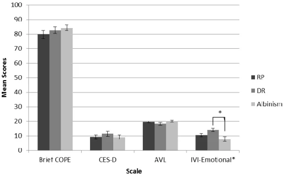
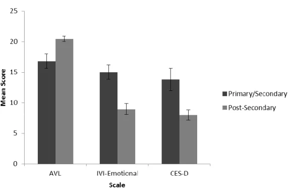
Figure 2.1. Mean scores on all psychological measures as a function of diagnosis ... 46
Figure 2.2. IVI-Emotional mean scores as a function of diagnosis with education level held constant ... 47
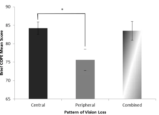
Figure 2.3. Brief COPE scores as a function of pattern of vision loss ... 48
Figure 2.4. Brief COPE mean scores by severity of impairment ... 50
Figure 2.5. IVI-Emotional mean scores as a function of visual status at the time of the study ... 51
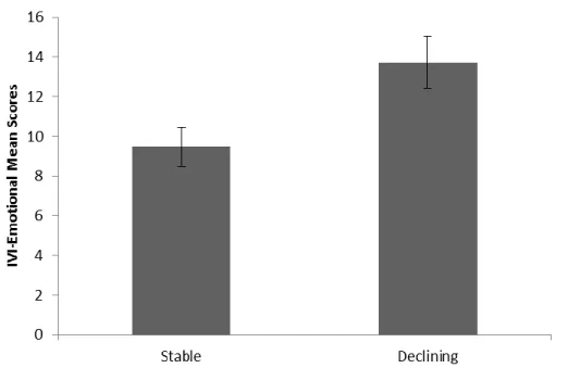
Figure 2.6. IVI-Emotional mean scores as a function of visual stability at the time of the study ... 52
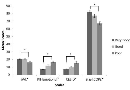
Figure 2.7. Mean scores of all psychological measures as a function of participants’ self- rating of health ... 55
Figure 2.8. Mean scores of psychological measures as a function of education level ... 57
Figure 2.9. IVI-Emotional mean scores as a function of marital status ... 58
Figure 2.10. Measures of affect as a function of family history of diagnosis ... 59
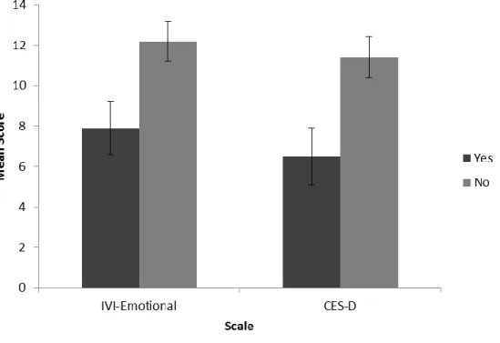
Chapter III Figure 3.1. Measures of psychological wellbeing and adaptation to vision loss/impairment as a function of diagnoses ... 75
Figure 3.2. Measures of perceived visual ability as a function of diagnosis with severity of impairment held constant... 77
LIST OF ABBREVIATIONS
AVL: Adaptation to Vision Loss Scale
CES-D: Centre for Epidemiological Studies Depression-10 Symptoms Index DR : diabetic retinopathy
INLB: Institut Nazareth et Louis-Braille IVI: Impact of Vision Impairment Profile
IVI-Emotional: Emotion related subscale of the Impact of Vision Impairment Profile MMRC: MAB-Mackay Rehabilitation Centre
RP: retinitis pigmentosa
To my parents, my brother, and all my family: You are the source of all my joy.
ACKNOWLEDGEMENTS
First, I'd like to express my gratitude to the School of Optometry of the University of Montreal and the Reseau vision of the FRSQ for the financial support that funded my studies.
I'd like to thank the MAB-Mackay Rehabilitation Centre and the Institut Nazareth et Louis-Braille for access to their clients for recruitment of participants for this study. Much
appreciation to Dr. Walter Wittich and Mme Claire Trempe for making the initial contact with all the participants.
I'd also like to thank Tanja Gninka, the lab manger in the Overbury lab, for all of her help and for keeping me sane during this process. And also, thanks to Carole Deharnais for testing some participants during an absence.
Merci à Marie-Chantal Wanet-Defalque, à Didier Chelin et à Anne Jarry de votre aide pour les traductions. Votre aide est bien appréciée.
I'd like to give a special thanks to my parents for being so supportive as I completed this degree. A special thanks too to my aunts, Elisabeth and Catherine, for their support and encouragement from a distance.
And finally, because I like to save the best for last, I'd like to thank my supervisor Dr. Olga Overbury, without whom this thesis would not have been possible. I thank her for her guidance and direction during this process, and for making it an enjoyable one. Mostly, I thank her for her kindness and patience as extenuating circumstances not related to this degree could have ended it all had it not been for her understanding. I appreciate all that you have done for me.
Chapter 1
INTRODUCTION
Retinitis pigmentosa (RP) is a hereditary vision disorder that involves the breakdown of the retinal pigment regeneration cycle, leading to the dystrophy of the photoreceptors beginning in the peripheral retina and moving to the fovea. Retinitis pigmentosa is characterized by a gradual loss of vision over a relatively extended period of time. The first symptoms of RP typically occur in adolescence or early adulthood with night blindness and mild peripheral field loss. As the disorder progresses, further peripheral loss is experienced and central vision may become affected as well. The final prognosis of RP remains relatively unpredictable with some individuals maintaining fairly good central vision, while others lose all light perception.
As with any cause of vision impairment, the psychological impact of RP can be significant. Depression and anxiety are found to be higher in individuals with acquired vision impairment, including those with RP. The stressors associated with the loss of vision resulting in an individual’s inability to continue performing daily activities, including reading, writing, driving, moving around independently, taking care of personal grooming, and earning a living, can lead to significant psychological distress. In the case of RP, due to the nature of the disease, individuals are forced to endure this loss over an extended period of time, without a predictable end. Relatively little research has been conducted in this area but, in the little that has been done, it has been suggested that individuals with RP may, in fact, adapt differently and not fare as well as individuals with other causes of vision loss.
The purpose of the following studies was to investigate whether individuals with RP differ from those with other visual diagnoses in their psychological wellbeing associated with, and adaptation to, vision loss/impairment and, if not, what factors are more closely associated with psychological outcomes. In order to do this, individuals with RP were compared to those with diabetic retinopathy (DR) and oculocutaneous albinism. Diabetic retinopathy shares many similar characteristics to RP, not so much in the symptoms themselves, but in the nature of the loss (unpredictable and extended over time) and in its psychological implications. Oculocutaneous albinism, on the other hand, is almost completely unlike RP and DR as it is a stable condition, with no changes in vision, but still involves a visual impairment with functional implications that can be significant. Factors independent of visual diagnosis that were considered included socio-demographic factors, functional vision, perceived functional vision, coping strategies, and personal identification with disability.
LITERATURE REVIEW
1. VISION IMPAIRMENT
Vision loss represents one of the most difficult and stressful life events an individual may encounter. In Canada, vision is within the top five conditions that cause significant disability among the elderly (Griffith, Raina, Wu, Zhu & Stathokostas, 2010). It is estimated that, in Canada, 6.7% individuals over the age of 65 years and up to 24.9% over the age of 85 years are visually impaired (Jin & Wong, 2008). In the developed world, the three most prevalent causes of permanent vision impairment are age-related macular degeneration (AMD), diabetic retinopathy (DR), and glaucoma (World Health Organization (WHO), 2009a). However, not all vision impairments are age-related. There exist a multitude of vision disorders that affect younger individuals either from birth or in early to mid-life.
Vision impairments can be grossly divided into two major categories: those that are congenital and those that are acquired. Congenital refers to disorders that are present at birth and are based on the physical characteristics of the organism at birth (i.e., lack of pigmentation and foveal underdevelopment in albinism). Congenital disorders can be genetically inherited or may occur spontaneously as a result of pre-natal environmental influences. Therefore, although a disorder may be genetic, it is not classified as congenital unless the physical effects are already present at birth. Some examples of congenital vision impairments due to environmental causes are retinopathy of prematurity, infections such as rubella and toxoplasmosis, or other unknown influences that may lead to hypoplasia of the retina, optic nerve and/or visual cortex. Some examples of hereditary congenital vision
impairments are ocular albinism, retinal blastoma, Leber's congenital amaurosis and aniridia (Crick & Khaw, 2003).
Acquired vision impairment, on the other hand, as the name suggests, is an impairment that develops after birth. This implies that, even if vision is lost when an infant is one month old, the visual system was functionally normal at birth. As previously mentioned, most individuals affected by visual impairment are affected by age-related as opposed to early- onset disorders. These typically include AMD, DR, glaucoma, and cataracts. There are other impairments, however, which are typically genetically inherited that can result in vision impairment as early as infancy (Leber’s hereditary optic neuropathy), childhood (Stargardt’s disease), or adolescence (retinitis pigmentosa). Many other vision disorders can also occur any time during life either due to trauma or infection (retinal detachment, corneal occlusion, cataracts, etc.) or as a secondary effect of a systemic disease such as diabetes or multiple sclerosis (Crick & Khaw, 2003).
Regardless of the cause, all vision impairments are classified by the functional effect and not based on biological and physiological characteristics. The World Health Organization (WHO) defines vision impairment as a visual acuity in the better eye with best correction of 20/60 (6/18) or worse, or a visual field in the better eye with best correction and 10 degrees or less from the point of fixation (WHO, 2009b). These definitions were established based on when a person’s function becomes significantly impaired due to the lack or loss of vision. Although these definitions exist for practical purposes, such as in the case of government assistance and tax exemptions, the visual experience and functional
implications for each individual may vary even if objetively measured vision may be similar.
1.1 Functional Impact of Vision Impairment
The functional impact of vision impairment varies depending on the cause of vision loss, the extent of loss and the individual reaction of each person. Although many vision disorders exist, the functional effects may be somewhat similar depending on how they affect the eye or visual system. In general, the functional effects of vision impairment can be divided into three major categories: peripheral vision impairment and central vision impairment (Faye, 1984), and generalized vision impairment.
1.1.1. Peripheral Vision Impairment
Disorders such as glaucoma and retinitis pigmentosa typically result in visual field loss beginning in the extreme periphery and moving toward the fovea. The major functional impact of these disorders, before the fovea is affected, is difficulty with orientation and mobility as the visual field is restricted to the point of inability to view enough of a visual scene to move freely within it. Reading, driving and other tasks involving tracking may also become difficult as the individual may frequently lose orientation and have difficulty re-establishing it. Although visual acuity may eventually be reduced in these individuals, tasks involving fine resolution vision are not usually affected in these disorders until late stages (Faye, 1984).
1.1.2. Central Vision Impairment
On the other hand, disorders such as macular degeneration, Stargardt’s, and albinism, affect the macula and fovea such that fine-detail resolution becomes impossible. In these cases, reading, writing, recognizing faces, driving and any other activity requiring a person to see fine-details becomes difficult. Mobility, apart from driving, is not generally severely affected in these disorders as the visual field remains intact. However, as contrast sensitivity is usually also affected in these situations, seeing the edges of steps or changes in level of terrain may become difficult, which can lead to falls (Faye, 1984).
1.1.3. Generalized Vision Impairment
Disorders such as diabetic retinopathy or retinal detachment can affect the retina in various locations. Therefore, the impact of the disorder will correlate with the location of the affected retina, and thus can have varying effects either on central vision or peripheral vision. Also, disorders such as cataracts, corneal opacities, amblyopia, albinism, aniridia and others can create general interference in vision due to glare, photophobia, and reduced contrast sensitivity, among others.
In all of the above cases, simple day-to-day activities can become severely impaired. Tasks such as cooking, cleaning, reading, watching TV, personal care, and participating in social activities can become difficult, if not impossible when vision impairment is severe. For younger individuals, school, work, and taking care of one’s family can also become
difficult. As a result, vision impairment can have indirect consequences on all aspects of an individual’s life, including psychological wellbeing.
1.2. Psychological Impact of Vision Loss/Impairment
Research has shown that vision impairment often results in significant psychological distress. Upton and colleagues (1998) reported high levels of anxiety and psychological distress in their participants, while Brody and colleagues (2001) found a depression rate among AMD sufferers at 32.5%, more than double the rate among an age-matched population. Elevated levels of depression rates in the visually impaired have been reported in many studies (Burmedi, Becker, Heyl, Wahl & Himmelsbach, 2002; Wahl, Schilling, Becker, & Burmedi, 2003). Vision-specific quality of life is understandably frequently poor in the visually impaired (Brody, Gamst, Williams, Smith, Lau, Dolnak et al., 2001; Seo, Yu & Lee, 2009; Wu, Nemesure, Hennis & Leske, 2009), but some studies have also found significant reduction in overall quality of life due to vision loss (Gillman, Simmel, & Simon, 1986; Good, 2008). Vision loss can also result in feelings of embarrassment (Nemshick, McCay & Ludman, 1986), loss of personal integrity (Taylor & Upton, 1988), and reduction in self-confidence (Tolman, Hill, Kleinschmidt & Gregg, 2005), often resulting in low self-esteem (Roy & Mackay, 2002). The psychological implications of vision loss have been shown to go so far as to threaten individuals’ perception of their identity as they may no longer be able to work in their chosen profession, thus feeling unable to provide for their family, and participate in activities that once contributed to their
personal identity (Estrada-Hernandez & Harper, 2007; Hayeems, Geller, Finkelstein, & Foden, 2005).
2. ADAPTATION TO VISION LOSS/IMPAIRMENT
The Oxford Dictionary of Psychology defines adaptation as “any process whereby behaviour or subjective experience alters to fit in with a changed environment or circumstance” (2009; p.11). The change in environment or circumstance in the case of this thesis refers to vision impairment. Also, adaptation is a neutral term that can result in negative or positive outcomes. Determining good or poor adaptation is usually done through measures of psychological wellbeing. Wellbeing is defined as “the condition of being contented in mind or health” (The Collins English Dictionary, 2004; p.987). In the case of research referred to in this text, wellbeing is limited to that of the mind, hence the use of “psychological wellbeing”. Many factors have been shown to contribute to individuals’ ability to adapt well, or not, to vision impairment. Some socio-demographic factors, functional ability, perceived functional ability, and coping styles and strategies have all been shown to impact the individual’s ability to adapt to vision loss/impairment.
2.1. Socio-Demographic Factors and Adaptation to Vision Loss/Impairment
A person’s age, gender, education level, susceptibility to depression, duration of vision impairment, the presence of co-morbidities, and ocular diagnosis have all been associated with adaptation to vision loss/impairment. Younger individuals have been shown to adapt
better to vision loss than their older counterparts (Lindo & Nordholm, 1999; Tolman, et al., 2005). Findings have suggested that men generally tend to adapt better to vision loss than women (Tolman et al., 2005). Those with higher education tend to adapt better, as well as those who have received vision rehabilitation services (Tolman et al., 2005). Depressive symptoms and susceptibility to depression have frequently been associated with poorer adaptation to vision loss (Brody et al, 2001; Burmedi et al., 2002; Hahm, Shin, Shim, Jeon, Seo, Chung et al, 2008; Reinhardt, Boerner & Horowitz, 2009; Tolman et al., 2005; Wahl et al., 2003). While some research suggests that the presence of co-morbidities may be associated with poorer adaptation to vision loss (Horowitz, Reinhardt, Boerner & Travis, 2003; Reinhardt, 1996; 2001), Coyne and colleagues (2004) and Rubin and Peyrot (1999) indicate that this may not be the case.
The type of ocular diagnosis has also been suggested to affect adaptation to vision loss. Some research has suggested that those with some ocular diagnoses, such as diabetic retinopathy and retinitis pigmentosa, may adapt more poorly to vision loss and impairment (Jangra, Ganesh, Thackray, Austin, Ulster, Sutherland, et al., 2007; Mangione, Lee, Pitts, Gutierrez, Berry & Hays, 1998). This research suggests that the nature of the progression of some visual disorders and their functional implications may result in more difficulty in adapting to vision loss (Fourie, 2007; Hahm et al., 2008; Jangra et al., 2007).
2.2. Functional and Perceived Functional Ability and Adaptation to Vision Loss/Impairment
Although some research suggests that adaptation to vision loss and psychological wellbeing is correlated with functional loss (Seo et al., 2009), there is a large body of evidence that suggests that severity of visual functional loss, typically measured as visual acuity, is not necessarily associated with adaptation and psychological wellbeing, but rather, is associated with changes and fluctuations in functional vision (Bittner, Edwards, & George, 2010; Brody et al., 2001; Wulsin, Jacobson & Rand, 1991). Wulsin and colleagues (1991) conducted a study examining visual function and psychological wellbeing and found that, although a lower visual acuity was correlated with poorer wellbeing with time, this was not the case at baseline. Upton and colleagues (1998) found similar results where severity of vision loss was not correlated with psychological wellbeing. They found that it was a recent loss in visual acuity, rather than severity of acuity loss, that was more strongly associated with wellbeing (See also Brody et al., 2001). Furthermore, studies have demonstrated that gradual loss over an extended period of time has particularly negative psychological consequences (Jangra, et al., 2007).
In addition to recent loss, fluctuations in vision have also been shown to be particularly detrimental to the wellbeing of those experiencing them. Bernbaum and colleagues (1988) found that participants with fluctuating vision had poorer scores on several psychosocial measures of wellbeing than those with stable vision or even blindness. Bittner and colleagues (2010) and Coyne and colleagues (2004) reported similar experiences in their
participants where fluctuations of vision from day-to-day were found to be extremely difficult to cope with and very anxiety provoking. Similar to the uncertainty of day-to-day visual fluctuations, the uncertainty of an unknown final prognosis of vision has also been found to result in significant anxiety and distress in individuals with diagnoses that are unpredictable (Jangra, et al., 2007; Nemshick et al., 1986)
2.3. Coping Styles and Strategies in Adaptation to Vision Loss/Impairment
Perhaps one of the most important influences on adaptation to vision loss, or any other life stressor, is one’s coping strategy. Coping is any cognitive or behavioural reaction to a given stressor in order to relieve anxiety and negative feelings associated with the stressor. However, coping strategies may or may not be beneficial in dealing with the stressor.
While several models of coping do exist (for example: The Assimilative and Accommodative Coping Model of Brandtstädter and Renner (1990), The Stress and Coping Model of Lazarus and Folkman (1984), The Health Realization/Innate Health Model of Mills (1995), the Perrson Model of Persson (1990), and the Life-Span Theory of Control by Heckhausen and Schulz (1995)), only three models have been investigated in more depth in the context of vision impairment: 1) The Stress and Coping Model, 2) The Assimilative and Accommodative Coping Model, and 3) The Life-Span Theory of Control, as well as one other model, Persson’s model of coping, having been investigated in a single study to date. Most research on coping in vision impairment tends to focus on the various coping strategies that individuals may use, and whether these result in positive or negative
outcomes, rather than on the development of coping models. Furthermore, this research typically aims to describe the coping strategies that this population uses, rather than to determine if these coping strategies are beneficial for successful adaptation (Benn, 1997; Bittner et al., 2010; Brennan, Horowitz, Reinhardt, Cimarolli, Benn, & Leonard., 2001; Lee & Brennan, 2006). Finally, even in the limited research that exists on coping with vision impairment, most of the studies focus on age-related vision loss and do not compare the coping strategies used by individuals with vision impairment due to different vision disorders.
The earliest studies investigating coping models in vision loss examined the most well-known coping theories developed by Lazarus and Folkman (1984), who divided coping strategies into two major types: problem-focused coping and emotion-focused coping. Problem-focused coping involves a strategy of coping aimed at proactively alleviating the problem at hand by tackling it directly. Problem-focused strategies may include seeking out information about the problem, seeking assistance from those that are aware of the problem and may be able to offer advice, attempting various possible solutions, etc. Emotion-focused coping, on the other hand, aims at alleviating the emotions associated with the problem, not so much the problem itself. This may involve avoiding the problem in order to reduce feelings of anxiety and depression, distracting oneself from the problem, venting about the problem, or focusing uniquely on the emotions in order to determine steps to be taken and choices to be made. Lazarus and Folkman suggested that problem-focused coping was the more effective of the two in successfully adapting to a stressor with a positive outcome.
Much research has supported this theory, including studies on vision impairment (Ben-Zur & Debi, 2005; Reinhardt et al., 2009; Wulsin et al., 1991). For example, Reinhardt and colleagues (2009) found that participants who utilized problem-focused coping had better outcome scores on measures of adaptation to vision loss and depressive symptoms. Conversely, they also found that emotion-focused coping was negatively associated with these scores. Similarly, Ben-Zur and Debi (2005) found that problem-focused coping was correlated with higher positive affect and better functioning, while emotion-focused coping was associated with negative affect and worse functioning. This may be problematic as research by Wulsin and colleagues (1991) found that use of emotion-focused coping was correlated with poorer visual acuity.
Some investigators have already suggested that coping strategies may not remain consistent throughout the lifespan. Lindo and Nordholm (1999) found that younger individuals with vision loss seem to cope better than their older counterparts. The authors suggest that it is due to a less prevalent feeling of hopelessness among the younger participants. Boerner (2004) also found differences in coping across the lifespan. In this study, accommodative coping became more prevalent in younger participants with an increase of functional disability, but this was not found in older adults where accommodative coping was present regardless of the level of functional disability. Boerner suggests that chronic impairment posed more of a risk for younger participants than for those who were older. She proposes that this may be because older adults expect and are already more accustomed to making changes due to their age and declining physical health. It may also be the case that it is not
expected for younger individuals to acquire chronic impairments and, thus, it is more difficult to accept when generally one is supposed to be healthy.
Research also suggests that coping strategies may change at different stages of disease progression. Boerner and colleagues (2006) found that problem-focused coping decreased with disease progression. They suggested that problem-focused coping may decrease with time and age as there may no longer be any action to be taken to achieve a cure or halt further loss, and individuals may have already consulted all possible services and information that could assist them with their impairment. Conversely, as use of problem-focused coping decreases, emotion-problem-focused coping may increase as an individual no longer has any action that may be taken, or may be older and unable to take any further action.
Finally, some studies also propose that coping may be different as a function of various vision disorders. It has been suggested that individuals with RP follow a different adjustment process compared to individuals dealing with other significant physical disorders. Research conducted by Strougo and colleagues (1997) found that RP patients suffered from significant anxiety and depression, while research by Siple Milles (2004) found that individuals with RP demonstrated a more frequent tendency for avoidance coping than average. Furthermore, regardless of their stage of adjustment, individuals with RP in this study demonstrated a consistent tendency for avoidance coping, which did not necessarily correlate with successful adjustment.
2.4 Self-Identity and Adaptation to Vision Loss/Impairment
It has also been suggested that individuals may adapt better to vision loss, and have more positive psychological wellbeing, if they are able to identify themselves as visually impaired versus sighted (Hayeems et al., 2005; Pollard, Simpson, Lamoureux & Keeffe, 2003). In a study conducted by Hayeems and colleagues (2005), important differences in life-style changes and device use were found between individuals who considered themselves as ‘sighted’ versus ‘visually impaired’. In this study, those who still identified themselves as ‘sighted’ resisted life-style changes and refused to use devices that could be helpful. Those who already identified themselves as ‘visually impaired’, on the other hand, were found to have already made life-style changes in accordance with their visual ability and were prepared to use assistive devices. Interestingly, a third group who considered themselves to be as ‘in transition’ admitted to the benefit of assistive devices but were not prepared to use them immediately. In another study by Pollard and colleagues (2003), participants who did not yet consider themselves as visually impaired were significantly less likely to seek and participate in vision rehabilitation services.
2.5 Stigma and Adaptation to Vision Loss/Impairment
It has also been suggested that one of the major factors for the resistance of beneficial change in lifestyle by visually impaired people is the fear of the stigma associated with visual impairment and blindness (Bittner et al., 2010; Hayeems et al., 2005; Lund & Gaigher, 2002; Roy & Mackay, 2002; Wan, 2003). In fact, in the above mentioned study by
Hayeems and colleagues (2005), participants who still considered themselves as sighted, but recognized the benefit of making life-style changes that more suited their needs, admitted that their fear of stigma was the primary reason for the lack of change. Fear of social stigma has also been reported by participants in other studies (Bittner et al., 2010; Estrada-Hernandez & Harper, 2007; Fourie, 2007; Jangra, et al., 2007; Nemshick & Ludman, 1986). Participants in Hayeems and colleagues’ (2005) study actually likened their use of assistive devices in public for the first time to ‘coming out’, presumably due to the extent to which they believed society stigmatizes the visually impaired and blind persons.
3. RETINITIS PIGMENTOSA
3.1 Description of the Disease
Retinitis Pigmentosa (RP) is a set of hereditary diseases involving the gradual degeneration of the rod and cone photoreceptors over a relatively lengthy period of time (Hartong, Berson & Dryja, 2006). As RP is a set of diseases, progression manifests itself at different rates and in different ways both physiologically and, potentially, psychologically. While there is extensive research in RP focusing on the biological and hereditary aspects of the disease, little research regarding the psychological adjustment has been conducted (Fourie, 2007).
It has been demonstrated in much research that RP progresses relatively slowly compared to other visual disorders. The disease typically begins with the death of the rod photoreceptors in the peripheral visual field, resulting in the initial symptoms of night blindness and peripheral field loss. The disease then progresses to the central visual field, with the eventual death of the cone photoreceptors, which results in blindness. However, this process typically unfolds over many years, even decades, and may, or may not, lead to blindness. Over 58 genes (The National Center for Biotechnology Information (NCBI): Gene Database, 2012) have already been identified as being implicated in RP, and it has already been suggested that disease progression of RP may be dependent on the type of inheritance pattern, or the specific gene associated with the given form of RP. As a result, individuals with RP are faced with a widely varying range of possible progression rates and final outcomes for their disease. Faced with this uncertainty, individuals with RP are forced to adjust to an unpredictable and typically lengthy period of gradual vision loss, a process almost unique among vision disorders (Hartong et al., 2006). Unfortunately, at this time, there is no treatment or cure for RP.
3.2 Functional and Psychological Implications
While the vast majority of individuals who are visually impaired in developed countries are the elderly, RP is generally diagnosed early in life, typically during late childhood or adolescence. Nemshick and Ludman (1986) found that 69% of their 307 participants were diagnosed with RP between the ages of 6 and 19 years. This early diagnosis has consequences not typically encountered by older individuals. For example, their
participants reported difficulties in school and work, as well as problems with social activities and family life. Although older individuals may encounter the social difficulties that those with RP might have, it is not during the formative years when friendships are developed, sometimes for life. Also, older individuals are not usually still supporting their family when their vision is affected, whereas those with RP often have young children to care for and support financially when the majority of their vision loss is taking place (Jangra, et al., 2007).
Another difficulty encountered by those with RP is the extended duration of vision loss. The progression of RP and the corresponding symptoms can take place over an extremely long period of time. Generally, visual symptoms in those with RP begin in adolescence with night blindness (nyctalopia). At this point, dystrophy of the photoreceptors, particularly the rods, is occurring, resulting in the onset of night blindness and visual field loss. However, as the loss in the visual field is to the extreme limits of the normal field, this loss is not typically noticed in this initial stage of RP. The deterioration of the visual field continues to progress toward the fovea over an extended period, sometimes spanning over several decades. Once the visual field is diminished to approximately 30 degrees, central vision also starts to become affected (Madreperla, Palmer, Massof & Finkelstein, 1990).
While visual acuity may not change until the very late stages of the disorder, changes in contrast sensitivity (Alexander, Barnes, Fishman, Pokorny, & Smith, 2004), color vision (Massof, Johnson, & Finkelstein, 1981; Nimsgern, Krastel, Auffarth, Eggers, & Lang, 1998), reading (Sandberg & Gaudio, 2006; Virgili, Pierrottet, Parmeggiani, Pennino,
Giacomelli, Steindler, et al., 2004), and motion (Alexander, Derlacki, & Fishman, 1999) have been reported. Finally, at the end stages of the disorder, the fovea degenerates and even central vision is lost.
However, to complicate matters even further, not everyone experiences the same rate of progression. Massof and Finkelstein (1979) indicate that various forms of RP progress differently. As multiple genes can cause RP, the physiological progression of the disease may occur differently depending on the biological mechanisms associated with each gene. Research concerning the progression rate of visual field loss has produced varying rates ranging from a visual field half-life of 3.8 years (Fishman, Bozbeyoglu, Massof & Kimberling, 2007) to 8.4 years (Madreperla et al., 1990), or an average annual loss of 2.6% (Berson, Rosner, Weigel-DiFranco, Dryja, & Sandberg, 2002) to 4.7% (Sandberg, Rosner, Weigel-DiFranco, Dryja, & Berson, 2007).
To further understand the difficult nature of RP, the pattern of the vision loss also needs to be taken into consideration. The first symptom of RP is typically night blindness. For those who are unaware of their diagnosis, this symptom can be confusing and anxiety provoking as it appears gradually and is situation specific. Often a person experiencing this may begin to avoid situations where scotopic vision is required, most often evening social events (Jangra, et al., 2007). This has been shown to lead to social isolation (Fourie, 2007; Jangra, et al., 2007), which may have particularly negative consequences during these socially formative years.
The progression of vision loss from periphery to central vision is also problematic. As is well known, the visual system favours central vision, as evidenced by cortical magnification of the fovea in the visual cortex, as most tasks in our day-to-day environment require detailed resolution (Bear, Connors & Paradiso, 2001). As a result, individuals losing peripheral vision may not even be aware of this loss in the initial stages. Later, even if this loss may become noticeable, if visual acuity remains good, a person can still function relatively well. At this point, mobility may become more difficult, however, and a person might begin to avoid situations where independent travel is necessary. Similar to night blindness, individuals with RP who have reduced mobility have indicated that they felt isolated as a result (Fourie, 2007; Jangra, et al., 2007).
The uncertainty of the final prognosis of RP is also an element found to be difficult for those with the disorder. While most individuals with RP stabilize within the legal blindness range, some fare quite well never reaching this stage, while others lose all vision. Multiple studies have found that the most frequent fear reported by participants with RP was that of losing their vision completely and the uncertainty of the final outcome (Hahm, et al., 2008; Jangra, et al., 2007; Nemshick & Ludman, 1986). Additionally, it has been reported that individuals with RP may experience fluctuations in vision on a day-to-day basis. This too has been shown to be anxiety provoking, causing psychologically distress (Bittner et al., 2010).
4. STUDY RATIONALE
The combination of the above-mentioned symptoms may explain the reported differences in coping and adjustment found between people with RP and those with chronic disabilities or other vision impairments. However, each individual symptom in itself is not unique to RP and all have been associated with difficulties experienced by individuals with other vision impairments. Why, therefore, should those with RP experience more difficulty coping and adjusting than people with other vision impairments? In order to examine this question, it is necessary to make a comparison to individuals with a vision impairment that has similar consequences if not the same physiological basis. It is also necessary to determine if the challenges experienced by those with RP are due to the loss of vision, and the consequent adjustment it necessitates, or if the difficulty lies in living with a vision impairment itself. In order to determine this, a comparison with a group of a similar age and vision characteristics but with a congenital condition, where no vision loss has occurred but rather poor vision has been present since birth, is also necessary. The two disorders that may allow for these comparisons are diabetic retinopathy and oculocutaneous albinism.
4.1 Diabetic Retinopathy
4.1.1 Description of the Disease
Diabetic retinopathy (DR) is a complication of diabetes mellitus that affects about a third of diabetics (Klein, 2007; Mitchell, Smith, Wang, Attebo, 1998), and accounts for
approximately 80% of legal blindness in individuals between the ages of 20 and 74 years (New Jersey Society of Optometric Physicians, 2004). Blood vessels throughout a diabetic’s body are damaged due to glucose fluctuations, particularly hyperglycemia, resulting in damage to blood-vessel walls, which can cause leakage from the vessels, having detrimental effects in the case of the retina. The initial stage of this damage in the retina is known as nonproliferative DR. During this stage, although there is already accumulation of glucose and fructose in the blood vessels, the damage is not usually sufficient to alter vision. Some individuals, however, develop macular oedema as blood vessels in the vicinity leak fluids and lipids into the macula causing swelling that can blur vision and reduce visual acuity.
In advanced stages, the growth of new, small, fragile blood vessels (neovascularization) begins as there is a lack of oxygen in the retina. When this occurs, DR is categorized as proliferative DR. These fragile blood vessels, if left untreated, can haemorrhage into the vitreous humour and the retina damaging the photoreceptors. Furthermore, the neovascularization can also cause retinal detachment due to traction, and neovascular glaucoma if the blood vessels grow into the angle of the anterior chamber.
4.1.2 Functional and Psychological Implications
Although nonproliferative DR is typically asymptomatic, proliferative DR can severely affect vision, often leading to complete blindness. Depending on the location of the haemorrhages, peripheral and/or central vision can be irreversibly affected. Typically,
individuals with DR will have multiple scotomas in both peripheral and central vision. This can have a wide variety of functional implications. If central vision is affected, as in AMD, individuals with DR will experience difficulties with tasks that require fine-detail vision, such as reading and writing, recognizing faces, driving, seeing bus numbers and street signs, etc. If peripheral vision is affected, as in RP, these individuals can experience difficulty with orientation and mobility tasks, such as avoiding obstacles and crossing roads, driving, and even reading.
As in RP, vision in DR tends to fluctuate, is unpredictable, and can decline over a very short period of time or over an extended period. Although the blood in the vitreous humour can clear up with time, haemorrhages can also occur repeatedly. If the photoreceptors in the area of the haemorrhage are not directly affected, vision may return in that given area. However, if the photoreceptors are damaged, vision is permanently affected or lost. Unfortunately, it is difficult to predict haemorrhages and whether vision will return after the blood has cleared up. The uncertainty regarding when and how severely vision will decline, as in RP, results in significant psychological distress and anxiety in individuals with DR (Coyne et al., 2004).
Also similarly to RP, DR can occur earlier in the lifespan. Although most cases of DR occur in late adulthood, it can occur in early or mid-adulthood as well. As previously mentioned, the impact of a visual impairment may have differing consequences at different stages of life.
However, DR is not identical to RP. Unlike RP, which has no current treatment or cure, DR can currently be treated in an attempt to slow progression of vision loss (although this does not cure the disease). The possibility of slowing disease progression may have some psychological effects that make coping with DR different than coping with RP. For example, Jangra and colleagues (2007) reported that their participants with RP expressed more dissatisfaction with the medical field than those with DR. They suggest that this may be the case due to the lack of any treatment for RP as opposed to DR. Also, unlike RP, DR is a result of a systemic disorder that may have other complications, and thus other stressors. Researchers in this area seem, however, to disagree as some studies suggest that the additional problems associated with DR affect the psychological wellbeing of patients (Devenney & O'Neill, 2011; Hirai, 2010) while other studies suggest that it does not (Rubin, & Peyrot, 1999). Furthermore, while RP occurs regardless of the sufferers’ actions, DR can be the result of their actions, or lack thereof. As DR is linked to fluctuations of glucose levels in the blood, those with DR may feel partially or fully responsible for their condition if, for example, they did not control their glucose levels sufficiently. Feelings of responsibility for the condition should not normally occur in RP and may have significant effects on overall wellbeing in those with DR, which would not be the case in RP.
Overall, research by Jangra and colleagues (2007) does seem to suggest that comparing those with DR and RP is valid because, although the precise nature of vision loss is not the same, the psychological experience may be similar given that both conditions are unpredictable and can progress over an extended period of time. Furthermore, although there may be some concern that the mere presence of diabetes and its potentially related
health issues may affect adaptation, Jacobson and colleagues (1985) have demonstrated that while the onset of proliferative DR leads to significant negative psychological distress, those with late-stage proliferative DR and those with diabetes but without DR did not demonstrate significant distress. These results suggest that, as long as the affected person experiences no major health conditions due to diabetes, the presence of disease itself does not necessarily lead to significant negative psychological distress.
4.2 Albinism
4.2.1 Description of the Disorder
Albinism is a set of hereditary, autosomal recessive conditions that result in hypopigmentation of the eyes, and in some cases the skin and hair as well. Albinism is divided into two major types: the rarer type that only occurs only in the eyes, known as ocular albinism, and the more common type that occurs in the eyes as well as the skin and hair, known as oculocutaneous albinism. As this condition can be the result of multiple genes, the amount of hypopigmentation can vary, with some individuals having near normal appearance and vision while others have no pigmentation resulting in white-blond hair, pale skin and light blue or grey eyes. The prevalence of albinism is estimated to be about 1 in 18,000 in the United States (Summers, 2009).
The cause of hypopigmentation in albinism is a lack of melanin which normally gives colour to the skin, hair and eyes. However, in the case of albinism, melanin is not present in
the body either due to a metabolic inability to produce melanin, or to a lack of tyrosine, which is a precursor necessary for the production of melanin.
This lack of melanin has consequences reaching much further than just the appearance of a person. During foetal development, melanin triggers the decussation of the optic nerve and foveal development. As a result of the lack of melanin in the case of albinism, the optic nerve is almost completely decussated, with little neural information reaching the ipsilateral visual cortex. This is believed to result in poor depth perception as the visual cortex cannot integrate information from both eyes within each hemisphere. The lack of melanin during development also results in an underdevelopment of the fovea (foveal hypoplasia), which results in a reduction of visual acuity. Visual acuity in albinism can range anywhere from 20/20 (6/6) to 20/400 (6/120), but is usually around 20/80 (6/24) (Summers, 2009).
However, a lack of melanin has even further effects on vision. Due to very little pigment in the iris of those with albinism, even when it constricts in an attempt to reduce incoming light into the eye, the iris is almost translucent, thus still letting most light into the eye. This results in significant light sensitivity (photophobia) and glare. Furthermore, as the retinal pigment epithelium (RPE) of the eye in albinism is also lacking pigmentation, light that is normally absorbed by the RPE bounces around in the eye further creating glare and a reduction in contrast sensitivity (Summers, 2009). Albinism is also associated with an involuntary movement of the eye (nystagmus), typically horizontal in nature, which can significantly reduce visual acuity (Meiusi, Lavoie & Summers, 1993), as well as strabismus and high astigmatism (Summers, 2009).
4.2.2 Functional and Psychological Implications
Due to the foveal hypoplasia, individuals with albinism have similar acuity restrictions as those with AMD. Although there is no scotoma as in AMD, this reduction in acuity results in difficulty with tasks requiring detailed vision such as reading, writing, driving, recognizing faces, and seeing computer displays, cell phone displays, street signs, store signs, etc. Although the visual field in individuals with albinism is normal, the glare and photophobia resulting from the iris transillumination and the lack of pigment in the retinal pigment epithelium results in reduced contrast sensitivity. This can reduce acuity further, particularly when contrast is already poor, such as with a newspaper. It also can make mobility difficult as it is more difficult to detect changes in elevation, such as steps, and more difficult to see signs, particularly with a bright sky or other bright lights in the background. Night travel can also be difficult due to glare as oncoming headlights from cars can temporarily blind an individual with albinism.
Unlike RP and DR, vision in albinism typically remains stable as the causes of vision impairment are present at birth and are not degenerative [However, it is worth noting that slight changes in visual function may occur when individuals with albinism are tired or sick as nystagmus during these times typically increases in amplitude, thus further reducing visual acuity (Summers, 2009).]. Furthermore, unlike with DR and most forms of RP, the person with albinism is visually impaired from birth, which means that there is never a point of vision loss.
These two factors, not having to deal with vision loss and the uncertainty of loss, represent two elements that can have devastating consequences for those with RP and DR, but with which those with albinism do not have to cope. In fact, multiple studies, and a literature review by Estrada-Hernandez and Harper (2007), have demonstrated that those with albinism have a normal range of psychological characteristics, such as intellectual abilities, coping, personality traits, self-concept and self-esteem, compared to the general population (Gold, 2002; Palmer, 2007).
There is, however, one point of difference. Those with oculocutaneous albinism have an innate visible difference that can lead to problems of social stigma, particularly in cultures identifying more with religious and myth-based beliefs than scientific and evidence-based beliefs (Estrada-Hernandez & Harper, 2007). In these contexts, it has been reported that affected individuals feel that myths regarding albinism affect their social interactions and even their parents’ approach to them versus their siblings (Lund & Gaigher, 2002). The few studies that have examined albinism in a Western context suggest that these individuals do not have poorer self-esteem due to social stigma (Gold, 2002).
5. STUDY OBJECTIVES
The two principal objectives of this thesis were: (1) to determine if individuals with RP adapt differently to vision loss than those with other ocular disorders and, if not, (2) to identify the factors (functional, socio-demographic and psychological) associated with
adaptation to and psychological wellbeing associated with vision loss/impairment in early and mid-adulthood.
In order to accomplish this, two experiments were conducted. The first investigated functional and socio-demographic factors that may be associated with adaptation to vision loss. Functional factors may include visual acuity, visual field size, recent vision loss, fluctuations of vision, and time since diagnosis. Socio-demographic factors include age, gender, education level, marital status, level of independence, and socio-economic status. The second experiment investigated psychological factors that may be associated with adaptation to vision loss. Such factors may include perceived functional ability, perceived impact of visual impairment, depression, coping strategies, visual identity (sighted vs visually impaired or blind), and concern about social stigma.
Chapter II
EXPERIMENT 1: Socio-Demographic Factors and Physiological Influences on Psychological Wellbeing and Adaptation In Young and Middle-Aged Adults with Vision Impairment
Retinitis Pigmentosa (RP) is a set of hereditary diseases involving the gradual degeneration of the rod and cone photoreceptors over a relatively lengthy period of time (Hartong et al., 2006). As RP is a set of diseases, progression manifests itself at different rates and in different ways physiologically and functionally, thus potentially resulting in varying psychological adjustment. While there is extensive research in RP focusing on the biological and hereditary aspects of the disease little research has been conducted regarding the socio-demographic influences and the effects of functional loss on psychological adjustment to the disease (Fourie, 2007).
Anecdotal evidence and some research studies suggest that those with RP may adjust differently to vision loss than those with other vision disorders (Jangra et al., 2007) or chronic disabilities (Strougo, Badoux & Duchanel, 1997). Guignard (1990) and Hayeems and colleagues (2005) propose that this may be due to the relatively unique pattern of vision loss in RP. Not only does vision loss in RP generally occur over an extended period of time, but the pattern of the loss itself may also be problematic. Vision loss in RP typically involves the gradual loss of peripheral vision moving toward the fovea, often eventually affecting central vision as well. Research has suggested that this is problematic as, perceptually and physiologically, the visual system places more importance on central vision (as evidenced in cortical magnification of the fovea in the visual cortex). Thus, those
with RP may underestimate the extent of their vision loss and may be less inclined to make necessary preparations and seek the assistance they need. In fact, research by Nemshick and Ludman (1986) found that, although half of their participants with RP recognized the eventual need to change occupation, only 1/5 had made any attempt to prepare for this eventuality.
Although it may be true that RP represents a combination of difficult elements in one eye disorder, each element is not necessarily unique in itself. Other eye disorders, such as glaucoma, progress with vision loss from periphery to the fovea, are unpredictable, such as in diabetic retinopathy (DR), and can occur over an extended period of time (both glaucoma and DR). Thus, the question of why RP should be so detrimental comes to mind. Do the individual elements themselves lead to the difficulty in adapting to RP, or is it the combination of these elements that causes this difficulty? Unfortunately, there is little research investigating this question and the research that does look at the functional implications of vision loss generally involves older individuals with disorders other than RP.
The limited research that has been conducted suggests that, although the severity of vision loss may be associated with vision-specific quality of life, severity of vision loss may not be associated with general quality of life and psychological wellbeing. Robbin and Murray (1988) found that the degree of vision loss in their participants did not correlate with adaptation. Similarly, Brody and colleagues (2001) found that visual acuity was not correlated with depressive symptoms. Rather, research suggests that both recent loss and
fluctuations in vision have more detrimental effects on wellbeing and adaptation than the severity of vision impairment. Brody and colleagues (2001) found that acuity loss, rather than the degree of loss was most associated with depression. Upton and colleagues (1998) also reported that recent vision loss (not duration or severity) was correlated with a poorer sense of wellbeing, more depressive symptoms, higher functional difficulties and higher negative affect. Furthermore, fluctuations in vision also seem to result in more anxiety and distress than poor, but stable vision (Bernbaum et al., 1988; Bittner et al., 2010). Bernbaum and colleagues (1988) even found that participants with fluctuating vision faired more poorly on several psychological wellbeing measures than blind participants.
Other factors that seem to be involved in whether a person adapts well or not to vision loss are level of education, participation in vision rehabilitation, gender and age. Tolman and colleagues (2005) found that participants with higher education expressed fewer depressive symptoms. Similarly, findings by Horowitz and colleagues (2003) and Reinhardt (1996; 2001) demonstrated better adaptation in those who had completed higher levels of education. Tolman and colleagues (2005) also found that those who did not participate in, or only received limited services from, a vision rehabilitation agency were also more likely to express depressive symptoms. This same study also suggested that women may be more at risk for depression than men.
Age also seems to play a role in how well an individual adapts to vision loss. Some research suggests that the experience of younger individuals with vision loss is different to those with age-related loss. However, results seem to be somewhat mixed. Tolman and
colleagues (2005) reported a higher frequency of depressive symptoms in older participants than their younger participants. Lindo and Nordholm (1999) also found that their younger participants coped better than their older participants, chiefly due to a more prevalent sense of hopelessness in the older people. Boerner (2004), on the other hand, found that younger participants were more at risk for poor psychological wellbeing due to chronic vision impairment as opposed to older individuals. In this case, Boerner suggests that this may be because older adults expect and are already more accustomed to making changes due to their age and declining physical health, whereas this is not a common expectation among younger individuals.
As previously noted, the majority of research on the impact of vision impairment is centered on the elderly, the findings of which are not informative concerning the issues faced by younger visually impaired individuals. Understanding the impact of demographic factors and the state of the person’s vision on psychological wellbeing in younger participants is crucial in order to appropriately assist them at their stage in life. Challenges that a younger individual will face may be considerably different than an older individual experiencing the same degree of vision impairment, and understanding which factors affect them the most will enable those working with younger people to better assist them in adapting successfully.
This experiment investigated whether those with RP adapt differently to vision impairment/loss to those with other visual diagnoses and, if not, to determine which socio-demographic factors (gender, marital status, employment status, etc.) and what elements of
vision loss (visual acuity loss, peripheral loss, duration since loss, etc.) have an impact on psychological wellbeing and adaptation to vision impairment in young to middle-aged adults. In order to identify what elements are unique to RP, a comparison of individuals with RP to those with other disorders sharing similar characteristics was necessary. Furthermore, in order to identify which elements may make adapting to vision loss difficult, it was necessary to isolate the difficulties inherent to vision impairment, and those due to adapting to the loss of vision. In order to do this, individuals with RP, DR, and albinism were interviewed and assessed using several questionnaires. Both RP and DR are acquired vision disorders that lead to frequent changes and unstable vision. Albinism, on the other hand, is a congenital vision disorder that remains stable, but can still lead a significant vision impairment.
The two principal hypotheses of this experiment were that (1) individuals with RP would not differ significantly from those with DR or albinism on measures of adaptation to, and psychological wellbeing associated with, vision loss/impairment, but rather that (2) demographic (ex: age, age at diagnosis, marital status, gender, level of education, employment status, etc.) and visual factors (ex: time elapsed since diagnosis, recent vision loss, the pattern of loss, etc.) would be better correlates of psychological wellbeing and adaptation to vision loss/impairment.
METHOD
Participants
Clients of the Institut Nazareth et Louis-Braille (INLB) and the MAB-Mackay Rehabilitation Centre (MMRC) who had been diagnosed with RP, DR or albinism were recruited for this study. Clients were included in the participant pool only if they were between the ages of 18 and 64 years, spoke either English or French fluently, and had no other ocular disorder or impairment other than their vision loss at the time of the study. The 61 participants (21 with RP, 21 with DR and 19 with albinism) ranged in age from 22 to 64 years, with a mean age of 43.89, SD = 12.83. Fifty-one percent of participants were male. The mean logMAR visual acuity across the three diagnoses was comparable, F(2, 59) = .303, p > .05. See Table 2.1 for other participant characteristics and basic demographic information across the three diagnoses.
Materials
Demographic information was collected by interview with participants and by consulting the client files at both the INLB and MMRC. A demographic questionnaire (See Appendix A) was used to collect information such as age, gender, marital status, living situation, family support, etc., directly from participants, while information regarding current vision (visual acuity, visual field, etc.) was retrieved from the appropriate client files.
Psychological wellbeing was measured using two scales: one measuring the general presence of depressive symptoms and the other the presence and frequency of negative emotions associated with vision impairment. To measure the general presence of significant