Université de Montréal
REVERSIBILITY OF AIRWAY REMODELING IN
EQUINE ASTHMA
Contribution of anti-inflammatory and bronchodilator therapies
par Michela Bullone
Département de sciences cliniques Faculté de médecine vétérinaire
Thèse présentée en vue de l’obtention du grade de
Philosophiae Doctor (Ph.D.)
en sciences vétérinaires
Mars 2016
Abstract
Airway remodeling and inflammation are the hallmarks of asthma. Both airway smooth muscle (ASM) mass and extracellular matrix (ECM) deposition are increased in the central and peripheral airways of asthmatic patients, which contribute to airway obstruction. Few studies have investigated the ability of current asthma medications to reverse airway remodeling, especially the increased ASM mass. Inhaled corticosteroids (ICS) and long-acting β2-agonist combinations (ICS/LABA) are more effective than ICS monotherapy to control asthma exacerbations. However, their efficacy at modifying bronchial inflammation and remodeling at the peripheral level of the lung is not well-described. In fact, most work has been performed using endobronchial biopsy samples obtained from asthmatic subjects, which completely disregard the alterations occurring in peripheral airways. Ethical considerations limit the possibility of biopsying the peripheral airways in humans due to the invasiveness of the procedure. Equine asthma, or heaves, is a naturally-occurring disease of adult horses and a recognized animal model of human asthma characterized by neutrophilic inflammation as well as ASM and ECM remodeling of peripheral airways.
This thesis has assessed the contribution of ICS and LABA, alone or combined, to the reversal of remodeling and inflammation in central and peripheral airways using the equine asthma model. To attain this goal, we have first optimized and validated the application of endobronchial biopsy and endobronchial ultrasound (EBUS) in the equine species. EBUS reliably estimates the bronchial ASM. Subsequently, asthmatic horses with ongoing airway remodeling and inflammation were treated with ICS, LABA, ICS/LABA, or antigen avoidance. Lung function, airway remodeling and inflammation were then weekly assessed for 3 months. Our results demonstrated a 30% decrease of peripheral ASM remodeling attained with ICS and ICS/LABA pharmacological treatment. A decrease of a similar magnitude of peripheral ASM was previously reported after 6 and 12 months of ICS monotherapy and antigen avoidance, respectively. A synergistic effect of ICS/LABA was observed on ECM deposition and alveolar neutrophils. ICS/LABA decreased the ECM fraction of the ASM layer both peripherally and centrally, while the same effect on the lamina propria was observed only
in central airways. Both ICS/LABA and ICS monotherapy decreased submucosal inflammation in central airways, while only ICS/LABA and antigen avoidance decreased bronchoalveolar neutrophilia.
In conclusion, our results suggest that the enhanced therapeutic effect of ICS/LABA over ICS monotherapy in asthmatic patients was associated with a reduction of ECM deposition, mainly observed within the large airways, and possibly also with a decreased alveolar neutrophilia. However, ICS/LABA did not provide any additional benefit to ICS monotherapy in terms of peripheral ASM remodeling as both induce a 30% decrease of the ASM mass in 3 months.
Keywords: asthma, animal model, horse, remodeling, airway smooth muscle, extracellular
Résumé
L’asthme bronchique est caractérisé par un remodelage et une inflammation des voies aériennes. La masse du muscle lisse ainsi que la déposition de matrice extracellulaire sont augmentées dans la paroi des bronches asthmatiques, ce qui contribue à l’obstruction respiratoire. Peu d’études ont évalué les effets des traitements utilisés dans l’asthme sur le remodelage bronchique, et surtout peu de données sont disponibles concernant les effets sur le muscle lisse. La combinaison de corticostéroïdes et de β2-agonistes à longue durée d’action administrée par inhalation permet de mieux contrôler les crises d’asthme par rapport à la monothérapie avec des médicaments corticostéroïdes. Cependant, l’action spécifique de la combinaison sur le remodelage et sur l’inflammation des bronches périphériques n’est pas décrite. Surtout, il reste à clarifier si l’administration de la combinaison est avantageuse par rapport à la monothérapie corticostéroïde. La plupart des études réalisées chez l’homme utilisent des tissus bronchiques obtenus par biopsie endobronchique, qui ne sont pas représentatifs du processus pathologique affectant les voies respiratoires périphériques. Leur inaccessibilité par des méthodes non invasives est la raison pour laquelle si peu de données existent sur la pathophysiologie des voies périphériques chez les patients asthmatiques. L’asthme équin, aussi connu comme « le souffle », est une pathologie obstructive des chevaux adultes considérée comme un modèle animal d’asthme humain. Elle est caractérisée par un remodelage des bronches périphériques et par une inflammation bronchoalvéolaire de type neutrophilique.
En étudiant le modèle équin, cette thèse a évalué la contribution des médicaments corticostéroïdes et de β2-agonistes à longue durée d’action, administrée comme monothérapies ou en combinaison, sur la réversibilité du remodelage et de l’inflammation de voies aériennes dans l’asthme bronchique. A cette fin, nous avons d’abord optimisé et validé l’application de la biopsie endobronchique et de l’échographie endobronchique chez le cheval adulte. Nos résultats indiquent que les échantillons obtenus par biopsie endobronchique sont inadéquats pour l’évaluation quantitative de la masse du muscle lisse chez le cheval. Cependant, ils permettent d’étudier les changements quantitatifs des structures épithéliales et de la lamina
propria, ainsi que les aspects qualitatifs du muscle lisse. L’échographie endobronchique, quant à elle, permet d'estimer la masse du muscle lisse bronchique, et ce, chez des chevaux sains et chez des chevaux asthmatiques. Cette thèse démontre aussi que le traitement de 12 semaines avec des corticostéroïdes induit une diminution significative de la masse du muscle lisse périphérique, qui n’est pas amélioré davantage par l’administration concomitante d’un β2 -agoniste à longue durée d’action. Cette diminution est toutefois incomplète. Un effet positif et synergique de la combinaison a également été observé au niveau de la déposition de matrice extracellulaire. La combinaison a produit une diminution significative de la quantité de matrices déposées dans la lamina propria et dans la couche du muscle lisse dans les bronches centrales, alors que l’effet été limité à la couche du muscle lisse dans les bronches périphériques. La combinaison n’améliore pas le contrôle de l’inflammation bronchique ni bronchiolaire par rapport aux monothérapies ; cependant, elle diminue la neutrophilie bronchoalvéolaire de façon synergique.
Mots-clés : asthme, modèle animal, cheval, remodelage, muscle lisse bronchique, matrice
Table of contents
Abstract ... i Résumé ... iii Table of contents ... v Acknowledgements ... xvii Introduction ... 1What this study adds to the field ... 3
Literature review ... 4
Normal anatomy and physiology of the lower respiratory tract ... 5
Structure and function of the airways ... 5
Structure and function of the lung parenchyma ... 10
Age-related changes of pulmonary structure and function ... 11
Mechanisms of airway patency ... 11
Mechanics of breathing ... 12
Functional anatomy of the lower respiratory tract ... 15
Mechanisms implicated in asthma pathobiology ... 17
Airway hyperresponsiveness ... 18
Airway remodeling ... 26
Airway inflammation ... 39
Functional consequences ... 42
The role of small vs large airways ... 43
Highlights and unanswered questions ... 44
Means of assessment of airway remodeling in asthma ... 45
Histology ... 45
Diagnostic imaging ... 48
Brief introduction to the endobronchial ultrasonography ... 50
Lung function ... 64
Highlights and unanswered questions ... 65
Reversibility of asthma and available treatments ... 66
Antigen avoidance ... 70 Bronchial thermoplasty ... 72 PHARMACOLOGICAL TREATMENTS ... 74 Drug delivery ... 74 Corticosteroids ... 75 β2-adrenergic bronchodilators ... 81 Combinations ... 84
Highlights and unanswered questions ... 86
Equine asthma ... 88
Rationale for using the equine model ... 89
Severe equine asthma model: state of the art ... 95
Hypothesis and objectives ... 97
General hypotheses ... 97 General objectives ... 97 Methods ... 99 Experimental design ... 100 Results ... 102 Article 1 ... 103 Abstract ... 105 Introduction ... 106
Material and Methods ... 108
Results ... 112
Discussion ... 116
References ... 123
Figures and tables ... 127
Article 2 ... 139
Abstract ... 142
Introduction ... 143
Material and Methods ... 144
Results ... 148
Conclusions ... 155
References ... 156
Figures and tables ... 160
Article 3 ... 164
Abstract ... 166
Introduction ... 167
Material and Methods ... 168
Results ... 173
Discussion ... 178
References ... 183
Figures and tables ... 187
Supporting information ... 205 Article 4 ... 209 Abstract ... 213 Introduction ... 215 Methods ... 217 Results ... 219 Discussion ... 224 Conclusions ... 228
Figures and tables ... 229
References ... 241 Supporting information ... 241 Article 5 ... 256 Summary ... 259 Introduction ... 260 Methods ... 261 Results ... 263 Discussion ... 265 References ... 270
Figures and tables ... 273
General discussion ... 283
Conclusions and perspectives ... 305
Perspectives ... 307 Bibliography ... 308 Annex I ... i Annex II ... v Article 6 ... vi Article 7 ... vii Curriculum vitae ... iv
Tables
Table 1. Normal function of airway components. ... 10
Table 2. Myocyte phenotypes. ... 36
Table 3. Differences between small and large airways ... 44
Table 4. US findings of the tracheobronchial wall. ... 59
Table 5. Efficacy of asthma treatments on rodent animal models and asthmatic patients. ... 69
Article 1 Table I. ... 127 Table II. ... 128 Table III ... 129 Article 2 Table 1. ... 160 Article 3 Table 1. ... 187 Table 2. ... 188 Table 3. ... 189 Table 4. ... 190 Table 5. ... 191 Table 6. ... 192 Table S1. ... 205 Table S2. ... 206 Article 4 Table 1. ... 229 Table 2. ... 230 Article 5 Table 1. ... 273 Table S1. ... 278
Figures
Figure 1. Bronchial anatomy. ... 6
Figure 2. Determinants of airway patency and hyperresponsiveness. ... 19
Figure 3. Summary of the alterations in ECM seen in asthma. ... 30
Figure 4. ECM fraction in the ASM layer. ... 34
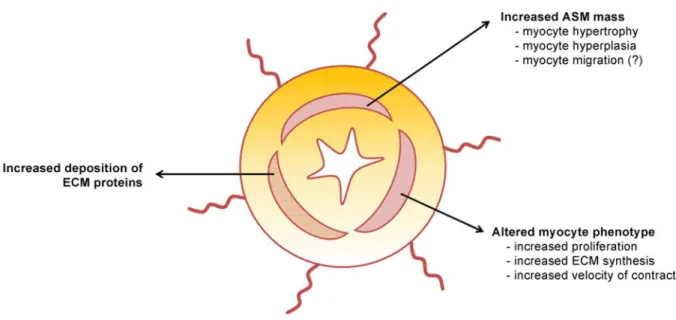
Figure 5. Summary of the alterations in ASM seen in asthma. ... 37
Figure 6. EBUS probes. ... 51
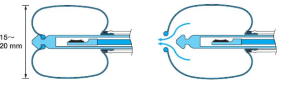
Figure 7. Pressure-dependent safety mechanism of the inflatable balloon. ... 52
Figure 8. Multilayered structure of the bronchial wall at EBUS. ... 54
Figure 9. Marginal echoes in EBUS images. ... 55
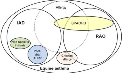
Figure 10. Equine asthma. ... 88
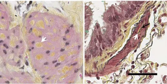
Figure 11. Heave line in an asthmatic horse. ... 93
Figure 12. Overall experimental design. ... 100
Article 1 Figure 1. ... 130 Figure 2. ... 131 Figure 3. ... 132 Figure 4. ... 133 Figure 5. ... 134 Figure 6. ... 135 Figure 7. ... 136 Figure 8. ... 137 Figure 9. ... 138 Article 2 Figure 1. ... 161 Figure 2. ... 162 Figure 3. ... 163
Article 3 Figure 1. ... 193 Figure 2. ... 194 Figure 3. ... 195 Figure 4. ... 196 Figure 9. ... 203 Figure 10. ... 204 Figure S1. ... 207 Article 4 Figure 1. ... 231 Figure 2. ... 232 Figure 3. ... 233 Figure 4. ... 234 Figure 5. ... 235 Figure 6. ... 236 Figure 7. ... 238 Figure 8. ... 239 Figure 9. ... 240 Figure E1. ... 253 Figure E2. ... 254 Article 5 Figure 1. ... 274 Figure 2. ... 275 Figure 3. ... 276 Figure 4. ... 277
Figure 13. Effect of bronchoconstriction and lamina propria thickness on endobronchial biopsy sampling. ... 285
Annex I
Figure 14 (Figure 1, Annex I). Correlation between lamina propria thickness and lung function data. ... ii Figure 15 (Figure 2, Annex I). Effect of bronchoconstriction on lamina propria thickness. ... ii Figure 16 (Figure 3, Annex I). Relationship between biopsy score and lung function. ... iii Figure 17 (Figure 4, Annex I). Effect of pharmacological treatments and antigen avoidance on
Abbreviations
AASM: non-corrected area of the airway smooth muscle (expressed in mm2) AHR: airway hyperresponsiveness
ASM: airway smooth muscle
ASM%: percentage of the endobronchial biopsy occupied by airway smooth muscle AP-1: activator protein 1
Atot: total area of the endobronchial biopsy (expressed in mm2) BAL: bronchoalveolar lavage
BALF: bronchoalveolar lavage fluid BM: basal membrane
C: control horses Cdyn: lung compliance
COPD: chronic obstructive pulmonary disease CT: computed tomography
ΔPL: variation of transpulmonary pressure EBB: endobronchial biopsy
EBUS: endobronchial ultrasound ECM: extracellular matrix EL: pulmonary elastance EUS: endoscopy ultrasound
FeNO: fractional exhaled nitric oxide FEV1: forced expiratory volume in 1 second HRCT: high-resolution computed tomography IAD: inflammatory airway disease
ICS: inhaled corticosteroids
ICS/LABA: inhaled corticosteroids and long-acting β2-agonist combination HE: horses with heaves (asthmatic horses) in exacerbation
HR: horses with heaves (asthmatic horses) in remission Ig: immunoglobulin
IL: interleukin
IOS: impulse oscillometry
L: endobronchial echographic layer LABA: long-acting β2-agonist
LYVE-1: lymphatic vessel endothelial hyaluronan receptor-1 MAPK: mitogen-activated protein kinase
MMPs: metalloproteinases
MRI: magnetic resonance imaging
NF-kappa β: nuclear factor kappa-light-chain-enhancer of activated B cells OCT: optical coherence tomography
PCNA: proliferating cell nuclear antigen PDE: phosphodiesterase
PET: positron-emission tomography Pi: internal perimeter (of the airway) PL: transpulmonary pressure
SMA: smooth muscle actin
SMMHC: smooth muscle myosin heavy chain
SPAOPD: summer-pasture associated obstructive pulmonary disease SPECT: single photon emission computed tomography
RAO: recurrent airway obstruction RL: pulmonary resistance
Th-1, Th2, or Th-17: polarized inflammatory response characterized by increased lymphocytes T-helper 1, 2, or 17
TGF-β: transforming growth factor beta TIMPs: tissue inhibitor of metalloproteinases TNF-α: tumor necrosis factor alpha
TSLP: thymic stromal lymphoprotein
US-TBNA: ultrasound-guided transbronchial needle aspiration : airflow
VEGF: vascular endothelial growth factor
VEGFR: vascular endothelial growth factor receptor VT: tidal volume
« On résiste à l'invasion des armées; on ne résiste pas à l'invasion des idées. »
Acknowledgements
The first person I would like to thank is Jean-Pierre Lavoie. Thank you for your patience, calmness, and perseverance. Thank you for your understanding. Thank you for your tireless guidance and encouragement through this serendipitous experience. I admire your deep knowledge and infinite curiosity, your managerial skills, and your synthetic ability (!). The way you approach the world around you has been inspiring to me, and has undoubtedly contributed to shape the person I am now. Thank you for all of this.
I would like to express my sincere gratitude to James G. Martin for his precious and always prompt advice. Your immense scientific culture and your humanity have been for me a source of motivation. I greatly appreciate your support and availability.
I am grateful to Yvonne Elce, Pierre Hélie, Philippe Joubert, and Daniel Jean for the substantial scientific contributions they have brought to the realization of this thesis. Our collaborations and exchanges have always been positive and formative to me.
I would like to thank Christine Théoret for accepting to review this thesis and preside over the jury. I am grateful to Ynuk Bossé for his commitment to the evaluation of this thesis. Your contribution will undoubtedly improve its content and enrich my knowledge.
I would like to recognize the professionalism and kindness of Christine Blondin, Diane Rodier, and Geneviève Michon, always available and helpful to solve many Michela-associated bureaucratic issues.
I am enormously grateful to my colleagues and friends who have shared with me these unforgettable and formative years. Particularly, I want to thank Mohamed Issouf for his genuine friendship and extraordinary listening skills. I cannot tell how much your presence has been supportive to me. I acknowledge that the experimental procedures described in this thesis have been realized thanks to the invaluable help of Catheryna Ouimet and Roger Fontaine (not to mention your contribution to my learning of the Quebec language and phonetics!). I will not forget to mention Mylène Chevigny for her patience and friendship, Amandine Vargas for her
inhuman efficiency and endless availability, Roxane Boivin for her frankness, Mathilde Leclère, Hélène Richard, Geneviève Michaud, Nicolas Heterman (without “H”), Mireille Godbout, Gabrielle Fillion-Bertrandt, Maude de Lagarde, Marion Allano, Emilie Gelinas-Lymburner, Juan Arango Sabogal, Aude Petlier, Gaelle Hirsh, Patricia Cano, Pamela Germim, and Patricia Mendosa.
Finally, from the bottom of my heart, I would like to thank my family and friends. Your unconditional love and endless support have been essential to my “survival” during these years. Thank you for being always so comprehensive with me. Thank you, Andrea. For your understanding, your ceaseless encouragement, and for your love; thank you for patiently waiting all this time.
Ad maiora!
Introduction
Central and peripheral airways sustain remodeling and inflammation in asthma (Bergeron, Al-Ramli et al. 2009). In particular, the airway smooth muscle (ASM) is increased and has the ability to induce an exaggerated bronchoconstriction in response to normally non-harmful stimuli, which is the main cause of airflow obstruction in asthma (Martin, Duguet et al. 2000). The amount, composition, and distribution of extracellular matrix (ECM) proteins within the bronchial wall can also profoundly affect lung function (Bosse, Riesenfeld et al. 2010). Multiple studies performed on asthmatic patients have investigated the effect of treatments on the reversibility of airway remodeling and inflammation, and most of them have studied samples of central airways obtained by endobronchial biopsies (Hoshino, Takahashi et al. 1999, Chakir, Shannon et al. 2003, Vignola, Riccobono et al. 2005). However, a growing body of evidence supports a central role of the small peripheral airways in disease pathobiology (Sturton, Persson et al. 2008, Contoli, Bousquet et al. 2010, Burgel 2011, Manoharan, Anderson et al. 2014). There is also increasing awareness that current therapeutic guidelines for asthma do not guarantee an adequate control of peripheral airway dysfunction (namely remodeling and inflammation) (Anderson, Zajda et al. 2012, Lipworth 2013). Peripheral airway assessment is challenging due to their anatomical inaccessibility, which is why their role in asthma has been overlooked for a long time. Equine asthma, or heaves, is a naturally-occurring disease of adult horses and a recognized animal model of human asthma (Leclere, Lavoie-Lamoureux et al. 2011). It is characterized by peripheral remodeling of the ASM and ECM, and by bronchoalveolar neutrophilic inflammation (Leclere, Lavoie-Lamoureux et al. 2011, Setlakwe, Lemos et al. 2014). Peripheral lung biopsies can be harvested from asthmatic horses for direct evaluation of small airway remodeling and inflammation (Lugo, Stick et al. 2002, Relave, David et al. 2008, Relave, David et al. 2010). Using this technique, our laboratory has previously shown that airway remodeling can be reversed by inhaled corticosteroids (ICS) monotherapy (Leclere, Lavoie-Lamoureux et al. 2012). However, the effect (a 30% reduction of ASM mass) was only partial as asthmatic horses still remained with twice as much ASM compared to control subjects even after 1 year of treatment. An insufficient deposition of the drug peripherally may explain the incomplete effect, and is
supported by the residual bronchoconstriction observed in ICS-treated animals (Leclere,
Lavoie-Lamoureux et al. 2012). In human asthma, ICS and long-acting β2-agonist
combinations (ICS/LABA) are more effective than ICS monotherapy to control disease exacerbations, but their efficacy compared to ICS on bronchial remodeling and inflammation are not well-described, particularly at the peripheral level of the lung. The objective of this thesis was to investigate and compare the effects of common anti-asthma medications on aspects of peripheral airway remodeling that cannot be studied in man due to ethical constraints. Specifically, we studied whether ICS/LABA enhances the reversibility of peripheral remodeling and the control of airway inflammation compared to ICS or LABA monotherapy. Of note, we simultaneously monitored the effect of these treatments on central airways, in order to identify whether the central remodeling or inflammation are related and may predict peripheral alterations. Due to the paucity of information available on the pathophysiology of peripheral airway remodeling in asthma and its reversibility with treatments, we believe that the results of this thesis are important and can be translated to human asthma.
What this study adds to the field
Airway wall remodeling is a characteristic finding in human asthma. It involves several components of the bronchial wall, with ASM and ECM likely mostly contributing to airway obstruction. These changes have been reported in both the central and peripheral airways in asthma, but few studies have investigated the effectiveness of commonly administered asthma treatments at inducing their reversal, especially in peripheral airways. The involvement of small airway dysfunction in asthma is increasingly recognized, but tools for studying the progression of disease at this level are not readily available. Also, how (and whether) peripheral remodeling and inflammation are reflected by changes observable in central airways remains unsettled. The studies described in this thesis have employed an equine model of neutrophilic asthma to address these questions. The results obtained provide evidence that the combination of ICS/LABA is not superior to ICS monotherapy for ASM remodeling reversibility or bronchial inflammation control. Bronchoalveolar neutrophilia is inhibited by ICS/LABA, while ICS and LABA monotherapies were ineffective. It remains to be determined whether ICS/LABA effectively increases peripheral deposition of the drug compared to ICS alone, or whether oral corticosteroids improve the reversibility of peripheral remodeling, before concluding that a portion of ASM remodeling is resistant to treatment. We have also shown that, peripherally at least, ASM remodeling is reversible in a shorter period of time compared to ECM remodeling, and that the reversibility of ECM remodeling appeared to be determined by the degree of airway wall inflammation. We could not identify remodeling or inflammatory features of central airways reflecting peripheral structure or function, indicating that central and peripheral bronchi act as separate compartments of the lung and not as a continuum. Finally, we have also significantly contributed to the advancement of veterinary medicine by optimizing respiratory research tools that can be employed in future studies and by describing new physiopathological mechanisms associated with equine asthma.
Normal anatomy and physiology of the lower respiratory tract
« Among the various cell types that populate the body, we might think of airway smooth muscle as the Hell’s Angel of cells, sitting on a Harley-Davidson, unshaven, a cigarette in one hand, a can of beer in the other, and a tattoo on its arm reading Born to Lose. » C.Y. Seow and J. J. Fredberg, J Appl Physiol (2001) 91:938-52
The lower respiratory tract extends from the larynx to the most distal portions of the lung parenchyma. Its role is to provide the tissues with oxygen and to remove their volatile metabolites as carbon dioxide, contributing to the maintenance of a balanced blood pH. Also, important metabolic and endocrine functions are accomplished in the lungs such as the activation of angiotensin I, or the removal of serotonin, bradykinin, as well as some prostaglandins and leukotrienes from the blood (Joseph, Puttaswamy et al. 2013).
Structure and function of the airways
Airways of the lower respiratory tract are considered as conducting airways when their walls do not contain alveoli and do not allow gas exchange, or as transitional airways when there are alveoli protruding from their walls and gas exchanges can occur. The formers comprise trachea, bronchi and bronchioles until terminal non-respiratory bronchioles, while the latter comprises the alveolar ducts. Distally to alveolar ducts there are alveolar sacs and alveoli.
These are not considered airways but, together with transitional airways and vessels, they form the respiratory tissue or lung parenchyma (West 2008). Airways can further be classified as cartilaginous (bronchi) or membranous (bronchioles) depending on the presence or absence, respectively, of cartilage within their wall. Also airways are classified as intra- or extra-pulmonary based on their anatomical location.
The basic microscopic anatomy of the conducting airways is similar along the bronchial tree, consisting of a pseudo-stratified epithelium overlying subepithelial tissue where connective tissue, vessels, glands and smooth muscle are present, with cartilage and adventitia located outermost (Figure 1). The bronchial epithelium is pseudo-stratified and ciliated. It is mainly composed of columnar ciliated and mucus-secreting goblet cells in trachea and proximal bronchi. Columnar ciliated cells and goblet cells decrease in quantity in smaller bronchi, and most of them are replaced by cuboidal epithelial cells and by Club cells (previously named Clara cells (Winkelmann and Noack 2010, Irwin, Augustyn et al. 2013)), respectively, in the bronchioles. The cuboidal bronchial respiratory epithelium completely disappears in the alveolar ducts. The bronchial epithelium lies on a basement membrane that provides attachment for epithelial cells, mainly composed of collagen IV, laminin, and proteoglycans, with elastic fibers within it.
The lamina reticularis is the deepest portion of the basal membrane, adjacent to the lamina propria of the submucosa. Collagen I, III, V, and tenascin amongst others form the lamina reticularis, which is strongly connected to the other portions of the basal membrane (namely the lamina rara and the lamina densa) by strands of collagen VII (Liesker, Ten Hacken et al. 2009). The lamina propria is made up mainly of connective tissue, in which vessels, nerves and elastic fibers are dispersed (Brinkman, Brooks et al. 1969). Airway vessels from the bronchial circulation are arranged in two plexuses within the bronchial wall: a submucosal plexus, running through the lamina propria between the ASM bundles and epithelium, and a peribronchial plexus, supplying the adventitia externally to the ASM layer (Carroll, Cooke et al. 1997). Collagen I and III are the main components of the lamina propria, with several proteoglycans and other glycoproteins. In the bronchial submucosa, beneath the lamina propria, there are smooth muscle fibers, cartilage, and tracheobronchial glands, all surrounded by supporting connective tissue. Caudally to terminal non-respiratory bronchioles, the lamina propria disappears, and only ASM and the elastic network continue in a spiral fashion surrounding the alveoli. Cartilage is disposed as parallel “C-shaped” rings along the trachea and main bronchi, and it becomes smaller and more irregular in segmental and lobular bronchi, completely disappearing in bronchioles (Fraser 2005).
Smooth muscle fibers are disposed as bundles either in the open portion of the cartilage rings (in the trachea, where they are recognized as the trachealis muscle), or between the lamina propria and the cartilage. Smooth muscle bundles make up roughly 3-5 and 14-18% of the central and peripheral bronchial wall in adult patients, respectively, with their orientation varying along the bronchial tree (Hale, Olsen et al. 1968, Dunnill, Massarella et al. 1969, Takizawa and Thurlbeck 1971, Sobonya 1984, Saetta, Di Stefano et al. 1991). Overall, they are more regularly and circumferentially oriented in the proximal airways, whilst they are arranged in branching and anastomosing bundles forming oblique and irregular spirals in the lower airways (Smiley-Jewell, Tran et al. 2002). Their disposition around smaller airways has been described as helical or geodesic (James and Carroll 2000). Of note, the orientation of the ASM bundles along the bronchial axis depends on the degree of lung inflation, with higher lung volumes increasing the angle of orientation as a consequence of the longitudinal stress
imposed on the airways. Taking this into account, the estimated angle of orientation of the ASM bundles has been described as small in central airways (<5th generation) and approximately 15 degrees in membranous airways. Furthermore, the distribution is often asymmetric, particularly in the more proximal airways, so that, in case of bronchospasm, ASM orientation tends to favor one direction of coiling over the other (Ebina, Yaegashi et al. 1990, Lei, Ghezzo et al. 1997). Ebina and colleagues (1990) showed that the thickness of ASM relative to airway diameter increases towards the periphery in man. Although the absolute volume of smooth muscle decreases with decreasing airway size, the volume fraction of smooth muscle in the airway wall increases in smaller peripheral airways, and this in spite of a reported tendency for the density of the bronchial smooth muscle bundles to diminish as one moves from the central to the peripheral airways (Lei, Ghezzo et al. 1997). The same group also showed that the thickness of ASM layer relative to the airway diameter could vary up to 10-fold within the same group of airways, possibly in relation to changes in smooth muscle thickness within a bronchial segment, subject size, sex or age, site sampled, degree of muscle contraction and possibly even racial-related differences. The bronchial smooth muscle bundles are surrounded by connective tissue whose ECM is composed of both collagenous and non-collagenous elements, such as elastin, proteoglycans, and other glycoproteins. They are secreted by structural cells such as fibroblasts, myofibroblasts, and smooth muscle cells themselves. Although the mechanisms regulating their turnover (synthesis, deposition, and degradation) within the lung are not completely elucidated, a strong body of evidence supports the balance of metalloproteinases (MMPs) and tissue inhibitor of metalloproteinases (TIMPs) within the bronchial tissue as the major determinant of ECM homeostasis.
Within the airways, each structural component has a recognized role contributing to the normal physiology of the lung (summarized in Table 1) with the only exception of the ASM, whose function, if any, is still unknown. A few theories have been proposed concerning the role of bronchial smooth muscle in ontology (Murphy, Summer et al. 2008):
1. in the developing fetal lung, the ASM has been shown to contract with a regular
peristaltic rhythm, generating distending pressures that could promote the development and maturation of the lung;
2. the contraction of ASM could serve to improve ventilation/perfusion matching and/or decrease dead space ventilation in case of reduced blood flow to a specific district of the lung;
3. during coughing episodes, ASM contraction may increase the velocity of gas movement to promote the expulsion of foreign bodies or noxious particulate materials;
4. the ASM may act as a damper, by balancing the hysteresis between small airways and alveolar units;
5. mucus propulsion (Bullowa and Gottlieb 1922);
6. lastly, the ASM may simply be an ‘‘evolutionary oversight,’’ that is, a vestige of the lung’s origin from an organ, the foregut, already programmed to develop smooth muscle, with no true physiologic purpose.
Furthermore, at present, there is no disease entity or appreciable physiological deficit associated with loss of ASM contraction (Seow and Fredberg 2001).
The lung is a relatively stable organ with low rates of cell turnover, particularly in the airways. With less than 1% of epithelial cells proliferating at any given point in time (Ayers and Jeffery 1988, Demoly, Simony-Lafontaine et al. 1994, Leigh, Kylander et al. 1995), there appears to be little need for local cellular self-renewal under normal circumstances. Nevertheless, following injury, the epithelium has great ability to repair in a very short time by means of processes of dedifferentiation, migration and proliferation of adjacent cells (Erjefalt and Persson 1997, Yahaya, Baker et al. 2011). Proliferation and possibly migration of subepithelial cells seem to be involved in this process as well, as documented by studies of induced epithelial damage in a sheep model and by the observation of increased quantity of subepithelial myofibroblasts in asthmatic patients after experimental allergen exposure (Gizycki, Adelroth et al. 1997, Yahaya, Baker et al. 2011). The proliferation rate of ASM cells in normal conditions is considered as negligible (Chernyavsky, Croisier et al. 2014).
Subepithelial cells proliferating in normal conditions are scarce and mostly represented by endothelial cells and cells in close proximity to the mucus glands (Yahaya, Baker et al. 2011). On the other hand, turnover of the ECM is impressively rapid; it is estimated that 10-15% of the lung ECM is renewed every day (Dunsmore, Lee et al. 1996, Roberts and Burke 1998).
Table 1. Normal function of airway components. Airway structure Normal function
Epithelium Physical barrier against inhaled particles
Lamina reticularis Structural support for epithelial attachment
Lamina propria Structural support of the bronchial wall
Determines mechanical properties of the bronchial wall May regulate ASM proliferative/synthetic function
Submucosal vessels Trophic support of the airways
Smooth muscle ?
Adventitia Sustains structure
Guarantees airway-parenchyma interdependence
Structure and function of the lung parenchyma
Within the functional respiratory tissue, which is composed of alveolar ducts, alveolar sacs, and alveoli, alveolar units are demarcated by septa composed of a continuous layer of squamous epithelial cells overlying a thin interstitium. Capillaries as well as connective tissue and several cells responsible for maintaining alveolar shape and defense lie within the septal interstitium. The alveolar epithelium consists primarily of type I and type II alveolar cells.
Type I cells are more numerous, they cover approximately 95% of the alveolar surface, and their function is related to the transport of substances in either direction across the air-blood barrier. Type II alveolar cells are generally located close to the junctions among different alveoli, they are responsible for production of surfactant and renewal of the alveolar epithelium by differentiation into type I cells. A basement membrane is present underneath the alveolar epithelial cells, intimately connected with the basement membrane of the capillary endothelium. Elastic and collagen fibers, as well as myofibroblasts, are irregularly distributed within the connective matrix between epithelial and endothelial basement membranes (Fraser 2005).
Age-related changes of pulmonary structure and function
The physiological aging process undoubtedly impacts the structure and function of the lungs. Anatomically, the airways and their components increase in volume during infancy until complete development of the body, with the smooth muscle occupying a slightly greater proportion of the airway wall in children compared to adults (James and Carroll 2000). The aging lung is characterized by higher levels of collagen deposition and decreased elastin expression, which contribute to the decreased elastic recoil observed with age (Thannickal, Murthy et al. 2015). Lung function decline with aging in healthy individuals is associated to alterations of the lung parenchyma and terminal conducting airways. Reduced mucociliary clearance and altered surfactant composition are also reported in aged individuals (Miller 2010, Ramly, Kaafarani et al. 2015).
Mechanisms of airway patency
When the lungs are removed from the thoracic cage, they immediately collapse due to their elastic recoil. Bronchial lumen is also significantly reduced when lungs are collapsed, and this independently of the presence or absence of cartilage in the airway wall, because of the highly
developed elastic network connecting all structures within the lung. Several mechanisms contribute to maintain airway patency in vivo and to oppose the collapsing force induced by reduced lung elastic recoil at low lung volumes. First of all, it is the intimate relationship between airway dimension and lung volume that prevents bronchial collapse even at very low lung volumes. This is known as airway or parenchymal tethering, or airway-parenchymal interdependence (Bates and Lauzon 2007). Airway stiffness itself as well contributes to airway patency in vivo. Lastly, the lining fluid exerts surface tension acting inwardly to close the airways, especially the smallest ones. The secreted surfactant, on the other hand, reduces the surface tension of the lining fluid and thereby contributes to airway patency (Veldhuizen and Haagsman, 2000).
Mechanics of breathing
The act of breathing is accomplished by respiratory cycles. For each human breath, one active inspiratory and one passive expiratory process occur, during which the air is forced in and out of the alveoli as a result of the changing intrapulmonary (or alveolar) pressures. Indeed, the contraction of the diaphragm (and other respiratory muscles) during inspiration expands the thorax which, in turn, induces lung expansion. Following the increase in lung volume, the alveolar pressure drops as a consequence of Boyle’s law and produces a pressure gradient that makes the air move from the mouth to the alveoli. As stated by Ohm’s law: the airflow between two points is directly proportional to the pressure difference across the two points. Similarly, during expiration, the lung passively recoils due to its elastic properties and its volume decreases. Alveolar pressure then increases compared to the pressure at the mouth, which allows air to move from the alveoli towards the atmosphere. Any alteration of the airway lumen (bronchoconstriction, space-occupying lesion or mucus) between two points of the bronchial tree is an obstacle for the (theoretically1) laminar airflow generated by the
1 In the airways there is usually a combination of both laminar and turbulent flow (called "transitional flow") in
pressure difference existing between them. In order to maintain a constant airflow ( ), the pressure gradient between the two points (ΔPL) must be increased proportionally to the resistance of the lung (RL):
= ∆
Lung resistance is expressed in cm H2O/L/s, and it is the sum of airway resistance (normally about 80% of RL) and tissue resistance (lung parenchyma and chest wall, counting for 20% of RL). The Poiseuille’s law states that airway resistance is directly proportional to the length of the airway and the viscosity of the gas, and inversely proportional to the radius to the fourth power (i.e.: a 2-fold decrease in bronchial radius increases airway resistance by 16-fold). Thus, the main determinant of airway resistance is airway diameter which, in turn, results from:
• ASM contraction (bronchospasm),
• airway wall thickening (edema and/or remodeling),
• airway lumen occlusion (i.e. by mucus and cell accumulation or space occupying lesions),
• lung volume (at higher volumes, the parenchyma pulls on the airways keeping them wide open).
For a given flow, resistance to air passage is thus greatest in individual small airways compared to large ones due to their small diameter. However, the total resistance to airflow contributed by the small airways taken together is very low (on average 10% in health) as they represent numerous parallel pathways, whose transversal area can be summed up. Under normal circumstances it has been calculated that the greatest resistance to airflow resides in the medium-sized bronchi (Macklem 1998).
The elastic recoil of the lung is fundamental for breathing. The slope of the pressure-volume curve represents lung compliance (Cdyn, expressed in L/cm H2O), which is a measure that
describes the ease by which the lungs stretch and expand in response to a given pressure. It is calculated using the formula:
= ∆ ∆
where V is lung volume and PL is pleural pressure. The compliance of the lungs demonstrates hysteresis; that is, the PL is different on inspiration and expiration for identical volumes. Also, compliance is volume dependent. Elastance (EL, expressed in cm H2O/L and also known as elastic resistance) is, by definition, the reciprocal of compliance (E = 1/Cdyn), and describes the pressure change that is required to elicit a unit volume change of the lungs. This is a measure of the resistance of a system to expand or to shrink. Elastance is an indirect expression of the work that has to be exerted by the inspiratory muscles to expand the lungs. An increased elastance needs to be counteracted by an increased power of the muscles during inspiration, leading to an increased work of breathing (work of breathing is the physical work that has to be carried out by the muscles of respiration to overcome the elastic and non-elastic resistance of the respiratory system). The elastance of the whole respiratory system depends on the elastance of the chest wall and that of the lungs. Changes in the elastance (and therefore the compliance) of the chest wall are uncommon. In contrast, the elastance of the lungs is affected by many respiratory diseases. Thus, variations in the elastance of the respiratory system are mainly due to alterations of the elastance of the lungs, which is governed by two main factors:
• the elastic recoil forces of the lung (mainly regulated by the properties of the pulmonary elastin network, but also by the altered deposition of fibrous tissue), • the surface tension at the air-alveolar interface (depending on the surfactant quantity
and composition).
Conversely to resistance, compliance and elastance of the lungs are mainly determined by the physical properties of the small airways and lung parenchyma.
Even in healthy subjects, heterogeneity exists among the behavior of different airways and alveoli. However, taken together, the behavior of the lungs can be compared to that of a balloon sealed over the end of a pipe, which can be simplified using a mathematical approach called the linear single-compartment model (Bates 1993). This model has a clear anatomical analogy to the lung; the pipe represents the conducting airways and the balloon represents the elastic parenchymal tissue. There is also a functional analogy, as the balloon can be inflated and deflated through the pipe in the same way that a lung inspires and expires. Mathematically, is it expressed as:
∆ = ∙ + ( ∙ )
where VT is tidal volume (in L) and K is the pulmonary end expiratory pressure (in cm H2O). This equation states that the pressure gradient required to increase lung volume by VT is proportional to the resistive and elastic properties of the lung, and it will increase at increasing RL and EL.
Functional anatomy of the lower respiratory tract
During breathing, the airway structures undergo dynamic morphological changes due to physical constraints and mechanical stress imposed by the changing intrathoracic pressures. Airways are subjected to two types of stretches or stresses: a radial stretch, acting outwardly all around the airway wall, and a longitudinal stretch, related to the elongation of the lung; both occurring during inspiration. The opposite occurs during expiration. Intra-thoracic airways reach a maximal and minimal cross-sectional area at inspiration and end-expiration, respectively. This is of particular interest during dynamic measurement of airway wall thickness or airway size.
Until recently, dynamic measurements of airway size or length were not possible, and general knowledge on this topic has been increasing exponentially in the last decade. With the advent of new imaging technologies, reliable measurements can now be performed. Airway wall area remains unchanged or undergoes minimal changes across the breathing cycle (Coxson,
Quiney et al. 2008), while airway wall thickness and lumen area vary. Specifically, airway lumen increases and bronchial thickness decreases during inspiration, as a result of bronchodilation. Significant changes are observed in the airway lumen area during quiet breathing in large (8-25 mm in diameter), intermediate (2-8 mm), and small airways (< 2 mm), with the latter being the site where this phenomenon is most remarkable (McLaughlin, Williamson et al. 2008). This occurs probably because they are more compliant than larger airways, and because of their strict interdependence with the pulmonary parenchyma. In patients with COPD, variations of the bronchial lumen up to 150-200% have been reported during deep breathing maneuvers (Matsuoka, Kurihara et al. 2008).
Mechanisms implicated in asthma pathobiology
« Breathing remains unaccountably easy. Indeed, it is this lightness of breathing in the healthy challenged lung, rather than the labored breathing that is characteristic of the asthmatic lung, that in many ways presents the greater challenge to our understanding of the determinants of acute airway narrowing. » J.J. Fredberg and S.A. Shore “The Unbearable Lightness of Breathing” J Appl Physiol (1999) 86:3-4
Asthma is a chronic disease, considered as multifactorial, diffuse, and heterogeneous in nature. Different pathophysiological mechanisms are involved in disease development, both intrinsic and extrinsic to the patient. Specifically, genetic predisposition or dysregulated immunity, as well as several environmental triggers have been shown to take part in asthma pathogenesis. Whether the intrinsic causes of asthma are inherited or acquired during life
remains a matter of debate (Wenzel 2012, Kudo, Ishigatsubo et al. 2013). Asthma is
considered a diffuse disease, as it affects the entirety of the lung. It is however heterogeneous, as different regions of the bronchial tree can be differently affected (Elliot, Jones et al. 2015). Pathophysiologically, asthma is defined as an obstructive and inflammatory disease. Both airway remodeling and excessive narrowing contribute to the airflow obstruction observed in asthmatic patients. An increased smooth muscle mass and reactivity to normal stimuli (hyperresponsiveness) are the most important determinants of airflow obstruction (Lambert,
Wiggs et al. 1993, Oliver, Fabry et al. 2007). While the leading role of bronchospasm during asthma attacks has long been recognized, the implication of inflammation to asthma pathophysiology was only described few decades ago, and its contribution to disease development and/or maintenance is still ill-defined. Several structural and immune cells contribute to the asthmatic response within the bronchi. Interestingly, among these cells, ASM cells have marked immunomodulating properties, making them potentially significant contributors to the inflammatory features observed in asthmatic airways (Damera, Tliba et al. 2009).
The relationship existing between bronchospasm, remodeling, and inflammation in asthma is far from clear. It has been speculated for some time that inflammation represents the initiating insult, causing remodeling and airway responsiveness (Kudo, Ishigatsubo et al. 2013). However, recent evidence argues against this theory and suggests that bronchospasm and remodeling themselves might in turn induce or facilitate inflammation (Damera, Tliba et al. 2009, Grainge, Lau et al. 2011).
Airway hyperresponsiveness
Airway hyperresponsiveness (AHR) is defined as an excessive decline of lung function upon ASM activation. AHR is a well-recognized feature of the asthmatic disease, however the primary contributors to this phenomenon remain to be elucidated (Martin, Duguet et al. 2000). Whether and to which extent ASM cell hypercontractility (abnormally increased degree of contraction) and hyperreactivity (contractile response in the presence of normally non-harmful stimuli) contribute to the development of AHR is still unknown. While ASM seems intuitively to be the key of such pathological behavior of the airways, a large amount of evidence supports that other non-contractile components of the airway wall as well as of the pulmonary parenchyma can contribute to the pathogenesis of AHR (Bosse, Riesenfeld et al. 2010). In other words: while ASM contraction is likely to be the central point, the load against
which the muscle contracts as well as the environment in which the muscle is placed must be taken into account (Figure 2) [65].
Smooth muscle-related determinants of AHR
Both static and dynamic modeling of pulmonary mechanics identify ASM as the most important determinant of airway narrowing and AHR (Lambert, Wiggs et al. 1993, Oliver, Fabry et al. 2007). Airway smooth muscle mass is increased in asthmatics and such increase is associated with the severity of the disease (James, Bai et al. 2009). However, whether such increase coexists with phenotypic changes affecting ASM contractile, proliferative or secretory properties and to what extent they would contribute to AHR in asthma remains unclear (Hirota, Nguyen et al. 2009). The possibility that functional anomalies of the ASM cells represent a central determinant of AHR and asthma pathogenesis must be kept in mind.
Figure 2. Determinants of airway patency and hyperresponsiveness. The arrows indicate
forces (pressures) acting on the bronchial wall. In red are indicated alterations observed in asthma.
Anatomical factors. ASM is organized in bundles running along the bronchial wall in a spiral
fashion (Bates and Martin 1990). In such spirals, ASM orientation seems to acquire a wide range of angles along the airways, following a distribution pattern likely conserved among species and among different airway generations (Smiley-Jewell, Tran et al. 2002). The magnitude of ASM obliquity along the bronchi has been judged as unlikely to result in physiologically important changes in airway length during bronchoconstriction (Lei, Ghezzo et al. 1997). Contrarily, airway caliber would be significantly affected (Bates and Martin 1990). It has been speculated that AHR might even result simply from a stiffening of the airway in the longitudinal direction (Bates and Martin 1990), a situation possibly reflecting the act of breathing at high lung volumes as during an asthma attack. Paradoxically, an in
silico model of the bronchial tree suggested that bronchial malfunction related to asthma
could be a « necessary consequence of the optimized efficiency of the structure of the human bronchial tree » (Mauroy, Filoche et al. 2004). In other words, an AHR could be the drawback of a bronchial tree anatomically optimized to be physiologically efficient.
Remodeling. The smooth muscle mass is increased in asthmatic airways (James, Elliot et al.
2012). As the force produced by the smooth muscle is proportional to its cross-section (Chin, Bosse et al. 2012), the asthmatic smooth muscle could cause AHR simply because a greater force would act against the mechanisms of airway patency. This could occur in the absence of mechanical alterations of the ASM in asthma, and is supported by mathematical models of the lung (Wiggs, Bosken et al. 1992, Lambert, Wiggs et al. 1993). However, the unchanged FEV1 (forced expiratory volume in 1 second) reported after ASM reduction obtained by bronchial thermoplasty in severe asthmatics (Pavord, Thomson et al. 2013, Denner, Doeing et al. 2015) argues against the hypothesis that AHR in asthma is just the result of the increased smooth muscle mass. The mechanical properties of ASM cells are influenced by their curvature. Particularly, cell stiffness is maximal at curvature similar to those expected in terminal membranous bronchioles (diameter 1.5 mm) and decreases at smaller or greater curvatures. Cell contractility instead is thought to be inversely proportional to airway dimension, as it decreases consistently as the curvature increases (Montaudon, Desbarats et al. 2007, West
alter the distribution of ASM cell orientation in asthmatic airways (Lei, Ghezzo et al. 1997), accentuating airflow heterogeneity during bronchospasm. In vitro, ASM cell alignment depends on the strain they are exposed to (Morioka, Parameswaran et al. 2011, Ghezzi, Risse et al. 2013). Airway smooth muscle orientation has the potential to influence the mechanics of airway narrowing by influencing the vector of forces acting on the airway wall during bronchoconstriction. That is, a portion of the forces developed by ASM contraction could potentially shorten the airway in addition to narrowing its lumen (Lei 1997).
Phenotype plasticity. Two different populations (phenotypes) of ASM cells are recognized in vitro, characterized by increased contractile or synthetic properties (Hirota, Nguyen et al.
2009), which were first described in 1997 (Ma, Li et al. 1997). Of note, the phenotype is not a constant property of the ASM cell. It can instead vary depending on the chemical and mechanical environment the cell is exposed to (Hirota, Nguyen et al. 2009). A large body of evidence supports the hypothesis that asthmatic ASM cells express a more proliferative/synthetic and less contractile phenotype compared to healthy ASM cells in vitro (Wright, Trian et al. 2013). Whether this is true also in vivo remains to be determined. The fact that hyperplasia is a recognized mechanism of increased ASM mass in asthmatic subjects, and that ASM cells are now recognized as immunomodulatory cells able to secrete a various array of pro-inflammatory molecules (James, Elliot et al. 2012, Wright, Trian et al. 2013) sustains this theory.
Length adaptation. Two very important and unique properties of the smooth muscle have
been described that highlight its adaptable nature. Length adaptability was first proposed in 1995 to explain that the force generated by smooth muscle contraction is (relatively) independent of its length (Pratusevich, Seow et al. 1995). In fact, when the smooth muscle length is changed, a decline of its strength is initially observed but it gradually recovers over time. In situ, length adaptation may be initiated by a change in transmural pressure (causing bronchoconstriction or bronchodilation), which is a primary physiological determinant of ASM length. When healthy subjects are prevented from taking deep breaths for 20-30 minutes, they indeed develop AHR (Skloot, Permutt et al. 1995). The recognized
bronchoprotective and bronchodilatory effects of deep inspirations observed in healthy subjects have been ascribed to this mechanism as well. However, despite the fact that bronchi of asthmatic subjects can dilate as much as those of healthy patients in vivo (Brown, Scichilone et al. 2001), the bronchoprotective effect of deep inspirations is impaired in asthmatics, suggesting that length adaptation alone is not the sole explanation of AHR in asthma. Recent studies performed using intact airway sections instead of isolated smooth muscle strips further support the inability of smooth muscle length adaptation alone to cause AHR, especially at the central level, because of the coupling existing between airway wall and ASM layer (Ansell, McFawn et al. 2015, Dowie, Ansell et al. 2016).
Force adaptation. Force adaptation has been described more recently as the ability of the
muscle to increase its force-generating capacity in response to stimulation (Bosse, Chin et al. 2009). Differently from length adaptation, force adaptation in vitro is not inhibited by oscillating maneuvers simulating breathing pattern (Pascoe, Jiao et al. 2012). Length and force adaptation can coexist and have additive effects (Bosse, Chin et al. 2010) . The significance of this finding in asthmatic airways, chronically exposed to bronchoconstrictive stimuli, appears straightforward but has not been proven yet.
Myosin isoforms. Smooth muscle myosin heavy chain (SMMHC) is a cytoplasmic structural
protein that is a major component of the contractile apparatus in smooth muscle cells. It is expressed as four different isoforms, generated by alternative splicing of the myosin gene. In particular, two of these isoforms regulate the velocity of contraction of the myosin head once it is attached to the actin filament. At a molecular level, they differ by the presence [(+)insert, also called SM-B] or absence [(-)insert, also called SM-A] of a 7–amino acid insert within the protein structure. The (+)isoform induces a propulsion velocity of the actin filament twice as fast as the (-)isoform, and it is overexpressed in human and equine asthmatic bronchial tissue compared to healthy subjects (Leguillette, Laviolette et al. 2009, Boivin, Vargas et al. 2014). A more rapid smooth muscle contraction could contribute to the blunted response of asthmatic patients to deep inspirations.
Fluidity. Muscle contractile properties are equilibrated dynamically. In vivo, non-activated
ASM is continuously exposed to force fluctuations (stress) that induce cell length variations (strains), which significantly inhibit its contractile capacity (Krishnan, Trepat et al. 2008). In asthmatics, ASM is activated by different stimuli likely reducing its strain and conferring quasi-static conditions. Using an immediate terminology (partly borrowed from the past), the group of Fredberg described the normal ASM as “fluid” and the asthmatic ASM as “frozen” (Krishnan, Trepat et al. 2008). One of the possible molecular explanations of this theory is that, during breathing, a number of cross-bridges between the myosin head and the actin filament are broken up mechanically and prematurely compared to the complete myosin duty cycle. This reduces the number of cross-bridges and the force-generating capacity of the muscle when it is in its fluid state (Fredberg, Inouye et al. 1999). In fact, when healthy individuals are restrained from taking deep breaths, their ASM “freeze” in a contracted position after only 15 minutes, becoming hyperresponsive to the same extent as asthmatic smooth muscle (Krishnan, Trepat et al. 2008).
The contractile properties of ASM have been studied in healthy subjects, in asthmatics and in animal models of asthma, in order to understand if any abnormality of the ASM could explain the exaggerated response to contractile stimuli observed in asthma. Main results of previous studies made on human tissues in static conditions have been variable and quite inconclusive, especially in light of the fact that ASM behavior in vivo is better represented by oscillatory conditions (Fredberg, Inouye et al. 1999). Furthermore, many studies included a too limited number of specimens to guarantee statistically sound comparisons between groups (de Jongste, Mons et al. 1987, Bramley, Thomson et al. 1994, Thomson, Bramley et al. 1996). Limitations of the early approaches used to study ASM mechanical properties are now recognized (Bai, Bates et al. 2004). Among others, as heterogeneity has been appreciated in ASM derived from different anatomical regions, most strikingly between trachea and bronchi (Bramley, Thomson et al. 1994, Ma, Li et al. 1997, Matusovsky, Kachmar et al. 2015), it seems inappropriate to compare specimens from different anatomical regions or to draw conclusions concerning the bronchial behavior from studies employing tracheal specimens. Having said that, comparing samples obtained from the same anatomical region between
asthmatics and non-asthmatics may however identify abnormalities in ASM behavior related to the disease. Two recent studies (Chin, Bossé et al. 2012, Ijpma, Kachmar et al. 2015) found no difference in the intrinsic properties of ASM in asthma, supporting the hypothesis that is it the environment where the muscle is located rather than the muscle itself, the main determinant of AHR in asthma.
Non-smooth muscle determinants of AHR
Lung volume and airway-parenchyma interdependence. Airways are intimately connected to
the pulmonary parenchyma, so that increasing lung volume stretches the bronchi both longitudinally and transversally, decreasing airway resistance. Such airway wall tension is primarily determined by non-contractile airway wall components when ASM is relaxed, but it is progressively transferred to the ASM during its active contraction (Martin, Duguet et al. 2000, Bosse, Riesenfeld et al. 2010). Lung expansion provides an elastic force contrasting that induced by ASM contraction. However, parenchyma and ASM are not directly linked; between them lies the external bronchial layer made of loose connective tissue, lymphatics and bronchial microvessels in small airways, in addition to cartilage and submucosal glands in large airways. This layer may attenuate force transmission between the two compartments, especially when its morphological and mechanical features are altered such as in asthma (Martin, Duguet et al. 2000, Dolhnikoff, da Silva et al. 2009).
Airway wall morphology and remodeling. The spatial arrangement and volumes of the
constituents of the airway wall are crucial in determining how the contraction of the ASM affects airway narrowing and airflow obstruction (Bosse, Riesenfeld et al. 2010). Specifically, the disposition and mechanical features of tissues lying around and within the ASM layer must be considered. Increasing the volume of tissues outside the ASM may lead to an uncoupling of the system, modifying the patterns of force transmission from lung parenchyma to the airway wall. On the other hand, the increased deposition of structures lying inside the
would be beneficial during bronchospasm), but also some space-occupying material protruding towards the airway lumen and reducing its caliber, facilitating airway obstruction (Lambert, Wiggs et al. 1993). Especially during ASM contraction, even small reductions of the airway lumen may increase dramatically airway resistance (Lambert, Wiggs et al. 1993). Remodeling of the ECM among and surrounding the ASM cells also deserves to be considered. Due to its relative stiffness, this ECM “cage” is thought to limit the contraction-induced cross-sectional widening of the smooth muscle bundle, and thus its excessive shortening. This theory is known as the radial constraint hypothesis (Meiss 1999), and it is supported by the reduced fraction of ECM proteins observed within the ASM layer of severe compared to moderate asthmatics (Pini, Hamid et al. 2007).
Heterogeneity. Structural and mechanical properties of the bronchial tree vary in time or
among different bronchial generations. Pathological entities affecting such properties – such as asthma –further increase the heterogeneity of the lung (Bosse, Riesenfeld et al. 2010). Using a mathematical model, it has been shown that airway resistance can significantly increase in response of uneven bronchial tone along the bronchial tree (Bates 1993). Airway smooth muscle mechanical heterogeneity is also recognized at tissue and cellular level (Ma, Li et al. 1997) which, in addition to anatomical heterogeneity, can produce AHR (Bosse, Riesenfeld et al. 2010). Any factor contributing to lung heterogeneity can finally lead to increased airway resistance and amplify the effect of contractile stimuli on respiratory physiology.
Inflammation. Evidence exists linking the increased inflammatory infiltrate into the ASM
layer and its increased proliferation (Ramos-Barbon, Fraga-Iriso et al. 2010). For this reason, inflammation could indirectly induce AHR by increasing the ASM mass in asthmatic airways. Furthermore, inflammatory cells may produce and secrete multiple bronchoconstrictor agents (Bossé 2014) that can contribute to overstimulate the asthmatic ASM cells.
Airway remodeling
Asthmatic airways are characterized by marked structural changes affecting most of their histologic components, and commonly referred to as airway remodeling. It significantly contributes to airflow obstruction in asthma, particularly during episodes of bronchoconstriction. Airway remodeling can affect small and/or large airways, possibly in relation with the asthma inflammatory phenotype (Elliot, Jones et al. 2015).
Remodeling has been considered for a long time as the consequence of chronic tissue inflammation and dysregulated repair. However, the similar degree of remodeling observed in recent compared to long-standing mild asthmatics (Boulet, Turcotte et al. 2000), the demonstration of bronchial remodeling in wheezing children (Saglani, Payne et al. 2007, O'Reilly, Ullmann et al. 2013), and the impressively quick remodeling response observed after antigen challenge in asthmatic patients (Kariyawasam, Aizen et al. 2007) argue against the fact that a chronic insult is necessary in order to develop airway remodeling. Different studies have shown that the degree of remodeling is related to the severity and not to the duration of the disease (Chetta, Foresi et al. 1997, James, Bai et al. 2009).
Due to the importance of the small airways in asthma pathogenesis and to the difficulty of sampling (Contoli, Bousquet et al. 2010, Burgel 2011), a lot of of attention has been focused on understanding whether the morphological changes observed in large and small airways are correlated (Postma, Brightling et al. 2015). To date, the information available about small airway remodeling comes from a limited number of studies performed on autopsy specimens which have provided precious insights into the contribution of the silent portion of the lung to the asthmatic disease.
Epithelium
inspired molecules that can reach the lungs. As such, it is a structure with important immunoregulatory roles, able to orchestrate complex innate and acquired responses to several triggers (Damera, Tliba et al. 2009). There is evidence supporting an impaired function of the airway epithelium in asthma (Swindle, Collins et al. 2009), in part mirrored by an altered structure. Increased susceptibility to injury and altered repair are considered to be important inducers of airway wall remodeling in asthma.
Bronchial epithelial fragility and detachment has been associated with asthma for long time, due to the high number of epithelial cells observed in sputum samples or within the bronchial lumen in histologic preparations. The evidence provided by in vitro studies of stressed and fragile airway epithelium in asthmatic airways (Swindle, Collins et al. 2009), incapable of adequate repair mechanisms (Puddicombe, Torres-Lozano et al. 2003), supports the fact that the macroscopic shedding of the bronchial epithelium often observed in endobronchial biopsy samples is now considered as being merely artefactual (Holgate 2011). The data obtained by Labonté and colleagues argue that the asthmatic epithelium could be macroscopically damaged only in the more proximal airways (Labonte, Laviolette et al. 2008), questioning whether the sampling site of endobronchial biopsy could have represented a confounder in previous studies.
Bronchial epithelial hyperplasia has been observed in severe compared to mild asthmatics and correlates with epithelial cell proliferation in some studies (Demoly, Simony-Lafontaine et al. 1994, Druilhe, Wallaert et al. 1998, Vignola, Chiappara et al. 2001, Cohen, E et al. 2007). However, two points need to be raised: first, the thickness of airway epithelium of severe asthmatics is not different from that of healthy subjects; second, severe asthmatics need corticosteroid treatment, which can profoundly modulate cell proliferation and apoptosis (Salmon, Koto et al. 1998, Dorscheid, Wojcik et al. 2001, Al-Wadei, Takahasi et al. 2005, Gallelli, Pelaia et al. 2010). In this perspective, whether epithelial hyperplasia does occur in severe asthma or it is the result of corticosteroid-induced alteration in epithelial cell cycle remains to be determined.
The observation of a thinner epithelium in mild asthmatics compared to healthy controls in the absence of increased cell apoptosis and occasionally in the presence of increased proliferation (Benayoun, Letuve et al. 2001, Vignola, Chiappara et al. 2001, Cohen, E et al. 2007) suggests that epithelial shedding and dysregulated repair could be a feature of the disease, successfully controlled by corticosteroid treatment. On the other hand, allergen challenge appears to increase epithelial proliferation in subjects with mild asthma without inducing epithelial shedding (Ricciardolo, Di Stefano et al. 2003), supporting a real thickening of airway mucosal layer in the more severe forms of the disease.
Mucostasis is an overlooked trait of asthmatic airways, certainly contributing to the obstructive nature of the disease. The amount of mucus within the airways results from the balance between the number of goblet cells and mucus glands, their synthetic and secretory activity, and the clearance ability of ciliated cells. Alterations in all of these mechanisms have been proposed as contributors to mucostasis in asthma. Because mucus is principally a mixture of mucin glycoproteins and plasma proteins, it is also possible that abnormalities at the vascular endothelial level could result in excess airway mucus (Fahy 2001). Even mild asthma is associated with goblet cell hyperplasia and increased (up to 3-fold) stored mucin in the bronchial epithelium, whereas in moderate asthmatics both stored and secreted pools are increased (Ordonez, Khashayar et al. 2001, Samitas, Zervas et al. 2011). These findings suggest that acute degranulation of hyperplastic goblet cells may represent a mechanism for exacerbations in mild and moderate asthma. This hypothesis however is not supported by a previous study showing a stable or increased quantity of mucus stored in the bronchial epithelium of mild asthmatics 1 or 48 hours after antigen inhalation (Hays, Woodruff et al. 2001). Chronic degranulation of goblet cells may contribute to chronic airway narrowing in moderate to severe asthma (Ordonez, Khashayar et al. 2001).
In response to acute or chronic airway injuries, the increase in percentage of goblet cells can occur by selective cellular proliferation, also called hyperplasia, or by cell transdifferentiation. The latter is defined as the conversion of a specific differentiated cell type into another type of differentiated cell. It belongs to a wider class of cell-type switches termed metaplasia


![Figure 8. Multilayered structure of the bronchial wall at EBUS. The five colored arrows on the left indicate the five layers of the bronchial wall (green: first layer [L1], white: second layer [L2], red: third layer [L3], yellow: fourth layer [L4], and b](https://thumb-eu.123doks.com/thumbv2/123doknet/12364937.329089/73.918.122.798.132.640/figure-multilayered-structure-bronchial-colored-arrows-indicate-bronchial.webp)