Université de Montreal
EFFECT 0F INTERFERON TAU ON THE SECRETION 0F E-CADHERIN AND MACROPHAGE MIGRATION INHIBITORY FACTOR FROM BOVINE
ENDOMETRIAL EPITHELJAL CELLS.
Par
Ana Maria Ocampo Barragân
Département de biorneédecine vétérinaire Faculté de médecine vétérinaire
Mémoire présenté à la Faculté des études supérieures en vue de l’obtention du grade
Maître ès sciences (M.Sc.) en sciences vétérinaires
option reproduction
Août, 2006
© Ana Marfa Ocampo Barragân, 2006 Université de Montreal
J
Université
(111
de Montréal
Direction des bibliothèques
AVIS
L’auteur a autorisé l’Université de Montréal à reproduire et diffuser, en totalité ou en partie, par quelque moyen que ce soit et sur quelque support que ce soit, et exclusivement à des fins non lucratives d’enseignement et de recherche, des copies de ce mémoire ou de cette thèse.
L’auteur et les coauteurs le cas échéant conservent la propriété du droit d’auteur et des droits moraux qui protègent ce document. Ni la thèse ou le mémoire, ni des extraits substantiels de ce document, ne doivent être imprimés ou autrement reproduits sans l’autorisation de l’auteur.
Afin de se conformer à la Loi canadienne sur la protection des renseignements personnels, quelques formulaires secondaires, coordonnées ou signatures intégrées au texte ont pu être enlevés de ce document. Bien que cela ait pu affecter la pagination, il n’y a aucun contenu manquant. NOTICE
The author of this thesis or dissertation has granted a nonexclusive license allowing Université de Montréal to reproduce and publish the document, in part or in whole, and in any format, solely for noncommercial educational and research purposes.
The author and co-authors if applicable tetain copyright ownership and moral rights in this document. Neither the whole thesis or dissertation, nor substantial extracts from it, may be printed or otherwise reproduced without the author’s permission.
In comptiance with the Canadian Privacy Act some supporting forms, contact information or signatures may have been removed from the document. While this may affect the document page count, t does flot represent any loss of content from the document
Ce mémoire intitulé
EfFECT 0F INTERFERON TAU ON THE SECRETION 0F E-CADHERIN AND
MACROPHAGE MIGRATION INFflBITORY FACTOR FROM BOVINE ENDOMETRIAL EPITHELIAL CELLS.
Présenté par
AnaMaria Ocampo Barragân
A été évaluée par un jury composé des persoimes suivantes
président-rapporteuse
Dr Alan K Goff, directeur de recherche
codirecteur
RÉSUMÉ
Jnterferon-tau (TFN-T) sécrété du concept allongé du bovin, exerce un effet paracrine sur la sécrétion d’une variété de protéines à partir de l’endométrium qui sont nécessaires pour l’adhésion de l’embryon, durant la période de préimplantation. L’objectif de ce travail était de déterminer si lIEN--r altérait les fonctions relatives à l’endomètre, en modifiant la sécrétion de protéines spécifiques importantes pour les fonctions utérines et/ou pour le développement de l’embryon. L’immunolocalisation du facteur d’inhibition de la migration du macrophage (MTF) était comparé sur des tissus endométrials provenant de vaches en cycles et enceintes. Les résultats démontrent une augmentation de la coloration de l’épithélium lurninal et glandulaire de vaches enceintes. Le traitement des cellules épithéliales de l’endomètre bovin (BEEC) in-vitro, avec 100 ng/rnl TFN-Tpour 24 h. démontre une plus forte accumulation du cytoplasme du MIF qtie les cellules contrôles. Donc les résultats in vivo observés sont probablement dû à cause de la stimulation du MIE par 1’ IFN-T. Le milieu du BEEC traité in vitro avec 100 ng/rnl WN-t pour 24 h. fut analysé par deux dimensions PAGE et Western blotting. Le résultat démontre l’apparence d’un point qui fut identifiée pour une forme soluble de E-cadherin.
Dans le présent ouvrage, en utilisant l’in vitro BEEC, il a été démontre que LFN-t stimule le clivage protéolytique de l’E-cadhenn et une subséquente accumulation dans le cytoplasme et accumulation du 3-catenin à la membrane plasmatique. Un effet autocrine de MIE fut
également observé sur E-cadherin et -catenin dans BEEC in vitro. Ces changements du MIE et E-cadherin causés par IFN--r, secrété par l’embryon jouent un rôle important dans l’attachement du trophoblaste sur le mur endornétrial.
iv
ABSTRACT.
Interferon-tau (1FN-T) secreted ftom the elongated bovine conceptus exerts a paracrine effect on the secretion of variety ofproteins from the endometrium that are necessary for adhesion ofthe embryo in the preimplantation period. The objective ofthis work was to determine if WN-t alters endometrial function by modifying the secretion of specific proteins important for endometrial function and/or embryo development.
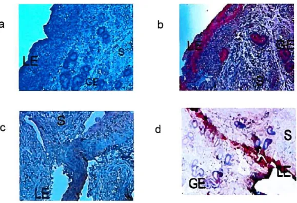
Immunolocalization of macrophage migration inhibitory factor (MIF
)
was compared in endometrial tissues from cyclic and pregnant cows compared to non pregnant cows. The resuits showed increased staining in luminal and glandular epithelium ofpregnant cows. Treatment of bovine endometrial epithelial cells (BEEC) in vitro with 100 ng’ml IFN-t for 24 h. showed stronger cytoplasmic accumulation ofMfF than control cells. Therefore, the observed in vivo results are probably due to stimulation of MW by LFN-r. Medium from BEEC in vitro treated with WN-T for 24 h was analyzed by two-dimensional PAGE and Western blotting. The resuit showed the appearance of a spot that was identified as the soluble form ofE-cadherin. In the present work using in vitro BEEC, it has been shown that IFN-T stimulates the proteolytic cleavage ofE-cadherin and subsequentaccumulation in cytoplasm, and of 13-catenin at the plasma membrane. An autocrine effect ofMIF was also observed on E-cadherin and f3-catenin in bovine endometrial epithelial celi in vitro. These data suggest that changes in MW and E-cadherin induced by IFN-t secreted by the embryo plays an important role in attachinent ofthe trophoblast to the endometrial wall.
TABLE 0F CONTENTS TITLE IDENTIFICATION 0F JURY ii RÉSUMÉ ifi SUMMARY iv TABLE 0F CONTENTS y
LIST 0F FIGURES vii
LIST 0F ARBREVIATIONS ix AKNOWLEDGEMENTS xiii DEDICATION xiv 1. INTRODUCTION 1 2. LITERATURE REVIEW 3 Conceptus deveiopment 3
Maternai preparation for implantation 7
Corpus Luteum formation $
Luteolysis 9
Embryo endometriai interactions 10
Recognition ofpregnancy 14
Adhesion mechanisms 15
Adhesion molecules and implantation 16
WNinduced protein secretionfrom endometrium 19
Endometrial cytokines 20
vi
MW in proliferation and differentiation 23
MW in adhesion 24
E-cadherin in epithelial ceils 25
Cadherin recruitment 32
Epithelial to mesenchymal transitions 33
Tntemalization and recycling of cadherin 34
Regulation of cadherins 35
Soluble E-cadherin 37
3. HYPOTHESIS AND OBJECTIVES 39
4. MATERIALS AND METHODS 39
Chemicals and Reagents 39
Preparation and culture of cells 40
Radioactive labeling of secreted proteins and 2D-PAGE 41
Protein sequencing 42 Western blotting 43 Immunohistochernistry 43 hnmunocytochemistry 44 Statistical analysis 45 5. RESULTS 46 6. DISCUSSION 68
MIF on secretion ofE-cadherin 72
7. CONCLUSION 76
LIST 0F FIGURES
Fïgure 1. Early pregnancy events in sheep 6
Figure 2. Apposition and adhesion phases ofblastocyst implantation in sheep 12
Figure 3. Cadherin domain layout 27
Figure 4. The dual function of 3-catenin in ceil adhesion and transcription. 30
Figure 5. Immunolocalization of MIE in bovine endometrium 47
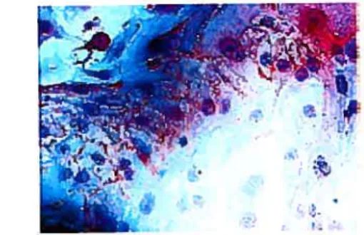
Figure 6. Immunolocalization ofMIF in bovine uterine endometrial celis 50
Figure 7. Identification ofproteins secreted from endometrial epithelial ceils
-and stimulated by IFN-T. 52
Figure 8. Irnmunolocalization ofE-cadherin and 13-catenin in bovine
endometrium. 55
Figure 9. Effect of WN-r on soluble E-Cadherin secretion 57
Figure 10. Effect of WN-t on E-cadherin in bovine uterine epithelial celis. 59
Figure 11. Expression of 13-catenin in bovine endometrial epithelial celis
for effect ofIFN-’t. 61
Figure 12. Response of MIE on E-cadherin in bovine endometrial
epithelial cell. 63
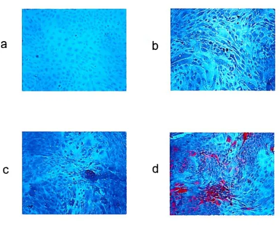
Figure 14. Effect ofMIF on 3-Catenin expression in bovine endometrial
epithelial cells. 67
Figure 15. Proposed scherne ofMIf-dependent signaling to MAPK and
cyc lin Dl transcription. 74
LIST 0F ABREVIATIONS
BEEC: bovine endometrial epithelial celis. bHLU: basic helix-loop helix.
CL: corpus luteum. COX: cyclooxygenase
CSFs: colony-stimulated factors. E-17v: estradiol -1713.
EC: endothelial celis.
EC1 -EC5: extracellular cadherin domains. ECM: extracellular matrix.
EGFR: epidermal growth factor receptor. ER: estrogen receptors.
ERK: extracellular signal-regulated kinase. ERK1I2: extracellular-signal regulated kinase 4. ET-1: endothelin- 1.
Fzd: Frizzled proteins
GAS: interferon-g-activated sequence. GCP-2: granulocyte chemotactic protein-2. GE: glandular epithelium.
GM-CSF: Granulocyte-macrophage colony-stimulating factor. hCG: Human corionic gonadotrophin.
IIGF/SF: hepatocyte growth factor/scatter factor.
IIGFRTKS: hepatocyte growth factor-regulated tyrosine kinase substrate. UAV: His-Ala-Val
X
1CM: innerceli mass. IFN-r: interferon-tau. IFN-R: interferon receptor. IFNs: interferons.
IL-8: interleukin-8. ILs: interleukins.
IP-1O: interferon-gamma-inducible protein 10 kDa IRF-1: interferon regulatory factor 1.
IRF-2: interferon regulatory factor 2. ISG17: interferon stimulating gene 17. ISGF: interferon-stimuÏated gene factor. ISGF3: stimulated gene factor three
ISRE: interferon-stimulated regulatory element. JAKs: tyrosine kinases, Janus kinases.
LE: luminal epithelium. Lgs: Legless/BCL9 LH: luteinizing hormone.
LUr: luteinizing hormone receptors. LLC: large luteal celis.
LPS: lipopolysacarides lipoproteins. L-selectin: celi-surface selectin.
LEF/TCF: lymphoid enhancer factor/T-ceÏl factor MAP: mitogen-activated protein.
MAPKs: mitogen-activated protein kinase MIIC: major histocornpatibility complex. MIF: Macrophage migration inhibitory factor. MLC: myosin light chain
MLCK: myosin light chain kinase MMPs: Matrix metalloproteinases.
MT1-MMP: Membrane type-1 matrix metalloproteinase. MUC-1: Mucin glycoprotein 1.
NBCS: newbom calfserum. Trp2: N-terminal b-strands oIFN-t: ovine interferon-tau. OPN: osteopontin.
OT: oxitocin.
OTR: oxitocine receptors. P: progesterone.
PBS: phosphate-buffered saline PGF2Q prostaglandin F2a.
PGE2 : prostaglandin E2.
PGFr: prostaglandin F receptors. PKC: protein kinase C
PLs: placental lactogens. PR: progesterone receptors. PRPs: prolactin-related proteins.
XII
Pygo: Pygopus
rMIf: recombinant MIE. RTK: receptor tyrosine kinase.
SIBLING: small integrin-binding ligand, N-linked glycoprotein. SLC: srnall luteal ceils.
STATs: signal transducers and activators of transcription. TBS: tris-buffered saline
TCF: transcription factors
TCF/LEF: T celi factor/lymphoid enhancer factor TGFs: transforming growth factors.
TNF-a: tumor necrosis factor c. TNFs: tumor necrosis factor. Tr: Trophoblast.
UCRP: Ubiquitin cross-reactive protein. VEGF: vascular endothelial growth factor.
AKNOWLEDGEMENTS.
Thanks to my “Aima Mater” University of Zacatecas, for this
gift.
Acknowledge to the University of Montreal, for permitting me
to do this work.
Sincerely thanks to my professor Dr Alan K Goff for his kind
help.
DEDICATION
for: Diana, Anabel
y
Luis Dario
“with
ail my affection
“.inhibitory factor from bovine endometrial epithelial celis.
INTRODUCTION
Early embryonic mortality causes a ioss of 600 million dollars per year in reduced weaning wcights and milk production (Austin, 2001) The embryo losses occur during the first 4 to 6 weeks of pregnancy in high producing dairy cattie, and during periods of nutritional or environmental stress, these losses can approach 80% (Ott, 2003a). The harmonic interactions between the hypothalamus, pituitary, ovary, uterus and conceptus are essentiai for normal embryo development and implantation. Disruptions in these interactions resuit in failure of the embryo to attach and embryo mortality. The basic events that occur in the utents in response to pregnancy must be studied and understood so that future technologies may be designed to detect eariy pregnancy and decrease eariy abortion.
The successful implantation of the ernbryo in the cow requires a succession of coordinated events; these inciude progesterone-induced development of the utenis, and conceptus and placental formation. The communication between the conceptus and the maternai endometrium in the preimplantation period happens via the secretion of growth factors, hormones and cytokines and by several molecules including adhesion signaling, transcription, ceil cycle and DNA replication proteins, that creates an environment propitious for attachment (Carson et al., 2000) When the conceptus and uterus are developing simultaneously, the attachment of trophectoderm to endometrium can occur. This event happens in ruminants, rodents and primates(Imakawa et al., 2004).
Steroid hormones secreted during the estrous cycle, estradioi -17r3 (E-173) and progesterone (P), prepare the utenis to receive the embryo. Thc appropriate secretions of
2 the endometrium in the preattachment period permit the elongation and development ofthe conceptus in ruminants (Bazer and Roberts, 1983).
In the mammalian uterus the establishment ofpregnancy is dependent on the corpus luteum (CL), which secretes P, the essentiai hormone of pregnancy. If the female does flot become pregnant, the CL regresses due to a pulsatile secretion ofprostaglandin f2a (PG F2a) from the endometrium, a process known as luteolysis and another new estrous cycle commences (Bazer and First, 1983). If the female becomes pregnant, the schronous deveiopment of the endometrium and conceptus resuits in the trophectoderm secreting interferon tau (IFN
T), which is the signal for the maternai recognition of pregnancy that prevents luteolysis,
and allows the attachrnent and subsequent implantation (Bazer et al., 1997).
Ruminant biastocysts develop for up to three weeks in the uterine lumen, before implantation, during this period maternal-conceptus communication is established (Yamada et ai., 2002). The trophobiast (Tr) secretes specific molecules such as placentai lactogens, prolactin-related proteins, IFN-r, and adhesion molecules such as integrins (Gumbiner, 1996; Hynes, 1987), glycoproteins and cadherins that serve as receptors for extracellular matrix ligand and act as modulator of cellular function (Lessey, 1994; Lessey et al., 1994a; Lessey et al., 1994b). After blastocyst elongation during the preimplantation period, LFN-t acts on the bovine endometrium and increases secretion of several proteins that support conceptus-endometrium development necessary for adhesion.
Endometrial proteins such as interferon-gamma-inducible protein 10 kDa (LP-10) regulate the establishment of apical interactions between trophobiast and epithelial ceils during early gestation (Nagaoka et al., 2003). Galectin-15 has an extracellular function to regulate Tr migration and adhesion to the endometriai epithelium, and intracelluiar function to regulate Tr ccli survival, growth and differentiation (Gray et al., 2004). Granulocyte
macrophage colony-stirnulating factor (GM-CSF) (Ernond et al., 2004) beta 2 microglobulin, interferon stimulate gene 17 (ISG17) (Jolrnson et al., 2002) osteopontin (OPN) I and OPN II may induce adhesion between luminal epithelium and tropliectoderm to facilitate superficial implantation (Jolmson et al., 1999a; Jolmson et al., 1999b). In the early pregnancy, WN-t from bovine conceptus, stimulate secretion of uterine endometrial cytokines; ubiquitin cross-reactive protein (UCRP), bovine granulocyte chemotactic protein-2 (GCP-2) (Staggs and Dooley, 1998; Teixeira et al., 1997) and macrophage migration inhibitory factor (MW) (Wang and Goff, 2003). Also WN-’c may affect the cleavage of the external domain of E-cadherin, that is transmembrane protein of the adherents junctions and is responsible for homophilic celi to cell adhesion (Wheelock and Johnson, 2003a; Wheelock and Johuson, 2003b).
The role of proteins secreted by the uterus in trophoblast-epithelium adhesion has flot been determined in domestic ruminants and remains a point of intense investigation. The effect of IFN-’c on cdl adhesion in bovine endornetrial epithelia! celis is flot understood. My goal was to determine if IFN-t lias an effect on the secretion of proteins such as MW and E cadherin and how these proteins are involved in the modification of epithelial cells for ce!! adhesion.
LITERATURE REVIEW Conceptus developmeut
In mammals aller mating and ferti!ization the zygote develops into a blastocyst as it migrates through the oviduct into the uterus. Close to the uterus, the so!id bail of ceils, the moru!a, becomes fluid-filled and a cavity blastocoele appears, which enlarges rapid!y and transforrns the moru!a to a blastocyst. The blastocyst lias peripheral layer of large ftattened celis, the trophectoderm or trophob!ast, and a knob of smaller celis to one side of the
4 central cavity, the so called inner ceil mass (1CM) (Russell et al., 2006). The 1CM will give rise mainly to the aduit organism, while the ceils of trophectoderm form the placenta and embryonic membranes (Koo et al., 2002; Maddox-Hyttel et al., 2003). After differentiation, the blastocyst hatches from the zone pellucida and acquires the ability to attach to the uterus (Brandao et al., 2004; Dalton et al., 1995; Hynes, 1987). This preimplantation stage varies in duration between species. In mice, implantation occur 4 days post coitum (McLaren, 1985), humans average 9 days (Lee and DeMayo, 2004), and in cow implantation does not occur until 30 days after fertilization (Xiang and MacLaren, 2002).
Tr celis express a number of extracellular matrix receptors and matrix-degrading activities that support interaction and invasion through the endometrium (Carson et al., 2000; Carson et al., 2002). During the development of the embryo, genes encoding for putative transcription factors, these transcription factors are expressed in 1CM or trophoblast lineages, such as Rex-I (Rogers et al., 1991), GAlA-3 (Ng et al., 1994), T-Box gene Eomesodermic (Hancock et al., 1999), the caudel related gene Cdx-2 (Beck et al., 1995), activating protein-2 gamma (Shi and Kellems, 1998), basic helix-loop helix (bHLH) (Sapin et al., 2000), Mash 2 (Rossant et al., 1998), Ets-2 that orchestrates modifications ofcellular adhesion (de Launoit et al., 1998; Meyer et al., 1997), and a transcription factor protocadherins encodes a transmembrane ceil adhesion molecule (Imakawa et al., 2004; Yamamoto et aI., 1998). Comparison of day 7 and day 14 embryos revealed that blastocyst expresion of most genes increased during this period, and a small number of genes exhibited decreased expression. Clustering analysis dernonstrated that trophoblast celis secrete specific molecules such as piacental lactogens (PLs), prolactin-related proteins (PRPs), IFN-T, and adhesion molecules that apparently ail play pivotai roles in the
preparation needed for implantation, since their expression was remarkably enhanced during the pre-implantation period (Ushizawa et al., 2005a). Expression of ovine nterferon tau (oLFN-T) gene is restricted to the trophoblast and is not detected in any other ceIl types in ruminants. Substantial secretion ofoIFN-t, starts on day 12-13 ofpregnancy, reaches the highest on day 16 —17, and then declines rapidly. Changes in the degree of DNA methylation could be one of the major mechanisms leading to downregtilation of the
oIfN-T gene during early gestation (Nojima et al., 2004; Ushizawa et al., 2006). Expresion of
gene eomesodermic, the caudel related gene Cdx-2, activating protein 2 gamma, bHLH, Mash 2, Hand I and Ets-2 increased up to implantation (Roberts et al., 2003). The change in expression of these genes propose novel molecules in trophoblast differentiation (Ushizawa et al., 2006; Ushizawa et al., 2005b).
6
ntryuit SliodZP EIonaalIol Appol1Ioi
Ut4IrU
I
j.
j.
L
f\
tn
p ___________ O Fimhrjuncti3n Ulorino boni
Oviduç(
— la
Q
5.
8
Days atter mating
Figure 1. Early pregnancy events in sheep. This schematic summarizes the relative changes in embryo/blastocyst development after fertilization in relation to position in the female reproductïve tract and circulating levels of ovarian steroid hormones. Fertilization occurs in the oviduct, and the morula stage embryo enters the uterus on day 4. The blastocyst is formed by day 6 and hatches from the zona pellucida on days 8—9. The blastocyst develops from a spherical to a tubular form by day 11 and then elongates to a filamentous conceptus between days 12 and 16. The elongation of the blastocyst marks the beginning of implantation, which involves apposition and
transient attachment (days 12—15) and firm adhesion by day 16. Taken from Spencer
Maternai preparatïou for implantation
The preparation of maternai tissue for implantation of conceptus implicates that the uterus undergoes dynamic changes, including differential and ordered activation or repression of gene expression and programmed changes in posttranscriptional and posttranslational modifications of mRNA and proteins (Carson et al., 2002). The steroid hormones secreted during estrous cycle E-1 7(3 and P, prepare the endometrium for the secretion of proteins and prostaglandins. The proteins nourish the embryo and the prostaglandins act on the corpus luteum and induce luteolysis if the animal does not have a viable ernbryo. The conceptus itself secretes growth factors, steroids, prostaglandins and cytokines depending on the species, which presumably act on the endometrium to prevent prostaglandin secretion, or directly on the ovary to stimulate protein secretion (Goff, 2002). ProstagÏandins are aiso involved in implantation. IFN-r elevated cycloxygenase (COX)-2 expression and selectively increased prostaglandin E2 (PGE2) secretion in epithelial celis at the tirne of pregnancy recognition, and may have a luteotropic effect (Asselin et al., 1997; Guzeloglu et al., 2004; Xiao et al., 1999).
In the estrous cycle, the coordinated secretion of hormones by the hypothalamus, ovary and utems prepares the endometrium for the implantation of embryo. Gonadotropin-releasing hormone produced by hypothalamus regutates the synthesis and release of luteinizing hormone (LH) and follicle-stimulating hormone (FSH) from the anterior pituitary gland. FSH and LH are synthesized and secreted by gonadotroph ceils of the anterior pituitary gland (Wilson et al., 2004). These hormones regulate gametogenesis, steroidogenesis and ovulation in mammalian ovaries.
8 The preovulatory LH surge initiates the differentiation of follicular ceils into luteal ceils (luteinization). The CL is a heterogeneous tissue containing endothelial celis, steroidogenic ceils as large luteal ceils (LLC) and small luteal celis (SLC), fibroblasts, smooth muscle celis and immune ceils (OTShea et al., 1989). The endothelial ceils contribute approximately 50% of the total cell population of the CL (Meidan and Girsh, 1997). Studies by Mamluk et aI., 1998 (Mamluk et al., 1998) showed that PGF2a is a major regulator of prostaglandin F receptor (PGfr) and luteinizing hormone receptor (LHr) expression in the two steroidogenic celi types, and ah three major cell types of the CL (steroidogenic and endothelial) express PGFr and LHr mRNA (Mamluk et al., 1998). PGf2a induces an elevation in luteal expression of endothelin-l (ET-1) (Girsh and Dekel, 2002) from endothelial celis, which may mediate the luteolytic action of PGF2a (Girsh et al., 1996; Levy et al., 2001).
The CL lasts for 17-18 days in the cyclic cow or for up to 200 days in the pregnant cow. Regression of the corpus luteum is essential for normal cyclicity as it allows the development of a new ovulatory follicle (Meidan and Girsh, 1997; Meidan et al., 1999). The CL produces P, required for the establishment and maintenance ofpregnancy (Schams and Berisha, 2004). P also seems to play a luteotropic role by stirnulating the synthesis of LHr in bovine CL (Jones et al., 1992). There is also evidence that P represses the onset of apoptosis in the CL by a progesterone receptor (PR) dependent mechanisrn (Rueda et al., 2000). During early diestrus, P from the newÏy formed CL stimulates accumulation of phospholipids in endometrial luminal epithehium (LE) and glandular epithehium (GE) that
cari liberate arachidonic acid for synthesis and secretion of PGF2Œ. During diestrus,
progesterone levels increase and act via PR to block expression of estrogen receptors tER) and oxytocin receptors (OTR) in the cndornetrial LE and GE (Spencer et al., 2004b).
Continuous exposure of the endometrium to P eventually down-regulates PR gene expression in the endometrial LE. The loss of PR terminates the P block to ERa and OTR formation. The increase in OTR expression is facilitated by increasing secretion of estrogen by ovarian follicles (Spencer and Bazer, 2002).
Luteolysis.
In ruminants and other large domestic animais PGF2u is the luteolysin secreted by the uterus that controls the length of the estrous cycle. Episodic release of PGF2a from the uterus reaches the CL through a counter current system between the uterine vein and the ovarian artery and induces luteolysis (Schams and Berisha, 2004). Luteolysis is initiated by increased expression ofERs and subsequently OTRs by the uterine endornetrial epithelium. Oxytocin (OT) stimulates PGF2a secretion by cow endometrial celis (Ohtani et al., 2004; Tysseling et al., 1998; Tysseling et al., 1996) through activating the OTR (a 7-transmembrane, G-protein-associated receptor), increasing inositol triphosphate turnover, cytosolic calcium concentration, and activation ofprotein kinase C (Duras et al., 2005). In the uterus, these events resuit in activation of COX-2 (Asselin and Fortier, 1996). In cattie and sheep, luteolysis appears to be initiated by an increase in endometrial sensitivity to OT due to the increase of the number of OTR (McCracken et al., 1996; McCracken et al., 1999), and the existence of positive feedback loop between endometrial PGF2u and luteal OT secretion (Parkinson et al., 1992).
In contrast to PGF2Œ, PGE2 may be a luteotropic agent and could be a luteo-protective signal to antagonize potential luteolytic effects of PGF2Œ. Before implantation, PGE2 may aiso be responsible for the increase in vascular permeability and secretion of growth factors and nutrients, and it may be involved in the local regulation of immune responses (Emond et al., 1998).
10 Emb ryo endometrial interactions
The synchronous development of blastocyst and endometrial luminal epithelium receptivity initiates an adhesion cascade that resuits in implantation. In ruminants the blastocyst sheds the zona pellucida (day 8) and elongates to a filamentous form. The elongation of the blastocyst marks the beginning of implantation, which involves apposition and transient attachment (days 12—15) and firm adhesion by day 16 in sheep (Spencer et al., 2004a). Apposition of the conceptus involves the trophectoderm becoming closely associated with the endometrial LE followed by unstable adhesion. Afler day 14, the filamentous conceptus appears to be immobilized in the uterine lumen. The elongating blastocyst maintains close contact with the endometrial LE, which appears to imprint its rounded shape on the trophectoderm in fixed specimens (Gharib-Harnrouche et al., 1993). On day 15, apposition occurred: most microvilli on the surface of the trophoblast disappeared. Between days 16 and 18, adhesion began as a result of the interpenetration of the uterine microvilli and cytoplasmic projections ofthe trophoblast celis (Guillomot et al.,
1981). Adhesion of the trophectoderm to the endometrial LE progresses along the uterine hom and appears to be completed around day 22 and 28, the establishment of an overali intimate epithelial contact of fetal binucleate celi with microvillar junction take place and continued binucleate ceil migration at the time of mature bovine placenta formation (King et al., 1982; Wathes and Wooding, 1980).
The endometrium in ruminants consists of LE, GE, several types of stroma (stratum compactum and stratum spongiosum), blood vessels and immune cells. In sheep, the endometrium has two distinct areas: aglandular caruncular and glandular intercaruncular. The caruncular consist of LE and compact stroma and are the sites of superficial implantation and placentation (Amoroso and Perry, 1975; Lawn et al., 1969). Placentation
in ruminants such as cows and goats are noninvasive, or the extent of invasion is very lirnited (Hashizurne et al., 2003; Hirata et al., 2003; Steven, 1975). Tr remain essentially in the uterine lumen and placentation involves onÏy superficial physical contact with the maternai tissue (Carter and Enders, 2004). In contrast placentation in humans and mice is highly invasive as the Tr penetrates into endometrial stromal tissue (Bischof and Campana, 2000; Bischof et al., 2000a; Bischof et al., 2000b).
(A) (B) Apposition Uenr.# J r __ T-r ê
ilrii
j”’ 1 t ff_a’.. ‘%% eÏ
MUC4 12 î r M.onFigure 2 Apposition and adhesion phases of blastocyst implantation in sheep. (A)
Preattachment involving shedding of the zona pellucida (phase 1) and precontact and
blastocyst orientation (phase 2). The antiadhesive mucin MUC-1 is present ou the endometrial LE, thereby preventing contact of the Tr with adhesive receptors such as integrins. Histotroph, sccreted from the endometrial LE and GE, nourishes the developing blastocyst. (B) Apposition and transient attachment (phase 3). After day 11, the tubular blastocyst elongates to form a filamentous conceptus. During this period, expression of MUC-1 declines on the LE, which exposes constitutively expressed integrins on the LE as well as trophoblast. Apposition occurs between the
trophoblast ami endometrial LE and between the Tr papillae ami GE ducts.
Elongation of the blastocyst probably requires apposition and transient attachment to the endometrial LE. (C) Adhesion (phase 4). Firm adhesion of the mononuclear cetis of the Tr to the LE occurs between days 15 and 16. Available evidence indicates that several molecules (GIyCA1’I-1, galectin-15 and osteopontin) interact witli receptors (integrins ami glycoconjugates) on the apical surfaces of the Tr and LE to facilitate adhesïon. Taken from (Spencer and Bazer, 2004).
14 Recognition of pregnancy
During the preimplantation period, pregnancy recognition signais from the conceptus to the maternai system are antiluteolytic and/or luteotrophic. The functional iife span of the CL is controiied by release of PGF2a from the uterus and IFN-T is the signal from the trophoblast acts in a paracrine or endocrine manner to interrupt endometrial production of luteolytic PGF2Π(Kim et al., 2003).
Maternai recognition of pregnancy in ruminants (sheep, cattie, goats) requires that the conceptus elongate from a spherical to a tubular and then filamentous form to produce IFN-’t, which is the signal that prevents development of the endometriai luteolytic mechanism (Kim et al., 2003; Spencer and Bazer, 2002). IFN-T is considered to prevent luteolysis by blocking the upregulation in OTR during diestrus (Spencer et al., 1996), through the activation and repression of genes responsive to Type I IFN (Green et al., 2005).
The IFN-r secreted by conceptus inhibits OTRs and protects the CL and maintains secretion of P (Bazer and First, 1983; Bazer and Roberts, 1983). IFN-r is released by the conceptus in the pregnant cow by day 12-28, reaches highest levels on days 15-19, after which time levels decrease from days 21 to 26 of pregnancy (Roberts, 1991; Roberts et al., 199 la; Roberts et al., 199 lb). In the sheep, the quantities of0IFN-T secreted are elevated (20—200 mg/day) by days 14-16 ofpregnancy and 1-2 mg!day by the day 12 ofpregnancy (Rooke et al., 2005). IFN-T is reduced as definitive attachrnent of trophectoderrn to the uterine epithelium is established (Day 21 or 25 of pregnancy in the sheep and cow, respectively) (Roberts et al., 1992; Spencer and Bazer, 2004).
The only maternai tissue immediately exposed to IFN-t is the epithelium that borders the endometrium (Rosenfeld et al., 2002). IFN-t binds to a dimeric interferon receptor (IfN-R)
in the celi membrane. The intracellular domain of the receptor binds tyrosine kinases, Janus kinases, (JAKs) which are activated afier interferon binding, and subsequently phosphorylate other proteins named signal transducers and activators of transcription (STATs). The STATs dimerize and bind two other proteins to form a trimeric interferon stimulated gene factor (ISGF) complex, which is translocated to the nucleus, where it binds an interferon-stimulated regulatory element (ISRE), resulting in the expression of the interferon regulatory factor 1 (IRF-l) gene. The product of this gene, in tum, activates expression of TRF-2, which interacts with other regulatory elements to control the expression of interferon-responsive genes, including the OTR and ER receptors (Demmers et al., 2001).
TFN-t possess similar antiproliferative and antiviral activities to other Type I IFN (Mathialagan and Roberts, 1994). IFN-T affects the synthesis of cytokines that contribute to the immunomodulation required to prevent rejection of the conceptus and stimulate blastocyst growth (Demmers et al., 2001; Gierek et al., 2006).
Adhesion mechanisms
Ce!! adhesion mechanisms are responsible for assembling cells together and, along with their connections to the interna! cytoskeleton determine the overal! architecture of the tissue (Gumbiner, 1992). Three general classes of proteins take part in cel! adhesion: the ce!l adhesion molecules/adhesion receptors, the celi-extracellular matrix (ECM) proteins, and the cytoplasmic plaque/peripheral membrane proteins (Gumbiner, 1996). The cell adhesion receptors are glycoproteins that mediate binding interactions at the extracellular surface and determine the specificity of celi-cel! and cell-ECM recognition. They comprise integrins, cadherins, immunoglobulins, selectins and proteoglycans (Bella and Berman, 2000). The ceil adhesion receptors recognize and interact with either other ce!! adhesion
16 receptors on neighboring celis or with proteins of the ECM. The ECM is typicaliy composed of large glycoproteins inciuding collagen, fibronectins, laminins and proteoglycans. Cytoplasmic plaque proteins serve to link the adhesion systems to the cytoskeleton, to regulate the functions of the adhesion molecuies, and to initiate transducer signais at the ceil surface by the adhesion receptors (Gumbiner, 2005). Signais generated locally by the adhesion receptors themseives are involved in the regulation of ceil adhesion. These reguiatory pathways are aiso influenced by extrinsic signais arising from the classic growth factor receptors (Gumbiner, 1996).Adhesion mechanisms are highly reguiated during tissue morphogenesis and are intimateiy reiated to the processes of celi motiiity and celi migration. Iii particular, the cadherins and the integrins have been impiicated in the control of ceil movement. Cadherin-mediated celi compaction; integrins mediated ceil spreading and motility on the extraceilular matrix (Wheelock and Johnson, 2003a; Wheelock and Johnson, 2003b).
Adliesion moecuJes and implantation
Endometrial epithelium synthesize and secrete or transport a complex array ofproteins and related substances termed “histotroph”, that is a mixture of enzymes, growth factors, cytokines, lymphokines, hormones and other substances that act as primary regulators of conceptus survival, deveiopment, production of pregnancy recognition signais, implantation and placentation (Bazer et al., 1979; Burton et al., 2002) These uterine secretions establishing synchrony between development of the conceptus and uterine receptivity that remodeling the endometrial LE for conceptus adhesion (Burghardt et al., 2002). The LE that is a simple, polarized ceil layer mediates cell-celi and ceii-extraceliuiar matrix interactions is normally nonadhesive; however, this characters is lost during
development of receptivity and begin apical adhesion between LE and Tr defines the onset of implantation (Lue et al., 2006).
In the early pregnancy, continuous exposure of the endometrium to progesterone down regulates the progesterone receptors in the epithelia, a process that is associated with loss of the ceil-surface mucin glycoprotein 1 (MUC-1) and induction of several secreted adhesion proteins (Carson et aÏ., 1998). The removal of mucin, that is an antiadhesive barrier is hypothesized to be necessary to expose other glycoproteins involved in the adhesion between Tr and LE (Aplin and Hey, 1995; Hey et al., 1995).Mucin is locally reduced at implantation sites, via the activity of celi-surface proteases that are triggered by the blastocyst or rnediated by paracrine signais from biastocysts (Brayman et al., 2004; Thathiah et al., 2004). In sheep the implantation adhesion cascade is initiated after down regulation ofMUC-1 (Johnson et aI., 2001).
A number of endometriaÏ proteins have been identified as potential regulators of blastocyst development and implantation in sheep, including glycosylated celi adhesion molecule 1, galectin-15, osteopontin, that binding to adhesion receptors such as integrin, cadherin and immunoglobulin and selectin, to proteins of ECM. These adhesion proteins are secreted for luminal epithelium and regulate for progesterone or TFN-T produced by the Tr during blastocyst elongation (Spencer et al., 2004a).
Galectins are proteins with a conserved carbohydrate recognition domain that bind b galactosides, thereby cross-linking glycoproteins as well as glycolipid receptors on the surface of ceils and initiating biologic responses (Cooper, 2002). Functional studies of other gaiectins have implicated these proteins in ceil growth, differentiation ami apoptosis
1$ Integrins comprise a family of heterodimeric intrinsic transmembrane glycoprotein receptors that mediate cellular differentiation, motility and adhesion (Giancotti and RuosÏahti, 1999; Munger et al., 199$; Oktay et al., 1999). The central role of integrins in the implantation adhesion cascade is to bind ECM Iigand(s) to cause cytoskeletal reorganization, stabilize adhesion, and mediate ceil migration, proliferation and differentiation through numerous signaling intemiediates(Burghardt et ai., 2002; Pfarrer, 2006). Altered expression of integrins is correlated with several causes of infertility (Lessey, 1994; Lessey et al., 1994a), nul! mutations of several integrins leads to peri implantation lethality (Hynes, 1996) and functional blockade of selected integrins reduces the number of implantation sites. In the sheep, receptivity to implantation does flot appear
to involve changes in either temporal or spatial pattems of integrin expression, but may
depend on expression of other glycoproteins and ECM proteins, such as galectin-15, OPN and fibronectin, which are ligands for heterodimers ofthese integrins.
OPN is a member of the small integrin-binding ligand, N-linked glycoprotein ($IBLTNG) family of genetically related ECM proteins recognized as key players in a number of diverse processes such as bone mineralization, cancer metastasis, cell-mediated immune responses, inflammation, angiogenesis and cell survival (Johnson et al., 2003a; Sodek et al., 2000).OPN has also been linked to pregnancy (Johnson et al., 2003b). Microarray profihing identified OPN as the most highly upreguïated ECM adhesion molecule in hurnan endometrium that is receptive to implantation (Carson et al., 2002). Multiple integrin receptors for OPN are present on trophoblasts and LE of humans and domestic animais, some ofwhich increase during the peri-implantation period (Damario et al., 2001; Lessey,
IFN induced protein secretion from endometrium
When the bovine conceptus makes contact with the uterine wall, IFN-r is secreted and has an immunomodulatory activity that offers protection for the embryo from the immune system of the mother. IfN-t is an apoptotic protein that induces the secretion of protein
and cytokines and prepares the endometrium for embryo attachment. IFN-’t possesses potent antiviral, antiproliferative, and immunomodulatory activities (Demmers et al., 2001). It can also alter the synthesis of endometrial proteins; retard the growth of the endometrium during the preimplantation period (Roberts et al., 1992).
IFN-r was shown to increase expression of beta 2 microglobulin, ISG1 7 (also known as Ubiquitin Cross Reactive Protein), which is expressed in the endometrium of Day 17 pregnant cows, prepares the uterine wall for the adhesion and implantation of the embryo (Austin, 2001; Hicks et al., 2003; Ott, 2003b; Ott et al., 1998). ISG17 controls cytosolic protein processing through the proteosome; osteopontin (Johnson, 1999), which promotes celi—celi attachment and may be involved in attachment ofthe blastocyst to the endometrial epithelial surface; and the antiviral Mx protein (Ott et al., 199$).
IFN-r stimulate the expression of endornetrial IP-10 that regulates the establishment of apical interactions between trophoblast and epithelial celis during early gestation (Nagaoka et al., 2003). Also Galectin-15 was discovered in the uterus ofsheep, and secretion in to the uterine lumen increased between days 14 and 16 of pregnancy and galectin- 15 mRNA was detected only in the endometrial LE and superficial ductal GE (Kuwabara et al., 2003; Purkrabkova et al., 2003). Galectin-15 is hypothesized to function extracellularly to regulate trophoblast migration and adhesion to the endometrial epithelium, and intracellularly to regulate trophoblast ce!! survival, growth, and differentiation (Gray et al., 2004).
20 Expression and secretion of OPN I and OPN II are induced by IFN-T in uterine glands during the periimplantation period. Also OPN is a potential mediator of implantation in sheep, as a bridge between integrin heterodimers expressed by Tr and uterine LE responsible for adhesion for initial conceptus attachment (Johnson et al., 2001; Johnson et al., 1999a; Johnson et al., 1999b) Also IfN-’r induces adhesion molecules UCRP and GCP 2 during early pregnancy (Staggs and Dooley, 199$).
In the periattachment period IFN-T increases the expression of GM-CSF in immune and nonimmune ceils of the bovine endometrium (Emond et al., 2004). Furthemore, IFN-T stimulates the production of PGE2 in bovine endometrial cells via the induction of COX-2, PGE2 increases the expression of GM-CSF, a cytokine that promotes conceptus growth and survival, from leukocytes and endometrial stromal cells (Emond et al., 2000; Emond et al., 2004).
Pleiotropic cytokine MIF was stimulate for IFN-t from uterine epithelial celis (Arcuri et al., 2001; Wang and Goff 2003). The pleiotropic activities of MIF are based upon transcriptional regulation of inflammatory gene products, modulation of cdl proliferation, differentiation, and cell cycle control, inhibition of apoptosis, and several metabolic effects (Calandra et al., 2000; Lue et al., 2006; Walter et al., 2000; Wang and Goff, 2003; Wang et al., 2003) MIF is released by bovine endometrial epithelial, but flot stromal, celis and is stimulated in response at IFN-T (Wang and Goff, 2003). MIF has an autocrine effect in adhesion; it stimulates the formation of integrin clusters and accumulation of cytoplasmic -catenin.
Endometrial cytokines
The cytokines are implicated in processes characteristic of inflammation, immunity, remodeling and the migration of various cellular components. In mammalian reproduction
processes like ovulation, blastocyst implantation and parturition resemble those of the inflarnmatory and reparative processes in which cytokines and chernokines act as autocrine and paracrine mediators. Actually, many reports have widely documented the involvement of cytokines in the intercellular signaling that affect reproductive events (Orsi et al., 2006). A recent study demonstrates the transcription of 16 different cytokines common in normal and turnor bearing ovaries (Burke et al., 1996).
The endometrium is source of cytokines; interleukins (ILs), tumor necrosis factors (TNFs), transforming growth factors (TGfs), colony-stimulating factors (CSFs), and interferons (IFNs) have been reported in cycling and pregnant endometrium (Arcuri et al., 1999; Arcuri et al., 2001). The cytokine interleukin-1 beta has a major effect on gene expression in stromal cells from human endometrium and plays a role in disorders of the endometrium, especially in implantation-related infertility and endometriosis (Rossi et al., 2005).
The recently recognized multifunctional cytokine MIF that modulates the immune response and acts as a growth and angiogenic factor (Baugh and Bucala, 2002), has possible functions in reproduction. MIF mRNA and protein have been identified in murine and human ovaries as well as in human follicular fluid. Iii rodents, MIF has been detected in the amniotic fluid and in the early embryo (Nishihira et al., 199$).
li human pregnancy, a sarcolectin-binding protein whose properties corresponded to those
of MIF lias been described in term placenta (Zeng et al., 1993) also MW is expressed by first-trimester human trophoblasts (Arcuri et al., 1999). Similarly, in human endometrium hCG induces MW synthesis and secretion by endometrial stromal celis, and results of nuclear transcription assays (run-on) revealed that hCG acts predominantly by up regulating MW gene transcription (Akoum et al., 2005; Kats et al., 2005). Human
22 endometrial epithelial ceils can also secrete MW (Chaisavaneeyakom et al., 2005). Thus, as demonstrated by immunohistochemistry, in the secretory-phase glandular epithelium, the protein is mainly located on the lurninal side, usually at the apical surface of the ceils (Paulesu et al., 2005). Moreover, abundant immunoreactive material is present in the glandular secretion (Arcuri et al., 1999; Paulesu et al., 2005), and it has been suggested that endometrial epithelial celis secrete MIF during the luteal phase of the menstrual cycle (Arcun et al., 2001).
MIF and the immune response
MIF bas immunosuppressive activity and it has bee shown that MIF is capable of inhibiting Natural Killer cells in cell-mediated cytolysis of both neoplastic and normal celis ofhuman endometrium (Apte et al., 199$). Also MIF represents an important effector of hCG-induced endometrial changes during embryo implantation, growth, and development (Akoum et al., 2005).
MW is present in human serum at concentrations ranging from 2-6 ng/ml. Macrophages contain large quantities of stored, pre-forrned MW (2-4 fg!cell) that is released in response in lipoproteins (LPS) stimulation. Physiological concentrations of glucocorticoids stimulate macrophage secretion of MW (Isidori et al., 2002; Nishihira et al., 1995). On release from macrophages, MIF can exert potent autocrine and paracrine effects, prornoting cdl activation, proinflammatory cytokine release and overriding glucocorticoid action at the site of inflammation (Chesney et al., 1999; Isidori et al., 2002; Liao et al., 2003). MIF is present in several tissues including T lymphocytes, anterior pituitary ceils, monocyte /macrophages, eosinophils, endothelium, various epithelial cell types, fibroblasts, and muscle cells (Baugh and Bucala, 2002).
MIF is involved in the regulation of innate and adaptative immunity. It is a counter regulator of glucocorticoid action within the immune system (Baugh and DonneÏly, 2003), and inhibits the random migration of macrophages and promotes tumor ceil growth. The pro-inflamatory effect of MIF may be explained by its ability to induce release of the pro inflammatory cytokine tumor necrosis factor a (TNF-a) by macrophages, and form a positive feedback ioop, as TNF-a is itself able to induce MIF secretion via tyrosine-kinase
dependent pathway (Mitcheil et al., 2002). MIF in proliferation and differentiation
MIF has the potential to suppress the action of the tumor suppressor gene p53, leading to ccli growth (Fingerle-Rowson et al., 2003). MIF inhibits p53 activity in macrophages via an autocrine regulatory pathway, resulting in a decrease in cellular p53 accumulation and subsequent function. This mechanism to explain its critical proinflammatory action of MW in conditions such as sepsis (Mitchell, 2002). MW induces tumor cell growth in concert with other growth factors. It stimulates the proliferation of fibroblasts and also in wound repair (Takahashi et al., 1998). MW could prornote both tumor celi growth and angiogenesis induced by lysophosphatidic acid via mitogen-activated protein kinase (MAPK) signaling pathways (Sun et aI., 2003). MIF is directiy associated with the growth of Iymphoma, melanoma, and colon cancer (Nishihira et al., 2003). Studies where treatments with either anti-MW immunoglobulin therapy andlor MW antisense oligonucleotide confer antitumor activity (Chesney et al., 1999) and the activity ofMIf is associated with cancer angiogenesis, progression, and metastasis (Leng et al., 2003; Stephan et al., 2006; Wymann et al., 1999).
24 MIF in adhesion
MIF is implicated in adhesion, and M1F secretion is induced by cell adhesion to fibronectin in quiescent mouse fibroblasts (Mitcheil et al., 1999). Therefore adhesion-mediated release of MLF subsequently promotes integrin-dependent activation of mitogen-activated protein (MAP) kinase, cyclin Dl expression, and DNA synthesis. MIF is secreted in a protein kinase C (PKC) dependent fashion as a consequence of ccli adhesion to the ECM and plays a significant role in integrin-mediated signalling to sustained MAP kinase activation cyclin Dl expression, and celi cycle progression (Liao et al., 2003). MIF stirnulates the proliferation of mouse fibroblasts (Mitcheli et al., 2002) throught the activation of the p44/p42 extracellular signal-regulated kinase (ERK) rnitogen-activated protein kinase (MAPK)s. Additional growth factors stimulate the rapid release of preforrned MIF from adherent, quiescent fibroblasts including the sustained activation of MAPK in serum stimulated fibroblasts is dependent upon MTF autocrine action (Ren et ai., 2003). Hence growth factors and adhesion are required for efficient signaling to sustained ERK activation and subsequent celi cycle progression (Roovers et al., 1999). This is through cyclin Dl transcription and the subsequent activation of specific cyclin-dependent kinases (Welsh et al., 2001).
MIF secretion is induced by a variety of stimuli including growth factors and integrin engagement (Liao et al., 2003). Extra-cellular MIF then binds to its putative membrane bound receptor, CD74 (Leng et al., 2003), which can then initiate the activation of Rho GTPase activity via an unknown mechanism. Increased Rho activity is then thought to promote the activation of Rho kinase and myosin light chain (MLC) phosphorylation. Hyperphosphorylated MLC, in turn, induces stress fiber formationlintegrin clustering and
subsequent focal adhesion kinase-dependent sustained MAPK activation, cyclin Dl transcription, and retinoblastoma tumor suppressor inactivation (Swant et al., 2005).
The activation of Rho by integrins and growth factors is essentiai for modulating the sustained activation of ERK and subsequent cyciin Dl transcription (Welsh, 2004). Rho and Rho kinase-dependent stress fiber formation have been tightiy linked to endothelial ceil reorganization and mature blood vessel formation (Hoang et ai., 2004), recruitment and clustering of integrins leading to focal adhesion formation (Roovers and Assoian, 2003). MW production following growth factor or extracellular matrix stimulation would also perform in cell repiication. In the case of neoplastic celis, internai (oncogenic) or external (growth factors, extracellular matrix) signals could serve to increase MW production that, in tum, may facilitate anchorage independence and loss of contact inhibition (Meyer-Siegler et ai., 2005; Meyer-Siegier et ai., 2004; Rumpier et al., 2003). MW may be implicated in downreguiation of E-cadherin because inflammatory cytokines can reduce E-cadherin mRNA levels, down-regulate E-cadherin surface expression, and induce a loss of E-cadherin-mediated adhesion (Jakob and Udey, 1998). IL-1, TNF-Πand LPS induced increased expression of major histocompatibility complex (MHC) class II Ag,
CD4O and CD86 compiex and decreased E-cadherin expression that was temporally related to dissociation ofaggregates (Jacob et al., 1999; Jakob et al., 1997; Jakob and Udey, 1998)
E-Cadherin in Epithelial celis
The epitheiium is composed of a single layer of polarized epithelial celis, the membrane lias one apical and one basolaterai dornain. In mammals, multiproteinjunctional complexes mediate the adhesion between epithelial cells, which are comprised of the apical tight junctions, the subapical adherens junctions and basoiateral desmosomes, linked to cytoskeletal filaments (Wang et al., 2006). The morphogenesis of the epithelium is
26 sustained by cadherins that are a superfarnily of calcium (Ca++) dependent celi-ceil adhesion molecules. E-cadherin that belongs to type 1 transmembrane protein of the adherens junctions, and are responsible for hornophilic ceil to celi adhesion (Wheelock and Johnson, 2003b). E-cadherin is a 120-kDa transmembrane gycoprotein (Takeichi, 1991) composed of five tandem extracellular cadherin domains (EC1—EC5), a single transmembrane domain and a distinct and highly conserved cytoplasmic tau that specifically binds catenins. Extracellular domains EC1 to EC4 are homologous cadherin repeats and include the well-known His-Ala-Val (HAV)-sequence that is conserved within the binding surface of the first domain (Renaud-Young and Gallin, 2002), and EC5 is a less-related membrane-proximal domain (Gooding et al., 2004).
Classical or type I cadherins (E-, N-, P-, R-, H-, EP cadherin) mediate adhesion at the adherens, ceil—ceil or ceil—matrix adhesive junctions that are linked to microfilaments. A predomain (usually less than 80 arnino acids) between the signal sequence and the start of the EC1 domain exists at the N terminus (Takeichi et al., 1990) and must be cleaved prior to adhesive function activation (Ozawa et al., 1990a; Ozawa et al., 1990b; Ozawa and Kernier, 1990). E-cadherins mediate both homotypic one type of cadherin on one celi surface interacting with the same type of cadherin on the surface of the opposing celi, and heterotypic ceil—ceil interactions. Homotypic adhesion involves N-terminal F3-strands (Trp2) (Gooding et al., 2004) and the conserved HAV sequence as essential components of the EC1 adhesion recognition site (Blaschuk et al., 1990a; Blaschuk et al., 1990b). T-cells expressing integrins aEb7/aM290b7 specifically interact with E-cadherin in the lymphocyte adhesion system (Cepek et al., 1994).
tciIu4ar cadhênn domains (.c1odomai) EC1 E2 EC3 EC4 EC5
- - - —
4_If 221 21 441
Figure. 3. Cadherin domain layout. A cadherin molecule (green) consists of five extracellular domains, a transmembrane domain and an intracellular domain, itself divided into a membrane proximal (residues 574—655) and catenin-binding (655—725) domain. In purpie, interacting proteins pl2Octn is bound to the membrane proximal region; b-catenin binds the catenin-binding domain, then binds a-catenin at its N terminus. a-catenin and vinculin form the direct link between the b-catenin—cadherin compiex and the actin cytoskeleton (orange). Residue numbers shown are based on the sequence and structure of the C-cadherin ectodomain. Taken from (Gooding et aI., 2004).
2$ The extracellular domain provides Ca++ dependent adhesion, and interactions between the cadherin cytoplasmic tau and the cytoskeleton significantly increase the strength of cadherin-mediated adhesion (Yap et al., 1997a; Yap et al., 1997b). The cytoplasmic carvoxil
tau
is associated with a group of closeïy related but distinct inner-membrane proteins, termed the catenins (Œ, f3 and p120)(Gumbiner and McCrea, 1993). Cytoplasmicdomain of E-cadherin interaction with a complex of f3-catenin (92 kDa) or plakoglobin (y catenin), which forrn the link to the actin cytoskeleton via OE-catenin (102 kDa) (Gooding et al., 2004; Nieset et al., 1997). The anchorage of catenines (cc-, f3-, y- and p120) to the actin cytoskeleton forrns staNe celI—celi contacts (D’Souza-Schorey, 2005). pl20ctn was originally identified as a substrate for receptor tyrosine kinases (Harrington and Syrigos, 2000) binds directly to the cytoplasmic dornain of cadherin and regulate actin cytoskeleton modulators such as RhoA, Rac and Cdc42 (Goodwin et al., 2003). The maturation of initial weak adhesive events between adjacent celis is consolidated by clustering of p120 and RhoA (Noren et aI., 2000). The pl20ctn—E-cadherin interaction may itselfbe sufficient to stabilize the E-cadherin complex (Wang et al., 2005) and Cdc42 (actin cytoskeleton modulators). The adhesion of E-cadherin-f3-catenin at the cytoplasmatic membrane provides stability at adherens junctions. The disassembly and loss of E-cadherin pemiits phosphorylation of f3-catenin that resuits in a decrease in the association of f3-catenin with
E-cadlierin and a-catenin, thereby weakening the adherens junctions (Huber et al., 2001). f3-catenin has also been found to serve as a key component in signalling processes during embryonic development and adult tissue horneostasis (Eger et al., 2000; Li et al., 2002a; Ponassi et al., 1999; Stockinger et al., 2001). Therefore f3-catenin lias emerged as a key effector of the Wnt pathway, and f3-catenin, or Armadillo in Drosophila, is a switch
associated with epithelial—mesenchymal transitions and cancer (Bienz, 2005; Eger et al., 2000). happropriate activation of 13-catenin in the intestinal epithelium, and in other tissues, ofien leads to cancer (Basta-Jovanovic et al., 2003; Polakis, 2000; Polakis, 2002). In the absence ofWnt signaling, f3-catenin is phosphorylated by glycogen synthase kinase-313, targeting it for ubiquitination and 26S proteosome-mediated degradation. Suppression of glycogen synthase kinase-313 following Wnt/Fzd (ftizzled) binding aÏlows f3-catenin to accumulate and function in the nucleus as a transcriptional co-activator with T celi factor/lymphoid enhancer factor (TCF/LEF) transcription factors (Nelson and Nusse, 2004; Winn et al., 2005). Ubiquitination of E-cadherin is mediated by Hakai, an E3 ubiquitin ligase, and has been proposed to signal the intemalization of cadherin molecules (Fujita et aÏ., 2002). It is also possible that Hakai-mediated ubiquitination might control the transport of E-cadherin to late endosomes and lysosomes for degradation, although this has flot been demonstrated (Cong et al., 2003; Serban et al., 2005; Yook et al., 2005).
Q Q w
8-.9
I
7-ÏJI
o ai ci ai ai .2 1. ai u 4-LC)ww
T O L)(. G©
O C C oo
Figure 4. Ihe dual function of b-catenin in celi adhesion and transcriptïon. b-catenin (b-cat) functions in ccli adhesion at the plasma membrane, by linking cadherins (E cad) to a-catenin (a-cat). Cytoplasmic leveis of b-catenin are tightty controlled by a destruction complex including the adenoma polyposis cou (APC) gene product, the scaffotd molecules axin (conductin homolog; also known as axin2), glycogen synthase kinase (GSK3b) and casein kinase (CM). b-catenin also functïons in transcription, and this switch can be regulated by tyrosine phosphorylation. b- catenin contains two crucial tyrosine resïdues at positions 142 and 654. Phosphorylation of tyrosine residue 654, for instance by c-src, leads to loss of E-cadherin-binding. The tyrosine kinases Fer, Fyn or Met can induce pliosphorylation of tyrosine residue 142 of b-cateniu. This induces ioss of binding to a-catenin and promotes the interaction with the nuciear co factor BCL9-2. The b-catenin—BCL9-2 complex locates to the nucleus and regulates, in conjunction with the LEF/TCF DNA binding proteins, the transcription of crucial target genes. Taken from (Brembeck et al., 2006)
32
Cadherin recruitment
E-cadherin is recruited exclusively to the lateral membrane domain, the site of cell-cell contact. The process begins with the engagement of opposing E-cadherin molecules at the tips of filopodial or lamellopodial projections. Following the formation of this initial cluster of E-cadherin molecules, assemble with adjacent extremity , generating a zipper
like structure, which then develops into a mature, linear celi-ceil contact (Adams et al., 1996), throughout this process, E-cadherin is transported from a cytoplasmic pool to the initial cluster (Hogan et al., 2004). Several proteins are known to interact with E-cadherin, including -catenin, pl20ctn, but appears that they flot are involved in the recmitment of E-cadherin to nascent ceil-ceIl contact sites (Fujita et al., 2002; Peinado et al., 2005).
The printing of small clusters of E-cadherin at the nascent celi-ceil contact sites, the homophilic ligation of E-cadherin induces the binding of C3G (a guanine nucleotide exchange factor for Rap 1) to the cytoplasmic tau of E-cadherin, which may in tum induce the activation of Rap 1. Activation of Rap 1 mediates the further recruitment of E-cadherin from the cytoplasmic or plasma membrane pool, facilitating the development of mature E cadherin-based cell-cell contacts. Rap activity is required for the recruitment of E-cadherin into nascent cell-cell contact sites but flot for the maintenance of mature E-cadherin (Cong et al., 2003; Wheelock and Johnson, 2003a). The interaction between C3G and E-cadherin
is increased during formation of new ceil-ceil contacts and decreased as ceil-celi contacts
matur this interaction may be modulated by the competition between C3G and r3-catenin
for E-cadherin (Hogan et al., 2004). The interaction between -catenin and E-cadherin is
the N terminus of C3G, which acts as an inhibitory domain suppressing the catalytic activity ofthe C terminus (Ichiba et al., 1999)..
Epithelial to mesenchymal transitions
Epithelial to mesenchymal transition, describes the morphological changes that occur in embryonic epithelia to individual migratory celis. The presence of E-cadherin at the celi surface is a key determinant in distinguishing epitheliai ceils from mesenchymal celis and
in establishing epitheliai ceil polarity within tissue (Palacios et al., 2005). Several
deveiopmentai processes in animais are controiied by the Wnt signaiing pathway. These comprise early embryonic patteming, epitheliai—mesenchyrnal interactions and maintenance of stem ceil compartrnents (Bienz and Clevers, 2003). The sharing of a critical component between two fundamental processes, ccli adhesion and ceil signaling, may reflect a need for coordinate control between them. The adhesion to maintain the tissue integrity and celi signaling is coupled to a loosening of adhesion between epitheiial ceils during epitheliai—mesenchymal transitions and other developmental processes (Bienz, 2005). The iinke between these two processes seemed to reside in Ç3-catenin, which potentially couples loss of ccli adhesion to increased Wnt signaling if diverted from the piasma membrane to the nucleus (Wang et al., 2006). Studies on cases of puimonary adenocarcinoma, have shown that the loss of expression of E-cadherin and -catenin occurs prior to the structural destruction of the alveolar waii by invasion of carcinoma celis (Genda et al., 2000).
There is a continuous process of reassembiy and disassernbly of epitheiiai ccli adherens junctions to maintain the dynamics of the epithelial monolayer. The intercellular adherens junctions are speciaiized subapical structures that function as principle mediators of ceii— celi adhesion. Their disassembly correlates with a ioss of ceil—celi contact and an
34 acquisition of migratory potential. Oncogenes and growth factors might trigger individual cellular responses, and the concerted action of several such responses will be required to bring about the decisive changes that lead to the pennanent dissolution of an adherens junctions (Palacios et al., 2005).
The development and regeneration of tissues is criticaiiy dependent on the balance of celi migration, celi—ceil adhesion, and ceil—matrix interactions (Vespa et al., 2005). Integrin mediated ceil-matrix adhesion and motility occur in the presence of cadherin-based intercellular adhesion (Grosheva et al., 2001; Larue and Bellacosa, 2005).A standard characteristic of epithelial tumor progression is the loss of the epithelial phenotype and acquisition of a motile or mesenchymal phenotype (Zhou et ai., 2004). These transitions are accompanied by the ioss of E-cadherin function by either transcriptional or posttranscriptional mechanisrns (Liu et al., 2006b; Vogelmaim et al., 2005). E-cadherin mediated adhesion inhibits receptor tyrosine kinase (RTK) activity. E-cadhenn was found to interact tbrough its extracellular domain with epidermal growth factor receptor (EGFR) and other receptor tyro sine kinases, thereby decreasing receptor mobility and ligand afflnity with the consequential ioss of ccli adhesion, and increased ceil migration and invasion (Andi and Rustgi, 2005).
Internalization and recycling of cadherin
Posttranscriptional and posttranslational modifications regulate the cadherin adhesive activity. The trafficking of E-cadherin together with the endocytic pathway represents a cellular process that can regulate stability of adherens junctions. E-cadherin is intemalized and then is recycled back to the laterai ceil surface for adherens junctions reassembly or can be transported to the lysosome for degradation. Actin remodeling and posttransiational modifications of junctionai components can also impact on the stability of the adherens
junctions (Wang et al., 2005) This traffiking of E-cadherin to the lysosome serves as a means to ensure that celis do not reform their celi-ceil contacts and remain motile (Baizac et al., 2005; Palacios et al., 2005).
The spreading of epithelial celis is mediated by the proteosome (Tsukarnoto and Nigam, 1999). Metalloproteases and y-secretase cleave the extracellular and transmembrane domains of E-cadherin, dissolving adhesive contacts (Ii et al., 2006; Wildeboer et al., 2006), suggesting that fragments generated by metalloproteases promote celi invasion through paracrine rnechanisms. Calpain and caspase-3 have been shown to target the cytosolic domains ofE-cadherin (Carragher et al., 2006; Raynaud et al., 2006; Wells et al., 2005). The proteolytic cleavage of E-cadherin has been linked to destabilization of the adherens junctions during the metastatic progression of various cancers. Proteolysis of E cadherin was reported almost two decades ago after the discovery that an 80-kDa E cadherin extracellular domain fragment was released into the culture media of tumor ceils (Billion et al., 2006; Chu et al., 2006; Kuefer et al., 2003). E-cadherin levels are increased in the serum of individuals affected with adenocarcinornas including prostate cancer (Kuefer et al., 2005).
Regulation of cadherins
Loss of the function or the expression of any of the elements of the E-cadherin!catenin complex makes the cell incapable of adhering, resulting in a loss ofthe normal architecture of tissues and acquisition of a motile or mesenchymal phenotype. The presence of E cadherin at the ceil surface is a key determinant that distinguishes epithelial celis from mesenchymal cells and establishes epithelial celi polarity within tissues. Silencing mutations in E-cadherin or its transcriptional repression have principally been attributed to the decrease in cellular levels of E-cadherin during epithelial to mesenchymal transitions
36
(Eger et al., 2004). In a significant percentage of invasive tumors, the genes encoding E cadherin as well as the associated catenins are normal, suggesting that posttranscriptional processes regulating adherens junctions stability may account for celi-ceil dissociation and acquisition of migratory potential (Larue and Bellacosa, 2005; Lu et aÏ., 2003). To maintain the dynamics of epithelial monolayers, E-cadherin is rapidly removed from the plasma membrane and then subsequently recycled back to the celi surface to reform new ceil-ceil contacts. Thus, the recycling of E-cadherin through the endosomal recycling pathway represents an effective mechanism for the remodeling of adhesive contacts in dynamic situations where ceil-celi contacts must be dissolved and refonried (Ivanov et al., 2004a; Ivanov et al., 2004b; Lock and Stow, 2005). When E-cadherin is intemalized and then shuttled to the lysosome instead of being recycled back to the lateral membrane the celis do flot reform their ceil-ceil contacts and remain motile (Kamei et al., 1999; Togashi et al., 2002). E-cadherin at celI-celi contact may stimulate or inhibit epithelial ceil proliferation in different settings (Liu et al., 2006b).
There is some evidence that cadherin adhesion may be regulated by tyrosine phosphorylation. In celis transfected with the v-src oncogene, the observed increased
tyrosine phosphorylation of f3-catenin and E-cadherin resulted in functional changes such
as decreased adhesion and increased migration, without affecting the overali expression of
either the catenins or the cadherins (Liu et al., 2006a; Liu and Li, 1998). EGfR as well as hepatocyte growth factor have been found to induce -catenin and y-catenin tyrosine phosphorylation (Nishioka et aÏ., 2003; Siater et al., 2005). Stability of cadherins are proportionate for proteins Rho. The Rho family are small GlPases regulate actin cytoskeletal dynamics in different ceil types.(Betson et al., 2002; Nakagawa et al., 2001).Rap 1 is actived upon adherensjunctions disassembly that is stimulate by E-cadherin
internalization and trafficking along the endocytic pathway and Rapl is associated and required for the formation of integrin-based focal adhesions (Balzac et al., 2005).
Soluble E-cadheriu.
Soluble E-cadherin is a 80 kDa fragment released during breakdown of extracellular domain of E-cadherin of the celi-ceil adhesion as it could be a tumor marker is found in serum from patients with lung cancer, Reduced expression of E-cadherin is present in tumorigenic and metastasis (Charalabopoulos et al., 2004; Jonsson et al., 2005).
In prostate cancer ceils hepatocyte growth factor/scatter factor mediate release of matrilysin furthermore matrilysin activate the extracellular cleavage of E-cadherin increased the concentration of soluble E-cadherin, resulting in the shedding of a soluble Mr $0,000 fragment in tissue culture medium. and the dissociation from the cadherinlcatenin complex. (Davies et al., 2001).
E-cadherin is involved in inducing cell cycle arrest, at least partially through up-regulation ofthe cyclin-dependent kinase inhibitor, p27 (Schrier et al., 2006). Loss ofE-cadherin lias been found in premalignant conditions in a number of organs including colorectal adenoma, oesophagus and gastric dysplasia. (Bailey et al., 1998). Soluble E-cadherin levels could predict disease recurrence in patients with gastric carcinorna that undenvent curative surgery. Serum soluble E-cadlierin was a good marker for predicting disease recunence in the first 3—6 months after surgery, with a median of 13 months before clinical recurrence (Chan et al., 2005). Reduced E-cadherin—catenin expression is associated with tumor dedifferentiation, infiltrative growth, and lymph node involvement (Park et al., 2006). It
lias been discovered that -catenin can bind flot only to E-cadherin but also to other


![Figure 14. Effect of MW on -catenin expression in bovine endometrial epitheha] ce]]s in culture](https://thumb-eu.123doks.com/thumbv2/123doknet/7712007.247435/83.918.192.785.171.654/figure-effect-catenin-expression-bovine-endometrial-epitheha-culture.webp)

