The slow oscillation as an intrinsic and emergent property of the
neocortex
Thèse
Maxime Lemieux
Doctorat en neurobiologie
Philosophiæ Doctor(Ph.D.)
Québec, Canada
© Maxime Lemieux, 2014
iii
Résumé
Le sommeil est présent chez pratiquement tous les animaux mais a atteint le plus haut niveau d’organisation chez les mammifères et les oiseaux avec le sommeil à ondes lentes et le sommeil paradoxal. De nombreuses études ont suggéré que le sommeil est généré par le cerveau pour ses propres besoins. L’oscillation lente est une caractéristique électroencéphalographique du sommeil à ondes lentes se traduisant par une alternance entre des états actif et silencieux du réseau thalamocortical. Elle a attiré le focus de plusieurs études étant donné son implication dans la plasticité synaptique et la consolidation de la mémoire. Plusieurs questions restent néanmoins en suspens. Quel est le rôle du thalamus dans l’oscillation lente? Quelles conditions mènent à l’état silencieux? Y a-t-il une variabilité entre espèces dans la synchronisation des ondes lentes? Dans la première étude de cette thèse, nous montrons que le thalamus est crucial à la genèse et à la propagation de l’oscillation lente alors que le cortex a la propriété intrinsèque de la restaurer en absence d’afférence fonctionnelle. Dans la seconde étude, nous nous intéressons aux conditions qui mènent à l’initiation des états silencieux dans le néocortex. Nous avons trouvé que l’inhibition dépendante du chlore est impliquée dans la terminaison des états actifs et que les afférences thalamocorticales jouent un rôle dans la synchronisation des états silencieux. Dans la troisième étude, nous comparons le niveau de synchronisation de l’oscillation lente dans les régions somatosensorielle et associative du néocortex chez le chat et le lapin. Nous rapportons que la synchronisation de l’oscillation lente corrèle avec le niveau de gyrification du cortex cérébral et le niveau hiérarchique dans le traitement de l’information d’une région néocorticale. Nous concluons que l’oscillation lente est une propriété intrinsèque du néocortex qui émerge du dialogue entre le néocortex et le thalamus, de la balance entre l’inhibition et l’excitation dans le réseau néocortical et dont la synchronisation a évolué avec le développement du cortex cérébral.
v
Abstract
Sleep is a defining feature of animals that achieved the highest degree of organization in mammals with two distinct types of sleep: the slow wave sleep (SWS) and the rapid eye movements sleep. A large body of evidences suggests that the sleep is generated by the brain to fulfill its own need. Among the electroencephalographic signatures of SWS and anesthesia, the slow oscillation (<1 Hz), a rhythmic alternation of active and silent states of the thalamocortical network, has attracted a lot of attention owing to its implication in synaptic plasticity and memory consolidation. Several questions remain unanswered on the mechanisms underlying the slow oscillation. For instance, what is the role of the thalamus in the slow oscillation? Which conditions lead to the onset of the silent state? Is there inter-species variability in the synchronization? In the first study of this thesis, we have investigated the respective contribution of the neocortex and the thalamus in the generation of the slow oscillation. We report that the thalamus is crucial to the generation and propagation of the active states of the slow oscillation while the neocortex has the intrinsic ability to recover the slow oscillation in absence of afferents. In the second study, we address the question regarding the conditions that lead to the onset of the silent state in the neocortex. We have found that chloride-mediated inhibition and functional thalamocortical afferents are involved in terminating the active states. In the third study, we compare the synchronization of the slow oscillation in the somatosensory and associative cortices of cats and rabbits. We have found that the synchronization of the slow waves correlates with the level of gyrification of the cerebral cortex and the hierarchical level of information processing of a neocortical region. We conclude that the slow oscillation is an intrinsic property of the neocortex that emerges from the dialogue between the neocortex and thalamus, the balance of inhibition and excitation in the neocortical network and that the synchronization of the slow oscillation evolved with the development of the cerebral cortex.
vii
Table of Contents
Résumé ... iii
Abstract ... v
Table of Contents ... vii
List of Tables... xiii
List of Figures ... xv
List of Abbreviations ... xvii
Acknowledgment ... xxi
Foreword ... xxiii
Chapter 1 General introduction ...1
1.1 A brief introduction to sleep ...1
1.2 Thalamocortical network ...2
1.2.1 General organization ...2
1.2.2 Neocortical pyramidal neurons ...4
1.2.3 Neocortical spiny stellate cells ...5
1.2.4 Neocortical interneurons (aspiny and sparsely spiny non-pyramidal neurons) ...6
1.2.6 Thalamic neurons ...7
1.2.7 Intrinsic properties of neurons ...8
1.2.8 Electrophysiological types of neurons ...9
1.3 Connectivity in the thalamocortical network ... 10
1.3.1 Thalamic connectivity ... 11
1.3.2 Thalamocortical afferents ... 12
1.3.3 Neocortical excitatory connectivity ... 14
1.3.4 Neocortical inhibitory connectivity ... 15
1.3.5 Neocortical efferents ... 17
1.3.6 Recurrent activity and the balance of excitation and inhibition ... 18
1.3.7 Synaptic and homeostatic plasticity ... 19
1.4 Brain rhythms in the states of vigilance ... 22
1.4.1 States of vigilance ... 22
1.4.2 Fast rhythms ... 22
1.4.3 Slow rhythms... 23
viii
1.4.5 Slow oscillation during anesthesia ... 29
1.4.6 Why studying the slow oscillation? ... 30
1.5 Mechanisms of states transition during the slow oscillation ... 32
1.5.1 Slow rhythm in the isolated neocortical network ... 32
1.5.2 Cortical contribution to the initiation the active state ... 35
1.5.3 Thalamic contribution to the initiation of the active state ... 37
1.5.4 Synchronization in the thalamocortical network ... 39
1.5.5 Terminating the active state (or initiating the silent state) ... 40
1.6 Goals of the thesis ... 43
1.6.1 Thalamic and neocortical contribution to the slow oscillation ... 43
1.6.2 Inhibition in the transition to the silent state ... 44
1.6.3 A comparative electrophysiological study of the slow oscillation ... 44
Chapter 2 The impact of cortical deafferentation on the neocortical slow oscillation... 47
2.1 Résumé ... 48
2.2 Abstract ... 49
2.3 Introduction ... 50
2.4 Materials and methods ... 51
2.4.1 Surgery ... 51
2.4.2 Electrophysiology ... 52
2.4.3 Data analysis ... 52
2.4.4 Computational models ... 53
2.5 Results ... 57
2.5.1 Thalamic inactivation disrupts cortical slow and fast activities ... 57
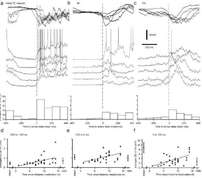
2.5.2 Time-dependent recovery of slow oscillation ... 59
2.5.3 Mechanisms of synchronization of slow oscillation and its recovery after thalamic inactivation ... 61
2.6 Discussion ... 64
2.6.1 Thalamic and neocortical contribution to the slow oscillation ... 64
2.6.2 The slow oscillation as the intrinsic property of the neocortex ... 66
2.7 References ... 68
2.8 Figure Legends ... 72
ix Chapter 3: Neocortical inhibitory activities and thalamocortical afferents contribute to the onset of
silent states of the neocortical slow oscillation ... 87
3.1 Résumé ... 88
3.2 Abstract ... 89
3.3 Introduction ... 90
3.4 Materials and methods ... 91
3.4.1 Surgery ... 91
3.4.2 Electrophysiological recordings ... 92
3.4.3 Thalamic inactivation and neocortical slabs ... 93
3.4.4 Electrical laminar stimulations ... 93
3.4.5 Data analysis and statistical tests ... 93
3.5 Results ... 95
3.5.1 Level of synchrony of the transition to the silent state ... 95
3.5.2 Disfacilitation and inhibition prior to the onset of the silent state ... 97
3.5.3 Laminar profile of stimulation efficacy to induce a silent state ... 99
3.5.4 Thalamic firing before the silent state ... 101
3.6 Discussion ... 101
3.6.1 Balance of excitation and inhibition ... 102
3.6.2 Synchronization of the cortical inhibition ... 104
3.6.3 Electrical stimulation ... 104
3.6.4 Sleep architecture, cortex and thalamus ... 105
3.6.5 Functional implications ... 105
3.7 References... 106
3.8 Figures ... 110
Chapter 4 The slow oscillation in the somatosensory and associative cortices of rabbits and cats: a comparative study of recurrent activity in the mammalian neocortex. ... 127
4.1 Résumé ... 128 4.2 Abstract ... 129 4.3 Introduction ... 130 4.4 Methodology ... 130 4.4.1 Surgery ... 130 4.4.2 Electrophysiological recordings ... 131
x
4.4.3 Data analysis ... 131
4.4.4 Statistical tests ... 133
4.5 Results ... 133
4.5.1 Composition of the power spectra ... 133
4.5.2 Properties of the slow oscillation ... 134
4.5.3 Coherence and synchronization of the slow oscillation ... 135
4.5.4 Propagation of the slow oscillation ... 136
4.5.5 Isolated neocortical slabs ... 137
4.6 Discussion ... 138
4.6.1 Methodological consideration... 138
4.6.2 Power spectral compositon in the different species-regions ... 139
4.6.3 Associative vs. somatosensory ... 139 4.6.4 Rabbit vs. cat ... 141 4.6.5 Concluding remarks ... 141 4.7 Literature cited ... 142 4.8 Figures ... 146 4.9 Table ... 158
Chapter 5 General discussion ... 159
5.0 Summary of the thesis ... 159
5.1 The roles of the thalamus ... 160
5.1.1 The rightful place of the thalamus ... 160
5.1.2 Technical limitations ... 161
5.1.3 Future studies ... 162
5.2 Is the slow oscillation the default state of the neocortex ... 162
5.2.1 Recovery of the slow oscillation ... 162
5.2.2 Limitations and future works ... 163
5.3 The silent state as a synchronous phenomenon ... 164
5.4 The fragile balance of excitation and inhibition ... 165
5.4.1 Chloride-mediated inhibition ... 165
5.4.2 Laminar stimulation ... 166
5.4.3 Future works ... 166
xi
5.5.1 Implications of the slow oscillation concordance ... 167
5.5.2 Emergence of the slow oscillation ... 167
5.5.3 Technical limitations ... 168
5.5.4 Future works ... 168
5.6 Concluding remarks ... 169
xiii
List of Tables
Table 2.1 Detailed geometry of thalamocortical network. ... 84
xv
List of Figures
Figure 1.1 Schematic representation of a typical neocortical pyramidal neuron ... 5
Figure 1.2 Electrophysiological identification of neocortical neurons ... 10
Figure 1.3 Types of thalamic nuclei based on their projections to the neocortex ... 11
Figure 1.4 Summary of intracortical excitatory connectivity mediating the recurrent activity ... 15
Figure 1.5 Brain rhythms during wake and sleep ... 25
Figure 1.6 Spontaneous active states in the isolated neocortical slab ... 33
Figure 1.7 The thalamus is not necessary to generate the slow oscillation ... 38
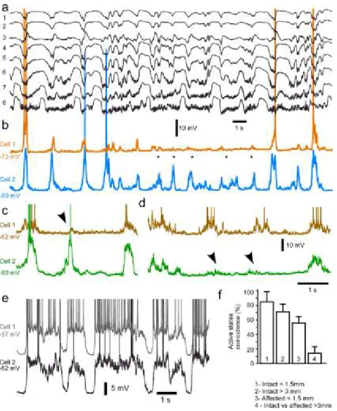
Figure 2.1 Effects of partial thalamic inactivation on the cortical slow oscillation ... 72
Figure 2.2 LFP power distribution in intact vs. affected by thalamic inactivation cortical regions ... 73
Figure 2.3 Intracellular activity in intact and partially deafferented cortical areas after inactivation of thalamic LP nucleus. ... 74
Figure 2.4 Alternating involvement in active states of closely located cortical neurons in the cortical area affected by thalamic inactivation. ... 76
Figure 2.5 LFP activities in acute and chronically isolated neocortical slabs. ... 77
Figure 2.6 Upregulation of synaptic activites following thalamic inactivation... 78
Figure 2.7 The geometry of thalamocortical network model. ... 79
Figure 2.8 Modeling study of the effect of thalamic deafferentation on the neocortical slow oscillation. ... 80
Figure 2.9 Impact of thalamocortical diffuse (matrix) vs. specific (core) projections and the target radii on the synchronization of the slow oscillation. ... 81
Figure 2.10 Modeling study of scaling the intracortical connectivity on the recovery of the neocortical slow oscillation. ... 82
Figure 2.11 Role of different biophysical features inthe recovery of the slow oscillation ... 83
Figure 3.1 Removing inputs to the neocortex decreases the synchrony of the silent state onset. ... 110
Figure 3.2 Synchrony of silent state onset at the cellular level. ... 112
Figure 3.3 Disfacilitation prior to the silent state onset ... 114
Figure 3.4 Decreased cortical firing prior to the onset of silent states. ... 116
Figure 3.5 Chloride inhibition at the transition to the silent state. ... 118
Figure 3.6 Long duration inhibition prior to the onset of the silent state under ketamine-xylazine anesthesia. 120 Figure 3.7 Long duration inhibition prior to the onset of silent state during natural sleep. ... 121
Figure 3.8 Depth profile of the efficacy to trigger a silent state. ... 122
Figure 3.9 Depth profile of neuronal response to electrical stimuli. ... 123
Figure 3.10 Thalamic firing in relation to cortical onset of the silent state. ... 124
Figure 4.1 Defining state in the LFP recording. ... 146
Figure 4.2 Proportion of frequency bands characteristic of ketamine-xylazine anesthesia. ... 148
Figure 4.3 Spatiotemporal coincidence of the slow oscillation ... 150
Figure 4.4 Cross-correlation of the slow oscillation ... 152
xvi
Figure 4.6 Active state duration in LFP recordings in the isolated neocortical slab. ... 156 Figure 4.7 Active state duration in intracellular recordings in slabs ... 157
xvii
List of Abbreviations
AC Alternative current DC Direct current EEG Electroencephalography EMG Electromyography EOG ElectrooculographyEPSP Excitatory postsynaptic potential
FRB Fast rhythmically bursting
FS Fast spiking
GABA Gamma-aminobutyric acid
GAD Glutamic acid decarboxylase
GFP Green fluorescent protein
HSP Homeostatic plasticity
HVA High voltage activated (calcium channels)
IA Transient potassium current
IH Hyperpolarization activated cationic current
IK Fast delayed rectifying potassium current
IK,Ca Calcium-activated potassium current
INap Persistent sodium current
IT Transient calcium current
IB Intrinsically bursting
i.m. Intra-muscular
i.v. Intra-venous
IPSP Inhibitory postsynaptic potential
KAc Potassium acetate
KCl Potassium chloride
LDT Lateral dorsal tegmentum
LFP Local field potential
LGN Lateral geniculate nucleus
LP Lateral posterior (nucleus of the thalamus)
LTD Long-term depression
LTP Long-term potentiation
LTS Low threshold spike
LVA Low voltage activated (calcium channels)
MD Medial dorsal (nucleus of the thalamus)
MEG Magnetoencephalography
MGN Medial geniculate nucleus
MUA Multi-unit activity
NaCl Potassium chloride
NBQX 2,3-dihydroxy-6-nitro-7-sulfamoyl-benzo[f]quinoxaline-2,3-dione
NMDA N-methyl-D-aspartate
NREM Non rapid eye movements
xviii
PBS Phosphate buffer saline
PFA Paraformaldehyde
PGO Ponto-geniculo-occipital (waves)
PPT Pedunculopontine tegmentum
QX-314 N-(2,6-Dimethylphenylcarbamoylmethyl)triethylammonium bromide
REM Rapid eye movements
RS Regular spiking
SD Standard deviation
STD Short-term depression
STDP Spike-timing dependent plasticity
STF Short-term facilitation
SWA Slow wave activity
SWS Slow wave sleep
TC Thalamocortical
Trans-ACPD Trans-(2R, 4R)-4-aminopyrrolidine-2,4-dicarboxylate
TTX Tetrodotoxin
Vm Membrane potential
VB Ventrobasal complex of the thalamus
xix
. Comment ne pourrais-je pas dédier cette
thèse à ma compagne, Geneviève, qui malgré son refus de poursuivre des études graduées a quand même du les endurer?
Je dédie également cette these à la mémoire de mon père, Pierre, qui est l’une de mes inspirations à suivre une vie dédiée à la recherche et qui aurait tant voulu être présent pour ce jour. “Life is what happens to you When you’re busy making other plans”
xxi
Acknowledgment
I’m above all grateful to my thesis supervisor, Igor Timofeev, who welcomed me in his lab five years ago. Over the years, I’ve learned under his supervision the fine art of in vivo electrophysiology. Throughout our discussions, I’ve learned to develop a keener sense of criticism, never to believe but always to understand.
I would also like to thank colleagues and friends without whom this project would not have been the same. Foremost, I have in mind Sylvain Chauvette from whom I’ve learned so much and Josée Seigneur who’s always so helpful, even when you don’t ask for it. I’m also grateful to Sergiu Ftomov who helped me get out of the Bermuda Triangle of electrical noise quite a few times. For all our discussions, sometimes extending in the late night, I want to thank my former partner in crime, Francis Lajeunesse, and for their kind support, Reza Zomorrodi and Courtney Pinard. Many thanks also to Soumaïa Boubou, Mathieu Blais-D'Amours, Magda Kusmierczak and Laszlo Grand for their help during this thesis. For their friendship over the last years, a special thank to Farida El-Gaamouch and Bruce Mesnage.
I want to thank my family for their support over the years, and especially my mother, Christine, who always believed in me and encouraged me through the years. And of course, I thank my beloved companion, Geneviève, who has been so supportive and understanding over the last four years
xxiii
Foreword
This thesis is a collection of three research articles (chapters 2-4). The first chapter is an introduction on the organization and rhythms generated by the thalamocortical network. The fifth chapter is a general discussion of the three studies. A special attention is given to the slow oscillation which is the scope of the three studies completed during the course of this thesis.
The first manuscript, “The impact of cortical deafferentation on the neocortical slow oscillation”, is published in the Journal of Neuroscience. This study was initially designed by me and my supervisor, Prof. Igor Timofeev, to investigate the role of the thalamus in the synchronization of the slow oscillation. To our surprise, we found that the inactivation of a restricted portion of the thalamus (lateral posterior nucleus) led to a dramatic decrease of the slow oscillation in the LFP recordings of a region of the suprasylvian gyrus. We further investigated with intracellular recordings this phenomenon in long experiments (36h) and we uncovered a partial restoration of the slow oscillation. In chronic experiments, we found a complete recovery in isolated neocortical slabs over a period of 2-3 weeks. These findings were supported by modeling experiments of the homeostatic plasticity in the thalamocortical network performed by the laboratory of Prof. Maxim Bazhenov. Both experimental and modeling studies were separately presented at SfN meeting in 2010 and were received with much excitement by our peers. We believed that the homeostatic nature of our results would have an impact not only on the thalamocortical field of research but also on neurosciences at large. We thus combined both experimental and modeling parts and submitted a manuscript in 2011 to Science and upon rejection, to Nature Neuroscience where the study was peer-reviewed for two months. Due to the controversial nature of our work (homeostatic issue), our work could not be published in its current form by the Nature Neuroscience. After several rounds of reviews, it was accepted in the Journal of Neuroscience
The second manuscript, “Neocortical inhibitory activities and thalamocortical afferents contribute to
the onset of silent states of the neocortical slow oscillation”, has been reviewed twice in the Journal of
Neurophysiology and we were invited to resubmit. I designed this study with my supervisor to address the question regarding the conditions that lead to onset of the silent state of the slow oscillation. This study combined LFP recordings in the intact and deafferented (thalamic inactivation, isolated neocortical slabs) neocortex, dual intracellular recordings (potassium chloride and potassium
xxiv
acetate), recording of multi-unit activity in the thalamus and laminar electrical stimulation in the slab. Due to the similarity to my results on the laminar stimulation in the neocortical slabs, we included in this study results from Dr. Sylvain Chauvette on intracellular response to laminar stimulations and an analysis of the reduction in firing in the cortical depth prior the onset of the silent state.. To provide control data on the synchronization of the silent states onset in paired intracellular recordings, I also used recordings made by Dr. Chauvette.
The third manuscript, “The slow oscillation in the somatosensory and associative cortices of rabbits
and cats: a comparative study of recurrent activity in the mammalian neocortex”, is in preparation for
submission to the Journal of Comparative Neurology. This study uses multisite LFP recordings to compare the synchronization of the slow oscillation in the neocortex of rabbits and cats. Experiments were conducted in the intact and isolated neocortical slabs of the somatosensory and parietal associative cortex. This study was performed to evaluate the possibility of replacing cats by rabbits as an in vivo model of the thalamocortical processes in the laboratory of Prof. Timofeev. A weakness of this study is by consequence the small sample of rabbits used. We consider adding to this study data from experiments on mice conducted of Dr. Maxim Sheroziya. The inclusion of those analyses will delay the submission of the manuscript.
In the course my PhD, I have also had the opportunity to contribute to a modeling study by Dr. Maxime Bonjean on the role of the neocortex in terminating the thalamic spindles. I performed and analyzed juxtacellular recordings in the cat motor cortex under barbiturate anesthesia to confirm the model in vivo. The reference to this publication : Bonjean M, Baker T, Lemieux M, Timofeev I, Sejnowski T, Bazhenov M (2011) Corticothalamic feedback controls sleep spindle duration in vivo. J Neurosci 31:9124-9134.
I have also co-supervised with Prof. Timofeev the preparation of a stereotaxic atlas of the ferret brain. Brains were collected during the in vivo experiments of Francis Lajeunesse in the course of his Ph.D. training. I thank Francis who stayed with me late in the night after his experiments to make stereotaxic measurements and perfusion. I’m also grateful to Marie-Josée Wallman who did all the histology and preliminary scanning for the atlas. However, after exploring several possibilities, we could not find a suitable avenue for high-resolution scanning of the sections intended for both the hard copy and web-based versions of the atlas. I have hope that we will eventually find a convenient
xxv
solution in the future and fulfill the project we settled for in 2011. As of today, there is still no published atlas of the ferret which is a model of neuroscience research. We believe the publication of our anatomical data will profit the scientific community working or planning to work on the ferret brain. Here is the list of manuscripts presented in this thesis
1- Lemieux M, Chen JY, Lonjers P, Bazhenov M, Timofeev I (2014) The impact of cortical deafferentation on the neocortical slow oscillation. J Neurosci 34:5689-5703.
2- Lemieux M., Chauvette S., Timofeev I. (in review in the Journal of Neurophysiology) Neocortical inhibitory activities and thalamocortical afferents contribute to the onset of silent states of the neocortical slow oscillation
3- Lemieux M., Timofeev I. (in preparation) The slow oscillation in the somatosensory and
associative cortices of rabbits and cats: a comparative study of recurrent activity in the mammalian neocortex
1
Chapter 1 General introduction
1.1 A brief introduction to sleep
We experience everyday periods of wakefulness and sleep. Wakefulness is a state which we can easily relate to because it is in this state of vigilance that we consciously experience life, interpreting various sensorial inputs from our environment or from our own body and executing a rich repertoire of motor tasks. Sleep escapes to our conscious experience or at the very least our recollection of it. Our cerebral cortex, the alleged siege of consciousness, is disconnected during sleep from the external world during this state of vigilance. The disconnection of the cerebral cortex from the external world is attributed to the thalamic gating (Munglani and Jones, 1992; McCormick and Bal, 1994; Timofeev et al., 1996; Del Felice et al., 2012).
A large body of evidences has suggested that the sleep is controlled by the brain to fulfill some of its need (Hobson, 2005). However, the nature of those needs is still uncertain to this date although the most accepted theory is that sleep is necessary to memory consolidation (Maquet, 2001; Steriade and Timofeev, 2003; Diekelmann and Born, 2010; Wang et al., 2011). One thing is sure, sleep is a neural feature shared by almost all animals. Throughout the animal kingdom, sleep is defined as a quickly reversible period of inactivity (in comparison to coma or hibernation) associated with a reduced responsiveness to external inputs (Zimmerman et al., 2008). Sleep is driven by a circadian process (process C) and by a homeostatic one (process S) depending on the duration of wakefulness and dissipating exponentially during sleep (Borbely, 1982).
Sleep is defined as a state of inactivity from primitive cnidarians like jellyfish (Kavanau, 2006) up to the much more evolved arthropods like the fruitfly (Shaw et al., 2000). This definition of sleep as a period of inactivity remains for basal vertebrates like the zebrafish (Yokogawa et al., 2007). The complexity of sleep has reached its climax in mammals and birds with the generation of the slow wave activity (SWA) probably due to the highly developed connectivity within their cerebral cortex (Rattenborg, 2006). Even among these two classes of vertebrate, the extent of sleep varies with the ecological niche (migrating birds or cetaceans) or the position in the food chain (predators sleep more than herbivores). In other words, it is a state of adaptive inactivity (Siegel, 2009).
We have attempted in this thesis to shed light on some mechanisms in the generation by the thalamocortical network of a key feature of sleep, the SWA or slow oscillation. We have studied the
2
contribution of the neocortex and the thalamus in generating the normal pattern of the slow oscillation and more specifically, in the transition between the active and the silent states of this slow rhythm. The slow oscillation is a phenomenon of recurrent activity occurring globally in the cerebral cortex and as such, we hypothesized that the extent of its synchronization may vary across neocortical regions. We also tested the hypothesis that the level of synchronization within a homologous cortical region may vary across species. Before we address these questions, we will describe in the following introduction the organization of the thalamocortical network in terms of its cellular components and connectivity among them to understand how brain rhythms (including the SWA) are generated.
1.2 Thalamocortical network
1.2.1 General organization
The thalamocortical network is made of the cerebral cortex and the thalamus, as its name implies. The cerebral cortex is a structure on the outer surface of the forebrain. Histologically, it is characterized by an external gray matter and an inner white matter that is formed by myelinated fibers arriving or leaving the cortex. The gray matter contains generally six layers (neocortex or isocortex) but can be made of four or five layers (paleocortex). Layer I, located at the surface of the cerebral cortex, contains mainly fibers. Layer II and III form the supragranular layers, layer IV is called the granular layers V and VI, the infragranular layers. The thalamus is located anteriorly to the midbrain, ventrally to the cerebral cortex and dorsally to the hypothalamus. It can be divided into a ventral and a dorsal part. The dorsal thalamus is formed by sensorial relay (or first order) and non-specific (higher order) nuclei. The ventral thalamus surrounds the exterior part of the dorsal thalamus and is formed by the reticular nucleus and the zona incerta.
The thalamocortical network can be divided into subsystems relating to a general function (sensorial processing, motor tasks, associative tasks and cognition). Based on clustering analysis of connectivity between the cortex and the thalamus, four major systems can be defined (Scannell et al., 1999). The first one is the visual system that contains the lateral geniculate nucleus (the primary recipient of retinal inputs to the thalamocortical network) and up to 32 neocortical areas in the macaque and 16 in the cat similarly organized in hierarchical levels of processing. The visual system is distributed in the occipital (containing the primary visual cortex), parietal, temporal and frontal lobe (the frontal eye field) and occupies half of the macaque cerebral cortex (Felleman and Van Essen, 1991). The second system is the auditory system formed by primary and second auditory cortex as
3
well as anterior and posterior auditory fields in the cortex (Rouiller et al., 1991). The cortical auditory regions are interconnected with medial geniculate nuclei and the posterior nucleus (Andersen et al., 1980).
The third system is the somato-motor system containing the somatosensory and the motor cortical areas and the ventrobasal complex of the thalamus (VB). The VB nucleus receives somesthetic inputs from the medial lemniscus, the spinothalamic tract (both transmitting information from the body) and the trigeminal tracts (related to the face). It transmits this information to the primary somatosensory areas (Brodmann areas 1, 2 and 3). High-order processing occurs in the posterior parietal cortex (Brodmann areas 5 and 7) which also integrates visual and auditory information (Hendry and Hsiao, 2003). The cortical areas are concerned with motor functions are the primary motor, the premotor and the supplementary motor area. These motor regions receive inputs from the somatosensory and associative areas (Schieber and Baker, 2003).
The fourth system is the fronto-limbic containing the prefrontal areas, the limbic areas and the anterior, midline and intralaminar nuclei of the thalamus (Scannell et al., 1999). The prefrontal cortex is a set of cortical areas connected with all sensory and motor systems and is involved in decision making, planning and guidance in social behaviors (Niedermeyer, 1998; Miller, 2000). The limbic areas were defined according to their anatomical features of cortical organization rather than on their functions and refer to the cingulate, retrosplenial, prelimbic, infralimbic, perirhinal, entorhinal and subicular regions of the cerebral cortex. These regions receive inputs from sensory cortical areas and are in close association with the hippocampus, the amygdala and the septum (Lopes da Silva et al., 1990).
Within each cortical areas, neurons form mini-columns vertically arranged (approximately 50 µm in diameter) and columns (300-600 µm in diameter) via their horizontal connections (Mountcastle, 1997). Depending on the cortical area, a column is associated with a specific function and is present in primary sensory areas (as well as those involved in higher level of processing), motor areas and even prefrontal areas.
In the following sections (1.2.2-1.2.8), we will describe the morphology and properties of neurons of the neocortex and the thalamus. These neurons were described in various mammalian species and
4
because these neurons are ubiquitous to the mammalian neocortex, we will not mention the studied species.
In summary, the cerebral cortex is organized in different levels of information processing and its basic unit is the radial column of cortical neurons. One of the questions we will address is whether the extent of synchrony of the slow oscillation within an area correlates with the hierarchical level of information processing.
1.2.2 Neocortical pyramidal neurons
Spiny pyramidal neurons are the principal cellular element of the neocortex as they account for 70-85% of the neurons in this brain region (DeFelipe and Farinas, 1992). A typical pyramidal neuron, as illustrated in figure 1.1can be distinguish by the pyramidal or ovoid shape of its soma from which arises a prominent apical dendrite reaching layer I (except for layer VI pyramidal neurons) and several basal dendrites. The dendrites contain spines which receive mostly asymmetrical synapses (Colonnier, 1968), commonly accepted as excitatory synaptic contacts. Spines accounts for roughly a third of the surface of a pyramidal neuron while the soma makes up only 2-4% (Mungai, 1967). Present in every layer, spines are more abundant in the layer where is located the cell body (Larkman, 1991), suggesting that the majority of excitatory inputs to a neuron comes from its respective layer. The axon of pyramidal cells points downward and leaves collateral extending horizontally, dorsally or ventrally. These cells use glutamate as neurotransmitter, which makes these neurons excitatory ones.
The pyramidal neurons are present throughout the cortical column except in layer I (DeFelipe and Farinas, 1992). The pyramidal neurons located in the supragranular layers (layers II and III) are smaller than those in infragranular layers (V and VI). Although layer IV contains mainly stellate cells, it also contains pyramidal neurons. The layer V contains the pyramidal neurons with the largest dendritic arborization, extending up to layer I just below the pia mater. Layer VI pyramidal neurons are of a size similar to those in layer V but their apical dendrites do not extend further than layer IV. Due to the abundance and the large size of pyramidal neurons, intracellular recordings in vivo are often biased toward this cellular type. In the course of our experiments, we will also stimulate the neocortical layers under the hypothesis that they have different impact on the neocortical recurrent activity.
5 Figure 1.1 Schematic representation of a typical neocortical pyramidal neuron
Adapted from DeFelipe and Farinas (1992).
1.2.3 Neocortical spiny stellate cells
The spiny stellate cells are non-pyramidal neurons found only in layer IV. Their soma is round and 5-6 dendrites originate from it and radiate in all directions, branching once or twice. The axon emerges from the deeper surface, descends shortly and divides in 2-3 branches that turn sharply to ascend in
6
layer II/III and sometimes even in layer I. One descending branch reaches layer V or layer VI in some cases. The major characteristic that distinguishes the spiny stellate cell from other non-pyramidal neurons is the presence of spines on dendrites (Jones, 1975).
1.2.4 Neocortical interneurons (aspiny and sparsely spiny non-pyramidal neurons) The absence of spine (aspiny) or the low presence of it (sparsely spiny) on these non-pyramidal neurons gives them a smooth aspect. They form a heterogenous population of neurons accounting for 15-30% of the neocortical neuronal population (DeFelipe and Farinas, 1992). These cells are immunoreactive for the glutamic acid decarboxylase (GAD), the synthesizing enzyme of the inhibitory neurotransmitter GABA, which makes these cells inhibitory interneurons (Ribak, 1978; Houser et al., 1983). There is a great diversity of interneurons in the neocortex that can be identified by their morphology and patterns of innervations (DeFelipe et al., 2013) as well as by their neurochemistry (Kawaguchi and Kubota, 1997). Although the classification varies depending on the authors, these cells can be broadly split in two groups based on their dendritic arborization: bitufted and multipolar. Furthermore, some classes of inhibitory neurons can be recognized based on the pattern of their axons.
The double bouquet is a typical example of bitufted cell. It is characterized by a vertical axonal bundle crossing radially layers II-V and with terminals making synapses on the soma, the dendritic shaft and the spines of non-pyramidal cells (Somogyi and Cowey, 1981). Double-bouquet cells can also be identified by their immunoreactivity for the vasoactive intestinal polypeptide (Kawaguchi and Kubota, 1996). The chandelier cell was named after the peculiar shape formed by its axon that descends from the lower pole of the soma and emits collaterals curving back to ascend. The axonal plexuses are located mostly beneath the soma of pyramidal neurons and form synapses on the initial segment of the axon (Somogyi, 1977; Fairén and Valverde, 1980; Peters et al., 1982). The dendritic arbor is bitufted with primary slender and sparse dendrites arising from the lower and upper poles of the fusiform soma.
The most commonly recognized type of multipolar interneuron is the basket cells. The ovoid soma of this cell emits long dendrites in every direction which branch once or twice. The term “basket” refers to the pattern made by their axon terminals around the soma and proximal dendrites of pyramidal neurons (Marin-Padilla, 1969; Jones, 1975; Kisvarday et al., 1987). The extent of their axonal
7
arborization is very large; it is even larger than any known dendritic arborization in the cortex (Somogyi et al., 1983). Another type of multipolar stellate cell is the Martinotti cell that sends its main axon and several collaterals to layer I. This type of interneurons is distinguishable by the expression of the somatostatin peptide (Kawaguchi and Kubota, 1996). The neurogliaform cell (also named spiderweb cell by Ramon y Cajal) is the smallest interneuron of the cortex and is frequently encountered in Golgi staining. It has several short dendrites radiating in every direction and branching once or twice; the axon emits several collaterals in a dichotomous manner and frequently changes course (Jones, 1975; Kisvarday et al., 1990).
This great diversity of inhibitory interneurons shapes the activity within the neocortical network, whether in response to a sensory stimulus or during spontaneous activity (Markram et al., 2004). Unfortunately, the lower proportion and smaller somatic size of these neurons have for consequence that they are difficult to record intracellularly in vivo. Alternative approaches (for example, pharmacological, genetic or alteration of ionic concentrations), are required to study their contribution to the network activity.
1.2.6 Thalamic neurons
In contrast to the diversity of cortical neurons, the morphology of the main cellular type of the thalamus, the relay cell or thalamocortical (TC) neuron, is much more homogenous among the nuclei of the dorsal thalamus, with some exception in the LGN (Jones, 2002) that is outside the scope of this thesis. These relay neurons come in different sizes and have a bushy appearance shaped by a variable number of radiating and dichotomously ramifying dendrites of up to 250 µm in length and bearing spines. Most of these cells project away from the thalamus although some cells in specific nuclei (but not in non-specific ones) were observed to have short local axons (Guillery, 1966; Scheibel and Scheibel, 1967). TC neurons are glutamatergic but there are also GABAergic cells in the dorsal thalamus. Accounting for approximately 30% of neurons in all nuclei of cats and monkeys, but restricted in the LGN in rodents, these local interneurons have a few thin dendrites with bulbous dilatations that look like axonal boutons [reviewed in (Jones, 2002)].
Neurons from the thalamic reticular nucleus have a round, ovoid or elongated soma. There have generally 3-6 elongated dendrites arising from the soma either from all around (multipolar) or from both poles (bitufted) of the soma and branching into second and third order dendrites. The dendrites
8
run parallel to the border of the nucleus. Spine-like varicosities are not present on first order dendrites but are on higher order ones. The axon of a thalamic reticular neuron leaves one or two collaterals in the thalamic reticular nucleus and curves toward the dorsal thalamus (Lübke, 1993). These cells are GABAergic and thus exclusively inhibitory neurons (Jones, 2002).
1.2.7 Intrinsic properties of neurons
The membrane potential of neurons is set by the distribution of potassium, sodium, calcium and chloride ions on each side of this membrane (Hille, 2001). Neurons are also rich in intrinsic voltage-gated channels for sodium, calcium and potassium ions that regulate the firing of neurons as well as allowing these cells to act as pacemaker and resonate with the network activity (Llinas, 1988). Sodium currents can be transient or persistent due to a different form of gating of the sodium channels. The transient opening of voltage-gated sodium channels generates the action potential which is the output the neuron (Hille, 2001). The persistent sodium current (INap) may amplify distal
dendritic excitation and be involved in subthreshold oscillating activity (Crill, 1996). INap is activated
during rhythmic firing (Stafstrom et al., 1984).
Voltage-dependent calcium currents also regulate the firing pattern of neurons. There are calcium channels activated at high voltage (HVA) and those activated at low voltage (LVA) (Llinas and Yarom, 1981). Another classification is based on the size of conductances and the rate of inactivation. Two types of HVA can be recognized based on the conductance size and kinetic of inactivation: the L-type with large conductance and slow rate of inactivation and the N-type with a lower conductance and a faster rate of inactivation. The LVA-type correspond to the transient calcium current (IT) which carries the smallest conductance and the faster rate of inactivation (Tsien et al.,
1988). The channels responsible of the IT are inactivated at depolarized level, de-inactivated at
hyperpolarized ones and activated at membrane potential more hyperpolarized by 15 mV than the firing threshold, making IT ideal for oscillatory activity (Huguenard, 1996).
Another voltage-dependent current, the hyperpolarization activated cationic current (IH), is
responsible for slowly depolarizing the membrane potential of hyperpolarized neurons (activated between -90 to -60 mV depending on the cell). This current plays an important role in shaping the oscillating behavior of neurons (McCormick and Pape, 1990). The IH and the voltage-dependent
9
hyperpolarizing currents relying on the efflux of potassium ions. There is a rich diversity of potassium currents with different properties: the fast delayed rectifier (Ik), the slow delayed rectifier activated via
muscarinic receptor (M current), the transient potassium current which is de-inactivated by hyperpolarization (IA) and calcium-activated potassium currents (IK,Ca) (Hille, 2001). Although some of
the channels carrying these currents can leak at the rest, there is a different family of channels not gated by voltage and dedicated to the potassium leak current (Goldstein et al., 2001).
The interplay of these intrinsic currents plays a major role in shaping oscillatory behavior. For instance, they underlie the dual mode of firing in TC neurons, that is tonic and bursting firing (Jahnsen and Llinas, 1984). The currents responsible for the bursting behavior (IH and the IT) are
also involved in an intrinsic pacemaker activity at 1-2 Hz present during SWS (McCormick and Pape, 1990). In this study, we will interfere with the bursting behavior of TC cells by blocking the IT with
QX-314 (Connors and Prince, 1982) to assess its impact on the neocortical activity. 1.2.8 Electrophysiological types of neurons
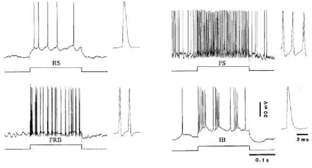
The morphology of a neuron also confers neurons a certain pattern in the firing. In the neocortex, there are four major types of firing pattern (or electrophysiological types) associated with a given morphology (McCormick et al., 1985; Connors and Gutnick, 1990) and more precisely with the dendritic structure and its electrotonic coupling with the soma (Mainen and Sejnowski, 1996). The most common type is the regular spiking (RS) neurons. In the original description of the RS type by Mountcastle and colleagues, the RS was said to present an adaptation in its firing rate (Mountcastle et al., 1969). The duration at half-amplitude of a RS action potential is generally 1-1.5 ms. The great majority of pyramidal neurons are RS. Fast spiking (FS) neurons increases steadily their firing rate with injected current. The action potential of FS neurons lasts in average less than 0.5 ms at half-amplitude. They are less frequently encountered than RS cells (Simons, 1978) and functionally correspond to inhibitory neurons (McCormick et al., 1985). However, not all inhibitory neurons are FS. For instance, Martinotti cells and most VIP-positive doublet bouquet cells are RS (Kawaguchi and Kubota, 1996). The intrinsically bursting (IB) neurons fire 3-5 action potentials at short intervals and each successive spike decreases in amplitude. About 15-20% of pyramidal neurons are IB cells and are generally found in layer V (McCormick et al., 1985; Timofeev et al., 2000a). Originally described as chattering cells in the supragranular layers of the visual cortex (Gray and McCormick, 1996), the fast rhythmically bursting (FRB) neurons fires thin spikes (0.3-0.4 ms at half-amplitude) at high
10
frequency (300-600 Hz) recurring rhythmically at 20-50 Hz. Although present in supragranular layers in cortical regions other than visual, FRB neurons are more numerous in infragranular layers (Timofeev et al., 2000a; Steriade et al., 2001). In our studies, most cells recorded intracellularly were of the RS type and to a lesser extent of the IB and FRB types. As no attempt was made to find differences between these cell types, data were pooled together.
Figure 1.2 Electrophysiological identification of neocortical neurons
Intracellular responses to depolarizing current pulse: regular spiking (RS), fast spiking (FS), fast rhythmically bursting (FRB) and intrinsically bursting (IB). Modified from Steriade et al. (2001).
1.3 Connectivity in the thalamocortical network
We have introduced in the preceding section the general organization of the thalamocortical network. We also mentioned that neurons are equipped with intrinsic conductances that permit certain oscillatory behavior. However, the full expression of oscillations observed in vivo requires synaptic activity. In our study, we were particularly interested in how the connectivity between the thalamus and the cortex as well as the intracortical connectivity could generate the slow oscillation. In this section, we will discuss the connectivity between the different elements of the thalamocortical network.
11
1.3.1 Thalamic connectivity
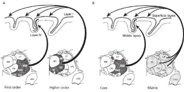
Two classes of dorsal thalamic nuclei are recognized based on their projections to the neocortex: specific or relay (VB, LGN, MGN) and non-specific or associative (intralaminar, midline and posterior) nuclei (Fig. 1.3). TC neurons of relay nuclei project in a topographic and specific manner to the neocortex while those of the associative nuclei send diffuse projections to the neocortex (Jones, 1998; Sherman and Guillery, 2002). Relay TC neurons are also considered as thalamic core neurons and non-specific TC neurons as thalamic matrix neurons (Jones, 1998).
Figure 1.3 Types of thalamic nuclei based on their projections to the neocortex
(A) Traditional view of the first- and higher-order nuclei. (B) Recent view of the core vs. matrix TC projections. Modified from Jones (1998).
TC relay neurons are excited by thick axons from sensory afferents and by thin axons from corticothalamic afferents. These afferents are organized in a topographic manner. The GABAergic inhibition is provided by thalamic reticular neurons and, although less well characterized, by local interneurons (Sherman and Guillery, 1996; Jones, 2002). There are also non-specific modulatory afferents: acetylcholine, noradrenaline and serotonine from the brainstem and histamine from the tuberomamillary nucleus (Sherman and Guillery, 1996). In VB, sensory afferents from the medial lemniscus and the spinothalamic tract accounts for 15% of the synapses on TC relay cells, corticothalamic for approximately half of those synapses and inhibitory synapses (GABAergic
12
terminals) for the remaining third of synapses. Sensory afferents are mainly located on proximal dendrites and corticothalamic preferentially on distal dendrites. Inhibitory synapses are located on all compartments (Liu et al., 1995).
As for relay nuclei, TC neurons from associative nuclei receive dense and complexly ramifying afferents from the spinal cord, the brainstem, the tectum, the basal forebrain and the cerebral cortex. An important difference between relay and associative nuclei is the origin of corticothalamic afferents. In the case of relay nuclei (such as LGN), corticothalamic axons originate from layer VI pyramidal neurons, are thin and form small terminals in the distal dendrites. These axons are also present in associative nuclei (for example, the pulvinar) and, in addition, layer V pyramidal neurons send thick axons forming large terminals located more proximally than those formed by layer VI afferents. Functionally, layer V inputs acts as a driver on the thalamus whereas layer VI ones are modulating (Sherman and Guillery, 1996, 2002), suggesting a greater influence of the neocortex on associative than on relay thalamic nuclei.
Neurons from the thalamic reticular nucleus and zona incerta are innervated by the axonal collaterals from both layer VI corticothalamic and thalamocortical neurons (Jones, 2002), which makes these cells at the center of the dialogue between the cortex and the thalamus. Although layer V axons cross the reticular nucleus, they do not leave collaterals (Sherman and Guillery, 1996; Jones, 2002). The contribution of thalamic reticular neuron is the inhibition of thalamic relay cells and as we will see later, is essential in shaping oscillatory behavior in the thalamocortical network.
1.3.2 Thalamocortical afferents
The thalamus is the gateway to the cerebral cortex for the sensorial inputs (except for the olfaction). Furthermore, it transmits information from one cortical region to another one (Sherman and Guillery, 1996, 2002). The primary excitatory cortical recipient of TC inputs is the spiny stellate cells in layer IV (Gilbert and Wiesel, 1979). Some pyramidal neurons in layer VI also receive direct TC inputs and they project onto layer IV stellate cells (Gilbert and Wiesel, 1979; White and Hersch, 1982; Burkhalter, 1989; Ahmed et al., 1994; Hirsch et al., 1998a; Lee and Sherman, 2009; Thomson, 2010). TC synapses can also be found on the basal dendrites of layer III and apical dendrites of layer V pyramidal neurons (Hornung and Garey, 1981; Ichikawa et al., 1985). Although TC synapses on
13
stellate cells have nothing outstanding in comparison to intracortical synapses in terms of position in the dendritic trees or size of the their postsynaptic targets (da Costa and Martin, 2011), these afferents are associated with the highest amplitude of postsynaptic response in spiny stellate cells (Stratford et al., 1996). As spiny stellate cells have a high probability of transmitter release at synapses of layer II/III pyramidal neurons (Silver et al., 2003), thalamic afferents thus have a strong excitatory influence on the neocortical network.
TC inputs also recruit inhibition in the neocortex. Synaptic contacts are found on sparsely spiny and aspiny non-pyramidal neurons in layer IV (Hornung and Garey, 1981), which are inhibitory interneurons as described above. Several classes of interneurons having their soma in layer IV or at the border of layer IV and V receive inputs from the thalamus and their likelihood of firing is even higher than excitatory cells (Porter et al., 2001; Cruikshank et al., 2007; Hull et al., 2009). An increase in thalamic firing in response to stimulation of several whiskers in the somatosensory system, for example, is accompanied by a higher firing of FS units and a decrease of firing of RS units in the cortex (Brumberg et al., 1996). This phenomenon, believed to generate the surround inhibition in the cortex, also shows how inhibition in the neocortical network can be efficiently recruited by TC inputs. Furthermore, disynaptic inhibition in the neocortex caused by thalamic stimulation has been evidenced in layer V pyramidal neuron and its threshold was lower than for intracortical stimulation (Gil and Amitai, 1996).
In other words, TC neurons are a major source of excitation to the neocortex and they also influence, perhaps even control, the level of inhibition in the neocortical network. Most importantly, thalamocortical neurons are involved in the communication between cortical areas. In that sense, the thalamus is much more than a relay for sensory information to the neocortex, it is potentially a hub for the intracortical communication.
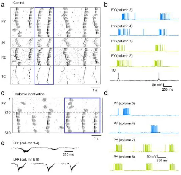
In this thesis, we have evaluated the contribution of an associative thalamic nucleus in propagating and synchronizing activity across different neocortical sites by pharmacologically inactivating the functional thalamocortical afferents. We chose as a model the associative cortex of the cat (area 5 and 7 located in the suprasylvian gyrus). We have targeted the lateral posterior (LP) nucleus of thalamus which provides most of the afferents to area 5 and 7 (Graybiel, 1972).
14
1.3.3 Neocortical excitatory connectivity
The activation of an excitatory synapse causes an excitatory postsynaptic potential cell (EPSP) that brings the postsynaptic cell closer to its firing threshold. The excitation in the neocortex is provided by pyramidal neurons and spiny stellate cells that make contacts on other pyramidal and stellate cells as well as on inhibitory interneurons. Most excitatory connections on pyramidal neurons are located on the dendritic spines (Colonnier, 1968) whereas those on interneurons (which are generally aspiny) are located mainly on the dendritic shaft (Winfield et al., 1981).
Within a cortical column, excitatory cells can communicate via intralaminar or interlaminar connections. Intralaminar connectivity refers to the communication between neurons located in the same layer. The probability of finding such connectivity between excitatory cells of layers II-III and layer V was evaluated to about 10-25%(Markram et al., 1997b; Gibson et al., 1999; Thomson et al., 2002). Interlaminar connectivity refers to a neuron making a connection with neurons of a different layer. It is very important as it forms the canonical flow of information starting in layer IV stellate cells relaying a signal to layer II/III pyramidal neurons which pass the input to layer V /VI pyramidal neurons (Gilbert and Wiesel, 1979). It is also involved in feedback excitation, that is deep layer neurons exciting back upper layer neurons. For instance, layer VI pyramidal neuron make excitatory and facilitating connections on layer IV neurons (Tarczy-Hornoch et al., 1999). There are several axonal processes of neurons in layer V and VI that are ascending to layers II/III (Burkhalter, 1989; Hirsch et al., 1998a). As summarized in figure 1.4, this ascending translaminar feedback excitation may be an essential substrate of recurrent (or sustained) activity within the neocortex.
Although we will not investigate directly the intracortical connectivity in this study, we will compare the duration of recurrent activity between different sites of a given neocortical area, different areas (associative vs. somatosensory) and different species (cat vs. rabbit).
15 Figure 1.4 Summary of intracortical excitatory connectivity mediating the recurrent activity
Schematic representation of translaminar excitatory connectivity in the neocortex. Afferents from the thalamus andcortico-cortical and cortico-thalamic efferents from a neocortical columnare also shown.
1.3.4 Neocortical inhibitory connectivity
The synaptic response of a neuron to the firing of presynaptic interneurons is the inhibitory postsynaptic potential (IPSP). There are two types of IPSP (Connors et al., 1988). The first one reverses at -75 mV, has a short duration (10-20 ms) and is associated with a 70-90 nS conductance. The second one reverses at -90mV, lasts much longer than the first type but has a lower conductance (10-20 nS), that is a lower current and by extension, a response of smaller amplitude. The first type is attributable to currents activated by GABAA receptors while the second is
characteristic of GABAB receptors activation. The activation of GABAA and glycine receptors is
16
are metabotropic; they activate a second messenger (a G-protein) that increases the conductance for potassium ions (Bormann, 1988). Most IPSPs recorded at the soma are identified by their reversal potential as the result of GABAA activation. The IPSPs elicited by FS and RS interneurons have
comparable amplitude (0.2-2.2 mV) and reversal potential suggestive of GABAA activation, but the
latter lasts longer (25-32 ms) than the former (10-20 ms) (Thomson et al., 1996).
Interneurons innervate all the compartments (soma, initial segment of the axon, dendritic shaft and spines) of pyramidal neurons and other interneurons (Hendry et al., 1983; Somogyi et al., 1983; Somogyi et al., 1998). Immunhistochemistry against GABA receptors at the electron microscopy level revealed that most (58%) GABA synapses are located on the dendritic shaft, a large portion (26.4%) on the spines and the balance on the soma (13.1%) and the axonal initial segments (2.5%) (Beaulieu and Somogyi, 1990). Considering that the soma accounts for 2-4 % of the surface of a pyramidal neuron (Mungai, 1967), it is fair to acknowledge that there is a lot of GABAergic synapses on the soma of pyramidal neurons (Freund et al., 1983). These inhibitory synapses on the soma are made by GABAA synapses as shown by a very strong immunoreactivity on the soma and proximal dendrites
of pyramidal as well as non-pyramidal neurons of all layers, except in layer I where it is fainter (Gu et al., 1993).
Although the largest inhibition comes from their respective layer, there is interlaminar inhibition. Reconstruction of layer III interneurons showed that their axons descend in layer V (Somogyi et al., 1983; Thomson and Morris, 2002), pointing out that interneurons from upper layers can inhibit neurons in deeper layer. The occurrence of EPSC in layer V FS and RS interneurons after stimulation of layer II/III by glutamate puff (Otsuka and Kawaguchi, 2009), showing that monosynaptic translaminar connection indeed exists. The other way around is also true as pyramidal neurons located in layer II-III are contacted by interneurons of layers V and VI (Kisvarday et al., 1987; Thomson and Morris, 2002; Katzel et al., 2011). However, a study in which channelrhodopsine-2 was transfected in GAD67 neurons (interneurons) of mice has suggested that the monosynaptic inhibition on layer V and VI pyramidal neurons comes almost exclusively from their layer (Katzel et al., 2011). Even though monosynaptic inhibition appears to be mainly intralaminar, the translaminar inhibition could also pass through disynaptic inhibition.
17
The cortical inhibition is mainly a local phenomenon (i.e., at the columnar level) as interneurons create a powerful zone of inhibition within 350 µm of their soma (Salin and Prince, 1996a). However, retrograde labeling revealed that a small proportion of inhibitory neurons (0.5-5%) in layers II/III as well as layers V and VI have been observed to project farther than 0.5-1 mm in the rat visual cortex (McDonald and Burkhalter, 1993) and somatosensory cortex (Fabri and Manzoni, 1996).In both cases, the proportion of long-range projecting interneurons was larger in layers V and VI than in layer II/III. Similar observations were made in the cat visual cortex (Albus and Wahle, 1994) as well as in the motor, somatosensory and visual cortices of GAD67-GFP knock-in mice (Tomioka et al., 2005). Aside from monosynaptic connectivity, long-range inhibition can also be disynaptic as long-range excitatory projections can recruit inhibition in distant cortical sites in a supralinear manner (Tucker and Katz, 2003).
Retrograde labeling after injection in the contralateral cortex and subsequent intracellular injections in slice to reveal the cellular morphology showed that approximately 5% of the labeled neurons were aspiny non-pyramidal neurons and thus presumably inhibitory neurons. These inhibitory neurons projecting to the contralateral hemisphere were identified as basket and chandelier cells located in layer II-V and there was also a some neurons in layer I (Martínez-García et al., 1994). This suggested that, in addition to a strong local inhibition and a smaller proportion from distant sites in the ipsilateral cortex, inhibition could also come from the contralateral cortex.
In this thesis, we have investigated with intracellular recordings the contribution of chloride-mediated inhibition (GABAA) in terminating the recurrent activity during the slow oscillation. We will also use
electrical stimulation to test whether there are differences among cortical layers regarding the contribution of inhibition in terminating the recurrent activity.
1.3.5 Neocortical efferents
A neocortical column receives and integrates information, but it also transmits it to other neural structures. One of the targets of a neocortical column is neighboring neocortical area and its contralateral equivalent. Neurons in layer II/III are mainly responsible for these projections to neighboring cortical areas (Gilbert and Kelly, 1975) and to the contralateral cortex via the the corpus callosum. The latter, called callosal or commissural neurons, were found to be most numerous in
18
layer III but also in layer V in several sensorial and associative cortical regions of the rats, cats and monkeys (Jacobson and Trojanowski, 1974; Wise and Jones, 1976). Layer V also contains neurons projecting to neighboring cortical areas (Lund, 1973), indicating that neurons in both supra- and infragranular layers project to other cortical areas, including those on the contralateral side.
Layer V pyramidal neurons are considered as the main subcortical projecting neurons. Among their targets are the thalamus (Sherman and Guillery, 1996), the superior colliculus (Holländer, 1974; Gilbert and Kelly, 1975), the pontine nuclei (Abdel-Kader, 1968) and the spinal cord (Schieber and Baker, 2003). Layer VI is also a projection layer and contains three classes of pyramidal neurons according to their projection: other pyramidal neurons, the thalamus (both the dorsal and reticular ones) and the claustrum (Thomson, 2010).
In this thesis, we have focused mainly on the suprasylvian gyrus of the cat which is divided into area 5 and 7. Area 5 receives cortical inputs from somatosensory regions (Avendaño et al., 1988) and area 7 from visual ones (Olson and Lawler, 1987). These associative regions project to the LP nucleus of the thalamus. Area 5 also projects to the ventral lateral group and to the medial division of the posterior group and area 7 to the pulvinar. In addition, both sends axons to the thalamic reticular nucleus and to the central lateral and the paracentral (intralaminar) nuclei (Robertson and Cunningham, 1981).
1.3.6 Recurrent activity and the balance of excitation and inhibition
As we have discussed in the preceding sections, there are intralaminar and interlaminar connections in the neocortex, both excitatory and inhibitory. Of particular significance, the axon layer V pyramidal neurons leave several collaterals in the granular and infragranular layers (Burkhalter, 1989). These collaterals have boutons making synaptic contact onto the spines and shafts of both apical and basal dendrites of other infragranular layers pyramidal neurons (Gabbott et al., 1987). It is reasonable to assume that these intracortical connections made by the efferent neurons of the neocortex can generate recurrent activity within the neocortex.
However, too much excitation within a network could lead to paroxysmal activity typical of seizures. There are mechanisms that prevent such over-excitation. Firstly, there is frequency-dependent depression of synaptic transmission (Galarreta and Hestrin, 1998). Secondly, there is the GABAergic inhibition. Inhibition keeps on a leash neurons by preventing them from firing too much. Inhibitory
19
conductances are dominant in a majority of neurons (Rudolph et al., 2007; Haider et al., 2012) and it is when it decreases that an action potential is likely to happen (Rudolph et al., 2007). Instead of hyperpolarizing the membrane potential, the shunting inhibition decreases the amplitude of excitatory responses due to a large increase in the membrane conductance. This increase in membrane conductance was attributed to the activation of GABAA receptors based on the reversal potential, the
large conductance and the short latency of response of cortical cells following a visual stimulus (Borg-Graham et al., 1998; Hirsch et al., 1998b). If the balance between excitation and inhibition is broken in favor of excitation, paroxysmal activity in the network is likely to follow. This has been observed for recurrent activity in neocortical slices (Sanchez-Vives and McCormick, 2000; Mann et al., 2009; Sanchez-Vives et al., 2010)
It is difficult to separate the cortical and thalamic contribution to the recurrent activity within the neocortex. As was mentioned previously, the thalamus receives input from the periphery but also from the cortex itself. Because they project widely to the cortex, thalamocortical neurons might also be involved in the recurrent activity in the neocortex. Modeling experiments have indeed shown that a thalamic drive on cortical pyramidal cells can reduce the duration of the silent state (Bazhenov et al., 2002). It remains to be demonstrated in vivo, but it would demonstrate that the recurrent activity is a property of the thalamocortical network as a whole.
We have tested in this study the impact of thalamocortical afferents on maintaining the recurrent activity in the neocortical network. We will also address the question about the balance of excitation and inhibition in terminating the recurrent activity.
1.3.7 Synaptic and homeostatic plasticity
The strength of synaptic connections between neurons is not fixed as it can be modified in an activity-dependent manner. This process is referred to as synaptic plasticity and translates in the potentiation or the depression of the amplitude of the postsynaptic response. The changes in amplitude can last on the timescale of milliseconds up to a second (short-term facilitation, STF, or short-term depression, STD) or for hours and days (long-term potentiation, LTP, or long-term depression, LTD) [reviewed in (Zucker, 1989; Byrne, 2003)]. There is also the medium-term plasticity which lasts from seconds to minutes (Timofeev et al., 2002).