Université de Montréal
Hyperactivations frontales en mémoire de travail
dans le trouble cognitif léger
Par Nicolas Moffat
Département de psychologie, Faculté des arts et sciences
Mémoire présenté à la Faculté des Arts et Sciences en vue de l‟obtention du grade de M.Sc. en psychologie
Avril 2015 © Nicolas Moffat, 2015
Résumé
Des études récentes ont rapporté que les individus âgés avec un trouble cognitif léger (TCL) ont de plus grandes activations en lien avec la réalisation d‟une tâche cognitive que des personnes âgées saines. Des auteurs ont proposé que ces hyperactivations pourraient refléter des processus de plasticité cérébrale compensatoires ayant lieu pendant la phase précoce de la maladie d‟Alzheimer. Des processus de compensations fonctionnelles pourraient émerger en réponse à une perte d‟intégrité structurelle dans les régions du cerveau normalement requises pour compléter une tâche. Dans ce mémoire, j‟ai évalué cette hypothèse chez des personnes avec TCL en faisant appel à une tâche de mémoire de travail comportant plusieurs niveaux de
difficulté ainsi qu‟aux techniques d‟imagerie par résonnance magnétique (IRM) structurelle et fonctionnelle. Des analyses de régression multiples ont été utilisées afin d‟identifier les régions cérébrales dont l‟activité variait en fonction de l‟intégrité neuronale telle que définie par le volume de l‟hippocampe. Les valeurs estimées des paramètres du signal de ces régions furent ensuite extraites afin de procéder à des analyses corrélationnelles sur la performance ainsi que sur le volume de différentes structures cérébrales. Les résultats indiquent des hyperactivations dans les régions frontales droites chez les participants TCL souffrant d‟une plus grande atteinte neuronale. De plus, le niveau d‟activation est négativement corrélé au volume de structures frontales et pariétales. Ces résultats indique la présence d‟une hyperactivation compensatoire dans la phase du TCL associée à la réalisation d‟une tâche de mémoire de travail.
Mots-clés: Maladie d‟alzheimer, trouble cognitive léger (TCL), fonctions exécutives, processus compensatoires, hyperactivation, imagerie à résonnance magnétique (MRI), analyses
Abstract
Recent studies have shown greater task-related activation in individuals with mild cognitive impairment (MCI) compared to healthy older adults. It has been suggested that these hyperactivations reflect compensatory processes of brain plasticity in the early phase of Alzheimer‟s disease (AD). Functional compensation processes could emerge in response to a loss of structural integrity in brain regions typically required for the task. In this masters
dissertation, I examined this hypothesis in MCI using a working memory task that incorporated a parametrical variation of difficulty level as well as functional and structural magnetic resonance imaging (MRI). Multiple regression analyses were used to identify areas of the brain in MCI in which functional activation varied as a function of neural injury as measured by hippocampal volume. Parameter estimates from these areas were then extracted and used for further correlational analyses with performance and structural volumes. Results indicate that MCI participants with high neural injury hyperactivated a subset of regions in the right frontal lobe. Furthermore, the level of activation was negatively correlated with the volume of frontal and parietal regions. These results indicate the presence of compensatory hyperactivations associated with a working memory task in persons with MCI.
Keywords: Alzheimer‟s disease (AD), mild cognitive impairment (MCI), executive functions, compensatory mechanisms, hyperactivation, magnetic resonance imaging (MRI), multimodal analyses.
Table des matières
Résumé ... 1
Abstract ... 2
Introduction ... 1
Les déficits executifs dans le trouble cognitive léger ... 1
Corrélats neuronaux de l‟atteinte des fonctions exécutives dans le TCL : études en neuroimagerie structurelle... 3
Modèle de plasticité cérébrale dans le TCL ... 3
Hyperactivations dans le trouble cognitif léger ... 5
But et hypothèses ... 7
Article ... 9
Conclusion ... 43
Comparaisons entre les groupes ... 43
Activations en fonction de l‟atteinte neuronale ... 44
Hyperactivations et performance ... 46
Hyperactivations et atrophie de régions exécutive... 47
Limites ... 48
Résumé, implications et perspectives futures ... 49
Tableaux et figures
Table 1 - Sociodemographic et neuropsychological data ... 29 Table 2 - List of structural regions based on the Desikan-Killiany Atlas for which the volume was used in
our mulimodal analyses. ... 29 Table 3 - Clusters that showed increased activity as a function of hypocampal volume in MCI during the
1-back condition with cluster size, peaks and corresponding t-values. ... 30 Figure 1 – Experimental design ... 31 Figure 2 – Task related activation and location of activation clusters representing areas of increased
activation in relation to smaller hippocampal volume in MCI. ... 32 Figure 3 - Correlation between changes in brain activation in response to an increase in difficulty (2 – 1
back) and performance in MCI. ... 33 Figure 4 – Multimodal correlations between parameter estimates in MCI (1-back) and the volume of the
Introduction
La maladie d‟Alzheimer (MA) est une maladie neurodégénérative qui se caractérise sur le plan histopathologique par une perte neuronale et synaptique causée par l‟accumulation de plaques amyloïdes et une dégénérescence neurofibrillaire. Le diagnostic de la maladie ne peut être confirmé qu‟à la suite d‟un examen histopathologique réalisé au décès du patient. Du vivant du patient, un diagnostic clinique est donné et celui-ci repose sur un déclin des fonctions
cognitives anormal tel qu‟évalué à l‟aide de tests neuropsychologique et corroboré par les proches. De plus, les déficits observés ne doivent pas être attribuables à une autre condition neurobiologique, à des effets de comorbidité ou à la prise de médicaments.
La maladie d‟Alzheimer est une maladie progressive. Le début des symptômes est
graduel et on sait maintenant que la maladie débute de nombreuses années avant le moment où le patient rencontre les critères cliniques de la démence. Le trouble cognitive léger (TCL) est reconnu comme étant une phase prodromale de MA. Pour cette raison, plusieurs études ont été effectuées afin de mieux caractériser cette phase sur le plan cognitif et neuronal.
Les déficits executifs dans le trouble cognitive léger
Les fonctions exécutives comprennent un ensemble de fonctions cognitives dont le rôle est de coordonner et de planifier l‟action. Plusieurs processus cognitifs de haut niveau pouvant être modulés en fonction de la réalisation d‟une tâche peuvent être considérés comme faisant partie des fonctions exécutives. La composante de contrôle de la mémoire de travail, la flexibilité mentale, la mise à jour ainsi que les processus d‟inhibition sont généralement considérés comme
faisant partie des fonctions exécutives. Malgré que la mémoire épisodique représente la première et la plus affectée des fonctions cognitives, un nombre grandissant de résultats suggèrent que les fonctions exécutives seraient elles aussi atteintes dans le TCL (Belleville et al, 2007; Belanger et Belleville, 2009; Johns et al., 2012). De plus, les études qui ont tenté d‟identifier des mesures permettant de distinguer parmi les personnes avec un TCL ceux qui vont ultérieurement progresser vers la MA et ceux qui vont rester stables ont rapporté que les fonctions exécutives étaient de bon prédicteurs de progression en association avec la mémoire épisodique (Chapman et al., 2011; Belleville et al., 2014).
Les réseaux neuronaux impliqués dans le contrôle exécutif sont relativement bien caractérisés. Ainsi, des études antérieures ont identifiées trois circuits exécutifs reposant sur différentes régions frontales et ayant des projections vers les lobes pariétaux et temporaux ainsi que des structures sous-corticales. Le premier circuit origine du cortex préfrontal dorsolatéral (dlPFC), une région versatile et associée à la fluence verbale, la flexibilité mentale, la résolution de conflits, la mémoire de travail ainsi que l‟inhibition. Le second circuit originerait du cortex orbitofrontal qui est quant à lui associé aux processus d‟inhibition. Enfin, un troisième circuit serait associé au cingulaire antérieur et serait lié aux mécanismes de motivation face à une tâche. (Alvarez et Emory, 2006; Cabeza et al., 2000; Cumming, 1993).
Vu son atteinte précoce dans le TCL, il est donc important de mieux comprendre les mécanismes sous-tendant la dysfonction exécutive des personnes avec un TCL. Or il existe relativement peu d‟études en neuroimagerie qui ont tenté de caractériser les changements
neuronaux associés aux fonctions exécutives dans le TCL alors que cela pourrait améliorer notre compréhension des facteurs entourant la progression vers la démence.
Corrélats neuronaux de l’atteinte des fonctions exécutives dans le TCL : études en neuroimagerie structurelle
Les trois circuits jouant un rôle dans le contrôle exécutif sont reconnus pour être altérés dans la MA et ils seraient aussi atteints dans la phase prodromale de la maladie, le TCL. Par exemple, différentes études ayant utilisé des mesures de volumétrie ou d‟épaisseur corticale ont montré des atteintes frontales, pariétales et temporales dans le TCL et la MA lorsque comparé à des sujets âgés sains (Chetelat et al, 2005; Julkunen et al., 2008, 2010; Singh et al., 2006). De plus, une atrophie de ces régions pourrait être responsable des déficits exécutifs observés. Ainsi, des études ayant corrélées la performance à des tests neuropsychologique avec des mesures d‟IRM structurelle ont montré que la présence d‟une atrophie des régions frontales et pariétales chez les participants TCL était associée à des déficits d‟attention et de mémoire de travail (Pa et al., 2010; Thomann et al., 2008) alors qu‟une atrophie des régions frontales et temporales était quant à elle associée à des déficits de fluence verbale et de raisonnements (Newman et al., 2007; Rabin et al., 2009). Ces études indiquent donc que les régions associées au contrôle exécutif sont
structurellement altérées dans la maladie d‟Alzheimer et que la perte de volume observée est associée à une atteinte dans différentes tâches exécutives. En particulier, l‟atteinte de mémoire de travail a été associée à une atrophie des régions frontales et pariétales.
Modèle de plasticité cérébrale dans le TCL
Les mesures d‟imagerie structurelle offrent des pistes intéressantes quant à l‟origine des déficits exécutifs. Elles ne sont toutefois pas suffisantes pour élucider les changements d‟activité cérébrale qui découlent des pertes neuronales subies dans les stades légers de la maladie et ne permettent pas de tirer de conclusion quant à leur possible rôle compensatoire. De récents
modèles suggèrent en effet que des mécanismes de plasticité cérébrale pourraient être présents dès les premières phases menant à la MA et que ces derniers supporteraient différents processus compensatoires (Clément and Belleville, 2010; Pruvolic et al., 2005; Sperling et al., 2010). Ces processus compensatoires expliqueraient l‟absence quasi complète de symptômes pendant la longue phase prodromale de la maladie. Ces modèles proposent que les processus
compensatoires seraient modulés par l‟ampleur de l‟atteinte neuronale et, par conséquent, par le stade de progression de la maladie. Au début de la maladie, l‟effet des lésions structurelles se traduirait par une plus grande difficulté subjective à la tâche qui pourrait être compensée par un plus haut niveau de recrutement des régions affectées ou faisant partir d‟un même réseau fonctionnel. Éventuellement, la progression des lésions structurelles atteindrait un seuil à partir duquel les ressources neuronales nécessaires à la tâche ne seraient plus en mesure d‟être mobilisées. Les processus compensatoires seraient par ailleurs associés à une plus grande activation fonctionnelle (hyperactivation) chez les participants TCL lorsque comparés à celles d‟un groupe contrôle dans des tâches nécessitant des régions structurellement affectées. Par conséquent, il est attendu que le niveau d‟atrophie cérébral à l‟intérieur de régions nécessaires à la tâche soit corrélé positivement au niveau d‟activation fonctionnel chez les personnes avec un TCL. Par ailleurs, un affaissement des processus compensatoires surviendrait lorsque la sévérité de l‟atteinte structurelle ne permettrait plus un recrutement compensatoire approprié. Cet
affaissement compensatoire serait associé à une incapacité à recruter les ressources neuronales nécessaire à l‟exécution de la tâche. Cela se manifesterait par un niveau d‟activation
fonctionnelle inférieur à celui de personnes âgées saines (hypoactivation). Il est intéressant de souligner que la plupart des études réalisées pendant la phase de démence de la MA ont rapporté
des hypoactivations c‟est à dire des activations moindre chez les personnes atteintes comparativement aux contrôles appariées (Prvulovic, et al., 2005).
Certains auteurs (Clément and Belleville, 2012; Prvulovic et al., 2005) ont proposé que le patron observé dans le décours progressif de la maladie –c‟est à dire une élévation des
activations suivies d‟une chute des activations- s‟apparente à celui qui serait observé en réponse à l‟augmentation de la difficulté d‟une tâche. Pour un même niveau de difficulté, l‟activation serait plus élevée chez les personnes avec un TCL ce qui représenterait une mobilisation des ressources neuronales plus élevée chez ces derniers. La capacité des personnes avec un TCL à répondre adéquatement à une augmentation de la difficulté de la tâche serait par ailleurs réduite. Par conséquent, une tâche suffisamment difficile pourrait mener à un affaissement des processus compensatoires et l‟observation d‟hypoactivations.
Hyperactivations dans le trouble cognitif léger
Certains résultats rapportés dans la littérature appuient les prédictions faites par les modèles compensatoires de la MA. En effet, quelques études ont rapporté des hyperactivations en lien avec des tâches de mémoire épisodique dans le TCL (Celone et al., 2006; Clement et Belleville, 2010; Clement et al., 2010; Dickerson et Sperling, 2008, 2010; Woodard et al., 2009). De manière générale, des hyperactivations sont observées lors de tâches épisodiques chez les participants TCL les moins touchés cliniquement alors que des hypoactivations sont parfois observées chez les sujets TCL plus touchées ainsi que chez les sujets avec une MA. De plus, Clément, Belleville et Gauthier (2013) ont rapporté des hyperactivations en lien avec une tâche d‟attention divisée dans plusieurs régions telles que le cingulaire antérieur bilatéral, le putamen
gauche ainsi que le gyrus inférieur gauche. Les activations plus élevées n‟étaient présentent que chez les TCL qui étaient moins touchés cliniquement et elles étaient corrélées positivement à leur performance.
Malgré des résultats prometteurs et en appui avec la présence de processus compensatoires dans le TCL, quelques questions doivent toutefois encore être abordées. D‟abord, ces études font principalement appel à des tâches de mémoire épisodique. Si les hyperactivations observées dans le TCL reflètent des mécanismes cérébraux généraux, ils devraient être observables lorsque les patients réalisent des tâches nécessitant d‟autres domaines cognitifs atteints. À notre connaissance, seule l‟étude de Clement et al (2013) s‟est attardée au processus compensatoires chez les TCL dans un domaine autre que celui de la mémoire épisodique. Ensuite, aucune de ces études n‟a évalué l‟impact du niveau de difficulté sur les hyperactivations qui reste donc inconnu. Or le degré de difficulté de la tâche devrait moduler les hyperactivations selon certains auteurs. Finalement, la plupart de ces études n‟ont pas fait appel à des mesures cérébrales structurelles permettant de documenter le degré d‟atteinte neuronale. Cette information est pourtant essentielle puisque le modèle prédit que l‟amplitude des
hyperactivations dans la phase précoce de la MA serait associée au niveau d‟atrophie structurelle de régions impliquées dans la tâche. Par conséquent, l‟inclusion de mesures structurelles pourrait nous fournir d‟importantes informations quant à l‟origine neurobiologique des différences d‟activation fonctionnelle observées. En outre, la performance clinique a le plus souvent été utilisée dans les études antérieures pour identifier le degré de sévérité de la maladie des patients. De plus, on sait que les groupes de TCL comprennent plusieurs personnes qui ne vont jamais progresser vers la MA. Or l‟imagerie structurelle procure des valeurs de références plus valides et moins indirectes quant à la présence et à la sévérité de la maladie. En particulier, le volume de
l‟hippocampe est une des première structure touchée dans le TCL et a été démontrée comme étant un excellent indicateur de l‟atteinte neuronale dans la MA (Du et al., 2001; Jack et al., 1999).
But et hypothèses
Le but du présent mémoire est d‟étudier les profils d‟activation cérébrale dans le TCL et chez des sujets âgés sains à l‟aide d‟une tâche exécutive incluant différents niveaux de difficulté. De plus, des mesures de volumétrie structurelle ont été utilisées afin d‟étudier l‟association entre les activations fonctionnelles et le volume de régions cérébrales impliquées dans la tâche.
L‟imagerie structurelle permet aussi de fournir un indice de sévérité pathologique. Contrairement aux études passées qui l‟évaluèrent à l‟aide de la performance clinique, nous avons utilisé le volume de l‟hippocampe. Cette mesure fut utilisée comme régresseur contre la variabilité des activations fonctionnelles au travers du groupe TCL.
La tâche utilisée était une variante du n-back (Gevins et Cutillo, 1993). Cette tâche offre de nombreux avantages. Il s‟agit d‟une tâche exécutive qui a largement été utilisée par le passé et dont les profils d‟activation cérébrale sont bien connus. Elle est assez simple à réaliser dans l‟IRM par des patients de sévérité clinique variée et est donc moins susceptibles aux effets planchers. Elle permet enfin une manipulation paramétrique du degré de difficulté en fonction du nombre du nombre d‟éléments à maintenir en mémoire.
Sur le plan des activations fonctionelles, notre première hypothèse est que des
hyperactivations seront présentes chez les sujets TCL par rapport aux sujets âgés sains et qu‟elles seront localisées dans des régions impliquées par la tâche. Les personnes avec un TCL devraient
donc montrer de plus grandes activations fonctionnelles que les contrôles appariés. Nous nous attendons aussi à ce que l‟amplitude des hyperactivations soit corrélée négativement avec le volume de régions frontales et pariétales connues pour être impliquées dans des tâches de nature exécutives. Nous prédisons aussi que les hyperactivations seront plus importantes chez les sujets TCL souffrant d‟une plus grande sévérité pathologique. Par conséquent, nous devrions observer une corrélation négative entre le niveau d‟activation et le volume hippocampique. Finalement, nous nous attendons à ce que les hyperactivations soient le plus importantes dans le premier niveau de difficulté de la tâche.
Article
Frontal lobe hyperactivation during working memory in persons with MCI
Nicolas Moffat1,2, Samira Mellah2, Oury Monchi4, Serge Gauthier3, Juan Manuel Villalpando2 and Sylvie Belleville1,2
1Départment de Psychologie, Université de Montréal, Montréal, Canada
2Centre de Recherche de l’Institut Universitaire de Gériatrie de Montréal, Montréal, Canada 3McGill Centre for Studies in Aging, Montréal, Canada
Abstract
Recent studies have shown greater task-related activation in individuals with mild cognitive impairment (MCI) compared to healthy older adults. It has been suggested that these hyperactivations reflect compensatory processes of brain plasticity in the early phase of Alzheimer‟s disease (AD). When there is loss of structural integrity of a brain region that is typically required for a specific task, functional compensation processes would emerge through increased activation of the structurally impaired regions or of proximal or distal regions. This hypothesis was examined in MCI using a working memory task that incorporated a parametrical variation of difficulty level as well as functional and structural magnetic resonance imaging (MRI). Multiple regression analyses were used to identify areas of the brain in MCI in which functional activation varied as a function of neural injury as measured by hippocampal volume. Parameter estimates from these areas were then extracted and used for further correlational analyses with performance and structural volumes. Results indicate that the task elicited load related activations in a number of frontal and parietal regions. However, no load by group interaction was observed. Multiple regression analyses identified two frontal clusters for which brain activation increased as a function of neural injury in MCI on the 1-back condition.
Furthermore, parameter estimates from these clusters were found to be negatively correlated with measures of frontal and parietal volumes in areas known to be implicated in executive tasks.
Introduction
Amnestic mild cognitive impairment (MCI) is believed to be a prodromal phase of Alzheimer‟s disease (AD) and for this reason, a large number of studies have been conducted to better characterize this phase at both the cognitive and neural level. Although episodic memory represents the earliest and most affected cognitive domain, accumulating evidence indicates that executive functions are affected as well (Belleville et al, 2007; Belanger and Belleville, 2009; Johns et al., 2012) and that the impairment of these functions might be key in predicting progression from MCI to dementia (Chapman et al., 2011; Belleville et al., 2014). The neuroimaging study of executive function deficits might thus provide insight regarding the underlying neural mechanisms of MCI to further ameliorate the characterization of the individuals who are likely to develop dementia.
The neural network responsible for executive control is well characterized. Previous studies have identified three circuits originating in fronto-subcortical regions and projecting to the parietal and temporal lobes, which contribute to executive functioning: the dorsolateral prefrontal cortex (DLPFC), the lateral orbital cortex and the anterior cingulate (Cabeza et al., 2000; Cumming, 1995). These circuits have been shown to be structurally impaired in MCI and AD (Chetelat et al, 2005; Julkunen et al., 2008, 2010; Singh et al., 2006) and atrophy in these regions might account for early executive function impairment. For instance, by using structural MRI measures, studies in MCI patients have shown that frontal and parietal atrophy are related to working memory and attentional deficits (Pa et al., 2010; Thomann et al., 2008) while frontal and temporal atrophy are associated with fluency and reasoning deficits (Newman et al., 2007; Rabin et al., 2009).
A few critical recent models have been proposed to suggest that brain plasticity might be present in the early stages of AD and that those would support processes of compensation
(Clément and Belleville, 2010; Pruvolic et al., 2005; Sperling et al., 2010). Thus, while structural measures provide information on the underlying neural origin of executive deficits in MCI, they do not provide information on the related changes in brain activity and on whether or not these changes may be compensatory in nature. Processes of compensation are modulated by the extent of neural loss and hence, by disease severity. In the early phase of the disease, the effect of structural lesions on behaviour would result in higher subjective task difficulty and be scaffolded through a larger recruitment of brain areas that are directly impaired or part of a functional network involved in the task. With disease progression, structural lesions would reach a threshold preventing the mobilization of the cortical resources necessary to complete the task. Compensatory processes would be associated with larger brain activation in patients than in controls when completing a task in which the structurally impaired region is involved. It is thus expected that during the MCI phase, the level of atrophy in task-related regions would be positively correlated with the level of brain activity, as measured with blood-oxygen level dependent (BOLD) fMRI. Compensation breakdown would be associated with a failure to successfully engage the neural resources required to complete the task, resulting in a decrease in brain activation below the level of healthy older adults (hypoactivation), as can be typically observed in the dementia phase of AD (Prvulovic et al., 2005). A similar pattern would be found when increasing task difficulty. Simple tasks would allow the engagement of compensatory processes but compensatory processes would no longer be possible with complex tasks.
Interestingly, a few studies have reported task-related hyperactivations in MCI using episodic memory tasks (Celone et al., 2006; Clement and Belleville, 2012; Clement et al., 2010; Dickerson and Sperling, 2008, 2010; Woodard et al., 2009). However there are many remaining questions to address. First, the presence of hyperactivation has been reported mostly with episodic memory tasks. If this a general phenomenon, it should be possible to observe it on a larger range of task-related activation. To our knowledge, only one study has investigated compensatory activations in a non-memory domain. Clément, Belleville and Gauthier (2013) reported task-related hyperactivations during a divided-attention task in a number of regions including a fronto-striatal cluster comprising the anterior cingulate bilaterally, the left putamen and the left inferior frontal gyrus. The increased activation was observed only in MCI individuals who were less clinically impaired and was positively correlated with their performance. One other important issue is that none of these studies have assessed the effect of task difficulty on patterns of hyperactivation. Thus, there is currently no information regarding the relationship between an increase in cognitive demand and presence of hyperactivations in MCI. Finally and more importantly, no structural brain measures were used in most of these previous studies. This is critical as the model predicts that structural atrophy should be correlated with the amount of activation in the compensatory phase of MCI. Thus, including structural measures could provide valuable information regarding the neurobiological origin of the observed differences in brain activation.
The aim of the present study was therefore to investigate the patterns of brain activation in MCI patients and in healthy older adults using a working memory task while integrating a component of task difficulty and volumetric measures to determine the association between brain activation and structural volume of brain areas known to be implicated in the task. The use of
structural imaging was also used to provide a proxy for disease severity. In past studies clinical performance was typically used to identify disease severity in MCI persons. Here, we looked at the volume of the hippocampus, which is one of the first structure to be affected in MCI and for which the volume has been shown to be a reliable marker of progression to AD (Du et al., 2001; Jack et al., 1999). This was used to assess the level the neuronal injury in MCI to be integrated as a regressor against the variability of functional activations across the MCI group.
To this end, we used a variant of the n-back task (Gevins and Cutillo, 1993) which has been used extensively in the past and which is known to provoke consistent functional activation patterns. Our first hypothesis was that hyperactivations would be present in MCI and thus
expected larger task-related activations in MCI patients than in healthy older adults. These hyperactivations were expected to be located in regions implicated in the task. We also
hypothesized that the amplitude of hyperactivations in MCI would be negatively correlated with the volume of frontal and parietal regions, areas involved in executive tasks. We also predicted that hyperactivations would be most salient in MCI patients with higher neuronal injury, and therefore anticipated a negative correlation between activation and hippocampal volume. We expected hyperactivations to be found on the 1-back condition which represents low executive load.
Participants
Seventeen individuals with MCI (10 women, 7 men) and sixteen healthy older adults (12 women, 4 men) were recruited to take part in this study. All participants were right-handed,
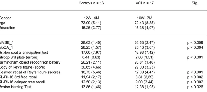
francophone and had normal or corrected vision. Sociodemographic and clinical data of both groups are shown in Table (1).
The MCI participants were referred to us from memory clinics and met the criteria proposed by Petersen (2001) for amnestic single- or multiple- domain MCI. These criteria include (a) cognitive complaint ideally corroborated by an informant; (b) performances at least 1.5 standard deviation below the average level of persons with similar age and education on a minimum of one clinical tests assessing memory, language or executive functions (See list of the clinical tests below); (c) absence of a significant impact on activities of daily living; and (d) absence of dementia. The healthy older adults were recruited from the community through advertisement in local newspapers.
Prior to study entry, MCI participants received an extensive medical and neurological examination to exclude the presence of any other significant systemic, neurological or
psychiatric conditions that could explain their impairment (exclusion criteria). Furthermore, all participants completed a comprehensive neuropsychological evaluation to assess global
cognitive functions (Mini-Mental State Examination [MMSE]; Folstein et al., 1975, and the Montreal Cognitive Assessment [MOCA]; Nasreddine et al., 2005), episodic memory (RL/RI free and cued word recall (Van der Linden et al., 2004); 3 min recall of the Rey‟s complex figure (Rey, 1959), executive functions (brixton spatial anticipation test; Burgess and Shallice, 1997); stroop-victoria (Regard, 1981) visual perception and praxia (4th subtest of Birmingham object recognition battery; Riddoch and Humphreys, 1993; and copy of the Rey Figure) and language (Boston Naming Test; Kaplan et al., 1983). This study was approved by the Human ethics board of the Institut universitaire de gériatrie de Montréal.
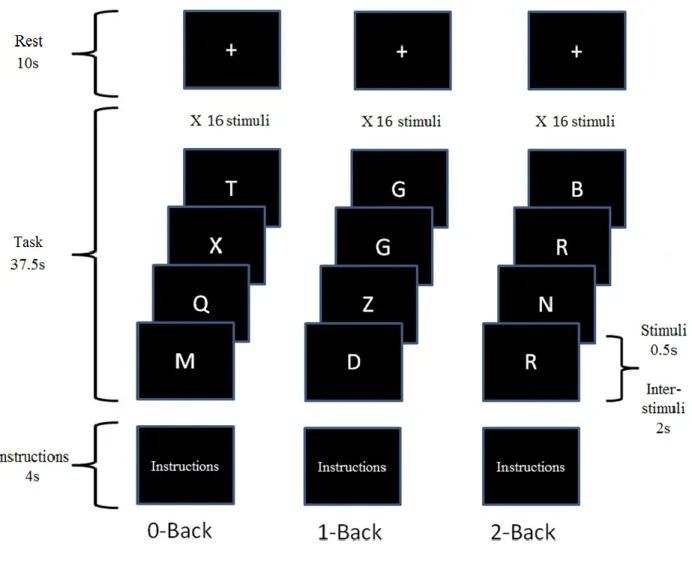
Working Memory Task
Functional images of the brain were acquired while participants completed a visual n-back working memory task. Illustration of a task sequence is displayed in Figure 1. The task included three conditions (0 back, 1 back and 2 back) presented in different blocks. For each condition, a list of random letters was presented sequentially and participants were asked to respond to all of them. In the 1-back condition, participants were asked to respond whether the letter was identical or not to the one preceding it. In the 2-back condition, participants were asked to determine whether the letter was identical to the one presented two items prior in the sequence. The 0-back condition served as a control in which participants were asked to identify whether the letter „X‟ was presented or not. Each condition consisted of five blocks presented in a pseudo randomized order. Each block comprised sixteen letters of which, five were targets. Letters were presented for 500 ms each with an inter-stimulus interval of 1500 ms. Each block included 5 seconds of instructions and a 15 seconds rest for a total block duration of 52 seconds. Stimuli were projected on a screen and reflected onto a mirror which was installed on the head coil in the fMRI machine. Participants responded to targets with their right thumb and to non-targets with their right index on a two-button response box.
Data Acquisition
MRI was performed using a SIEMENS 3T Magnetom TRIO System (Erlangen, Germany) at the Unité de Neuroimagerie Fonctionnelle (UNF) of the Institut Universitaire de Gériatrie de Montréal (IUGM). Functional magnetic images were acquired with gradient-echo echo-planar sequences sensitive to blood oxygen level-dependant contrast (repetition time/echo
time = 2500/30 msec, flip angle = 90o, 41 trans-axial slides per volume, voxel size = 3 x 3 x 3 mm, field of view = 192 mm, matrix = 64 x 64). A 3-dimensional structural image was acquired using a sagittal T1-weighted magnetization prepared rapid gradient echo sequence (repetition time/echo time = 2530/3.5 msec, flip angle = 7.0o, 176 slices, voxel size = 1 x 1 x 1 mm, field of view = 256 mm, matrix = 256 x 256).
Clinical and Behavioural Analysis
Clinical and socio-demographic data were compared across groups using independent t-tests (for continuous variables) and chi-squares (for categorical variables). Measures of task performance included mean accuracy and the ratio of correct answers minus the ratio of false alarms (Hit-FA) to correct for response bias. Accuracy and Hit-FA were also compared using two separate repeated-measure ANOVAs with Group (healthy controls, MCI) as a between-subject factor and Condition (0-back, 1-back, 2-back) as a within-between-subject factor. All behavioral data were analysed using the SPSS statistical package (Version 18).
Structural analysis
Cortical reconstruction and volumetric segmentation was performed using FreeSurfer 5.3 analysis suite. To describe it briefly, this processing includes motion correction of individual T1-weighted images, removal of non-brain tissue using a hybrid watershed/surface deformation procedure (Segonne et al., 2004), automated Talairach transformation, segmentation of the cortical and subcortical white matter and of the gray matter volumetric structures (Fischl et al.,
2004, Desikan et al., 2006), intensity normalization (Sled et al., 1998), tessalation of the gray matter/white matter boundary and automated topology correction (Segonne et al., 2007).
Individual data was inspected at each step of the processing streams and manual corrections were applied when necessary. Cortical volumetric data were extracted for each subject according to the Desikan-Killany atlas (Desikan et al., 2006). Cortical and hippocampal volumes were corrected as a proportion of the total intracranial volume.
Analysis of Task-Related Activations
Data were analyzed with MATLAB 8.1 (www.mathworks.com) using the statistical parametric mapping software SPM8 (www.fil.ion.ucl.ac.uk/.../spm8/). Images were converted to analyse format and unwarped. They were then realigned to the middle slice and a mean image was calculated for each subject. Mean images were then coregistered to each subject‟s structural data and normalised into the Montreal‟s Neurological Institute (MNI) stereotactic space. Images were smoothed using an 8 mm Gaussian kernel and low frequency noise was filtered using a high-pass filter of 160 sec. The instruction blocks were modeled as a condition of no-interest. First level analysis were performed using the 0-back condition as a baseline and individual contrast maps (1-back, 2-back and 2-1 back) were calculated for each subjects. A random effect analysis was then performed using a flexible factorial analysis of variance (ANOVA) with Group (MCI, healthy older adults) as a between-subjects factor and Task Difficulty (1-back, 2-back) as a within-subjects factor to assess load effect and possible interactions.
Additional analyses were performed in order to take into account the possible
positively modulated by disease severity across the different contrasts. To this end, multiple regression analyses were performed on each group‟s individual subject‟s statistical T-maps using hippocampal volume as a regressor. Functional analyses were performed using a threshold of p < 0.001 (uncorrected) and only clusters with a minimum of 15 contiguous voxels and significant at p < 0.05 (FWE-corrected) are reported. Both age and education were used as covariates.
Multimodal Analyses
Correlational analyses were performed in order to assess whether the task-related
activation in MCI patients was related to the cortical volume within the regions implicated in the task. To this end, parameter estimates (β values) from functional clusters showing an increase in activation with disease severity were extracted using Marsbar (Brett et al., 2002) and correlated with structural volumes from regions that demonstrated significant load-related activity (see Table 2 for the full list of regions). Possible volumetric differences of these regions across the groups were tested using independent-sample t-tests. Given our hypothesis that the influence of neuronal injury on brain activity in MCI individuals would be present under low memory load, we limited the analyses to the 1-back condition. Because of the large number of regions, we limited the comparison of parameter estimates to structural regions located in the same hemisphere and used a corrected statistical threshold of p < 0.005.
Results
Sociodemographic and Neuropsychological Data
Sociodemographic information and performance on neuropsychological tests are
presented in Table 1. Groups did not differ on age [t (31) = 0.23, p > 0.05] or education [t (31) = 0.80, p > 0.05] and the sex distribution was equivalent (χ2 = 0.97, N.S.). Persons with MCI obtained lower scores than healthy controls in a task requiring inhibition control (Stroop). In addition, persons with MCI showed a lower performance on a number of tasks evaluating
episodic memory. Examination of the individuals‟ clinical profile in the MCI group revealed that eight persons met the criteria for the amnestic single domain subtype (only memory deficit) whereas nine persons met the criteria for the amnestic multiple domain subtype (impairment of memory and another cognitive domain).
Behavioural Data on n-back
MCI individuals performed the task with an accuracy of 96% for the 0-back condition, 90% for the 1-back condition and 80% for the 2-back condition whereas controls had an
accuracy of 98% for the 0-back condition, 93% for the 1-back condition and 86% for the 2-back condition. The ANOVA on behavioral data showed a main effect of Condition [F (2, 30) = 57.42, p < 0.001]. Post-hoc comparisons indicated that all conditions differed from one another. Neither the Group [F (2, 30) = 3.30, p > 0.05], nor the Group by Condition interaction [F (2, 30) = 0.98, p > 0.05] reached significance indicating that performance of MCI participants was equivalent to that of controls on all conditions. Response accuracy was also examined by subtracting the ratio of false positives to the ratio of correct response (HIT-FA). Again, a
condition effect was also observed [F (2, 30) = 64.48, p < 0.001], but there were no significant interaction [F (2, 30) = 0.60, p > 0.05] or differences between the groups [F (2, 30) = 2.86, p > 0.05]. Post-hoc comparisons indicated that all conditions differed from one another.
Structural Analyses
Mean hippocampal volume across both groups was shown to be statistically different [t (30) = 2.142, p<0.04]. There was a positive correlation between hippocampal volume and score on the MMSE in the MCI group [A: r2 = 0.601, p < 0.007] but not in controls [A: r2 = 0.052, p < 0.425].Furthermore, no volumetric differences for the regions used in our multimodal analyses reached significance.
Analysis of Task Related Activation
Load effect
There were significant load-related bilateral activations in a number of regions including the dorsolateral prefrontal cortex (BA 8, 9) and the superior and inferior parietal cortex (BA 7, 40). Additional activation was also observed in the precuneus bilaterally as well as in the left insula and bilateral cerebellar regions. These activations are consistent with the distribution of regions traditionally observed in studies using a variant of the n-back task (for review, see Owen et al., 2005). No group or load by group interaction was observed.
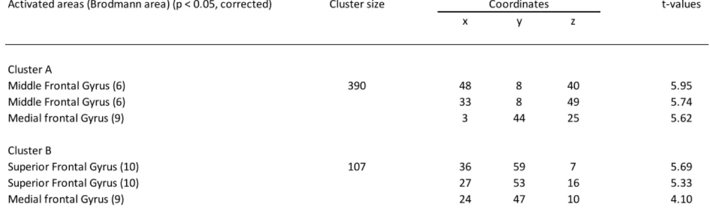
Multiple regression analyses
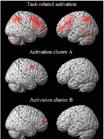
To assess the relation with disease severity, we performed whole-brain functional multiple regression analyses using the corrected hippocampal volume as a regressor. This revealed two significant clusters for which functional activity in MCI was positively correlated with disease severity in the 1-back condition (Figure 2). These clusters comprised a wide range of regions located mainly in the DLPFC with peaks described in Table (2). No other contrast showed significant results.
In order to examine whether the observed clusters of activation were the result of
compensatory brain activation, β-values of both clusters were extracted for all three contrasts and correlated with performance. In the control group, β -values were found to significantly correlate with performance on the 2-back condition for cluster B [r2 = 0.534, p < 0.016] although a
tendency was also observed for cluster A [r2 = 0.407, p < 0.059]. No correlation was found between parameter estimates and performance for MCI participants in either condition when assessed separately. Interestingly, the parameter estimate increase across conditions strongly correlated with MCI performance in both the 1-back [A: r2 = 0.662, p < 0.002; B: r2 = 0.674, p < 0.002] and 2-back conditions [A: r2 = 0.597, p < 0.006; B: r2 = 0.629, p < 0.003] as shown in figure 3. Furthermore, a negative correlation was observed between activation in the 1-back condition and performance in the 2-back condition in the MCI group [A: r2 = -0.444, p < 0.037; B: r2 = -0.514, p < 0.017] but not in controls [A: r2 = 0.003, p < 0.496; B: r2 = 0.353, p < 0.090].
Multimodal Analyses
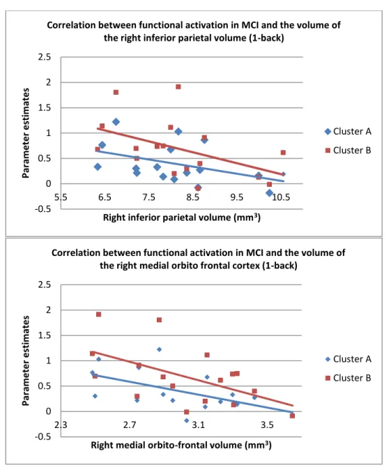
In order to examine the relationship between brain activation and brain volume in MCI, correlational analyses were performed between the parameter estimates extracted from the previously mentioned activation clusters and the corrected volume of a number of regions of interest that showed task-related activation. Results (shown in figure 4) indicate that the amplitude of activation in MCI was negatively correlated with the volume of the right inferior parietal volume for cluster B [A: r2 = -0.572, p < 0.016; B: r2 = -0.667, p < 0.005]. There was also negative correlation between activation for cluster B and the right medial orbitofrontal volume although it did not reach significance when correcting for multiple comparison [A: r2 = -0.616, p < 0.009; B: r2 = -0.652, p < 0.006].
Discussion
The primary aim of this study was to investigate the functional brain activation associated with an executive task in persons with MCI and in healthy older adults. More precisely, our goal was to assess the presence of hyperactivation that is, a larger activation in persons with MCI than in healthy older adults. Our results indicate the presence of a task-load effect in a set of regions that are typically associated with working memory. Activation was found in a number of frontal and parietal regions. Although significant load-related activations were found across both groups in regions associated with the task, no group effect was observed. A possible interpretation of the absence of hyperactivation at the group level could be the high variability of functional response within the patient group. Not all persons with MCI progress to dementia and those who will
progress are likely to vary in their disease severity. For this reason, multiple regression analyses were used to identify regions in which neural activity varied as a function of neural injury.
Results from the multiple regression analyses revealed two significant clusters primarily located in the right DLFPC in which brain activation was negatively correlated with
hippocampal volume in the 1-back condition. In other words, those were regions in which MCI with higher neural injury consistently showed higher task-related activation under low executive load. Interestingly, the DLPFC is a very versatile brain area that has been shown to be activated in a number of memory and executive tasks (Reuter-Lorenz and Park, 2014). Its capacity to “scaffold” or to show increased activation in response to task difficulty has been observed in healthy older adults when compared to younger ones (Spreng et al., 2010). Some authors have argued that the increase in DLPFC activity observed with aging could be a compensatory mechanism caused by normal neural loss that would negatively impact the efficiency of more specialised areas of the brain. Although hyperactivation of the DLFPC in MCI is not as well documented, the same mechanism could be used in response to neural injury specific to the disease. Overall, these result supports our view that larger activation in MCI is associated with increased severity of the disease as indexed by hippocampal volume and that this is found under low executive load.
We also measured whether more activation was associated with better performance. To this end, we extracted the parameter estimates of the significant clusters across both groups and conditions and ran additional correlational analyses with performance on the task. Our results showed a significant positive correlation between functional activation and performance in the control group on the 2-back condition, suggesting that older adults who perform better on the task recruited those areas under a high executive load for task completion. Performance in MCI
participants, however, was not correlated with brain activation for either condition separately. We then investigated the ability in MCI patients to increase neural recruitment in response to task difficulty by measuring the slope of the parameter estimates between the 1-back and the 2-back conditions. Interestingly and for both clusters, the increase in functional activation between the two conditions was highly correlated with performance. These results are coherent with our view that the MCI participants with higher neural injury hyperactivated these regions under low executive load and did not have the neural resources to successfully respond to an increase in task difficulty. This is further supported by the fact that the MCI participants who showed the most brain activation in the 1-back condition were also those whose performance scores dropped the most on the 2-back condition. Although hypoactivation has been observed in the DLPFC in response to high task difficulty (Cappell et al., 2010), this effect was not present in the current study. Furthermore, no difference in performance was observed between the groups. It is thus likely that the MCI participants with high neural injury hyperactivated the right DLPFC in the 1-back condition. Also, although their capacity to increase neural recruitment in response to the 2-back condition was limited, it was sufficient to maintain the ability to complete the task albeit not as efficiently. Interestingly, our results diverge from those of previous studies on
compensatory mechanisms that have traditionally found hyperactivations in MCI participants with lower neural injury. This discrepancy could be due to the fact that those studies relied on an episodic memory task which is a cognitive function that is affected earlier in the disease. Since executive functions are typically affected at a later stage it is likely that, in our study, the MCI participants with lower neural injury did not require the recruitment of additional neural resources to complete the WM task normally.
Another goal of this study was to investigate whether the hyperactivations observed in the 1-back condition were related to neural loss in frontal and parietal regions as measured by structural volumetry. Our results showed that higher DLFPC neural recruitment was linked with lower structural volumes in both the right inferior parietal and medial orbitofrontal cortex. This is consistent with previous studies which have found that the metabolic (Habeck et al., 2012) and structural changes (Grambaite et al., 2011; Nho et al., 2012) of these regions to be associated with lower scores on tests of executive function. Interestingly, evidence suggests a high level of connectivity between those regions. For instance, inputs from the orbitofrontal cortex which has traditionally been associated with reinforcement learning have been shown to be integrated with inputs from the DLFPC through the striatum (Haber & Knudson, 2010). Functional connectivity between the posterior parietal lobe which supports visuospatial processing and the DLFPC has also been demonstrated in adults (Choi et al., 2012). In a recent study, Jarbo and Verstynen (2015) have also found the DLFPC, the inferior parietal cortex and the medial orbitofrontal cortex to be part of the same structural and functional network with projections organised primarily in a rostral-caudal fashion. Given the fact that both the inferior parietal and medial orbitofrontal cortex are specialised brain areas, neural loss would likely affect their efficiency and add “noise” to the network. The inverse relationship observed in our study between the volume of those regions and hyperactivations in the DLPFC supports the view that increased frontal neural recruitment could play a compensatory role in the case of neural injury in MCI.
There were a few limitations to this study. First and foremost, no group or group x condition differences were found on activation level. We argue that this was probably due to the variability of brain response within the MCI group. Not all individuals with MCI will progress to dementia and it is expected that non-progressors will not suffer from neuronal injury.
Furthermore, compensatory models propose that compensatory hyperactivation should increase in the active compensation phase as a function of neuronal damage. This view is supported by the large number of studies in which group differences were only observable after splitting the MCI group into two groups according to the severity of their symptoms as measured by clinical instruments such as the MMSE or CDR (e.g. Celone et al., 2006 and Clement and Belleville, 2010). Importantly, the methodology used in these previous studies relied on determining an arbitrary cut-off based on clinical measures. Here, we decided to use multiple regression
analyses to identify regions in which the level of recruitment varied as a function of neural injury of the hippocampus, a recognized biomarker of AD. With this procedure, larger brain activation was observed MCI participants as a function of their level of neural injury. Our results suggest that the hyperactivation of the DLPFC was associated with neural loss in executive areas that shared the same network. However, the correlational nature of our analyses limits our ability to infer causality.
Summary
The goal of this study was to investigate the nature of hyperactivations and its relationship with structural atrophy in MCI and healthy older adults during a WM task. We found a number of frontal and parietal regions for which brain activation increased as a function of executive load. Furthermore, a subset of right frontal regions concentrated in the DLPFC was found to hyperactivate as a function of neural injury in MCI under low executive load. The successful scaffolding of these brain regions in response to an increase in task difficulty was related to better performances in healthy older adults. Our results suggest that MCI with high
neural injury exhibited similar scaffolding mechanisms but relied on them more heavily under low executive load which limited their ability to adequately increase neural recruitment in the face of an increase in task difficulty. The level of hyperactivation in MCI was also found to be related to a loss in structural volume of executive areas that were part of the same network. Neural loss in those areas likely negatively impacted the efficiency of the network which we believe was partially offset by increased recruitment of the DLPFC.
Table 1 - Sociodemographic et neuropsychological data
Table 2 - List of structural regions based on the Desikan-Killiany Atlas for which the volume was used in our mulimodal analyses.
Controls n = 16 MCI n = 17 Sig.
Gender 12W. 4M 10W. 7M
Age 73.00 (5.11) 72.43 (8.35)
Education 15.25 (3.77) 15,38 (4,97)
MMSE_1 28.63 (1.40) 26.63 (2.47) p < 0.009
MoCA_1 28.25 (1.57) 25.13 (3,67) p < 0.004
Brixton spatial anticipation test 17,00 (7,97) 16,93 (7,42)
Stroop 3rd plate (errors) 0.44 (0.63) 2,00 (1.51) p < 0.001
Birmingham object recognition battery 26,21 (2,11) 26.81 (1.40) Copy of Rey's figure (score) 30.65 (4,66) 29.00 (3,25)
Delayed recall of Rey's figure (score) 18,75 (5,46) 12,09 (4,47) p < 0.001
RL/RI-16 3rd free recall 11,94 (2,17) 8.31 (3,59) p < 0.002
RL/RI-16 delayed free recall 12.50 (2,13) 9.00 (3.44) p < 0.002
Boston Naming Test 13.86 (1,46) 12.38 (1,93) p < 0.026
Caudal middle frontal Inferior parietal Lateral orbito frontal Medial orbito frontal Pars orbitalis Pars triangularis Rostral middle frontal Superior frontal Superior parietal Frontal pole
Table 3 - Clusters that showed increased activity as a function of hypocampal volume in MCI during the 1-back condition with cluster size, peaks and corresponding t-values.
Activated areas (Brodmann area) (p < 0.05, corrected) Cluster size Coordinates t-values
x y z
Cluster A
Middle Frontal Gyrus (6) 390 48 8 40 5.95
Middle Frontal Gyrus (6) 33 8 49 5.74
Medial frontal Gyrus (9) 3 44 25 5.62
Cluster B
Superior Frontal Gyrus (10) 107 36 59 7 5.69
Superior Frontal Gyrus (10) 27 53 16 5.33
Figure 2 – Task related activation and location of activation clusters representing areas of increased activation in relation to smaller hippocampal volume in MCI.
Figure 3 - Correlation between changes in brain activation in response to an increase in difficulty (2 – 1 back) and performance in MCI.
-0.8 -0.6 -0.4 -0.2 0 0.2 0.4 0.6 0.8 0.65 0.7 0.75 0.8 0.85 0.9 0.95 1 Par am e te r e sti m ate s Performance
Correlation between changes in functional activation and performance (1-back) Cluster A Cluster B -0.8 -0.6 -0.4 -0.2 0 0.2 0.4 0.6 0.8 0.65 0.7 0.75 0.8 0.85 0.9 0.95 1 Par am e te r e sti m ate s Performance
Correlation between changes in functional activation and performance (2-back)
Cluster A Cluster B
Figure 4 – Multimodal correlations between parameter estimates in MCI (1-back) and the volume of the right inferior parietal and right medial orbitofrontal cortices.
-0.5 0 0.5 1 1.5 2 2.5 5.5 6.5 7.5 8.5 9.5 10.5 Par am e te r e sti m ate s
Right inferior parietal volume (mm3)
Correlation between functional activation in MCI and the volume of the right inferior parietal volume (1-back)
Cluster A Cluster B -0.5 0 0.5 1 1.5 2 2.5 2.3 2.7 3.1 3.5 Par am e te r e sti m ate s
Right medial orbito-frontal volume (mm3)
Correlation between functional activation in MCI and the volume of the right medial orbito frontal cortex (1-back)
Cluster A Cluster B
References
Bélanger, S., & Belleville, S. (2009). Semantic inhibition impairment in mild cognitive impairment: a distinctive feature of upcoming cognitive decline?. Neuropsychology,
23(5), 592. doi: 10.1037/a0016152
Belleville, S., Chertkow, H., & Gauthier, S. (2007). Working memory and control of attention in persons with Alzheimer's disease and mild cognitive impairment. Neuropsychology,
21(4), 458. doi: 10.1037/0894-4105.21.4.458
Belleville, S., Gauthier, S., Lepage, É., Kergoat, M. J., & Gilbert, B. (2014). Predicting decline in mild cognitive impairment: A prospective cognitive study. Neuropsychology, 28(4), 643. Doi : 10.3233/JAD-130842
Brett, M., Anton, J. L., Valabregue, R., & Poline, J. B. (2002). Region of interest analysis using an SPM toolbox 8th International Conference on Functional Mapping of the Human Brain. Sendai, Japan, p. abstract, 497.
Burgess, P.W., & Shallice, T. (1997). The Hayling and Brixton Tests. Thurston, UK: Thames Valley Test Company.
Cabeza, R., & Nyberg, L. (2000). Imaging cognition II: An empirical review of 275 PET and fMRI studies. Journal of cognitive neuroscience, 12(1), 1-47. doi:
10.1162/08989290051137585
Cappell, K. A., Gmeindl, L., & Reuter-Lorenz, P. A. (2010). Age differences in prefontal recruitment during verbal working memory maintenance depend on memory load.
Celone, K. A., Calhoun, V. D., Dickerson, B. C., Atri, A., Chua, E. F., Miller, S. L., ... & Sperling, R. A. (2006). Alterations in memory networks in mild cognitive impairment and Alzheimer's disease: an independent component analysis. The Journal of
neuroscience, 26(40), 10222-10231. doi: 10.1523/JNEUROSCI.2250-06.2006
Chapman, R. M., Mapstone, M., McCrary, J. W., Gardner, M. N., Porsteinsson, A., Sandoval, T. C., ... & Reilly, L. A. (2011). Predicting conversion from mild cognitive impairment to Alzheimer's disease using neuropsychological tests and multivariate methods. Journal of
clinical and experimental neuropsychology, 33(2), 187-199. doi:
10.1080/13803395.2010.499356
Clément, F., & Belleville, S. (2012). Effect of disease severity on neural compensation of item and associative recognition in mild cognitive impairment. Journal of Alzheimer's
Disease, 29(1), 109-123. doi: 10.3233/JAD-2012-110426
Clément, F., Belleville, S., & Mellah, S. (2010). Functional neuroanatomy of the encoding and retrieval processes of verbal episodic memory in MCI. Cortex, 46(8), 1005-1015. doi: 10.1016/j.cortex.2009.07.003
Clément, F., Gauthier, S., & Belleville, S. (2013). Executive functions in mild cognitive impairment: emergence and breakdown of neural plasticity. Cortex, 49(5), 1268-1279. doi: 10.1016/j.cortex.2012.06.004
Chetelat, G., Landeau, B., Eustache, F., Mezenge, F., Viader, F., de La Sayette, V., ... & Baron, J. C. (2005). Using voxel-based morphometry to map the structural changes associated with rapid conversion in MCI: a longitudinal MRI study. Neuroimage, 27(4), 934-946. doi: 10.1016/j.neuroimage.2005.05.015
Choi, E. Y., Yeo, B. T., & Buckner, R. L. (2012). The organization of the human striatum estimated by intrinsic functional connectivity. Journal of neurophysiology, 108(8), 2242-2263. doi: 10.1152/jn.00270.2012
Cummings, J. L. (1995). Anatomic and Behavioral Aspects of Frontal‐Subcortical Circuitsa.
Annals of the New York Academy of Sciences, 769(1), 1-14.
Dickerson, B. C., & Sperling, R. A. (2008). Functional abnormalities of the medial temporal lobe memory system in mild cognitive impairment and Alzheimer's disease: insights from functional MRI studies. Neuropsychologia, 46(6), 1624-1635. doi:
10.1016/j.neuropsychologia.2007.11.030
Desikan, R. S., Ségonne, F., Fischl, B., Quinn, B. T., Dickerson, B. C., Blacker, D., ... & Killiany, R. J. (2006). An automated labeling system for subdividing the human cerebral cortex on MRI scans into gyral based regions of interest. Neuroimage, 31(3), 968-980. doi: 10.1016/j.neuroimage.2006.01.021
Du, A. T., Schuff, N., Amend, D., Laakso, M. P., Hsu, Y. Y., Jagust, W. J., ... & Weiner, M. W. (2001). Magnetic resonance imaging of the entorhinal cortex and hippocampus in mild cognitive impairment and Alzheimer's disease. Journal of Neurology, Neurosurgery &
Psychiatry, 71(4), 441-447. doi: 10.1136/jnnp.71.4.441
Fischl, B., Salat, D. H., van der Kouwe, A. J., Makris, N., Ségonne, F., Quinn, B. T., & Dale, A. M. (2004). Sequence-independent segmentation of magnetic resonance images.
Neuroimage, 23, S69-S84. doi: 10.1016/j.neuroimage.2004.07.016
Folstein, M. F., Folstein, S. E., & McHugh, P. R. (1975). “Mini-mental state”: a practical method for grading the cognitive state of patients for the clinician. Journal of psychiatric
Gevins, A. S., & Cutillo, B. C. (1993). Neuroelectric evidence for distributed processing in human working memory. Electroencephalogr. Clin. Neurophysiol, 87, 128-143. Grambaite, R., Selnes, P., Reinvang, I., Aarsland, D., Hessen, E., Gjerstad, L., & Fladby, T.
(2011). Executive dysfunction in mild cognitive impairment is associated with changes in frontal and cingulate white matter tracts. Journal of Alzheimer's Disease, 27(2), 453-462. doi: 10.3233/JAD-2011-110290
Habeck, C., Risacher, S., Lee, G. J., Glymour, M. M., Mormino, E., Mukherjee, S., ... & Alzheimer‟s Disease Neuroimaging Initiative. (2012). Relationship between baseline brain metabolism measured using [18F] FDG PET and memory and executive function in prodromal and early Alzheimer‟s disease. Brain imaging and behavior, 6(4), 568-583. doi : 10.1007/s11682-012-9208-x
Haber, S. N., & Knutson, B. (2010). The reward circuit: linking primate anatomy and human imaging. Neuropsychopharmacology, 35(1), 4-26. doi: 10.1038/npp.2009.129
Jack, C. R., Petersen, R. C., Xu, Y. C., O‟Brien, P. C., Smith, G. E., Ivnik, R. J., ... & Kokmen, E. (1999). Prediction of AD with MRI-based hippocampal volume in mild cognitive impairment. Neurology, 52(7), 1397-1397. doi: 10.1212/WNL.52.7.1397
Jarbo, K., & Verstynen, T. (2015). Converging structural and functional connectivity of
orbitofrontal, dorsolateral prefrontal, and posterior parietal cortex in the human striatum.
The Journal of Neuroscience, 35(9), 3865-3878. doi:
Johns, E. K., Phillips, N. A., Belleville, S., Goupil, D., Babins, L., Kelner, N., ... & Chertkow, H. (2012). The profile of executive functioning in amnestic mild cognitive impairment: disproportionate deficits in inhibitory control. Journal of the International
Neuropsychological Society, 18(03), 541-555. doi: 10.1017/S1355617712000069
Julkunen, V., Niskanen, E., Muehlboeck, S., Pihlajamäki, M., Könönen, M., Hallikainen, M., ... & Soininen, H. (2008). Cortical thickness analysis to detect progressive mild cognitive impairment: a reference to Alzheimer's disease. Dementia and geriatric cognitive disorders, 28(5), 404-412. doi: 10.1159/000256274
Kaplan, E.F., Goodglass, H., & Weintraub, S. (1983). The Boston Naming Test. (2nd ed .). Philadelphia, PA: Lea & Febiger.
Nasreddine, Z. S., Phillips, N. A., Bédirian, V., Charbonneau, S., Whitehead, V., Collin, I., ... & Chertkow, H. (2005). The Montreal Cognitive Assessment, MoCA: a brief screening tool for mild cognitive impairment. Journal of the American Geriatrics Society, 53(4), 695-699. doi: 10.1111/j.1532-5415.2005.53221.x
Newman, L. M., Trivedi, M. A., Bendlin, B. B., Ries, M. L., & Johnson, S. C. (2007). The relationship between gray matter morphometry and neuropsychological performance in a large sample of cognitively healthy adults. Brain imaging and behavior, 1(1-2), 3-10. doi: 10.1007/s11682-007-9000-5
Nho, K., Risacher, S. L., Crane, P. K., DeCarli, C., Glymour, M. M., Habeck, C., ... &
Alzheimer‟s Disease Neuroimaging Initiative (ADNI. (2012). Voxel and surface-based topography of memory and executive deficits in mild cognitive impairment and
Alzheimer‟s disease. Brain imaging and behavior, 6(4), 551-567. doi: 10.1007/s11682-012-9203-2
Pa, J., Possin, K. L., Wilson, S. M., Quitania, L. C., Kramer, J. H., Boxer, A. L., ... & Johnson, J. K. (2010). Gray matter correlates of set-shifting among neurodegenerative disease, mild cognitive impairment, and healthy older adults. Journal of the International
Neuropsychological Society, 16(04), 640-650. doi: 10.1017/S1355617710000408
Petersen, R. C., Doody, R., Kurz, A., Mohs, R. C., Morris, J. C., Rabins, P. V., ... & Winblad, B. (2001). Current concepts in mild cognitive impairment. Archives of neurology, 58(12), 1985-1992. doi: 10.1001/archneur.58.12.1985.
Prvulovic, D., Van de Ven, V., Sack, A. T., Maurer, K., & Linden, D. E. (2005). Functional activation imaging in aging and dementia. Psychiatry Research: Neuroimaging, 140(2), 97-113. doi: 10.1016/j.pscychresns.2005.06.006
Rabin, L. A., Saykin, A. J., West, J. D., Borgos, M. J., Wishart, H. A., Nutter-Upham, K. E., ... & Santulli, R. B. (2009). Judgment in older adults with normal cognition, cognitive
complaints, MCI, and mild AD: Relation to regional frontal gray matter. Brain imaging
and behavior, 3(2), 212-219. doi: 10.1007/s11682-009-9063-6
Regard, M. (1981). Cognitive Rigidity and Flexibility: A Neuropsychological Study. (Unpublished Ph.D. dissertation). Canada: University of Victoria.
Reuter-Lorenz, P. A., & Park, D. C. (2014). How does it STAC up? Revisiting the scaffolding theory of aging and cognition. Neuropsychology review, 24(3), 355-370. doi:
10.1007/s11065-014-9270-9
Rey, A. (1983) Test de copie d’une figure complexe: manuel. Paris: Les éditions du centre de psychologie appliquée.
Riddoch, M.J., Humphreys, G.W. (1993). BORB: Birmingham Object Recognition Battery. Hove, UK, Lawrence Erlbaum Associates.
Ségonne, F., Dale, A. M., Busa, E., Glessner, M., Salat, D., Hahn, H. K., & Fischl, B. (2004). A hybrid approach to the skull stripping problem in MRI. Neuroimage, 22(3), 1060-1075. doi: 10.1016/j.neuroimage.2004.03.032
Ségonne, F., Pacheco, J., & Fischl, B. (2007). Geometrically accurate topology-correction of cortical surfaces using nonseparating loops. Medical Imaging, IEEE Transactions on,
26(4), 518-529. doi: 10.1109/TMI.2006.887364
Sled, J. G., Zijdenbos, A. P., & Evans, A. C. (1998). A nonparametric method for automatic correction of intensity nonuniformity in MRI data. Medical Imaging, IEEE Transactions
on, 17(1), 87-97. doi: 10.1109/42.668698
Singh, V., Chertkow, H., Lerch, J. P., Evans, A. C., Dorr, A. E., & Kabani, N. J. (2006). Spatial patterns of cortical thinning in mild cognitive impairment and Alzheimer's disease. Brain,
129(11), 2885-2893. doi: 10.1093/brain/awl256
Sperling, R. A., Dickerson, B. C., Pihlajamaki, M., Vannini, P., LaViolette, P. S., Vitolo, O. V., ... & Johnson, K. A. (2010). Functional alterations in memory networks in early
Alzheimer‟s disease. Neuromolecular medicine, 12(1), 27-43. doi: 10.1007/s12017-009-8109-7
Spreng, R. N., Wojtowicz, M., & Grady, C. L. (2010). Reliable differences in brain activity between young and old adults: a quantitative meta-analysis across multiple cognitive domains. Neuroscience & Biobehavioral Reviews, 34(8), 1178-1194.
doi:10.1016/j.neubiorev.2010.01.009
Thomann, P. A., Toro, P., Dos Santos, V., Essig, M., & Schröder, J. (2008). Clock drawing performance and brain morphology in mild cognitive impairment and Alzheimer‟s disease. Brain and cognition, 67(1), 88-93. doi: 10.1016/j.bandc.2007.11.008
Van der Linden, M., Adam, S., Agniel, A., Baisset-Mouly, C., Bardet, F., Coyette, F., et al. (2004) L’évaluation de troubles de la mémoire: présentation de quatre tests de mémoire
épisodique (avec étalonnage). Marseille: Solal.
Woodard, J. L., Seidenberg, M., Nielson, K. A., Antuono, P., Guidotti, L., Durgerian, S., ... & Rao, S. M. (2009). Semantic memory activation in amnestic mild cognitive impairment.
Conclusion
La maladie d‟Alzheimer est d‟apparition graduelle et se caractérise par la présence d‟une longue phase prodromale. Des auteurs ont proposé que les processus de plasticité neuronale pourraient être actifs pendant cette phase et qu‟ils se reflèteraient par la présence
d‟hyperactivation compensatoire. Le but principal de ce travail était de tester cette hypothèse en faisant appel à une tâche de mémoire de travail. De manière complémentaire aux travaux effectués par le passé, nous avons étudié l‟impact d‟une hausse du niveau de difficulté sur le patron d‟activations fonctionnelles chez les sujets TCL et les sujets âgés sains. Afin de rendre compte de l‟impact possible de l‟hétérogénéité du degré de sévérité pathologique au sein du groupe TCL, nous avons effectué des analyses de régressions multiples. Cela nous a permis d‟identifier des régions cérébrales dont l‟activité variait en fonction de l‟atteinte neuronale telle que caractérisée par le volume hippocampique. Des analyses de corrélation furent ensuite effectuées entre le niveau d‟activation des clusters précédemment identifiés et la performance à la tâche afin de déterminer si les hyperactivations pouvaient jouer un rôle compensatoire. Finalement, nous avons examiné la relation entre les hyperactivations obtenues et le volume de régions cérébrales impliquées dans la tâche. Les résultats obtenus seront discutés plus bas puis seront suivis des limites et des implications futures.
Comparaisons entre les groupes
Le premier objectif de ce mémoire était d‟investiguer l‟activation fonctionnelle cérébrale associée à une tâche de mémoire de travail dans le TCL et chez des personnes âgées saines. Plus précisément, nous cherchions à mieux caractériser la présence d‟hyperactivations, soit des