Characterization of UDP-glucose:protein transglucosylase genes from
potato
Flavia A. Wald
1, Ralph Kissen
2, Patrick du Jardin
2and Silvia Moreno
1,∗1Plant Biochemistry Laboratory, Instituto Leloir (formerly: Instituto de Investigaciones Bioqu´ımicas Fundaci´on Campomar), I.I.B.B.A.-CONICET, Av. Patricias Argentinas 435, (1405) Buenos Aires, Argentina (∗author for correspondence; e-mail smoreno@leloir.org.ar); 2Plant Biology Unit, Gembloux Agricultural University, 5030 Gembloux, Belgium
Received 26 July 2002; accepted in revised form 27 February 2003
Key words: autocatalytic glycosyltranferase, cell wall, polysaccharide synthesis, potato, UDP-glucose
Abstract
Many plant autocatalytic glycosyltransferases are implicated in plant polysaccharide biosynthesis. Cloning of cDNAs encoding potato (Solanum tuberosum L.) UDP-Glc:protein transglucosylase (UPTG, EC 2.4.1.112) and expression of the cDNA clone E11 in Escherichia coli have been previously reported. Here, we studied the functional expression of a second cDNA of the enzyme (E2 clone). Northern blots analysis, with specific cDNA probes for the two UPTG isoforms, showed a differential expression pattern of mRNA levels in different potato tissues. Moreover, both UPTG recombinant enzymes showed different kinetic parameters. The recombinant protein encoded by E2 clone has an apparent Imax for UDP-Xyl and UDP-Gal, significantly higher than for UDP-Glc. The Kmvalues for UDP-Glc were 0.45–0.71 µM and the values for UDP-Xyl and UDP-Gal were slightly higher than
that of the UDP-Glc (1.2–2.71 µM) for both UPTG recombinant enzymes. The present study revealed further evidence for the proposed role of UPTG in the synthesis of cell wall polysaccharide. It was found a correlation between UPTG transcript levels and the growing state of the tissues in which there was an active synthesis of cell wall components. Southern blot analysis indicates that at least three genes encoding UPTG are present in potato genome. Phylogenetic analysis of both UPTG recombinant proteins showed that they are members of the RGP subfamilies from dicots.
Introduction
Glycosylation reactions have a large biological im-portance for both prokaryotes and eukaryotes and are dependent on a class of enzymes named glycosyltrans-ferases. The substrate specificity of those glycosyl-transferases are now beginning to be established, little is known yet about the regulation of plant glycosyl-transferase genes or the localization of the enzymes that encode at the cellular and subcellular levels. Thus, although a very good understanding exists of the com-position of commercially important plant polymers such as cellulose, pectic substances, hemicelluloses and starch, little is known about the enzymes involved in the early stages of polysaccharide synthesis.
Redundancy is a common feature of many of these glycosyltransferases. Different genes, post-translational modifications and different conforma-tional isoforms have been reported (Winter et al., 1997; Renz et al., 1998). Moreover, the expression of several glycosyltransferase genes, such as sucrose syn-thase in higher plants, could be enhanced in specific tissues and favour optimization of different functions (Koch et al., 1992; Chourey et al., 1998). There-fore, the elucidation of the physiological role of these enzymes involved in the metabolism of plant polysac-charides turned out very complex. The cloning of a gene that encode a putative component of the cellulose synthase complex, represented a remarkable achieve-ment in this field (Arioli et al., 1998). Concerning starch, the enzymes responsible for its elongation and
branching have been extensively characterized and their coding genes were isolated and cloned, however, the initiation of the polymer received relatively little attention (Myers et al., 2000).
Plant cells, unlike animal cells, are surrounded by a cell wall. Studies aimed at identifying the protein re-sponsible for biosynthesis of the cell wall polysaccha-rides have revealed the presence of a small multigene family of autocatalytic glycosyltransferases in pea epi-cotyls (Dhugga et al., 1991), Arabidopsis (Delgado et al., 1998) and potato (Bocca et al., 1999). In an-imal tissues, polysialyltransferase-1 (Hart, 1997) and glycogenin (Alonso et al., 1995), which performs the first steps of the glycogen synthesis, provide further examples of those autocatalytic proteins. Many ques-tions remain unanswered regarding the precise func-tion of the self-glycosylating proteins in tissue plants, however, the substrate specificity and the localization of some of them seem to agree with a role in the syn-thesis of hemicellulosic polysaccharide (Dhugga et al., 1997). Faik et al. (2000) reported that in Nasturtium an autoglycosylating polypeptide(s) fruit could facilitate the channelling of UDP-activated sugars from the cy-toplasm through Golgi vesicle membranes to luminal sites, where they can be used for xyloglucan synthesis. Thus, this new family of glycosyltranferases opens the door to the understanding of cell wall polysaccharide synthesis.
In potato, UDP-Glc:protein transglucosylase (UPTG, UDPglucose:protein 4α-glucosyltransferase, EC 2.4.1.112) undergoes self-glucosylation from UDP-Glc and Mn2+ (Ardila and Tandecarz, 1992). Then, it was purified to apparent homogeneity from potato tubers, and a Km value of 0.7 µM for the
glucosyl donor UDP-Glc was found (Bocca et al., 1997). Using a polyclonal antibody raised against the UPTG protein from potato (Solanum tuberosum L.), we isolated ten positive clones after the screening of a cDNA expression library prepared from swelling stolon tips poly(A) RNA. The cDNA clone E11 dis-played UPTG activity in Escherichia coli and two categories of UPTG-related sequences from plants have been reported (Bocca et al., 1999). The first cate-gory of sequences shares 86–93% identity with UPTG and includes the reversible glycosylating polypeptides (RGP) (Dhugga et al., 1997). The second one shares 44–47% identity at the amino acid level (Bocca et al., 1999).
UPTG has also been reported to be present in developing maize endosperm (Rothschild and Tande-carz, 1994) in which the presence of an UPTG
in-hibitor protein has been described (Rothschild et al., 1996). This UPTG inhibitor was later identified as the SS1 isozyme of sucrose synthase (Wald et al., 1998). In maize kernels SS1 is important in the main-tenance of cell wall integrity (Chourey et al., 1998). Thus, these results point towards a possible function of UPTG in cell wall polysaccharide synthesis. Although UPTG and related proteins have been suggested to play a role in the synthesis of cell wall polysaccha-rides, many questions should be addressed about the physiological role of the self-glycosylating proteins and their regulation (Dhugga et al., 1997; Bocca et al., 1999).
We are studying the expression of the UPTG gene in potato in order to identify putative genes involved in the regulation of polysaccharide synthesis. In this work, we characterized and expressed in E. coli a sec-ond cDNA (E2 clone) of potato which shares 89% identity with cDNA clone E11. We have also reported that the two closely UPTG isozymes exhibit differ-ent kinetic properties, and a differdiffer-ential expression pattern of its mRNA levels in different potato tissues was observed. Moreover, a correlation between UPTG transcript levels and the growing state of the tissues was observed. This study presents further evidence for the proposed role of UPTG in the synthesis of cell wall polysaccharides.
Materials and methods
The potato (Solanum tuberosum L. cv. Désirée) plants used were grown in a greenhouse under natural light supplemented with fluorescent light. Plantlets were grown aseptically in flasks containing Murashigue and Skoog medium (Murashigue and Skoog, 1962) sup-plemented with 2% sucrose and 0.2% gelrite (Kelco) and kept at 22–24◦C under a 16 h photoperiod (40– 60 µE m−2 s−1) with cool white fluorescent tubes (Osram). For plantlet multiplication, nodal cuttings with a single axillary bud were transferred to fresh medium every 21–28 days. Tubers were produced by an in vitro tuberization system as previously described (Moreno and Tandecarz, 1996).
UDP-[14C]Glc (11 100 GBq/mol), UDP-[14C]Gal (11 100 GBq/mol) and UDP-[14C]Xyl (11 100 GBq/ mol) were obtained according to Thomas et al. (1968).
Expression of the recombinant protein in Escherichia coli
Single colonies of E. coli XL1Blue cells, transformed by either the plamisd pE11 (clone E11), plasmid pE2
(clone E2) or the parental (pBluescript) plasmids, were
used to inoculate 2 ml LB (Luria Bertani broth) con-taining 100 µg/ml ampicillin and incubated at 37◦C overnight with shaking (Bocca et al., 1999). Of the overnight culture 1 ml was sub-cultured in 50 ml of the same medium and grown to A600 0.4 before
the expression of the recombinant UPTG was in-duced by adding 0.1 mM IPTG. After growing for 12 h at 30 ◦C, cells were pelleted and re-suspended in 1 ml lysis buffer (25 mM Tris pH 7.6, 100 mM KCl, 0.1 mM EDTA, 12.5 mM MgCl2, 10%
glyc-erol and 0.1% NP-40) in the presence of protease inhibitors (1 mM DTT, 0.1 mM sodium metabisulfite and 0.1 mM PMSF). Lysozyme was added to a final concentration of 0.5 mg/ml. After 30 min of incu-bation the suspension was sonicated and centrifuged at 10 000× g for 20 min. The supernatant was used for biochemical assays, SDS-PAGE and western blot analysis.
SDS-PAGE and western blots
SDS-PAGE analysis was performed on 10% acry-lamide gels according to Laemmli (1970). For western blot analysis, proteins were transferred onto nitro-cellulose membranes (Towbin et al., 1970), blocked with 10% non-fat milk in PBS 0.2% NP-40 and incu-bated with anti-potato tuber UPTG antiserum (1:1000 dilution) for 60 min. Immunoreactive bands were visualized by the ECL system (Amersham).
UPTG glycosylation assays
For analysis of glycosylation 50–100 µg protein from the soluble extract of the E. coli cultures was incu-bated at 30◦C for 30 min with UDP-[14C]Glc, UDP-[14C]Gal or UDP-[14C]Xyl (0.2 nmol, 100 000 cpm) and 10 mM MnCl2 in a final volume of 100 µl.
Transfer of sugar was measured as incorporation of ra-dioactivity into a 10% w/v trichloroacetic acid (TCA) pellet as described (Bocca et al., 1999). Alternatively, the precipitated proteins (50 µg) were separated by SDS-PAGE, stained with Coomassie blue R-250, and subjected to autoradiography for 2 weeks.
Analysis of RNA and DNA
Plant material was harvested and frozen immediately in liquid nitrogen. Total RNA was isolated according to Logemann et al. (1987). Total RNA (20 µg per lane) was subjected to electrophoresis in a formalde-hyde gel and blotted onto a Hybond-N+ membrane (Amersham) essentially as described (Sambrook et al., 1989). After UV cross-linking, the membranes were prehybridized with a solution containing 1% BSA, 0.5 M sodium phosphate, 1 mM EDTA, 7% SDS at 65◦C and hybridized overnight at 65◦C in the same solution with the labelled UPTG probe. Then they were washed twice at room temperature for 5 min with 40 mM sodium phosphate, 1 mM EDTA, 1% SDS, and then for 15 min at 65◦C. Alternatively, prehybridiza-tion of the blots was performed for 2 h at 65◦C in a solution composed of 6× SSC, 5× Denhardt’s so-lution, 0.5% SDS and 10 mg/ml salmon sperm DNA. The gel blots were hybridized with a 1.4 kb UPTG cDNA insert from pE11 (complete E11 cDNA probe) and with UPTG1- and UPTG2-specific probes. The UPTG1-specific probe is a 330 bp cDNA fragment corresponding to the amino acid sequence 338–367 encoding UPTG1, and the UPTG2-specific probe is a 242 bp fragment corresponding to the amino acid sequence 341–365 encoding UPTG2. Probes were la-belled with [32P]dCTP (prime-a-gene labelling kit, Promega). Alternatively, hybridization was performed in a fresh solution (same composition) for 16 h at 65◦C with [32P]dCTP-labelled probes (Oligolabeling Kit, Pharmacia). After hybridization the blots were first washed four times for 5 min at room tempera-ture with a 2× SSC solution, then twice for 30 min at 65◦C with a 2× SSC/0.5% SDS solution and finally twice for 30 min at room temperature with a 0.1× SSC solution. Signals were detectable after 2–4 days of ex-posure at−70◦C of the blot to X-ray film (Biomax MR, Kodak) with an intensifying screen. The filter was then stripped off the probe (90 ◦C, 0.1× SSC, 0.1% SDS) and re-hybridized with a control potato 18S rRNA to verify equal RNA loading on the gel and equal transfer to the membrane. Quantification of RNA was performed with Scion Image and expressed as arbitrary units relative to RNA loading.
For DNA gel blot analysis, restricted DNA was separated on a 1% agarose gel (10 µg/lane) and blot-ted onto nylon membranes (Hybond-N, Amersham). A fragment of the E11cDNA clone corresponding to the amino acid sequence 143–276 recognizing both genes
(universal probe) and gene-specific DNA fragments were labelled with32P as described above.
Results
Functional expression of a second recombinant UPTG isoform in E. coli
We have described the isolation of two cDNAs of potato stolon tips (E11and E2 clones), GenBank ac-cession numbers AJ223252 and AJ310910, respec-tively); after their sequencing, 89% of identity was observed between them (Bocca et al., 1999). The E11 clone was characterized and its authenticity as UPTG was confirmed. In order to investigate if the cDNA clone E2 codes for a second UPTG isoform, the functional expression in E. coli was performed. The optimal functional expression of UPTG protein in soluble extracts of E. coli cultures was obtained after induction at 0.1 mM IPTG during 16 h. These conditions were similar to those previously described for the expression of cDNA clone E11 (Boca et al., 1999). The term UPTG1 was proposed for the protein encoded by the E11 clone.
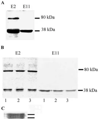
Figure 1A shows the immunoblot analysis of solu-ble protein extracts prepared from E. coli cells trans-formed with the plasmid pE2 (E2 clone) with a poly-clonal antibody raised against the potato UPTG. Solu-ble protein extract from E. coli cells transformed with the E11 clone is also shown. The extracts from the E2 clone showed the presence of a protein with similar mobility to the protein expressed by the E11 clone (ca. 38 kDa). Another band of ca. 80 kDa was also ob-served in the E2 clone. It is important to mention that, as described (Bocca et al., 1999), the 38 kDa band was absent in the extract of the vector (pBluescript) without inserts.
To assay if the E2 clone encodes a function-ally active UPTG, extracts of E. coli were incu-bated with UDP-[14C]Glc, UDP-[14C]Xyl or UDP-[14C]Gal, since UPTG1 can be glycosylated by these UDP sugars. When these UDP sugars were assayed, the radioactivity incorporation levels obtained for the E2 clone were similar to those found for the E11 clone extracts whereas control extracts presented an in-significant incorporation of radioactivity into the TCA precipitates (Table 1). Figure 1B shows the radioactive products, examined by SDS-PAGE and autoradiogra-phy, upon incubation with the three radioactive sugar donors. E. coli extracts of the E2 clone (Figure 1B,
Figure 1. A. Western blot of soluble extracts of E. coli harbour-ing the plasmid pE2 (E2) or pE11 (E11) with an antibody raised against the purified potato tuber UPTG. B. Fluorography after SDS-PAGE of soluble extracts of the E2 clone (E2) or the E11 clone (E11), incubated with (1) UDP-[14C]Glc, (2) UDP-[14C]Xyl or (3) UDP-[14C]Gal. C. Doublet band corresponding with the 38 kDa subunit of the UPTG recombinant protein. Activities were assayed as described in Materials and methods. The lines indicate the induced proteins.
left) showed a radioactive band with the same mobility as that of the polypeptide recognized in the western blot (38 kDa). Moreover, no differences with respect to the radioglycosylation of the 38 kDa subunit of the protein encoded by the E11 clone was observed (Figure 1B, right). It is well known that the extract of vector without insert does not present any radioactive band, as reported (Bocca et al., 1999). Thus, the E2 clone encoded a second UPTG recombinant enzyme and both UPTG protein isoforms showed the same substrate specificity. The term UPTG2 is proposed for the protein encoded by the E2 cDNA clone.
E2 clone extract showed also a 80 kDa glycosy-lated band upon incubation with the three radioactive sugar donors, which is recognized by the anti-UPTG antibody (Figure 1A, B, left). Moreover, a third clone showing UPTG activity, isolated from a potato leaf library (H6 clone), also presented the radioglyco-sylation of 80 kDa, 120 kDa and 150 kDa bands
Table 1. Glycosylation of recombinant UPTG by UDP-[14C]Glc, UDP-[14C]Xyl and UDP-[14C]Gal.
Plasmid Radioactivity in trichloroacetic acid precipitates (cpm) UDP-[14C]Glc UDP-[14C]Xyl UDP-[14C]Gal
Control 265± 35 60± 12 139± 25
E11 clone 3390±170 1080± 86 1800±156 E2 clone 5100± 24 4350±370 3600±290 Control: pBluescript alone.
recognized by the potato UPTG antibody (data not shown). The nature of the 80 kDa band present in the E2 clone extract remains unclear, but it is possible that the high-molecular-weight bands are oligomers of the enzyme.
Another interesting point is the fact that the 38 kDa subunit of both UPTG recombinant proteins showed a doublet band with slightly different electrophoretic mobility after electrophoresis in SDS-PAGE (Fig-ure 1C). Similar results have been reported for the UPTG protein in partially purified preparations of potato tuber (Rothschild et al., 1996; Bocca et al., 1997; Wald, 2001). The results presented in Figure 1 provide strong evidence that the UPTG doublet may arise from a post-translational modification, since it is present after translating the protein from a sin-gle UPTG mRNA. Moreover, this modification must occur after the translation in the recombinant system. UPTG1 and UPTG2 exhibited different kinetic properties
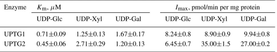
It is of interest to study the kinetic properties of both UPTG isoforms, so the apparent Kmand Imax
(max-imum label incorporation) for both recombinant pro-teins were determined (Table 2). The Km values
ob-tained for UDP-Glc are in the range of 0.45–0.71 µM, which is comparable with that of the native enzyme ex-tracted from potato tuber (Bocca et al., 1997). The val-ues obtained for UDP-Xyl and UDP-Gal are slightly higher than for UDP-Glc (1.2–2.71 µM) for both en-zymes. No variation in the Imax values for UDP-Glc
was observed between UPTG1 and UPTG2. Surpris-ingly, the apparent Imax for UDP-Xyl and UDP-Gal
increased 4-fold for UPTG2 compared to the values obtained for UPTG1. The elevated glycosylation rates for xylose and galactose with similar Kmvalues
sug-gested that UPTG2 could be a more active isoform compared to UPTG1.
Figure 2. mRNA UPTG levels in an in vitro potato culture. To-tal RNA (20 µg) was extracted from non-tuberizing stolons (white bars), stolon-bearing immature tubers (grey bars) or stolon-bearing mature tubers (black bars) after 15 or 35 days of culture on MS containing different concentrations of sucrose (A) and from potato plantlets after different culture times (B). RNA gels blots were hy-bridized with [32P]-labelled full-length E11 cDNA probes, which cross-hybridized with E11 and E2 transcripts. Levels of mRNA were quantified as described in Materials and methods.
Differential expression of UPTG1 and UPTG2 mRNA in potato tissues
RNA hybridization studies, with a full-length UPTG insert as a probe (cDNA clone E11), have indicated that UPTG mRNA was expressed in all potato tis-sues, with expression being highest in stolons (Bocca et al., 1999). Here, we analysed by northern blot-ting with the same probe UPTG expression in vitro in different potato tissues (tuberizing potato culture and micropropagated plantlets). We found similar UPTG mRNA levels in non-tuberizing stolons (15 days of culture on 3% of sucrose) and stolon-bearing imma-ture tubers (15 days of culimma-ture on 6% of sucrose) (Figure 2). However, unexpectedly, higher levels of UPTG mRNA were seen in stolons-bearing immature than in mature tubers (Figure 2A). UPTG mRNA
lev-Table 2. Kinetic parameters for UPTG1 and UPTG2. Values represent averages of data from several independent experiments (n= 3).
Enzyme Km, µM Imax, pmol/min per mg protein UDP-Glc UDP-Xyl UDP-Gal UDP-Glc UDP-Xyl UDP-Gal UPTG1 0.71±0.09 1.25±0.13 1.67±0.17 8.24±0.8 8.90±0.9 9.94±0.8 UPTG2 0.45±0.06 2.71±0.29 1.20±0.13 6.45±0.7 35.00±1.5 27.00±0.2
Figure 3. Differential expression of UPTG1 and UPTG2 mRNA in different potato plant tissues. Different tissues of potato plants growing in soil – leaves (L), senescent leaf (Ls), rachis (Rs), stem (S), stolon (St), tubers (T), dormant tubers (Td) and roots (R) – were analysed by northern blotting with specific probes. A 330 bp frag-ment corresponding to the amino acid sequence 338–367 encoding the UPTG1 (UPTG1-specific probe) and a 242 bp fragment corre-sponding to the amino acid sequence 341–365 encoding the UPTG2 (UPTG2-specific probe) were used as probes. A specific probe for 18 S rRNA was used as control (C).
els remained unresponsive to the appearance of tubers and also remained unresponsive to changes in sucrose concentration (a known tuberization inductor).
According to the assumption that UPTG is in-volved in the cell wall polysaccharide synthesis and because it is well known that during plant develop-ment there is an active synthesis of cell wall com-ponents, we analysed UPTG mRNA levels in micro-propagated plantlets during culture at different stages (Figure 2B). We found a greater UPTG transcript in growing plantlets than in mature plantlets. A correla-tion between UPTG transcript levels and the growing state of the plantlets may exist since a decrease in UPTG expression along the culture time was clearly observed.
The different kinetic properties of the two UPTG isoforms may be indicative of their different functions in vivo. Next, we investigated their transcript levels in different potato plant tissues (Figure 3). Since the identity between UPTG1 and UPTG2 was very high, specific probes for E2 and E11 clones were designed to distinguish one from the other. A 330 bp fragment corresponding to the amino acidic sequence 338–367
encoding the UPTG1 (UPTG1-specific probe) and a 242 bp fragment corresponding to the amino acidic sequence 341–365 encoding the UPTG2 (UPTG2-specific probe) were used as probes (see Materials and methods). These probes, under the conditions performed, did not hybridize between them (data not shown). By northern blot analysis, UPTG2 transcripts were detected in all tisues analysed albeit mRNA levels were higher in leaves and stems (Figure 3A). In contrast, UPTG1 transcripts were detected pre-dominantly in stolons, tubers and roots (Figure 3B). Interestingly, higher mRNA levels were observed for UPTG2 than for UPTG1 in all organs analysed. These results indicated that there is a differential expression between the two UPTG clones in potato tissues. Southern blot analysis
Southern blot analysis was performed with genomic DNA extracted from potato leaves and digested with EcoRI or XbaI. Specific probes for UPTG1 and UPTG2 and a 401 bp fragment corresponding to the amino acid sequence 158–292 encoding the UPTG1 as a universal probe were used. The universal probe, which was amplified by PCR, corresponding to an UPTG sequence shared in more than 90% sequence identity with respect to the homologous sequences present in the data bank. The presence of only one band with the UPTG1-specific probe suggested the presence of a single- or a low-copy gene, since UPTG1 probe does not present a restriction site for EcoRI and XbaI. By contrast, two bands were observed for UPTG2, which could mean the presence of a third iso-form recognized by the UPTG2-specific but not the UPTG1-specific probe. The genomic DNA digested with EcoRI with the universal probe showed three DNA fragments, one of them (4.3 kb) being recog-nized by UPTG1- and UPTG2-specific probes- and another two faint fragments (Figure 4). This result in-dicated that there are at least three copies of UPTG genes in the potato genome.
Figure 4. DNA gel blot analysis of genomic DNA from potato. Genomic DNA (6 µg/lane) was digested with EcoRI (E), or XbaI (X). The fragments were separated by agarose gel electrophore-sis, blotted and were hibridized with 32P-labelled UPTG1- and UPTG2-specific probes and with a universal probe for UPTG cor-responding to the amino acid sequence 143–276. Lines indicate the two faint fragments recognized by the universal probe.
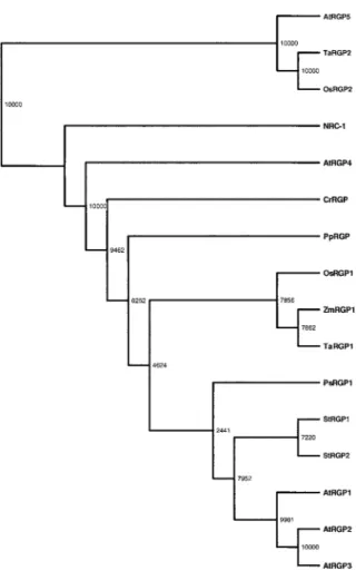
Phylogenic analysis of UPTG
The phylogenetic tree based on multiple sequence alignment of UPTG related proteins is shown in Fig-ure 5. Because this analysis revealed that both UPTG recombinant enzymes are members of the RGP sub-families from dicots, the UPTG1 and UPTG2 terms were changed to StRGP1 and StRGP2 in the clado-gram. It is interesting to note that the sequences of RGP from dicots are separated from those of mono-cots. The sequences AtRGP4, PsRGP1, PpRGP, Cr-RGP and NRC-1 are more separated from the other RGPs. The AtRGP5, TaRGP2 and OsRGP2 sequences form another group, well separated from the other RGPs.
Discussion
Many autocatalytic glycosyltransferases, whose func-tion may be tightly connected to cell wall
polysaccha-Figure 5. Cladograms of the amino acid sequences showing ho-mology with UPTG from potato. The amino acid sequences were aligned with the multiple alignment program CLUSTAL X (Thomp-son et al., 1994) with TREEVIEW software. Abbreviations: RGP, reversibly glycosylated polypeptides; StRGP1 (AJ223252), StRGP2 (AJ310910), RGP from Solanum tuberosum formerly named UPTG; AtRGP1–AtRGP4 (AF013627, AF013628, AF034255, AF329280) from Arabidopsis thaliana; AtRGP5 (Q9FFD2) for a translation of a genomic sequence of Arabidopsis thaliana; PsRGP1 (U31565) from Pisum sativum; ZmRGP1 (U89897) from Zea mays; Os-RGP1 (AJ011078), OsRGP2 (Y18623) from Oryza sativa; TaOs-RGP1 (Y18626) and TaRGP2 (Y18625) from Triticum aestivum; CrRGP from Chlamydomonas reinhardtii; PpRGP from Physcomitrella patens; NRC-1 (Q9HR03) is the translation of a genomic sequence from Halobacterium sp. The numbers indicated the amino acid se-quences following an incremental pattern with the known sequence IDs as guides.
ride synthesis, were isolated from several plant species and characterized (Dhugga et al., 1991; Dhugga et al., 1997; Delgado et al., 1998; Bocca et al., 1999). However, the roles of these proteins were not yet suf-ficiently clarified. Comparisons among EST related to UPTG of potato suggested that there are many kinds of that protein in each plant (Bocca et al., 1999).
Consid-ering the importance of the elucidation of the proposed role of the protein in the metabolism of plant polysac-charides, the isolation and the functional analysis of each UPTG gene are required. In this work, differ-ent UPTG cDNAs from potato were characterized by northern blot analysis, with differential cDNA probes for both UPTG isoforms (UPTG1 and UPTG2). We observed that the mRNA of the two UPTG genes could be expressed in different tissues of potato plants. A high expression of mRNA of UPTG2 was found in leaves and stems, while UPTG1 seems to be expressed predominantly in sink tissues such as stolons and tu-bers (Figure 3). The expression of UPTG1 mRNA reminds of the data of Delgado et al. (1998) who also found high RGP1 mRNA expression in roots and a lower expression in stems and leaves from Arabidopsis plants.
Regarding the question whether UPTG expression is regulated by cell growth control, our results sug-gest a correlation between UPTG transcript levels and the growing state of the tissues. We found a greater UPTG transcript in growing tissues such as in shoots of micropropagated plantlets at short times and in bearing young tubers as compared to stolon-bearing mature tubers (Figure 2A). The increment in the mRNA levels observed in stolon-bearing young tubers may arise from the growing state of the tubers since the stolons have stopped elongation. In earlier years, UPTG has been implicated in starch synthesis, although after its cloning the enzyme was suggested to be involved in cell wall polysaccharide synthesis (Bocca et al., 1999). The data of Figure 2 agree with the last suggestion since it is well known that in elon-gating plant tissues there is an active synthesis of cell wall components.
We found that the tuberization process per se has no direct effect on UPTG expression since a similar mRNA level upon tuber formation was found (Fig-ure 2A). Also, the UPTG mRNA levels remained unresponsive to changes in sucrose concentration. On the other hand, as in photosynthetic tissues, most of the UPTG transcript hybridized with the UPTG2-specific probe (Figure 3), it would suggest that UPTG2 can be the isozyme present in the growing state of the plantlets that disappeared in mature plantlets (Fig-ure 2).
It appears like the two UPTG isoforms carrying out the same biochemical function in different tissues. It is possible that their differential expression in vivo may be indicative of a different substrate preference. Moreover, the higher glycosylation rates for xylose
and galactose presented by UPTG2 isoform suggest that at least this isoform could be involved in xy-loglucan synthesis at the Golgi apparatus in actively elongating tissues. According to these data we found that the cDNA clone E11 and cDNA clone E2 have dif-ferent kinetic parameters and a significant difference in the Imax was obtained for UDP-Xyl and UDP-Gal for UPTG2 (Table 2). Is important to note that even though we used a partially purified preparation for kinetic parameter determinations, the Km and Imax values for UPTG1 showed to be closely similar, which would indicate the absence of interfering components in the bacterial extracts. The Kmfor UDP-Glc of the
recombinant enzymes (Table 1) is strikingly similar to the previously reported Km of the pure enzyme
isolated from potato tuber (Bocca et al., 1997). Appar-ently, the expression system did not affect the labelling of the protein, however the results obtained with the fusion protein might or might not be similar to the native protein.
A different degree of labelling was found for dif-ferent sugar nucleotides (Table 1). In fact, the E2 clone apparently prefers xylose and the Vmaxof E2 for
UDP-xylose/UDP-galactose is 4 times higher than that of E11 (Table 2). The reaction period of 30 min would allow enough time for saturation, thus it is probable that the UPTG enzyme stopped the reaction before the protein was fully labelled. On the other hand, as the reversibility of UPTG glycosylation has been re-ported previously (Bocca et al., 1999), a steady state between glycosylated or deglycosylated UPTG forms may occur.
By southern blotting we found at least three UPTG genes (Figure 4). The presence of only one band with the UPTG1-specific probe is consistent with a single-or a low-copy uptg1 gene. We previously suggested the presence of one locus on chromosome V designed uptg1 gene after mapping with the UPTG1 probe as a RFLP marker probe (Bocca et al., 1999). In contrast, the presence of two bands for the UPTG2-specific probes suggests the existence of another UPTG iso-form. The two bands that appeared with the universal probe after digestion with EcoRI could indeed repre-sent another two UPTG isoforms prerepre-sent at one locus each. The map of the potato UPTG2 locus will resolve this point.
UPTG1 and UPTG2 are members of the RGP subfamilies from dicots according to the phylogenic analysis based on the multiple sequence alignment of 16 sequences related to UPTG from different plants, algae, bryophytes and archebacteria (Figure 5). In
order to avoid confusion with myriad acronyms, the UPTG1 and UPTG2 terms were changed to StRGP1 and StRGP2 in the cladogram. It is interesting to men-tion the fact that the dicot RGP subfamily is separated from the monocot subfamily, such as OsRGP1, Zm-RGP1 and TaZm-RGP1. The group closer to NRC-1 is formed by AtRGP5, TaRGP2 and OsRGP2 and is well separated from the RGP group. Inside this group monocots and dicots seem also to be separated.
Both UPTG clones exhibited protein subunits as a doublet that may arise from a post-translational modification of the protein, since, it is present after translating the protein from a single mRNA in the re-combinant system. The presence of two species with slightly different electrophoretic mobility was also re-ported for potato UPTG (Tandecarz et al., 1995) and for RGP proteins in pea and Arabidopsis (Dhugga et al., 1991; Delgado et al., 1998). Preliminary re-sults indicated that the potato 38 kDa subunit gets phosphorylated in potato extracts (Wald, 2001). This result is not surprising since with the program NetPhos we have predicted 20 (6 Ser, 6 Thr and 8 Tyr) and 19 (7 Ser, 5 Thr and 7 Tyr) putative phosphorylation sites in the sequence of UPTG1 and UPTG2, respec-tively (Blom et al., 1999). It is well known that some other enzymes involved in sugar metabolism, such as sucrose synthase and sucrose phosphate synthase, are phosphorylated and they are detectable as both soluble and membrane-associated forms (Huber and Huber, 1992; Winter et al., 1997). In this way, UPTG has been found in the membranous fraction of potato tu-bers as well as in the soluble fraction of the potato cells (Moreno and Tandecarz, 1982; Moreno et al., 1986; Bocca et al., 1997).
Although the physiological role of the UPTG en-zyme is far from clear, this work provided further evidence for the role of UPTG in the synthesis of cell wall polysaccharide. The sugar donor specificity, kinetic parameters, its mRNA expression in potato tis-sues and phylogenic aspect point to a possible role of the UPTG protein in the synthesis of cell wall polysaccharides.
Acknowledgements
We thank S. Raffo and Marta Bravo for technical assistance and Diana S. Tolmasky for reading the manuscript. This work was supported by grants from SECYT, Argentina (PICT 01-06986). F.W. was
sup-ported by FOMEC fellowships, Argentina. R.K. is supported by a Belgian F.R.I.A. fellowship.
References
Alonso, M.D., Lomako, J., Lomako, W.M. and Whelan, W.J. 1995. A new look at the biogenesis of glycogen. FASEB J. 9: 1126– 1137.
Ardila, F.J. and Tandecarz, J.S. 1992. Potato tuber UDP-glucose:protein transglucosylase catalyses its own glucosylation. Plant Physiol. 99: 1342–1347.
Arioli, T., Peng, L., Betzner, A.S., Burn, J., Wittke, W., Herth, W., Camilleri, C., Hofte, H., Plazinski, J., Birch, R., Cork, A., Glover, J., Redmond, J. and Williamson, R.E. 1998. Molecular analysis of cellulose biosynthesis in Arabidopsis. Science 279: 717–720.
Blom, N., Gammeltoft, S. and Brunak, S. 1999. Sequence and structure-based prediction of eukaryotic protein phosphorylation sites. J. Mol. Biol. 294: 1351–1362.
Bocca, S.N. 1998. Biochemical and molecular characterization of the autocatalytic glycosyltransferase UDP-Glucose:protein transglucosylase enzyme from Solanum tuberosum. Doctoral thesis.
Bocca, S.N., Rothschild, A. and Tandecarz, J.S. 1997. Initia-tion of starch biosynthesis: purificaInitia-tion and characterizaInitia-tion of UDP-glucose:protein transglucosylase from potato tubers. Plant Physiol. Biochem. 35: 203–210.
Bocca, S.N., Kissen, R., Rojas-Beltrán, J.A., Noël, F., Gebhardt, C., Moreno,S., du Jardin, P. and Tandecarz, J.S. 1999. Molecular cloning and characterization of the enzyme UDP-glucose:protein transglucosylase from potato. Plant Physiol. Biochem. 37: 809– 819.
Chourey, P.S., Taliercio, E.W., Carlson, S.J. and Ruan, Y.L. 1998. Genetic evidence that the two isozymes of sucrose synthase present in developing maize endosperm are critical, one for cell wall integrity and the other for starch biosynthesis. Mol. Gen. Genet. 259: 88–96.
Delgado, I.J., Wang, Z., de Rocher, A., Keegstra, K. and Raikhel, N.V. 1998. Cloning and characterization of AtRGP1. A re-versibly autoglycosylated Arabidopsis protein implicated in cell wall biosynthesis. Plant Physiol. 116: 1339–1349.
Dhugga, K.S., Tiwari, S.C. and Ray, P.M. 1997. A reversibly glycosylated polypeptide (RGP1) possibly involved in plant cell wall synthesis: purification, gene cloning and trans-Golgi localization. Proc. Natl. Acad. Sci. USA 94: 7679–7684. Dhugga, K.S., Ulvskov, P., Gallagher, S.R. and Ray, P.M. 1991.
Plant polypeptides reversibly glucosylated by UDP-glucose: pos-sible components of Golgi β-glucan synthase in pea cells. J. Biol. Chem. 226: 21964–21977.
Faik, A., Desveaux, D. and Maclachlan, G. 2000. Sugar-nucleotide-binding and autoglycosylating polypeptide(s) from nasturtium fruit: biochemical and potential functions. Biochem. J. 347: 857–864.
Hart, G.W. 1997. Dynamic O-linked glycosilation of nuclear and cytoskeletal proteins. Annu. Rev. Biochem. 66: 315–335. Huber, J.L. and Huber, S.C. 1992. Site-specific serine
phosphory-lation of spinach leaf sucrose-phosphatate synthase. Biochem. J. 283: 877–882.
Koch, K., Nolte, K.D., Duke, E.R., McCarty, D.R. and Avigne, W.T. 1992. Sugar levels modulate differential expression of maize sucrose synthase genes. Plant Cell 4: 59–69.
Laemmli, J.K. 1970. Cleavage of structural proteins during assem-bly of head bacteriophage T4. Nature 227: 680–685.
Logemann, J., Schell, J. and Willmitzer, L. 1987. Improved method for the isolation of RNA from plant tissues. Anal. Biochem. 163: 16–20.
Moreno, S. and Tandecarz, J.S. 1996. Analysis of primer indepen-dent phosphorylase activity in potato plants: high levels in tubers and sucrose dependence activity in microtubers and cultured stem explants. Cell. Mol. Biol. 42: 637–643.
Moreno, S. and Tandecarz, J.S. 1982. Potato tuber glucosyltrans-ferases: partial characterization of the solubilized enzymes. FEBS Lett. 139: 313–316.
Moreno, S., Cardini, C.E. and Tandecarz, J.S. 1986. α-Glucan synthesis on a protein primer, uridine diphosphoglucose:protein transglucosylase. I. Separation from starch synthetase and phos-phorylase and a study of its properties. Eur. J. Biochem. 157: 539–545.
Murashigue, T. and Skoog, F.A. 1962. A revised medium for rapid growth and bioassay with tobacco tissues cultures. Physiol. Plant. 15: 473–479.
Myers, A.M., Morell, M.K, James, M.G. and Ball, S.G. 2000. Recent progress toward understanding biosynthesis of the amy-lopectin crystal. Plant Physiol. 122: 989–997.
Renz, A., Schikora, S., Schmid, R., Kossman, J. and Beck, E. 1998. cDNA sequence and heterologous expression of monomeric spinach pullulanase: multiple isomeric forms arise from the same polypeptide. Biochem J. 331: 937–945.
Rothschild, A. and Tandecarz, J.S. 1994. UDP-glucose:protein transglucosylase in developing maize endosperm. Plant Sci. 97: 119–127.
Rothschild, A., Wald, F.A., Bocca, S.N. and Tandecarz, J.S. 1996. Inhibition of UDP-glucose: protein transglucosylase by a maize endosperm protein factor. Cell. Mol. Biol. 42: 645–651. Sambrook, J., Fritsch, E.F. and Maniatis, T. 1989. Molecular
Cloning: A Laboratory Manual, 2nd ed. Cold Spring Harbor Laboratory Press, Plainview, NY.
Tandecarz, J.S., Ardila, F.J., Bocca, S.N., Moreno, S. and Roth-schild, A. 1995. On the initiation of starch synthesis. In: H.G. Pontis, G.L. Salerno and E. Echeverria (Eds.) Current Top-ics in Plant Physiology, vol. 14, American Society of Plant Physiologists, Rockville, MN, pp. 107–114.
Thomas, J.A., Kaith, K., Schlender, K. and Larner, J. 1968. A rapid filter paper assay for UDPglucose-glycogen glucosyltransferase, including an improved biosynthesis of UDP-[14C]glucose. Anal. Biochem. 25: 486–499.
Towbin, J., Staehelin, T. and Gordon, J. 1979. Electrophoretic trans-fer of proteins from polyacrylamide gels to nitrocellulose sheets: procedure and some applications. Proc. Natl. Acad. Sci. USA 76: 4350–4354.
Wald, F.A. 2001. Biochemical and molecular characterization of an autocatalytic glycosyltransferase UDP-Glucose:protein transglu-cosylase. Doctoral thesis.
Wald, F.A., Rothschild, A., Moreno, S. and Tandecarz, J.S. 1998. Identification of a UPTG inhibitor protein from maize en-dosperm: high homology with sucrose synthase protein, Cell. Mol. Biol. 44: 397–406.
Winter, H., Huber, J.L. and Huber, S.C. 1997. Membrane associa-tion of sucrose synthase: changes during the graviresponse and possible control by protein phosphorylation. FEBS Lett. 420: 151–155.
![Table 1. Glycosylation of recombinant UPTG by UDP-[ 14 C]Glc, UDP-[ 14 C]Xyl and UDP-[ 14 C]Gal.](https://thumb-eu.123doks.com/thumbv2/123doknet/6150349.157365/5.892.250.650.143.247/table-glycosylation-recombinant-uptg-udp-glc-udp-xyl.webp)