Université de Montréal
Role of the ASPP Family in the Regulation of p53-Mediated Apoptotic
Death of Retinal Ganglion Cells after Optic Nerve Injury
Par
ARIEL WILSON
Département de Pathologie et biologie cellulaire Faculté de Médecine
Thèse présentée à la Faculté des études supérieures en vue de l’obtention du grade de Philosophiæ Doctor (Ph.D.)
en pathologie et biologie cellulaire
Février 2014
ii
Université de Montréal
Faculté des études supérieures et postdoctorales
Cette thèse de doctorat intitulée :
« Role of the ASPP Family in the Regulation of p53-Mediated Apoptotic
Death of Retinal Ganglion Cells after Optic Nerve Injury»
Présentée par: ARIEL WILSON
a été évaluée par un jury compose des personnes suivantes:
Jean-François Bouchard, Ph.D. Président-rapporteur Adriana Di Polo, Ph.D. Directrice de recherche Alex Parker, Ph.D. Membre du jury Andrea Leblanc, Ph.D. Examinatrice externe Louis-Éric Trudeau, Ph.D. Représentant de la doyenne de la FES
iii RÉSUMÉ
Le glaucome est la première cause de cécité irréversible à travers le monde. À présent il n‟existe aucun remède au glaucome, et les thérapies adoptées sont souvent inadéquates. La perte de vision causée par le glaucome est due à la mort sélective des cellules rétiniennes ganglionnaires, les neurones qui envoient de l‟information visuelle de la rétine au cerveau. Le mécanisme principal menant au dommage des cellules rétiniennes ganglionnaires lors du glaucome n‟est pas bien compris, mais quelques responsables putatifs ont été proposés tels que l‟excitotoxicité, le manque de neurotrophines, la compression mécanique, l‟ischémie, les astrocytes réactifs et le stress oxidatif, parmis d‟autres. Indépendamment de la cause, il est bien établi que la perte des cellules rétiniennes ganglionnaires lors du glaucome est causée par la mort cellulaire programmée apoptotique. Cependant, les mécanismes moléculaires précis qui déclenchent l‟apoptose dans les cellules rétiniennes ganglionnaires adultes sont mal définis. Pour aborder ce point, j‟ai avancé l‟hypothèse centrale que l‟identification de voies de signalisations moléculaires impliquées dans la mort apoptotique des cellules rétiniennes ganglionnaires offrirait des avenues thérapeutiques pour ralentir ou même prévenir la mort de celles-ci lors de neuropathies oculaires telles que le glaucome.
Dans la première partie de ma thèse, j‟ai caractérisé le rôle de la famille de protéines stimulatrices d‟apoptose de p53 (ASPP), protéines régulatrices de la famille p53, dans la mort apoptotique des cellules rétiniennes ganglionnaires. p53 est un facteur de transcription nucléaire impliqué dans des fonctions cellulaires variant de la transcription à l‟apoptose. Les membres de la famille ASPP, soit ASPP1, ASPP2 et iASPP, sont des protéines de liaison de p53 qui régulent l‟apoptose. Pourtant, le rôle de la famille des ASPP dans la mort des cellules rétiniennes ganglionnaires est inconnu. ASPP1 et ASPP2 étant pro-apoptotiques, l‟hypothèse
iv
de cette première étude est que la baisse ciblée de ASPP1 et ASPP2 promouvrait la survie des cellules rétiniennes ganglionnaires après une blessure du nerf optique. Nous avons utilisé un modèle expérimental bien caractérisé de mort apoptotique neuronale induite par axotomie du nerf optique chez le rat de type Sprague Dawley. Les résultats de cette étude (Wilson et al. Journal of Neuroscience, 2013) ont démontré que p53 est impliqué dans la mort apoptotique des cellules rétiniennes ganglionnaires, et qu‟une baisse ciblée de ASPP1 et ASPP2 par acide ribonucléique d‟interference promeut la survie des cellules rétiniennes ganglionnaires.
Dans la deuxième partie de ma thèse, j‟ai caractérisé le rôle d‟iASPP, le membre anti-apoptotique de la famille des ASPP, dans la mort anti-apoptotique des cellules rétiniennes ganglionnaires. L‟hypothèse de cette seconde étude est que la surexpression d‟iASPP promouvrait la survie des cellules rétiniennes ganglionnaires après axotomie. Mes résultats (Wilson et al. PLoS ONE, 2014) démontrent que le knockdown ciblé de iASPP exacerbe la mort apoptotique des cellules rétiniennes ganglionnaires, et que la surexpression de iASPP par virus adéno-associé promeut la survie des cellules rétiniennes ganglionnaires.
En conclusion, les résultats présentés dans cette thèse contribuent à une meilleure compréhension des mécanismes régulateurs sous-jacents la perte de cellules rétiniennes ganglionnaires par apoptose et pourraient fournir des pistes pour la conception de nouvelles stratégies neuroprotectrices pour le traitement de maladies neurodégénératives telles que le glaucome.
Mots clés : cellule ganglionnaire de la rétine, mort neuronale, protéine stimulatrice d‟apoptose de p53, axotomie du nerf optique
v SUMMARY
Glaucoma is the leading cause of irreversible blindness worldwide. At present, there is no cure for glaucoma, and current therapies are often inadequate. Loss of vision in glaucoma results from the death of retinal ganglion cells, the neurons that send visual information from the retina to the brain. The principal mechanism leading to retinal ganglion cell damage during glaucoma is not well understood, however, putative culprits have been proposed including excitotoxicity, neurotrophin deprivation, mechanical compression, ischemia, reactive astrocytes and oxidative stress. It is well established that retinal ganglion cell loss during glaucoma is caused by apoptotic programmed cell death, however, the precise mechanisms that lead to apoptotic death of adult retinal ganglion cells are poorly defined. To address this point, I put forth the central hypothesis that the identification of signaling pathways involved in apoptotic retinal ganglion cell death would offer therapeutic avenues to slow or prevent retinal ganglion cell death during ocular neuropathies such as glaucoma.
In the first part of my thesis, I characterised the role of Apoptosis Stimulating Protein of p53 family (ASPP) proteins, which are regulators of p53, in the apoptotic death of retinal ganglion cells. p53 is a nuclear transcription factor implicated in cellular functions ranging from transcription to apoptosis. ASPP family members ASPP1, ASPP2 and iASPP are p53 binding proteins that belong to a family of protein regulators of p53-dependent apoptotic death. However, the role of ASPP family members in retinal ganglion cell death is unknown. As ASPP1 and ASPP2 are pro-apoptotic, the hypothesis of our first study was that the knockdown of ASPP1 and ASPP2 gene expression would lead to retinal ganglion cell survival after an optic nerve lesion. We used a well-characterized experimental model of neuronal apoptosis induced by optic nerve axotomy in Sprague Dawley rats. The results of this study
vi
(Wilson et al. Journal of Neuroscience, 2013) demonstrated that p53 is implicated in retinal ganglion cell death, and that targeted knockdown of ASPP1 and ASPP2 by short interference ribonucleic acid promotes retinal ganglion cell survival. The knockdown of ASPP2 correlates with a reduction in the levels of pro-apoptotic p53 regulated targets PUMA and Fas/CD95.
In the second part of my thesis, I characterized the role of the anti-apoptotic member of the ASPP family, iASPP, in the apoptotic death of retinal ganglion cells. The hypothesis of this second study is that the overexpression of iASPP would promote retinal ganglion cell survival after axotomy. The data (Wilson et al. PLoS ONE, 2014) demonstrate that the targeted knockdown of iASPP by short interference ribonucleic acid exacerbates retinal ganglion cell death, and that the overexpression of iASPP by adeno-associated virus promotes retinal ganglion cell survival. The overexpression of iASPP correlates with a reduction in protein levels of PUMA and Fas/CD95.
In conclusion, the findings presented in this thesis contribute to a better understanding of the pathological mechanisms underlying retinal ganglion cell loss by apoptosis and might provide insights into the design of novel neuroprotective treatments for neurodegenerative diseases such as glaucoma.
Key words : retinal ganglion cell, neuronal death, apoptosis-stimulating protein of p53, axotomy
vii TABLE OF CONTENTS
RÉSUMÉ ... III SUMMARY ... V TABLE OF CONTENTS ... VII LIST OF FIGURES ... XII LIST OF ABBREVIATIONS ... XIV ACKNOWLEDGEMENTS ... XVIII
CHAPTER 1 ... 1
I. GENERAL INTRODUCTION ... 1
I.1. THE RETINA AS AN EXPERIMENTAL MODEL SYSTEM ... 2
I.1.1. Retinal cytoarchitecture ... 2
I.1.2. The retina as a model system to study neurodegenerative mechanisms ... 5
I.2. GLAUCOMA MODELS ... 5
I.2.1. Definition, risk factors and pathological features of glaucoma ... 5
I.2.2. Implication of the aqueous humour in experimental models of glaucoma ... 6
I.2.3. Experimental models of glaucoma ... 9
I.2.3.1. Primary-angle closure glaucoma ... 9
I.2.3.2. Open-angle glaucoma ... 9
1.2.4 Glaucoma model caveats ... 10
I.3.OPTICNERVEAXOTOMYMODEL ... 11
I.3.1. Acute optic nerve injury and RGC death ... 11
I.3.2. Pattern of RGC cell death in the axotomy model ... 13
I.4. P53 AND ITS REGULATORS ... 15
I.4.1. The p53 family ... 15
viii
I.4.2.1. p53 in ocular development ... 18
I.4.2.2. p53 in post-mitotic neurons ... 19
I.4.3. p53 regulation... 20
I.4.3.1. The ASPP family ... 21
I.4.3.2. MDM2/MDMX ... 26
I.4.3.3. YY1 ... 27
I.4.3.4. P300/CBP ... 28
I.5.APOPTOTICPATHWAYSACTIVATEDINRGCS ... 29
I.5.1 The intrinsic apoptotic pathway ... 32
I.5.1.1. Pro-apoptotic kinases ... 32
I.5.1.2. The Bcl-2 family ... 34
I.5.1.3. Mitochondrial dysfunction ... 38
I.5.1.4. Caspases... 41
I.5.1.5. Calcium-dependent mechanisms ... 44
I.5.2. The extrinsic apoptosis pathway... 46
I.5.2.1. Tumour Necrosis Factor alpha (TNFα) ... 46
I.5.2.2. Fas ligand and Fas/CD95 ... 49
I.6. TOOLS FOR GENE DELIVERY TO INJURED RGCS ... 50
I.6.1. Virus vector-based approaches to target RGCs or surrounding cells ... 51
I.6.1.1. Adeno-associated virus (AAV) ... 52
I.6.1.2. Adenovirus... 54
I.6.1.3. Lentivirus ... 55
I.6.2. DNA- and RNA-based technologies to modify RGC gene expression ... 56
I.6.2.1. Naked DNA ... 57
I.6.2.2. Small Interference RNA (siRNA) ... 58
I.7.OBJECTIVESOFTHETHESIS,HYPOTHESESANDEXPERIMENTAL APPROACHES... 59
ix
CHAPTER 2 ... 61
II. FIRST ARTICLE: “ASPP1/2 REGULATE P53-DEPENDENT DEATH OF RETINAL GANGLION CELLS THROUGH PUMA AND FAS/CD95 ACTIVATION IN VIVO” ... 61
II.1. ABSTRACT ... 63
II.2. INTRODUCTION ... 64
II.3.MATERIALSANDMETHODS ... 65
II.3.1. Experimental animals ... 65
II.3.2. Axotomy-induced RGC death assay ... 66
II.3.3. Reverse transcription and quantitative real time PCR (qPCR) ... 67
II.3.4. Short interfering RNA (siRNA) ... 68
II.3.5. Intravitreal injections ... 69
II.3.6. Retinal immunohistochemistry ... 69
II.3.7. Western blot analysis ... 70
II.3.8. Statistical analyses ... 71
II.4. RESULTS ... 71
II.4.1. Axotomized RGCs die in a p53-dependent manner ... 71
II.4.2. ASPP1 and ASPP2 are expressed by adult RGCs... 72
II.4.3. Selective knockdown of retinal ASPP1 or ASPP2 by intravitreal siRNA delivery ... 73
II.4.4. ASPP1 and ASPP2 knockdown protects RGCs from axotomy-induced death 74 II.4.5. siASPP2 protects axotomized RGCs through downregulation of the p53 pro-apoptotic targets PUMA and Fas/CD95... 75
II. 5. DISCUSSION ... 77
II.6.REFERENCES ... 82
x
CHAPTER 3 ... 101
III. SECOND ARTICLE: INHIBITOR OF APOPTOSIS-STIMULATING PROTEIN OF P53 (IASPP) IS REQUIRED FOR NEURONAL SURVIVAL AFTER AXONAL INJURY ... 101
III.1. ABSTRACT ... 103
III.2. INTRODUCTION... 104
III.3.MATERIALANDMETHODS ... 105
III.3.1. Experimental Animals ... 105
III.3.2. Optic nerve axotomy ... 106
III.3.3. Short interfering RNA (siRNA) ... 106
III.3.4. Recombinant AAV Serotype 2 Vectors ... 106
III.3.5. Intravitreal injections... 107
III.3.6. Retinal immunohistochemistry ... 107
III.3.7. Western blot analysis ... 108
III.3.8. Immunoprecipitation ... 109
III.3.9. Quantification of RGC survival ... 110
III.3.10. Statistical analyses ... 111
III.4. RESULTS ... 111
III.4.1. iASPP is abundantly expressed by injured RGCs but its activity decreases after axonal damage ... 111
III.4.2. Retinal iASPP knockdown exacerbates RGC loss after axonal damage .... 112
III.4.3. AAV-mediated iASPP overexpression selectively increases iASPP activity in RGCs ... 113
III.4.4. AAV.iASPP protects RGCs from axotomy-induced death ... 114
III.4.5. iASPP downregulates p53 activity and the expression of pro-apoptotic targets PUMA and Fas/CD95 ... 115
III.5.DISCUSSION ... 115
III.6.REFERENCES ... 121
xi
CHAPTER 4 ... 137
IV. GENERAL DISCUSSION ... 137
IV.1. THE ROLE OF P53 IN RGC APOPTOTIC DEATH ... 138
IV.2. THE ROLE OF ASPP FAMILY MEMBERS IN RGC SURVIVAL ... 139
IV.2.1. Retinal expression pattern of ASPP family members ... 139
IV.2.2. Putative role of ASPP family members in ocular neuropathies ... 140
IV.2.3. Regulation of ASPP family apoptotic activity by phosphorylation ... 141
IV.2.4. Summary of the role of ASPP family members in RGC apoptosis ... 142
IV.3.NOVELROLEOFPUMAINRGCAPOPTOSIS ... 144
IV.4.USEOFAAVANDSIRNATOREGULATERGCSURVIVALINVIVO .... 145
IV.5. GENERAL CONCLUSIONS ... 148
REFERENCES ... 151
APPENDIX A ... 176
CONTRIBUTION TO THE ARTICLES ... 176
APPENDIX B ... 177
xii LIST OF FIGURES
CHAPTER 1
Figure 1. A schematic diagram of the retina demonstrating the principal cell types involved in
retinal signaling ... 4
Figure 2. Schematic representation of the aqueous humour circulation within the eye ... 8
Figure 3. p53 family structural motifs ... 17
Figure 4. ASPP family apoptotic signaling pathway ... 23
Figure 5. Amino acid sequences and domain organisation of ASPP and p53 proteins .... 25
Figure 6. Extrinsic and intrinsic apoptotic pathways regulating RGC death ... 30
Figure 7. The Bcl-2 family of proteins ... 36
CHAPTER 2 Figure 1. Axotomized RGCs die in a p53-dependent manner ... 94
Figure 2. ASPP1 and ASPP2 are expressed by adult RGCs ... 95
Figure 3. Expression of ASPP family members after optic nerve axotomy ... 96
Figure 4. Selective knockdown of retinal ASPP1 or ASPP2 by intravitreal siRNA delivery ... 97
Figure 5. ASPP1 and ASPP2 knockdown protects RGCs from axotomy-induced death . 98 Figure 6. siASPP2-mediated knockdown of PUMA, Fas and Noxa depends on p53 transcriptional activity ... 99
Figure 7. siASPP2 protects RGCs through downregulation of the p53 pro-apoptotic targets PUMA and Fas/CD95 ... 100
CHAPTER 3 Figure 1. iASPP is expressed by adult RGCs, amacrine and horizontal cells. ... 131
Figure 2. Expression of iASPP protein and phosphoserine levels after optic nerve axotomy. ... 132
xiii
Figure 3. Selective knockdown of iASPP by siRNA exacerbates axotomy-induced RGC death ... 133 Figure 4. Targeted overexpression of iASPP in RGCs by AAV2 modulates iASPP
phosphoserine levels post-axotomy. ... 134 Figure 5. Overexpression of iASPP increases RGC survival post-axotomy. ... 135 Figure 6. Overexpression of iASPP leads to downregulation of pSer15-p53, PUMA and Fas/CD95 levels after axotomy………136
CHAPTER 4
xiv LIST OF ABBREVIATIONS
AAV adeno-associated virus
Ad adenovirus
ADAM a disintegrin and metalloproteinase ADAM17 ADAM metallopeptidase domain 17
AMPAR α-amino-3-hydroxy-5-methyl-4-isoxazolepropionic acid receptor AIF apoptosis-inducing factor
Apaf-1 apoptotic protease activating factor 1 ASK1 apoptosis signal regulating kinase 1
ASPP apoptosis stimulating protein of p53 (or ankyrin-repeat, SH3 and Proline- rich domain containing protein)
Bad Bcl-2-associated death promoter Bak Bcl-2 homologous antagonist/killer Bax Bcl-2-associated X protein
Bcl-2 B-cell lymphoma 2
Bcl-XL B-cell lymphoma-extra large BDNF brain-derived neurotrophic factor Bid BH3 interacting domain death agonist
BIRC2 baculoviral IAP repeat-containing protein 4 (also called cIAP1) BIRC4 baculoviral IAP repeat-containing protein 4
Ca2+ calcium
cAMP cyclic adenosine monophosphate CBA chicken beta actin
CBP CREB-binding protein
xv CMV cytomegalovirus
CNS central nervous system CNTF ciliary neurotrophic factor
CREB cAMP response element-binding protein DBD DNA binding domain
DIABLO direct inhibitor of apoptosis (IAP)-binding protein with low pI DNA deoxyribonucleic acid
EndoG endonuclease G
ERK 1/2 extracellular signal-regulated kinases 1/2
FG fluorogold
GLAST glutamate aspartate transporter GMP good manufacturing practice HIV human immunodeficiency virus
HTRA2 high-temperature-requirement protein A2 IAP inhibitor of apoptosis
ICE interleukin-1-converting enzymes ITR inverted terminal repeats
JNK c-Jun N-terminal kinase
Kb kilobase
kDa kilodalton
LTR long terminal repeat LV lentivirus
MAPK mitogen-activated protein kinase
MAPKKK mitogen-activated protein kinase kinase kinase MDM2 mouse double minute 2
xvi
MDMX mouse double minute X (also called MDM4) miRNA microRNA
mRNA messenger RNA Noxa latin for damage
OD oligomerization domain ONH optic nerve head
OPA-1 optic atrophy 1
PACG primary angle-closure glaucoma PBS phosphate-buffered saline PI3K phosphatidylinositide 3-kinase PKC protein kinase C
PNS peripheral nervous system POAG primary open-angle glaucoma PTPC permeability transition pore complex PUMA p53 upregulated modulator of apoptosis qPCR quantitative polymerase chain reaction RBPMS RNA-binding protein with multiple splicing RGC retinal ganglion cell
RING really interesting new gene RISC RNA-induced silencing complex RPE retinal pigment epithelium ROS reactive oxygen species RNA ribonucleic acid
RNAi RNA interference
xvii SAM sterile alpha motif
SAPK stress activated protein kinase shRNA short hairpin RNA
siRNA short interfering RNA
SMAC second mitochondria-derived activator of caspases syn1 synapsin 1
TA transactivation domain TACE TNFα-converting enzyme TM trabecular meshwork
TNFα tumour necrosis factor-alpha
TRAIL TNF-related apoptosis-inducing ligand TRPV1 transient receptor potential vanilloid type 1 Trk tropomyocin receptor kinase
µm micrometer
XIAP X-linked inhibitor of apoptosis protein YY1 ying yang 1
xviii ACKNOWLEDGEMENTS
I would like to acknowledge and extend my heartfelt gratitude to my supervisor Dr. Adriana Di Polo. Under her tutelage, I acquired a great deal of knowledge and invaluable skills that will be incredibly useful in my future endeavours. By giving me the opportunity to participate in the research conducted in her laboratory, she offered me the chance to broaden my intellectual horizons and build my professional network. Her proficiency and intuitive vision are truly inspiring.
I would like to thank our collaborators Dr. Gilbert Bernier from the HMR Research Center, Dr. Elena Feinstein from Quark Pharmaceuticals, Dr. Philip Barker from the MNI, Dr. William Hauswirth from the University of Florida and Dr. Nicholas Brecha from UCLA for their expertise and contributions to the articles in this thesis.
I am grateful to my thesis jury members Dr. Andrea Leblanc, Dr. Alex Parker and Dr. Jean-François Bouchard, and thank them for their time and insightful comments. I am also very appreciative of my advisory committee members Dr. Nicole Leclerc and Dr. Karl Fernandes for their guidance over the course of my PhD.
To the current and former Di Polo lab members Ali, Mathieu, Dara, Nico, Koky, Yoko, Jessica, Barb, Momo and Florence, I would like to thank you for your wonderful presence, you certainly made my time spent in the lab incredibly enjoyable and memorable. To my department friends Laura, Joel, and Mathieu, may science continue to guide you to great places!
I am infinitely grateful to my family for their encouragement, and my husband Marc for his patience and moral support.
xix
The eye owes its existence to the light. Out of indifferent animal organs the light produces an organ to correspond to itself...
J W von Goethe
To my mother, for her joy and understanding To my father, for his boundless curiosity To my sister Alexandra, for her encouragement To my husband Marc, for his endless support
1 CHAPTER 1
2
I.1. THE RETINA AS AN EXPERIMENTAL MODEL SYSTEM
When the Roman physician Galen of Pergamon observed the superficial blood vessels spanning across the retina, he concluded that the purpose of the retina was to feed the eye, which he described in his manuscript De Usu Partium Corporis Humani (On the Usefulness of the Parts of the Body) in the second century A.D. That beneath these blood vessels lay nervous tissue only became apparent when Johannes Kepler‟s optical description of the eye identified the retina as the sensitive receptor of the eye (Kepler, 1604). William Bowman, a British surgeon, furthered this realisation by stating that “the eyeball [...] consists primarily and essentially of a sheet of nervous matter visually endowed”, the retina (Bowman, 1849). Furthermore, the exquisite drawings of the Spanish histologist Santiago Ramón y Cajal laid the groundwork for the understanding of how the retina functions (Ramón y Cajal, 1899).
Cajal teased out the inter-relationships between neurons and traced the interweavings of neural communities. The quality of Cajal‟s observation revealed that the nervous system was made up of many independent, but interlinked, cells. Cajal not only identified a diversity of neurons, but also hypothesized that neurons receive electrical impulses through incoming dendrites, and conduct signals through outgoing axons (Ramón y Cajal, 1899). His findings revealed that the nervous system was to be understood not just through the behaviours of individual neurons, but through their connections with other cells.
I.1.1. Retinal cytoarchitecture
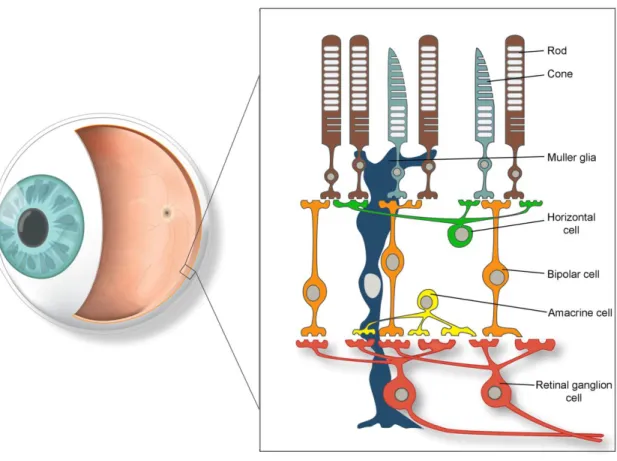
The retina is comprised of many cell types: photoreceptors, bipolar cells, horizontal cells, amacrine cells, retinal ganglion cells (RGC) and Müller glia (Figure 1). There are two types of photoreceptors in the retina: rods, which function as low light photoreceptors, and
3
cones, which are responsible for colour vision. Photoreceptors are light-sensitive cells that have an outer segment, composed of membranous disks that contain photopigment and lies adjacent to the pigment epithelial layer, and an inner segment that contains the cell nucleus and gives rise to synaptic terminals that contact bipolar or horizontal cells. Absorption of light by the photopigment (rhodopsin or cone opsins) in the outer segment of the photoreceptors initiates a cascade of events that changes the membrane potential of the photoreceptor, and therefore the amount of neurotransmitter released by the photoreceptor synapses onto the cells they contact. The synapses between photoreceptor terminals and bipolar or horizontal cells occur in the outer plexiform layer. The cell bodies of photoreceptors make up the outer nuclear layer, whereas the cell bodies of bipolar cells lie in the inner nuclear layer. The short axonal processes of bipolar cells make synaptic contacts on the dendritic processes of RGCs in the inner plexiform layer. The much larger axons of the RGCs form the optic nerve and carry retinal nerve impulses to the brain.
Horizontal and amacrine cells have their cell bodies in the inner nuclear layer and are primarily responsible for lateral interactions within the retina. The processes of amacrine cells, which extend laterally in the inner plexiform layer, are postsynaptic to bipolar cell terminals and presynaptic to the dendrites of ganglion cells. Müller glia, which represent the principal glial cells of the retina, span radially across the entire retina, with their endfeet enveloping RGCs and displaced amacrine cells in the ganglion cell layer. Their cell bodies are found at the level of the inner nuclear layer.
4
Figure 1. A schematic diagram of the retina demonstrating the principal cell types involved in retinal signaling.
The main retinal cell types are depicted as follows, with their respective attributed colour in parenthesis: rod photoreceptor (brown), cone photoreceptor (light grey), Müller glia (dark blue), horizontal cell (green), bipolar cell (orange), amacrine cell (yellow), retinal ganglion cell (red). Source: adapted from Wilson et al., Gene Therapy. 2012
5
I.1.2. The retina as a model system to study neurodegenerative mechanisms
RGCs are classified in 10 to 15 subtypes based on morphology or physiology in mammalian species (Masland, 2001a, b, Rockhill et al., 2002) but, despite their diversity, RGCs display the typical properties of CNS neurons, and comprise a cell body, dendrites and an axon. The RGC axons converge at the optic nerve head (ONH) to form the optic nerve which projects to four main targets: 1) the lateral geniculate nucleus of the thalamus, 2) the superior colliculus, 3) the pretectal nucleus, and 4) the suprachiasmatic nucleus. In rodents, a majority of axons project to the superior colliculus, whereas in humans the lateral geniculate nucleus is the predominant projection site (Linden and Perry, 1983, Purves, 2001). The optic nerve is the second of twelve paired cranial nerves but it is part of the CNS as it is derived from an evagination of the diencephalon during embryonic development and, as a consequence, the fibers are covered with myelin produced by oligodendrocytes, rather than Schwann cells found in the PNS. The accessibility of the retina renders it an ideal model in which to study neurodegenerative processes. One such disease is glaucoma, models of which will be discussed in the following section.
I.2. GLAUCOMA MODELS
I.2.1. Definition, risk factors and pathological features of glaucoma
Glaucoma refers to a group of chronic optic neuropathies characterized by progressive optic nerve damage and selective loss of RGCs, which functionally translates to progressive visual field defects leading to irreversible blindness. It is estimated that more than 60 million people suffer from glaucoma worldwide (Quigley and Broman, 2006) and according to the World Health Organization, glaucoma is the second leading cause of blindness after cataracts,
6
accounting for 12% of total cases of blindness globally (Foster and Resnikoff, 2005). Furthermore, glaucoma is the first irreversible cause of blindness worldwide, as vision loss from cataracts is reversible. Age and ethnic background are important risk factors for developing glaucoma. Individuals over the age of 40 are at a much higher risk of developing this ocular neuropathy (Coleman and Miglior, 2008). Individuals of African descent are at higher risk of developing glaucoma and have the highest rate of blindness due to this disease (Leske et al., 2007). Elevated intraocular pressure (IOP) is also an important risk factor, and the only modifiable one, for developing glaucoma. Ocular hypertension is characterized by an IOP in the human eye of over 21 mmHg, whereas a value of 15 mmHg is considered normal (Quigley et al., 1994). Although IOP is regarded as an important risk factor, it is not an accurate predictor of glaucoma since over 30% of glaucoma patients have an IOP in the normal range (Nemesure et al., 2007).
Glaucoma is characterized by damage to the neural components of the visual pathway including the retina, the optic tract, and the brain at the level of the lateral geniculate nucleus and the visual cortex. One of the determining features of glaucoma pathology is the selective loss of RGCs, which is characteristic of all glaucoma patients (Kendell et al., 1995, Quigley, 1999).
I.2.2. Implication of the aqueous humour in experimental models of glaucoma
As IOP is an important risk factor linked to glaucoma pathogenesis, many animal models have been developed based on inducible high IOP, which results in ONH damage and gradual RGC death. Incidentally, the production, circulation and drainage of the aqueous humor -the clear fluid which fills the anterior and posterior chambers of the eye- are
7
determining factors in the regulation of IOP levels. To understand how these animal models of glaucoma induce IOP increase, we will review the anatomical structures of the eye involved in the production and circulation of the aqueous humour.
The aqueous humour, produced by the ciliary epithelium in the ciliary body, provides nutrients for the lens and removes waste as it flows through the pupil into the anterior chamber of the eye (Krupin et al., 1986). The aqueous humor fills the anterior chamber and provides nutrients to the cornea as well. Within the anterior chamber, the cornea and iris join and it is here where the drainage of the aqueous humour takes place (Figure 2). The angle with which the cornea and iris join is of particular interest, as the two major categories of glaucoma are defined by the formation of an open or closed angle between the cornea and the iris: primary open-angle glaucoma (POAG) and primary angle-closure glaucoma (PACG).
Aqueous fluid flows toward the angle where it enters the trabecular meshwork (TM) (Tamm, 2009), which filters and directs the aqueous fluid into the Schlemm‟s canal (Johnstone, 2004). Several models of inducible and spontaneous glaucoma rely on a blockade of the aqueous humour drainage and will be discussed in subsequent sections.
8
Figure 2. Schematic presentation of the aqueous humour circulation within the eye. Aqueous humour is produced by the ciliary body and enters the anterior chamber through the pupil. The trabecular meshwork (TM) is located in the angle between cornea and iris and provides an outlet for aqueous drainage by directing it into the Schlemm's canal. Reproduced with permission from (Kwon et al., 2009), Copyright Massachusetts Medical Society.
9 I.2.3. Experimental models of glaucoma
I.2.3.1. Primary-angle closure glaucoma
Primary-angle closure glaucoma (PACG) is characterized by the blockade of the aqueous humour drainage and/or its circulation. This results in increased IOP and consequent damage to the retina and optic nerve. Several breeds of dogs are prone to developing spontaneous glaucoma (Reinstein et al., 2009) however the high cost of purchasing and housing dogs, and complications of handling dogs in large experimental groups have resulted in the limited use of this model. A model of laser photocoagulation to induce the closure of the anterior chamber angle has been adapted for mice (Aihara et al., 2003). This model employs a diode laser which creates burn spots causing the iris root to attach to the peripheral cornea, which consequently obstructs the aqueous outflow and results in an elevation of IOP (Aihara et al., 2003). The IOP elevation in this model is accompanied by significant loss of RGC axons (Mabuchi et al., 2003). However, the small size of mice eyes demands a high level of dexterity by the experimenter, as excessive or misplaced laser burns could result in an inflammatory response and retinal damage.
I.2.3.2. Open-angle glaucoma
Primary open angle glaucoma (POAG) is the most common form of glaucoma worldwide. POAG is characterized by changes in the optic nerve head (also called optic disk), damage to RGC axons in the optic nerve, and loss of RGCs in the retina (Quigley, 2005). POAG is not necessarily associated with IOP elevation, however high IOP is a major risk factor for developing POAG (Leske et al., 2003).
10
In primate models of POAG, Rhesus or Cynomolgus monkeys are subjected to laser photocoagulation whereby burn spots are created on the circumference of the TM, resulting in moderate IOP increase (Wang et al., 1998). Loss of RGCs and visual deficits are well documented in this model (Hood et al., 1999, Morgan et al., 2000, Yücel et al., 2000, Hare et al., 2001, Yücel et al., 2003). However, the high cost of monkeys, their limited availability and difficulty to work with are drawbacks of this model.
The laser photocoagulation technique has also been used in rodent models of POAG (Ueda et al., 1998, Levkovitch Verbin et al., 2002, Gross et al., 2003, Ji et al., 2005). Another rodent model of POAG is the Morrison rat model, in which a hypertonic saline solution is injected into an episcleral vein of Brown Norway rat eyes. Hypertonic saline disrupts the structure of the TM and gradually reduces the aqueous outflow, resulting in IOP elevation (Morrison et al., 1997). Another method of reducing the aqueous outflow is by cauterizing the episcleral veins (Shareef et al., 1995). In this model, two to three veins are isolated and blocked using an ophthalmic cautery instrument. Obstruction of the TM and elevation of IOP can also be achieved by injection of sterile latex microspheres (Weber and Zelenak, 2001, Urcola et al., 2006) or microbeads into the anterior chamber of rodent eyes (Sappington et al., 2010, Chen et al., 2011). Finally, a hereditary mutation in the DBA/2J mice leads to iris pigment dispersion and adhesion of the iris to the cornea, which results in significant elevation of IOP by 6 months of age (John et al., 1998).
1.2.4 Glaucoma model caveats
The aforementioned glaucoma models each have their advantages and disadvantages. Chronic animal models of glaucoma rely on OHT induction to ultimately cause RGC
11
neurodegeneration, and yet not all glaucoma patients exhibit an increase in IOP. Furthermore, signaling pathways activated in chronic experimental models of glaucoma, much like the disease, are complex and variable. The complexity and variability in these models are not ideal for the detection of precise molecular mechanisms leading to apoptotic RGC death, a crucial aspect in the pathophysiology of all forms of glaucoma. The identification of key molecular mechanisms involved in neuronal death in vivo often requires a model that allows reliable, reproducible and predictable time-course of RGC death.
I.3. OPTIC NERVE AXOTOMY MODEL
Following traumatic injury to the mammalian CNS, neurons die by apoptosis, necrosis, or autophagy. Neurons that have their axons sheared open will undergo fundamental cellular and biochemical changes. Notably, the physical separation of the cell body from its target effectively prevents retrograde transport of neurotrophic factors thought to be required for survival (Oppenheim, 1991). Furthermore, calcium influx destabilizes the cytoskeleton and contributes to the activation of apoptotic pathways and the ensuing cellular degradation (Trump and Berezesky, 1995). Neurons that survive are severely impaired and rendered inactive, at least transiently, by the dramatic variations in their ionic and metabolic environment. In brief, adult mammalian CNS neurons are unable to regenerate an axon and their soma dies or atrophies after lesion (Ramon y Cajal, 1928).
I.3.1. Acute optic nerve injury and RGC death
The optic nerve transmits visual input from the retina to the visual cortex where image processing occurs. Damage to the optic nerve will affect this transmission of visual information, thus compromising vision. Optic nerve afflictions include: 1) glaucoma, in which
12
the optic nerve at the optic nerve head are damaged; 2) optic neuritis, an inflammation of the optic nerve which leads to the degradation of the myelin sheath enveloping the optic nerve; 3) cancerous tumours such as a pituitary tumour which compresses the nerve at the level of the optic chiasm, and 4) acute trauma such as an orbital fracture. Although a full transection (axotomy) is unlikely to occur and traumatic injury of the optic nerve is very rare (Steinsapir and Goldberg, 1994), the optic nerve axotomy produces a well characterized time course of RGC death, permitting the accurate evaluation of neuroprotective and regenerative strategies. In addition, RGCs die by apoptosis after optic nerve axotomy as they do in glaucoma (Garcia-Valenzuela et al., 1995) and other CNS diseases. However, because a larger number of RGCs die abruptly after axotomy, the molecular mechanisms that promote neuronal apoptosis might be more readily identified in this simpler model of RGC death. Furthermore, the initial wave of apoptotic RGC death is more reproducible in an axotomy model than in experimental rat glaucoma models, allowing for a spatiotemporal correlation between pro-apoptotic gene expression and RGC loss in vivo. Thus, strategies to promote cell survival in this system may be extrapolated to other neurodegenerative diseases affecting other neuronal populations. For these reasons, the optic nerve axotomy model was selected for the studies presented in this dissertation. Importantly, the axotomy model has proved extremely useful in the study and treatment of traumatic CNS injuries. Indeed, optic nerve axotomy has been extensively used to study the molecular mechanisms underlying retinal neuron death. For example, this model was used to detect the activation of apoptosis initiator caspase 9 (Kermer et al., 2000, Koeberle, 2009) and cleavage of effector caspase 3 in RGCs (Kermer et al., 1999, Hu et al., 2012) after optic nerve lesion, and subsequent TUNEL reactivity (Berkelaar et al., 1994, Quigley et al., 1995). This model has also been useful to evaluate the effect of neuroprotective strategies in
13
vivo; for example with the demonstration that caspase inhibitors reduced RGC loss following optic nerve axotomy (Kermer et al., 1998, Chaudhary et al., 1999, Ahmed et al., 2011, Monnier et al., 2011). Moreover, this was an effective model to investigate the signal transduction pathways involved in RGC survival, including TrkB signaling (Cheng et al., 2002b), the Erk1/2 pathway (Pernet et al., 2005, Almasieh et al., 2011), and the opposing effects of TrkA and p75 receptor signaling pathways (Lebrun-Julien et al., 2009).
I.3.2. Pattern of RGC cell death in the axotomy model
Transection of the adult rat optic nerve leads to a bi-phasic pattern of cell death. The first phase is prolonged, as within the first 4 to 5 days after lesion only a negligible number of RGCs die (Berkelaar et al., 1994, Peinado-Ramón et al., 1996). The second stage is rapid, however, and is characterized by massive cell death. Indeed, seven days after axotomy, only 50% of RGCs survive and less than 10% remain 14 days after injury (Berkelaar et al., 1994, Quigley et al., 1995). Interestingly, approximately 5% of RGCs remain up to 20 months after transection (Villegas-Perez et al., 1993), however, the molecular basis for this apparent resilience is currently unknown.
Following optic nerve axotomy, RGC death has been described as apoptotic (Berkelaar et al., 1994, Garcia-Valenzuela et al., 1994). Indeed, RGCs die by apoptosis in optic nerve acute lesion models, such as the optic nerve axotomy or crush models (Berkelaar et al., 1994), experimental glaucoma (Quigley et al., 1995), human glaucoma (Kerrigan LA, 1997), an observation that has been confirmed by in vivo real-time visualization in ocular hypertensive rat eyes (Cordeiro et al., 2004). Apoptosis is an energy consuming process that requires de novo protein synthesis. Apoptosis is a common mechanism of neuronal loss in the injured or
14
degenerating visual system. The hallmark structural features of apoptosis are cellular round-up, retraction of pseudopodia, reduction of cellular volume, nuclear fragmentation, modification of cytoplasmic organelles, plasma membrane blebbing, and engulfment by resident phagocytes (Kerr, 1972, Kerr et al., 1995). The apoptotic process can be triggered by various stimuli and involves the death receptor and/or mitochondrial apoptotic pathways.
Autophagy, traditionally defined as a non-apoptotic type of programmed cell death, involves lysosomal degradation of dysfunctional cellular components. Mutations in the Toll-Like Receptor 4 (TLR4) gene, a sensor for autophagy, have been associated with normal tension glaucoma in a Japanese cohort study (Shibuya et al., 2008). There is evidence that autophagy occurs in glaucoma, as LC3-B and Beclin-1 upregulation as well as an accumulation of autophagosomes were detected in RGCs after IOP elevation in a hypertensive rat glaucoma model (Park et al., 2012), and in a rhesus monkey hypertensive glaucoma model (Deng et al., 2013). In addition, an upregulation of genes linked to autophagy was detected in ocular hypertensive astrocyte samples (Tezel et al., 2012). Autophagy is also activated following optic nerve transection, and was compellingly revealed to have a neuroprotective role following axotomy (Kim et al., 2008, Rodriguez Muela and Boya, 2012).
Although apoptosis is the active process by which RGCs die after axonal damage, a small number of cells die by necrosis due to mechanical or inflammatory damage inflicted by the injury (Thanos et al., 1993, Bien et al., 1999). Necrosis typically occurs after toxic insult, hypoxia, energy depletion or other exogenous insults. It results in a swelling of the cell body and mitochondria, followed by perforation of the cell membrane resulting in the leakage of cellular contents and consequent inflammatory response. It is likely that there is a continuum
15
between axotomy-induced apoptotic and necrotic cell death, as both modes share common characteristics (Nicotera et al., 1999). While the orchestral role of p53 in apoptotic pathways is well established, recent findings have reported a novel role in necrosis, whereby p53 opens the mitochondrial permeability transition pore to trigger necrosis (Vaseva et al., 2012). An understanding of p53 function in RGC death is therefore warranted. We opted to focus on apoptotic cell death in our studies because of the central role apoptosis plays in RGC death during glaucoma. Furthermore, despite potential crosstalk amongst apoptotic, necrotic and autophagic pathways, there is no strong evidence of necrosis occurring in glaucoma (Osborne et al., 1999b), and the understanding of autophagic processes in glaucomatous neurodegeneration is incipient.
I.4. p53 AND ITS REGULATORS
I.4.1. The p53 family
The p53 transcription factor belongs to a family of proteins called the p53 family, comprised of three evolutionarily conserved transcription factors, p53, p63 and p73. The p53 family proteins are involved in many important cellular functions, including tumour suppression (p53 and p73), epithelial cell stratification (p63), and CNS development (p73). Furthermore, p53, p63 and p73 genes can independently mediate apoptosis (Sheikh and Fornace, 2000). All p53 family members are expressed during retinal development, but their levels are downregulated in the adult eye (Vuong et al., 2012). Upregulation of p63 and p73, but not p53, are observed in human retinoblastoma tumour samples, an eye cancer affecting the retina (Adithi et al., 2008).
16
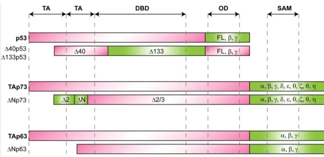
All three members of the p53 family share significant homology at the genomic and protein levels. They share common structural motifs, including a transactivation domain (TA), a DNA-binding domain (DBD) and an oligomerization domain (OD). In addition, p63 and p73, but not p53, contain long C-termini containing a sterile alpha motif (SAM), which is a protein-protein interaction domain (Chi, 1999) (Figure 3). p63 and p73 genes have been found to encode several proteins whose structure and functions are similar yet not identical to those of p53 (Kaghad et al., 1997, Yang et al., 1998). p53, p63 and p73 encode differentially spliced mRNAs, with most of splicing occurring at the 3‟ end for p63 and p73, creating proteins with varying C-termini lengths (Kaghad et al., 1997, De Laurenzi et al., 1998, De Laurenzi et al., 1999, Ueda et al., 1999). Not only do all three p53 family members express multiple splice variants, they also contain different internal promoters in addition to their proximal promoters, yielding truncated or full length variants, respectively. This allows for a multitude of transcript variants arising from p53, p63 and p73 gene transcription. Altogether, the p53 gene can transcribe ten different variants: p53 (α, β, γ), ∆40p53 (α, β, γ), ∆133p53 (α, β, γ) and ∆p53 (Courtois et al., 2004, Bourdon et al., 2005, Mills, 2005, Rohaly et al., 2005) (Figure 3). In the case of p63 and p73, the promoter and differential splicing options yield α, β, γ, δ, ε, δ, ε, and φ isoforms for both full length and truncated forms (Figure 3). Furthermore, p73 has 3 additional truncated forms due to an additional amino-terminal splicing site (Moll and Slade, 2004). As the N terminus is crucial for the transactivation of target genes, full-length isoforms of p53, p63 and p73 can be functionally distinguished from the transactivation-compromised ∆N isoforms that show anti-apoptotic and dominant–negative properties.
17
Figure 3: p53 family structural motifs. The overall protein architecture of the p53 family is highly conserved, and consists of a central sequence-specific DNA binding domain (DBD), an N-terminal transactivation domain (TA) and a C-terminal oligomerization domain (OD). Both p63 and p73 have a sterile alpha motif (SAM) domain implicated in protein–protein interactions, whereas p53 does not. The highest degree of homology is seen within the DBD, where >97% of all tumour-associated p53 mutations are located. All three genes express many differently spliced isoforms, and contain a second intronic promoter that generates N-terminally truncated proteins (∆133p53, ∆Np63 and ∆Np73). Alternative splicing of C-terminal exons yields many different isoforms (α, β, γ, δ, ε, δ, ε, and ζ) with still incompletely understood DNA-binding properties, transcriptional activities and biological functions. Source of image adapted by permission from Macmillan Publishers Ltd: [Nature Reviews Cancer] (Stiewe, 2007), copyright (2007).
18 I.4.2. The p53 transcription factor
I.4.2.1. p53 in ocular development
The role of p53 during ocular development has been assessed in p53 knockout mice of varying strains, which revealed striking differences. Indeed, in two strains, the C57BL x CBA and 129/Sv x C57BL/6 mice, there were no reported ocular developmental defects (Donehower et al., 1992, Jacks et al., 1994). However, abnormalities were detected in the p53 knockout mice with C57BL/6 and BALB/c OlaHsd backgrounds. Indeed, cataract formation was detected in p53-null BALB/c OlaHsd mice, as well as aberrant hyaloid vasculature, which is a transient embryonic vasculature that regresses with the formation of adult retinal vessels (Reichel et al., 1998). In addition to abnormal hyaloid structure, p53-null mice in a C57BL/6 background also exhibited retinal folding and underdeveloped (hypoplastic) optic nerves (Ikeda et al., 1999). It was hypothesized that the phenotype discrepancy between p53-null mice with 129/Sv or C57BL/6 backgrounds was caused by protective and compensatory alleles for p53 loss present in 129/Sv mice (Ikeda et al., 1999).
p53 transcript levels during murine retinal development peak at embryonic days E17-E18 and decrease gradually until postnatal day P15, where they remain low in the post-mitotic retina (Vuong et al., 2012). Likewise, p53 protein was readily detected at E18, after which time point protein levels began to decrease, and were undetectable by P7 (Vuong et al., 2012). This decrease in p53 levels in the murine retina coincides with the developmental time point that retinal cells exit the cell cycle, differentiate, and become postmitotic (Cepko et al., 1996). p53 is not required for developmental programmed death of RGCs, as mice deficient for one
19
or both alleles of p53 do not exhibit developmental changes in RGC number in comparison to wild-type animals (Li et al., 2002).
I.4.2.2. p53 in post-mitotic neurons
The tumour suppressor and nuclear transcription factor p53 mediates the apoptosis of post-mitotic neurons exposed to a wide range of insults such as DNA damage, neurotrophic factor deprivation, oxidative stress, ischemia and excitotoxicity (Culmsee and Mattson, 2005). Stress signals lead to activation and accumulation of p53, which then promotes the transcription of pro-apoptotic genes (Michalak et al., 2005). Neuronal apoptosis induced by p53 has been well documented in models of neurodegenerative disorders including Alzheimer‟s disease, Parkinson‟s disease, Huntington‟s disease and amyotrophic lateral sclerosis (de la Monte et al., 1997, González de Aguilar et al., 2000, Martin, 2000, Duan et al., 2002, Tamagno et al., 2003, Ryan et al., 2006), suggesting a key role for this transcription factor in the regulation of neuronal viability after neural injury. Given its critical role in the control of cell death, several mechanisms exist to ensure a tight regulation of p53 activity. The level of p53 protein is normally kept low in most cell types, including neurons (Soussi, 2000), via rapid and continuous degradation following ubiquitination by the E3 ubiquitin ligases Mdm2 and MdmX (Wade et al., 2010). Additional control of p53 function is exerted via post-translational acetylation, phosphorylation or methylation; and by interactions with protein partners including the apoptosis-stimulating proteins of p53 (ASPP) family (Iwabuchi, 1994, Nagase et al., 1998, Boehme and Blattner, 2009).
p53 has been shown to exert an important pro-oxidant activity in the retina (Chatoo et al., 2009). Inactivation of the p53 gene, or reduced p53 expression, has been shown to protect
20
RGCs against retinal ischemia or excitotoxic death (Rosenbaum, 1998, Li et al., 2002). Pro-apoptotic genes induced by p53 including Gadd45a and Ei24 are upregulated in the retina after optic nerve transection and in experimental glaucoma models (Levkovitch-Verbin et al., 2006). Attempts to link single nucleotide polymorphisms in the p53 gene with glaucoma have been contradictory: while some studies found a correlation in Caucasian and Chinese populations (Ressiniotis et al., 2004, Daugherty et al., 2009) an association has not been found in other ethnic groups (Acharya et al., 2002, Dimasi et al., 2005, Mabuchi et al., 2009, Saglar et al., 2009, Silva et al., 2009). A recent study demonstrated that loss of WDR36, a gene of unknown function identified as causative for glaucoma (Monemi et al., 2005), leads to activation of the p53 stress-response pathway in zebrafish (Skarie and Link, 2008). These studies suggest that defects in p53 pathway genes may increase the risk of certain populations to develop glaucoma (Fan et al., 2010). Furthermore, p53 has been implicated in age-related macular degeneration (AMD): AMD is generally believed to start with retinal pigment epithelium (RPE) cell death, and p53 is upregulated in RPE cells in response to high-energy light exposure, inducing apoptosis (Westlund et al., 2009, Bhattacharya et al., 2012). In retinoblastoma, Mdm4 is overexpressed in response to loss of RB1, leading to degradation of p53 (Laurie et al., 2006, Wallace, 2006).
I.4.3. p53 regulation
There are over 100 known p53 binding proteins, whose interactions with p53 along with p53 post-translational modifications frequently dictate p53 function, for example whether it induces cell cycle arrest or apoptosis (Braithwaite et al., 2005). These regulating mechanisms range from inhibition to coactivation of p53. The identification of p53 in 1979 has led to nearly 70,000 peer-reviewed publications, and was named the „Molecule of the
21
year‟ in 1993 by the journal Science (Koshland, 1993). p53 is mutated in nearly half of all human cancers (Petitjean, 2007), a trait rarely found in p63 and p73 family members (Strano et al., 2007). Not only does p53 act as a tumour suppressor, it is activated in response to various stress signals. p53 activation occurs via multiple mechanisms including increased p53 protein concentration, often caused by decreased p53 degradation (Ashcroft et al., 2000), nuclear translocation (Liang and Clarke, 2001), and post-translational protein modifications including phosphorylation and acetylation (Jayaraman and Prives, 1999). Furthermore, p53 can also be activated by non-covalent modifications such as electrostatic interactions with other proteins (Benyamini and Friedler, 2011). p53 is modified by as many as 50 individual posttranslational modifications, which mediate precise protein-protein interactions, an array of modifications that is interdependent (Meek and Anderson, 2009).
The p53 interactome is continuously being updated, however relatively few of these p53 binding partners have been studied in the CNS. The following sections on p53-regulating proteins focus on the ASPP family, and additional p53 regulating proteins whose roles have been assessed in the retina.
I.4.3.1. The ASPP family
The ASPP proteins constitute a recently discovered family of proteins that bind and modulate p53-dependent apoptosis (Trigiante and Lu, 2006). Their name is an acryonym either based on the domain organization of the proteins (Ankyrin-repeat, SH3, and Proline-rich domain containing Protein) or their function (Apoptosis-Stimulating Protein of p53). The founding member of the family, ASPP2, was initially identified as 53BP2 (p53 binding protein 2) in a yeast two-hybrid screen, using the p53 DNA binding core domain as bait (Iwabuchi,
22
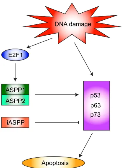
1994). ASPP1 was identified later in a homology search (Nagase et al., 1998). Functional studies revealed that p53-induced apoptosis was substantially enhanced in the presence of ASPP1 or ASPP2 (Lopez et al., 2000, Ao et al., 2001, Samuels-Lev, 2001) and that complexes with ASPP1 or ASPP2 increased the affinity of p53 for promoters of pro-apoptotic genes (Samuels-Lev, 2001, Bergamaschi et al., 2006). Furthermore ASPP1 and ASPP2 selectively enhance the apoptotic-promoting ability of p53 without affecting p53 cell cycle arrest functions (Bergamaschi et al., 2003). This preferential activation of apoptotic genes in the presence of ASPP1 and ASPP2 was also observed for p63 and p73 (Bergamaschi et al., 2004). The third member of the ASPP family, iASPP, was originally identified as an inhibitor of the nuclear factor kappa (NFk) (Yang, 1999). iASPP is the p53 inhibitor within the ASPP family, blocking apoptosis by repressing the transactivation potential of p53 (Bergamaschi et al., 2003, Bergamaschi et al., 2006) (Figure 4). Notably, the ability of iASPP to inhibit p53 mediated apoptosis is conserved from C. elegans to humans.
23
Figure 4. ASPP family apoptotic signaling pathway. The pro-apoptotic ASPP1 (ankyrin-repeats-, SH3-domain- and proline-rich-region-containing protein 1) and ASPP2 are induced by the E2F1 transcription factor and cooperate with the p53 transcription factor and its family members p63 and p73 in transactivating pro-apoptotic genes to promote apoptosis. The inhibitory family member iASPP functions as a transrepressor of the same genes that ASPP1 and ASPP2 transactivate. (Image generated by Ariel Wilson).
24
The regions of ASPP family members that interact with p53 have been mapped to their C-termini (Patel et al., 2008, Robinson, 2008). The N termini of ASPP1 and ASPP2 confers their apoptotic activity (Samuels Lev et al., 2001), and show no sequence similarity with the N terminal of iASPP (Bergamaschi et al., 2003). ASPP1/2 and iASPP bind to the core domain of p53 (Robinson, 2008), and iASPP also binds to the proline-rich region of p53 (Bergamaschi et al., 2006) (Figure 5).
Pro-apoptotic ASPP1 and ASPP2 are frequently downregulated in tumours, and anti-apoptotic iASPP is frequently upregulated (Bergamaschi et al., 2003, Jiang et al., 2011, Li et al., 2011, Li et al., 2012, Mak et al., 2013, Zhao et al., 2013). Post-translational modifications of ASPP family members in cancer cells have been discovered. Indeed, ASPP2 is phosphorylated by the Ras/MAPK pathway in an osteocarcinoma cell line (Godin Heymann et al., 2013), and Cyclin B1/CDK1 phosphorylates iASPP in melanoma cells (Lu et al., 2013). Other mechanisms of ASPP regulation have been uncovered, such as the study of ASPP1 and ASPP2 epigenetic regulation which revealed that in tumour cells expressing wild-type p53, ASPP1 and ASPP2 promoters are hypermethylated and subsequently downregulated in hepatocellular carcinoma tumours (Zhao et al., 2010) as well as in breast cancer and lung carcinoma tumour cell lines (Liu et al., 2005). Furthermore, microRNA downregulation of iASPP protein levels was detected in a cerebral ischemia mouse model (Liu et al., 2013).
25
Figure 5. Amino acid sequences and domain organisation of ASPP and p53 proteins. A) Domain organization of ASPP2, ASPP1 and iASPP. Individual domains are as follows: ubiquitin-like (ULD), glutamine-rich (Gln), proline-rich (Pro), ankyrin repeats (ANK), and Src homology 3 (SH3). B) Domain organization of p53. Individual domains are the transcription activation (TAD), the proline-rich (Pro), the DNA binding or core (CD), the linker (L), the oligomerization or tetramerization (OD), and the basic (BD) domains. Source of image: adapted from Ahn et al., 2009, originally published in the Journal of Biological Chemistry © the American Society for Biochemistry and Molecular Biology.
26
I.4.3.2. MDM2/MDMX
The murine double minute 2 (MDM2) and murine double minute X (MDMX) (also known as MDM4) inhibit p53 activity by engaging its N-terminus transactivation domain (Momand et al., 1992, Kussie et al., 1996, Laurie et al., 2006). MDM2 and MDMX are RING (Really Interesting New Gene) domain proteins, and many proteins containing a RING domain have been shown to play a key role in the ubiquitination pathway (Joazeiro and Weissman, 2000). However, despite their similar structures, only MDM2 has intrinsic E3 ubiquitin ligase activity, conferring MDM2 the ability to target p53 for degradation by the proteasome (Honda et al., 1997). Although MDM2 alone can inhibit p53, its RING-dependent heterodimerization with MDMX has an important role in p53 inhibition. Indeed, MDM2 inhibits the transactivation ability of p53 (Momand et al., 1992), and MDMX stabilizes MDM2 by preventing it to self-ubiquitinate (Stad et al., 2001).
Within the retina, the role of MDM2 was assessed in RPE cells, which are essential for photoreceptor function by regulating cell homeostasis and serving as blood-retinal barrier (Strauss, 2005). Inhibition of MDM2 in primary RPE cultured cells resulted in an increase of pro-apoptotic targets, sensitizing RPEs to apoptosis (Bhattacharya et al., 2011). In aged RPEs, the MDM2/p53 pathway interaction is disrupted, leading to an age-dependent increase in apoptosis (Bhattacharya et al., 2012). Furthermore, MDM2 has been studied in the context of retinoblastoma. Mutations in the tumour suppressor RB1 gene are pivotal in the development of this early childhood cancer of the retina. Indeed, loss of RB1 function in the developing retina leads to MDM2 and MDMX amplification, contributing to p53 pathway inactivation (Laurie et al., 2006), which facilitates retinal cell transformation and tumorigenesis.
27
I.4.3.3. YY1
Yin yang 1 (YY1) is a transcription factor that belongs to the Polycomb family, a group of chromatin modulators that are critical to homeobox gene regulation during development. First identified in 1991 (Shi et al., 1991), YY1 is a highly conserved and multifunctional transcription factor. Its name, representing interconnected yet seemingly opposing forces, stems from the fact that YY1 can act as an activator or repressor of transcription (Shi et al., 1997). YY1 has been implicated in cell proliferation, differentiation and apoptosis (Gordon et al., 2006), and is crucial for embryonic development as YY1 deficiency results in peri-implantation lethality, i.e. lethality occurring in the period between blastocyst formation and uterine implantation (Donohoe et al., 1999).
YY1 negatively regulates p53 protein levels and activity (Sui et al., 2004). YY1 acts as an MDM2 cofactor, facilitating MDM2-p53 interaction, and YY1 downregulation results in p53 accumulation due to a decrease in p53 ubiquitination levels. In contrast, YY1 overexpression increases p53 ubiquitination and degradation (Sui et al., 2004). YY1 is ubiquitously expressed in the CNS during early embryonic development (Kwon and Chung, 2003), and is highly expressed in the adult neural retina (Bernard and Voisin, 2008). In mouse embryos and Xenopus oocytes, YY1 was shown to activate Otx2, a transcription factor that controls photoreceptor cell fate (Kwon and Chung, 2003, Nishida et al., 2003, Takasaki et al., 2007). In the adult retina, YY1 is mainly expressed by photoreceptors at the level of the inner segments and nuclei in the intact chicken retina, and weakly in the inner nuclear layer (Bernard and Voisin, 2008).
28
I.4.3.4. P300/CBP
p300 and CREB Binding Protein (CBP) are highly homologous nuclear proteins that play a key role in transcriptional regulation. The interaction of p53 with p300 and/or CBP (p300/CBP) regulates the ability of p53 to bind to its cognate DNA sequences and activate transcription. It is specifically the acetyltransferase activity of p300/CBP that has been implicated in the regulation of p53 function. Indeed, in response to DNA damage, transcription coactivators p300/CBP bind to and acetylate p53, which stimulates the DNA binding activity of p53 (Gu and Roeder, 1997, Prives and Hall, 1999, Grossman, 2001).
There is evidence that p300/CBP are involved in retinal function. For instance, mutations in CBP or p300 are responsible for a subset of Rubinstein-Taybi syndrome cases (Schepis et al., 2001, Zimmermann et al., 2007), a disease in which retinal dystrophy and glaucoma are common (van Genderen et al., 2000). The study of p300/CBP in animal models revealed that p300 and CBP knockout mice are embryonic lethal (Yao et al., 1998, Tanaka et al., 2000). The development of photoreceptor-specific p300/CBP conditional knockout mice was instrumental to demonstrate a key role for p300/CBP in photoreceptor gene expression (Hennig et al., 2013). Furthermore, p300 and CBP are expressed by adult RGCs, and adenoviral-mediated overexpression of p300 in RGCs has been shown to promote optic nerve regeneration (Gaub et al., 2011). Although p53 C-terminal acetylation by p300 is known to be involved in the fine-tuning of p53 stress responses (Krummel et al., 2005), these modifications do not appear to play a crucial role in RGC death. Indeed, in an acetylation-deficient missense mutant mouse model, the loss of p53 acetylation at its C terminus by CBP/p300 was not required for p53 transactivation (Krummel et al., 2005).
29
The ASPP family, MDM2/MDMX, YY1, and CBP/p300, along with additional p53 regulating proteins, are implicated in the apoptotic function of p53. The signaling cascades occurring during apoptosis in RGCs will be discussed in the subsequent section.
I.5. APOPTOTIC PATHWAYS ACTIVATED IN RGCs
The apoptotic machinery is present in all cells and is therefore considered to be an intrinsic suicide program. Neuronal survival relies on a detailed interaction between the cell and its environment. The neuron is thought to be continuously on the verge of apoptosis, requiring survival signals to prevent its death (Raff, 1992, Raff et al., 1993). Neurotrophins, cytokines, growth factors such as brain derive neurotrophic factor (BDNF) and ciliary neurotrophic factor (CNTF), and other peptide ligands stimulate transmembrane receptors, which activate intracellular pathways and ultimately inhibit this internal death program (Jacobson et al., 1997) (Figure 6). The apoptotic pathway can be induced either through a mitochondrial pathway (Green and Reed, 1998), or by stimulation of cell surface death receptors (Ashkenazi, 1998), which are referred to as the intrinsic and extrinsic apoptotic pathways, respectively.
31
Figure 6. Extrinsic and intrinsic apoptotic pathways regulating retinal ganglion cell death. The apoptotic death of RGCs can be triggered by various stimuli and involves extrinsic and intrinsic pathways. Extrinsic signals include the death-receptor ligands TNF-α, Fas-L, and TRAIL, and their respective receptors, which induce RGC death. Lack of neurotrophic factors may result in deficits of pro-survival pathways including Erk1/2 and PI3K. The intrinsic pathway converges on the Bcl-2 family members: pro-apoptotic (Bax, Bad, Bid) or anti-apoptotic (Bcl-2, Bcl-XL), which regulate the mitochondrial outer membrane permeabilization. These proteins control the release of cytochrome c into the cytosol which can activate the caspases, executioners of apoptosis. Source: from Wilson et al., Gene Therapy. 2012
32 I.5.1 The intrinsic apoptotic pathway
I.5.1.1. Pro-apoptotic kinases
The mitogen-activated protein kinases (MAPKs) are a large family of protein Ser/Thr kinases that relay extracellular signals to the intracellular milieu and, as such, are central regulators of many cellular functions (Cargnello and Roux, 2011). The conventional MAPKs family comprises extracellular signal-regulated kinases 1 and 2 (Erk1/2), c-Jun N-terminal kinases (JNKs), p38 isoforms and Erk5. In contrast to the RGC survival response associated with activation of the Erk1/2 pathway, pro-apoptotic MAPK are typically activated by a variety of stress signals and have been proposed to contribute to RGC death. One such MAPKs subfamily are the JNKs, also known as stress-activated protein kinases (SAPKs), which play central roles in the regulation of signal transduction in the mammalian brain (Brecht et al., 2005). The elucidation of JNKs function in vivo has been challenging due to the presence of ten different JNK isoforms that result from alternative splicing (Kyriakis et al., 1994) and display differential specificity towards their target proteins (Gupta et al., 1996, Kallunki et al., 1996, Chang and Karin, 2001). C-jun, a transcription factor activated by JNK phosphorylation, mediates the transcription of pro-apoptotic genes (Curran and Franza, 1988) (Figure 6). The JNK/c-jun pathway is upregulated in RGCs after optic nerve axotomy or crush (Herdegen et al., 1993, Hull and Bahr, 1994, Robinson, 1994, Isenmann and Bähr, 1997, Kreutz et al., 1999, Takeda et al., 2000, Yang et al., 2007), during excitotoxic damage (Bessero et al., 2010), in rodent models of experimental glaucoma (Levkovitch-Verbin et al., 2005, Kwong and Caprioli, 2006, Yang et al., 2007), and in human glaucoma (Tezel et al., 2003). Of interest, long-term activation of c-Jun has been observed in optic nerve head astrocytes of monkeys subjected to experimental ocular hypertension (Hashimoto 2005). Short