Respiratory Medicine 175 (2020) 106202
Available online 10 November 2020
0954-6111/© 2020 Elsevier Ltd. All rights reserved.
Clinical and biological factors associated with irreversible airway
obstruction in adult asthma
Sophie Graff
a,*, No¨emie Bricmont
b, Catherine Moermans
a, Monique Henket
a, Virginie Paulus
a,
Françoise Guissard
a, Renaud Louis
a, Florence Schleich
aaDepartment of Respiratory Medicine, CHU Liege, GIGA I3 Research Group, University of Liege, Belgium
bDepartment of Pediatrics, division of respirology, CHU Liege, GIGA I3 Research Group, University of Liege, Belgium
A R T I C L E I N F O Keywords: Asthma Airway inflammation Airflow obstruction Eosinophils Lung diseases A B S T R A C T
Background and objective: Airway remodeling, as many other factors, may lead to lung function decline and irreversible airflow obstruction (IRAO) in asthma. This study was undertaken in order to highlight predictors of incomplete reversibility of airflow obstruction in adult asthmatics to identify patients with poorer prognosis and improve their care, and decrease morbidity.
Methods: A retrospective study was conducted in 973 asthmatics recruited from the University Asthma Clinic of Liege. Patients with IRAO (post-BD FEV1/FVC<0.7 & FEV1<80% predicted) were compared to patients with reversible airway obstruction (RAO) (post-BD FEV1/FVC≥0.7 & FEV1≥80% predicted). TGF-β was measured in sputum supernatant of 85 patients.
Results: Seventeen percent of asthmatics presented with IRAO. These patients were significantly older, more smokers, with a lower proportion of female, a longer disease duration, were more poorly controlled with a lower quality of life. This sub-population of asthmatics also showed more often elevated blood and sputum eosinophils and neutrophils, and higher exacerbation and hospitalisation rates in the previous year. The multivariable analysis revealed male gender, longer disease duration, cigarette smoking, ACQ score, sputum eosinophils and neutrophils, ICS dose and OCS maintenance, BMI, and asthma onset as variables independently linked to IRAO. Total TGF-β levels appeared higher in patients with IRAO (n = 38) compared to patients with RAO (n = 47). Conclusion: These data show that risk factors for IRAO are male gender, smoking, a longer disease duration, uncontrolled asthma, eosinophilic or neutrophilic airway inflammation, lower BMI, and later asthma onset. Moreover, TGF-β levels are higher in IRAO.
Sophie GraffFlorence SchleichNo¨emie BricmontCatherine Moer-mansMonique HenketVirginie PaulusFrançoise GuissardRenaud Louis
1. Introduction
Asthma is a chronic inflammatory airway disease commonly asso-ciated with reversible airway obstruction. On average, asthma patients have lower lung function than healthy individuals [1] and their lung function (FEV1) decline can be greater over time [2,3]. Most of patients
with mild to moderate asthma can be controlled with regular medica-tion. However, asthma patients are at risk of developing structural changes resulting in persistent airflow limitations [1,2,4] despite anti-inflammatory therapies. The features of remodeling include sub-epithelial reticular basement membrane (RBM) thickening, hypertrophy and hyperplasia of airway smooth muscle (ASM) cells, angiogenesis and
goblet cell hyperplasia [5], responsible for airway narrowing. A small proportion of non-smoking asthmatics present with irreversible airway obstruction (IRAO) which can be considered another form of lung function decline in asthma [6]. IRAO is defined as a significantly reduced ratio between FEV1 and FVC after bronchodilation [7,8].
Studies on risk factors and prevalence of IRAO in asthma are limited. Moreover, no consensus is reached on a definition of IRAO [9]. Yet, predictors for IRAO including smoking [10], longer disease duration [10,11], male gender [10,12], aspirin sensitivity [13], greater airway hyperresponsiveness (AHR) [14], less chronic rhinitis [15], adult onset [14], and FeNO [9,10,14] have been reported.
Persistent airway obstruction partly relates to airway remodeling the histological substrate of which is a sub-epithelial fibrosis [16] [–] [18]. TGF-β is known to be the prominent mediator in airway fibrosis but has been poorly investigated in the context of asthma with fixed airway * Corresponding author. Department of Respiratory Medicine, CHU Sart-Tilman, GIGA +4; CHU - B34, Avenue de l’Hˆopital, 11, 4000, Li`ege, Belgium.
E-mail address: sgraff@uliege.be (S. Graff).
Contents lists available at ScienceDirect
Respiratory Medicine
journal homepage: http://www.elsevier.com/locate/rmed
https://doi.org/10.1016/j.rmed.2020.106202
obstruction [19]. The active TGF-β present at steady state is the bio-logically active form while total TGF-β1 is the active form plus the latent TGF-β liberated by acidification. Assessing both the active and latent TGF-β simultaneously is useful to assess how TGF-β is involved in the pathogenesis of the disease.
This study was undertaken in order to compare patients with irre-versible airway obstruction to patients with reirre-versible airway obstruc-tion, and highlight predictors of incomplete reversibility of airflow obstruction in adult asthmatics to identify patients with poorer prog-nosis and improve their care, and decrease morbidity.
2. Methods
A retrospective study was conducted on adult asthmatics at stable state with post-bronchodilation (BD) spirometry measurements and successful sputum induction recruited from the University Asthma Clinic
of Liege, Belgium.
Patients were allocated in two distinct groups based on post-BD FEV1/FVC and post-BD FEV1 measurements. Patients with IRAO
(FEV1/FVC<0.7 and FEV1<80% predicted) were compared with pa-tients with reversible airway obstruction (RAO) (post-BD FEV1/FVC
≥0.7 and FEV1≥80% predicted). Patients that did not fit into one of these categories (i.e. FEV1/FVC <0.7 and FEV1 ≥ 80% predicted or FEV1/FVC ≥0.7 and FEV1<80% predicted) were not included (Fig. 1).
All procedures were performed in the context of clinical practice and the retrospective data collection was conducted with approval from the ethics committee of CHU Li`ege (2005/181) in accordance to the Helsinki Declaration.
Quality of Life was assessed using self-administered Asthma Quality of Life Questionnaire (AQLQ) [20] and Asthma control by the Juniper Asthma Control Questionnaire (ACQ7) [21] and an Asthma Control Test (ACT) [22]. Subjects were characterized as atopic if they had at least one
Abreviation list
ACQ asthma control questionnaire ACT asthma control test
AQLQ asthma quality of life questionnaire ATS American Thoracic Society BD bronchodilation
BEC blood eosinophil count BNC blood neutrophil count CRP C-reactive protein
ERS European Respiratory Society FeNO fractional exhaled nitric oxide
FEV1 forced expiratory volume in 1 s
FVC functional vital capacity ICS inhaled corticosteroids IgE immunoglobulin E
IRAO irreversible airway obstruction LABA long-acting ß2 agonists
LAMA long-acting muscarinic antagonists LTRA antileukotrienes
OCS oral corticosteroids
RAO reversible airway obstruction TGF-β transforming growth factor beta
Fig. 1. Study design: FEV1, forced expiratory volume in 1 s (% predicted); FVC, Forced vital capacity; post-BD, post-bronchodilation; RAO, Reversible airway
positive specific Immunoglobulin E (IgE) test (0.35 kU.L-1; Phadia, Groot-Bijgaarden, Belgium) for at least one common aeroallergen.
Patients underwent Fractional exhaled Nitric Oxide (FeNO) mea-surements at flow rate of 50 mL/s according to the ERS/ATS recom-mendations [23] (NIOX, Aerocrine, Sweden) followed by spirometry with bronchodilation, sputum induction on the same day. Sputum in-duction and processing were performed as previously described [24] using the whole expectorate.
Cell counts were estimated on samples centrifuged (Cytospin) and stained with Hemacolor® Staining set after counting 500 non-squamous cells (Merck chemical, Overijste, Belgium). Sputum cytology was analyzed and 4 phenotypes were defined: the eosinophilic phenotype with 3% sputum eosinophil count (and < 76% neutrophil count), the neutrophilic phenotype with 76% sputum neutrophil count (and <3% eosinophil count), and the mixed granulocytic phenotype being a com-bination of the above [25]. The paucigranulocytic phenotype was defined as an inflammatory cell count below these thresholds.
Routine laboratory of the University Hospital of Liege performed blood cell count and analysis of C-reactive protein (CRP), fibrinogen, and total IgE levels (ImmunoCAP system (Phadia AB, Uppsala; Sweden)).
Inhaled corticosteroids (ICS) dosages and treatment with anti-leukotrienes (LTRA), Long-acting muscarinic antagonists (LAMA), Long- acting beta-2 agonists (LABA), Anti-IgE, anti-IL5 and oral corticosteroids (OCS) were recorded and used in the analyses.
In a subpopulation of 85 non-smoking, OCS naïve patients, with either an eosinophilic or a neutrophilic inflammatory phenotype, both active (present at steady state) and total (present at steady state & latent form liberated after acidification) Transforming growth factor beta (TGF-β) were measured in sputum supernatant [19]. Briefly, trans-formed mink lung cells (TMLC, gift of Daniel Rifkin, New York Uni-versity medical center, NY) stably transfected with plasminogen activator inhibitor-1 (PAI-1) promoter fused to the firefly luciferase re-porter gene, were cultured with sputum supernatant. To activate the latent TGF-β, the supernatants were incubated with 1 N H Cl for 10 min and neutralized by 1.2 N NaOH/0.5 M HEPES. The TMLC were cultured in DMEM with 10% of FBS and were plated at the density of 15,000 cells/well in a 96-well plate. The supernatants were then added (final dilution 4X) and incubated 16–20 h. Recombinant human TGF-β 1 (4 ng/ml, R&D Systems, Minneapolis, USA) were used as positive controls. Each condition was done with and without anti-hTGF-β 1 antibody (1
μg/ml, R&D Systems, Minneapolis, USA). Each sample was done in
triplicate and results were expressed as relative light units. 2.1. Statistical methods
Variables independently associated to IRAO were identified by lo-gistic regression. Independent variables such as atopy, gender, cigarette smoking (Pack Year), Body Mass Index, asthma onset, disease duration, FeNO, ACQ score, ICS dose, OCS maintenance, hospitalizations and exacerbations during the last 12 months, blood eosinophil (BEC) and blood neutrophil (BNC) counts (/mm3), sputum eosinophils and
neu-trophils counts (%) were included in the univariate model. FEV1/FVC
<70 and FEV1<80% predicted was used as the dependent variable. A multivariable analysis was done including all independent variables. In order to test the robustness of the analysis, the same logistic regression analysis was performed with the never-smoking patients. Factors affecting FEV1/FVC ratio were evaluated with a conventional linear
regression using the same independent variables as in the logistic regression. FEV1/FVC ratio was used as the dependent variable.
We constructed receiver-operating characteristic (ROC) curves for all continuous variables independently associated with IRAO to determine the cut-off which best identified IRAO (FEV1/FVC <0.7 and FEV1<80% predicted) in asthma. Optimal cutoff points were determined by the method of the nearest point to (0,1). A p value < 0.05 was considered statistically significant. Statistical analysis was done using STATA
version 14.0 (Statistical Software, College Station, TX: StataCorp LP).
3. Results
3.1. IRAO patients’ characteristics
In our database, 1138 patients recruited between January 2005 and March 2019 had post-BD spirometry measurement and successful sputum induction. Out of these, 973 of these patients were allocated in two distinct groups. A hundred and ninety-six asthmatics (17% of total population, 196/1138) presented with IRAO (FEV1/FVC<0.7 &
FEV1<80% predicted) and 777 patients (68% of the total population 777/1138) with RAO (FEV1/FVC ≥0.7 & FEV1>80% predicted). De-mographic, clinical and inflammatory characteristics of these patients are presented in Table.1. Patients with IRAO were significantly more often male (p = 0.034), older (p < 0.0001), more (ex)smokers (p < 0.0001), with a longer disease duration (p < 0.0001), were more poorly controlled (ACT and ACQ scores) (p < 0.0001) with a lower quality of life (AQLQ score) (p < 0.0001), treated with higher ICS daily dose (p < 0.0001) and more often with OCS maintenance (p < 0.0001), LABA (p < 0.0001), LAMA (p < 0.0001), and LTRA (p = 0.001). This sub- population of asthmatics also presented more often with diffuse (blood and sputum) eosinophilic and neutrophilic inflammation (p < 0.0001), and significantly higher IgE (p = 0.007) and markers of inflammation (fibrinogen (p = 0.008), CRP (p = 0.02) and blood leukocytes (p < 0.0001)). Exacerbation and hospitalisation rates in the previous year (p <0.001) were also higher in this sub-population.
3.2. Factors associated with IRAO
The univariate model of the logistic regression (Table.2) showed a positive association between FEV1/FVC<0.7 & FEV1<80% predicted and male gender (OR:0.71 (95%CI 0.52–0.97)), Pack-Year (1.77 (1.44–2.16)), disease duration (1.02 (1.02–1.04)), ACQ score (3.25 (2.71–3.91)), ICS dose (2.07 (1.78–2.42)), OCS maintenance (4.75 (3.12–7.22)), exacerbation (1.39; (1.24–1.56)) and hospitalisation rate (2.35 (1.70–3.25)) in the previous year, BNC (/mm3) (1.0 (1.0–1.0)),
BEC (1.41 (1.23–1.63)), and sputum eosinophil count (/mm3) (1.36 (1.18–1.56)).
The multivariable analysis revealed male gender (OR female: 0.38 (95%CI 0.20–0.72)), pack-year (1.76 (1.19–2.61)), disease duration (1.06 (1.04–1.09)), ACQ score (2.63 (1.94–3.56)), sputum eosinophils (1.73 (1.22–2.47)), sputum neutrophils (1.53 (1.15–2.04)), ICS daily dose (1.60 (1.21–2.13)), BMI (0.70 (0.54–0.92)), age of onset (2.20 (1.27–3.83), and OCS therapy (2.67 (1.11–6.39)) (Table.2).
3.3. IRAO in never-smokers
In the never-smoking patients (n = 521) (Supplementary Table S1), seventy-six (15%) presented with IRAO compared to 445 (85%) with RAO. Results of the multivariable logistic regression showed ACQ (OR:2.27 (95%CI:1.47–3.48)), sputum eosinophils (1.85 (1.08–3.14)), disease duration (1.06 (1.02–1.09)) were independently associated with the outcome.
3.4. Factors affecting FEV1/FVC ratio
The univariate analysis of the linear regression analysis (Table.3) revealed a negative association between FEV1/FVC ratio and cigarette
smoking (Pack Year) (Reg. coeff:-0.17 (95%CI:-0.21 to − 0.13)), ACQ score (− 4.26 (− 4.80 to − 3.71), BEC (− 0.0017(-0.0031 to − 0.0002), BNC (− 0.0004 (− 0.0006 to − 0.0002)), sputum eosinophil count (%) (− 0.13(-0.17 to − 0.09)), ICS daily dose (− 3.04(-3.63 to − 2.43)), OCS maintenance therapy (− 8.30(-10.6 to − 5.99), exacerbation (− 1.54 (-2.07 to − 0.99)), and hospitalisation (− 4.35(-5.74 to − 2.96)) rates over the last 12 months, and disease duration (− 0.15 (− 0.19 to − 0.096)). A
positive association was observed for the female gender (2.48 (0.98–3.98)). The multivariable analysis (Table.3) showed that vari-ables independently associated to FEV1/FVC ratio were Body Mass Index
(0.42 (0.26–0.58)), smoking (Pack-Year) (− 0.11(-0.16 to − 0.06)), fe-male gender (3.26 (1.61–4.90)), ACQ (− 2.57(-3.34 to − 1.81)), BNC (− 0.0002 (− 0.001 to − 0.00003)), ICS daily dose (− 1.11 (− 1.87 to − 0.35)), hospitalisation rate over the last 12 months (− 2.04 (− 3.81 to − 0.26)), disease duration (− 0.23 (− 0.29 to − 0.17)), sputum eosinophil count (− 0.08 (− 0.14 to − 0.02)), and age of onset (− 3.39 (− 4.88 to − 1.90)).
3.5. Roc curves
Constructing a ROC curve, revealed that the ACQ score was able to identify IRAO with the best cut-off point of 2.36 providing a % 72 sensitivity and 77% specificity (AUC 0.8260, p < 0.0001, Fig. 2). We also tested other potential markers for IRAO, such as FeNO, disease duration, sputum neutrophil count, sputum eosinophil count, and BNC, but found AUC of 0.4898 (p = 0.6642), 0.6326 (p < 0.0001), 0.6133 (p =0.0698), 0.5419 (p < 0.0001), and 0.6542 (p < 0.0001) respectively. These markers are not able to discriminate between IRAO and RAO. 3.6. TGF-ß activation levels
TGF-β levels were measured in 85 asthmatics. Thirty-eight patients with IRAO were compared to 47 patients with RAO. In the IRAO group, 21 patients presented with eosinophilic asthma and 17 were classified as having neutrophilic asthma. Twenty-four patients were eosinophilic and 23 were neutrophilic in the RAO group. These 85 patients presented with the same characteristics as the total population in this study
(Supplementary Table S2).
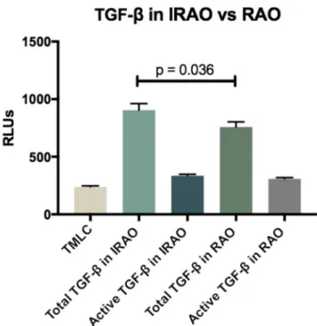
Active TGF-β levels were not different in IRAO and RAO groups (p = 0.2775). Total TGF-β levels were significantly higher in IRAO compared to RAO group (p = 0.0363) (Fig. 3 & Supplementary Table S3).
4. Discussion
In a general population of asthmatics, we found that risk factors for fixed airway obstruction are: male gender, smoking (pack-year), longer disease duration, poor asthma control, sputum eosinophils and neutro-phils, ICS daily dose, BMI, later onset, and OCS therapy. Focusing on non-smoking patients we confirmed that ACQ score, disease duration
Table 1
Demographic, functional, inflammatory, and treatment characteristics.
Characteristics RAO IRAO p-value
N. (%) 777/1138 (68) 196/1138 (17) Women (%) 478 (62) 104 (53) 0.034 Age (yr) 48 ± 16 56 ± 13 <0.0001 Height (cm) 168 ± 9 169 ± 10 0.6043 Weight (Kg) 75 ± 16 75 ± 16 0.9189 BMI (kg/m2) 27 ± 5 26 ± 5 0.6017 Atopy (Y) (%) 416 (57) (n=730) 95 (52) (n=182) 0.278 Smoking status: (%) (n=775) (n=192) <0.0001 Never Smokers 443 (57) 72 (38) Current Smokers 140 (18) 40 (21) Ex-Smokers 192 (25) 80 (42) Pack-years: <0.0001 Current smokers 14 (7-26) 27 (11-41) Ex-smokers 14 (6-26) 19 (8-36)
Age of onset (yr) 32 ± 22 (n=585) 32 ± 21 (n=158) 0.9756 Disease duration (yr) 16 ± 16 (n=585) 24 ± 19 (n=158) <0.0001
Pre-BD FEV1 (L) 2.854 ± 0.809 1.559 ± 0.588 <0.0001 (% pred) 94 ± 14 54 ± 14 <0.0001 Post-BD FEV1 (L) 3.04 ± 0.84 1.69 ± 0.60 <0.0001 (% pred) 100 ± 12 58 ± 14 <0.0001 Post-BD FVC (% pred) 103 ± 13 81 ± 16 <0.0001 Post-BD FEV1/FVC (% pred) 81 ± 7 58 ± 8 <0.0001 PC20M (mg/mL) 1.54 (0.18-13.13) (n=549) 0.64 (0.44-0.93) (n=17) 0.4120 Reversibility (%) 7.00 ± 8.41 9.53 ± 14.07 0.0013 ACT score 16.4 ± 5.1 (n=767) 12.8 ± 5.2 (n=188) <0.0001 ACQ score 1.62 ± 1.02 (n=749) 3.08 ± 1.14 (n=185) <0.0001 AQLQ score 4.73 ± 1.29 (n=766) 3.81 ± 1.29 (n=195) <0.0001 FeNO (ppb) 23 (13-44) (n=762) 22 (13-44) (n=187) 0.6642 Sputum eosinophil count
(% of non-squamous cells) 1.2 (0.2-7.8) 3.7 (0.5-26.5) <0.0001 (/μL) 20.94 (0.68-
150.53) 77.34 (8.96- 788.76)
<0.0001 Sputum neutrophil count
(% of non-squamous cells) 57.0 (35.4-77.2) 63.1 (35.4-83.0) 0.0698 (/μL) 711.5 (260.0- 2060.0) 1297.0 (377.0- 3626.0) 0.0002 Inflammatory phenotypes: <0.0001 Paucigranulocytic 315 (41%) 34 (17%) Eosinophilic 252 (32%) 97 (50%) Neutrophilic 167 (21%) 55 (28%) Mixed granulocytic 43 (6%) 10 (5%) Total serum IgE (kU/L) 104 (34-295)
(n=733) 139 (64-384) (n=177) 0.0072 Blood leukocytes (x 103/ μL) 7.10 (5.88-8.39) (n=761) 8.43 (6.76-10.13) (n=186) <0.0001 Fibrinogen (g/L) 3.23 (2.71-3.72) (n=734) 3.38 (2.87-3.93) (n=167) 0.0080 CRP (mg/L) 1.90 (0.84-4.54) (n=734) 2.44 (1.15-6.26) (n=167) 0.0190 Blood eosinophils (n=761) (n=185) (%) 2.4 (1.4-3.9) 3.0 (1.4-5.6) 0.0031 (/μL) 168 (92-283) 249 (119-433) <0.0001 Blood neutrophils (n=761) (n=185) (%) 53.9 (47.8-60.8) 59.1 (51.7-65.9) <0.0001 (/μL) 3761 (2935-4910) 4766 (3641- 6374) <0.0001 ICS dose (μg/d) 4 (0-1000) 1500 (800-2000) <0.0001 ICS category: (%) <0.0001 Steroid naïve 347 (47) 29 (11) Low dose 99(13) 14 (7) Medium dose 135 (18) 40 (21) High dose 162 (22) 107 (56) LABA, N (%) 403 (52) (n=774) 167 (86) (n=195) <0.0001 LTRA, N (%) 175 (23) 67 (34) 0.001 LAMA, N (%) 13 (2) (n=773) 53 (27) (n=195) <0.0001 Table 1 (continued)
Characteristics RAO IRAO p-value
OCS therapy, N (%) 55 (7) (n=773) 52 (27) (n=195) <0.0001 Biotherapies: 0.055 Anti-IgE 8 (1) 5 (3) Anti-IL5 11 (1) 6 (3) Hospitalizations in previous year 0 (0-1) (n=664) 1(0-2) (n=160) <0.0001 Exacerbations in previous year 0 (0-0) (n=676) 0 (0-1) (n=170) <0.0001 Comparison between RAO (Post-BD FEV1/FVC≥0.7 & post-BD FEV1≥80 pre-dicted) and IRAO Post-BD FEV1/FVC<0.7 & Post-BD FEV1<80 predicted) asthmatics.
Data are presented as mean ± SD or median and IQR. PC20 M is presented as geometric mean (min-max). BMI, Body Mass Index; BD, bronchodilation; FEV1, forced expiratory volume in 1s; FVC, Forced vital capacity; PC20 M, provocative concentration of metacholine causing a 20% fall in FEV1; ACT, Asthma control test; ACQ, Asthma Control Questionnaire; AQLQ, Asthma Quality of Life Ques-tionnaire; FeNO, fractional exhaled nitric oxide; ppb, parts per billion; CRP, C reactive protein; ICS, inhaled corticosteroid; LABA, Long-acting ß2-agonist; LTRA, Leucotriene receptor antagonist; LAMA, Long-acting muscarinic antago-nist; OCS, oral corticosteroid; Low-dose ICS: <500 μg/d; moderate-dose ICS : >500–1000 μg/d; high-dose ICS : > 1000 μg/d beclomethasone dipropionate – chlorofluorocarbon.
and sputum eosinophils are associated with IRAO. We also found that total TGF-β levels were significantly higher in IRAO.
There were significantly more ex-smokers in the IRAO group. Smoking was also a risk factor for IRAO and responsible for a decrease in the FEV1/FVC ratio in the multivariable analyses, and seems to be a
major risk factors for IRAO [9,26,27]. Cigarette exposure is responsible for neutrophilic inflammation and neutrophil apoptosis leading to damage-associated molecular pattern (DAMPs), which may amplify smoking induced airway inflammation [28,29]. Due to this neutrophilic inflammation in smokers or changes in glucocorticoid receptor sensi-tivity, treatment regimens for smoking asthma require higher doses of inhaled corticosteroids [30]. Cigarette smoking also contributes to severity and exacerbations. This might explain why sputum neutrophil count, ICS doses and OCS therapy were not independently associated to IRAO anymore in the never smoker population. One might be tempted to assume IRAO is simply due to smoking habit in our IRAO population.
However, the sub-analysis on never smokers with IRAO confirmed that ACQ score, sputum eosinophils and disease duration are also associated with irreversible airway obstruction. According to guidelines [31], it is required to treat comorbidities such as smoking habit in order to prevent lung function decline in asthma. Since therapy for remodeling is not on the market yet, this study adds evidence to the fact that quitting smoking is clearly a first step to help these patients.
Patients presenting with neutrophilic inflammation represent only one quarter of our study population while 50% had an eosinophilic phenotype. These rates are similar to what has been reported in severe asthma [32] that can be defined by FEV1<80% predicted. In our study IRAO occurred almost twice more often in patients with increased per-centages of sputum eosinophils. Eosinophilic airway inflammation has the potential to induce airway remodeling [33] and is present in 41% of a general population of asthmatics [34]. We previously found that pa-tients exhibiting elevated blood and sputum eosinophils were
Table 2
Factors associated with IRAO. Results of the logistic regression *N = 973.
IRAO Total population
UNIVARIATE MULTIVARIABLE
OR 95%CI P-value OR 95%CI P-value
Gender (M) 0.71 0.52–0.97 0.031 0.38 0.20–0.72 0.003 BMI 1.05 0.91–1.21 0.505 0.70 0.54–0.92 0.010 Age of onset 0.99 0.81–1.21 0.911 2.20 1.27–3.83 0.005 Pack-Year 1.77 1.44–2.16 <0.0001 1.76 1.19–2.61 0.005 Atopy 0.82 0.60–1.14 0.245 0.84 0.43–1.64 0.617 ACQ 3.25 2.71–3.91 <0.0001 2.63 1.94–3.56 <0.0001 ICS 2.07 1.78–2.42 <0.0001 1.60 1.21–2.13 0.001 OCS 4.75 3.12–7.22 <0.0001 2.67 1.11–6.39 0.028 BEC 1.41 1.23–1.63 <0.0001 1.12 0.82–1.53 0.468 BNC 1.0 1.0–1.0 0.023 1.00 0.99–1.00 0.272 SEC 1.36 1.18–1.56 <0.0001 1.73 1.22–2.47 0.002 SNC 1.10 0.96–1.26 0.148 1.53 1.15–2.04 0.004 FeNO 0.97 0.83–1.13 0.681 0.74 0.54–1.02 0.067 Exacerbations 1.39 1.24–1.56 <0.0001 0.97 0.78–1.20 0.776 Disease duration 1.02 1.02–1.04 <0.0001 1.06 1.04–1.09 <0.0001 Hospitalizations 2.35 1.70–3.25 <0.0001 1.12 0.60–2.07 0.720
IRAO, irreversible airway obstruction, Post-BD FEV1/FVC<0.7 & post-BD FEV1<80 predicted. Number of observations = 571. OR, odds ratio; CI, confidence interval; BMI, Body Mass Index (categories: ≤23; >23 to ≤26; >26 to ≤30; >30); FeNO, fractional exhaled nitric oxide (categories: ≤ 12; >12 to ≤ 25; >25 to ≤50; >50); ACQ, Asthma Control Questionnaire; ICS, inhaled corticosteroids (categories: Naïve; 0 to≤ 500; >500 to ≤ 1600; >1600); OCS, oral corticosteroids; Pack-Year categories: ≤0; >0 to ≤20; >20); Age of onset (categories: ≤12; >12 to ≤40; >40); BEC, blood eosinophil count (categories: ≤150; >150 to ≤300; >300 to ≤400; >400); SEC, sputum eosinophil count (categories: ≤0.2; >0.2 to ≤3; >3 to ≤10; >10); SNC, sputum neutrophil count (categories: ≤38; >38 to ≤59; >59 to ≤76; >76).
Table 3
Results of the linear regression model *N = 973. FEV1/FVC Total population*
UNIVARIATE MULTIVARIABLE
Coeff. 95%CI P-value Coeff. 95%CI P-value
Gender (M) 2.48 0.98 to 3.98 0.001 3.26 1.61 to 4.90 <0.0001 BMI 0.08 − 0.07 to 0.23 0.308 0.42 0.26 to 0.58 <0.0001 Age of onset 0.02 − 0.92 to 0.95 0.970 −3.39 − 4.88 to − 1.90 <0.0001 Pack-Year − 0.17 − 0.21 to − 0.13 <0.0001 −0.11 − 0.16 to − 0.06 <0.0001 Atopy 1.04 − 0.49 to 2.57 0.182 0.52 − 1.28 to 2.31 0.572 ACQ − 4.26 − 4.80 to − 3.71 <0.0001 −2.57 − 3.34 to − 1.81 <0.0001 ICS − 3.04 − 3.63 to − 2.43 <0.0001 −1.11 − 1.87 to − 0.35 0.004 OCS − 8.30 − 10.6 to − 5.99 <0.0001 −2.74 − 5.59 to 0.11 0.060 BEC − 0.0017 − 0.0031 to − 0.0002 0.022 0.001 − 0.003 to 0.004 0.756 BNC − 0.0004 − 0.0006 to − 0.0002 <0.0001 −0.0002 − 0.001 to − 0.00003 0.048 SEC − 0.13 − 0.17 to − 0.09 <0.0001 −0.08 − 0.14 to − 0.02 0.007 SNC − 0.02 − 0.05 to 0.004 0.102 −0.03 − 0.07 to 0.001 0.060 FeNO − 0.13 − 0.84 to − 0.57 0.705 0.31 − 0.55 to 1.16 0.481 Exacerbations − 1.54 − 2.07 to − 0.99 <0.0001 0.03 − 0.67 to 0.74 0.922 Disease duration − 0.15 − 0.19 to − 0.096 <0.0001 −0.23 − 0.29 to − 0.17 <0.0001 Hospitalizations − 4.35 − 5.74 to − 2.96 <0.0001 −2.04 − 3.81 to − 0.26 0.024
Number of observations = 319. OR, odds ratio; CI, confidence interval; BMI, Body Mass Index; FeNO, fractional exhaled nitric oxide (categories: ≤ 12; >12 to ≤ 25; >25 to ≤50; >50); ACQ, Asthma Control Questionnaire; ICS, inhaled corticosteroids (categories: Naïve; 0 to≤ 500; >500 to ≤ 1600; >1600); OCS, oral corticosteroids; BEC, blood eosinophil count; BNC, blood neutrophil count; SEC, sputum eosinophil count; SNC, sputum neutrophil count.
characterized by poorer lung function [35]. In our study, blood eosin-ophil count was not associated with IRAO. Indeed, eosineosin-ophils must be attracted into the airways to induce remodeling [35]. Moreover, sputum eosinophil but not blood eosinophil counts were predictors of improvement of lung function with anti-IL5 therapy in severe eosino-philic asthmatics [36]. Eosinophils are the main source of TGF-β, a cytokine with profibrotic properties. We confirmed Cianchetti [37]’s results showing that TGF-ß identified a subgroup of severe asthmatic patients with IRAO. In studies evaluating efficacy of Mepolizumab (Anti-IL5), they found TGF-β expression in eosinophils and signs of airway remodeling are reduced in parallel [38]. TGF-β is one of the main factors implicated in airway remodeling in asthma. We found that total TGF-β levels were significantly higher in IRAO. Only a small fraction of TGF-β were biologically active in our samples and did not differ between groups. What differed was the pool of latent TGF-β as total TGF-β (which represent the active and latent TGF-β) was significantly different. The
airways may serve as a reservoir of latent TGF-β in the condition of IRAO representing a pool of “utilizable” TGF-β insuring a ready source for local activation. Indeed, TGF-β in the latent complex is the predominant form of the molecule and was shown to possess an extended plasma half-life compared with active TGF-β [39].
To our knowledge, this is the first report using this gene reporter assay in a context of IRAO and sputum samples.
Though a higher ACQ score is certainly a consequence of IRAO, we think it is interesting to mention that IRAO can be suspected in a patient with a poor asthma control. Indeed, remodeling induces reduction in airflow calibre, and more asthma symptoms [5]. ACQ score can be ob-tained easily and could be very useful for general practitioners to perform in order to find out if the patient is presenting with IRAO (cut-off point of 2.36) in which case, IRAO can be suspected and this patient must be sent to a pulmonologist to exclude residual type 2 inflammation in the absence of which overtreatment with ICS can be avoided [40]. As previously shown, men have a higher risk of being IRAO than women [14,27,41]. Indeed, males tend to present with fewer symptoms, are poorer perceivers, despite active eosinophilic inflam-mation, thus tend to be less compliant to their medication [42]. Not surprisingly, disease duration is associated with IRAO [9].
One limitation of this study is that we based our definition of IRAO on BTS [43]/NICE [44] guidelines. Using the lower limit of normal (LLN) FEV1/FVC ratio reduces the misclassification of airway
obstruc-tion [45]. Unfortunately, visits prior 2018 did not include LLN values in our database. Since we purposely eliminated patients that did not fit in either IRAO nor RAO group, our patients in the IRAO group are truly asthmatics with persistent airway obstruction characteristics. As a ter-tiary care centre, the prevalence of IRAO might have been overestimated in our study.
5. Conclusion
We were able to show many risk factors independently associated to IRAO all together, previously mentioned separately in different studies. Risk factors for IRAO in a general population of asthmatics are male gender, smoking, a longer disease duration, uncontrolled asthma, eosinophilic or neutrophilic airway inflammation, lower BMI, and later asthma onset. Not surprisingly, sputum eosinophils are biomarkers for remodeling as they reflect local inflammation as opposed to blood eo-sinophils which reflect systemic inflammation. Moreover, TGF-β levels are higher in IRAO.
Disclosure
Dr. LOUIS reports grants and personal fees from GSK, grants and personal fees from AZ, grants and personal fees from Novartis, grants from Chiesi, outside the submitted work. The other authors declare no conflict of interest related to this paper.
Funding
This work was supported by the European Union (Interreg EMR Muse Rhine 5a) and Interuniversity Attraction Poles Program.
Declaration of competing interest
The authors declare that they have no known competing financial interests or personal relationships that could have appeared to influence the work reported in this paper.
Acknowledgements
We thank Prof. Daniel Rifkin, New York University medical center, NY, for the gift of Transformed mink lung cells (TMLC).
Fig. 2. ROC curve showing the best cut-off of ACQ score to identify IRAO in
asthma. Sensitivity 72%, specificity 77%, cut-off: 2.36, p < 0.0001, AUC: 0.8260.
Fig. 3. Comparison of (total and active) TGF-β levels in IRAO and RAO. RAO,
Appendix A. Supplementary data
Supplementary data related to this article can be found at https://do
i.org/10.1016/j.rmed.2020.106202.
References
[1] J.K. Peat, A.J. Woolcock, K. Cullen, Rate of decline of lung function in subjects with asthma, Eur. J. Respir. Dis. 70 (1987) 171–179.
[2] P. Lange, J. Parner, J. Vestbo, P. Schnohr, G. Jensen, A 15-year follow-up study of ventilatory function in adults with asthma, N. Engl. J. Med. 339 (1998) 1194–1200.
[3] S. Graff, S. Demarche, M. Henket, V. Paulus, R. Louis, F. Schleich, Increase in blood eosinophils during follow-up is associated with lung function decline in adult asthma, Respir. Med. (2019) 60–66.
[4] T.R. Bai, D.A. Knight, Structural changes in the airways in asthma: observations and consequences, Clin. Sci. 108 (2005) 463–477.
[5] H. Fehrenbach, C. Wagner, M. Wegmann, Airway remodeling in asthma: what really matters, Cell Tissue Res. 367 (2017) 551–569.
[6] C. Hudson, H. Turcotte, M. Laviolette, G. Carrier, L.-P. Boulet, Characteristics of bronchial asthma with incomplete reversibility of airflow obstruction, Ann. Allergy Asthma Immunol. 78 (1997) 195–202.
[7] C.I. Panhuysen, J.M. Vonk, G.H. Ko¨eter, J.P. Schouten, R. van Altena, E. R. Bleecker, D.S. Postma, Adult patients may outgrow their asthma: a 25-year follow-up study, Am. J. Respir. Crit. Care Med. 155 (4) (1997 Apr) 1267–1272, https://doi.org/10.1164/ajrccm.155.4.9105065. PubMed PMID: 9105065. [8] C.S. Ulrik, V. Backer, Nonreversible airflow obstruction in life-long nonsmokers
with moderate to severe asthma, Eur. Respir. J. 14 (1999) 892–896.
[9] L. Zhang, L. He, J. Gong, C. Liu, Risk factors associated with irreversible airway obstruction in asthma: a systematic review and meta-analysis, Biomed. Res. Int. (2016), 2016Available from: https://www.ncbi.nlm.nih.gov/pmc/articles/PM C4828538/.
[10] D. Bumbacea, D. Campbell, L. Nguyen, D. Carr, P.J. Barnes, D. Robinson, K. F. Chung, Parameters associated with persistent airflow obstruction in chronic severe asthma, Eur. Respir. J. 24 (2004) 122–128.
[11] P. Sexton, P. Black, L. Wu, F. Sommerville, M. Hamed, P. Metcalf, J. Kolbe, Fixed airflow obstruction among nonsmokers with asthma:A case-comparison study, J. Asthma 50 (2013) 606–612.
[12] J.H. Lee, T. Haselkorn, L. Borish, L. Rasouliyan, B.E. Chipps, S.E. Wenzel, Risk factors associated with persistent airflow limitation in severe or difficult-to-treat asthma: insights from the TENOR study, Chest 132 (2007) 1882–1889. [13] K. Mascia, T. Haselkorn, Y.M. Deniz, D.P. Miller, E.R. Bleecker, L. Borish, Aspirin
sensitivity and severity of asthma: evidence for irreversible airway obstruction in patients with severe or difficult-to-treat asthma, J. Allergy Clin. Immunol. 116 (2005) 970–975.
[14] A. ten Brinke, A.H. Zwinderman, P.J. Sterk, K.F. Rabe, E.H. Bel, Factors associated with persistent airflow limitation in severe asthma, Am. J. Respir. Crit. Care Med. 164 (2001) 744–748.
[15] A.-S. Jang, J.-S. Park, J.-H. Lee, S.-W. Park, D.-J. Kim, S.-T. Uh, Y.-H. Kim, C.- S. Park, Asthmatics without rhinitis have more fixed airway obstruction than those with concurrent rhinitis, Allergy Asthma Immunol. Res. 2 (2010) 108. [16] P.K. Jeffery, Remodeling and inflammation of bronchi in asthma and chronic
obstructive pulmonary disease, Proc. Am. Thorac. Soc. 1 (2004) 176–183. [17] J.C. Hogg, F. Chu, S. Utokaparch, R. Woods, W.M. Elliott, L. Buzatu, R.
M. Cherniack, R.M. Rogers, F.C. Sciurba, H.O. Coxson, P.D. Par´e, The nature of small-airway obstruction in chronic obstructive pulmonary disease, N. Engl. J. Med. 350 (2004) 2645–2653.
[18] K.F. Chung, I.M. Adcock, Multifaceted mechanisms in COPD: inflammation, immunity, and tissue repair and destruction, Eur. Respir. J. 31 (2008) 1334–1356. [19] L. Godinas, J.-L. Corhay, M. Henket, J. Guiot, R. Louis, C. Moermans, Increased
production of TGF-β1 from sputum cells of COPD: relationship with airway obstruction, Cytokine 99 (2017) 1–8.
[20] E.F. Juniper, G.H. Guyatt, R.S. Epstein, P.J. Ferrie, R. Jaeschke, T.K. Hiller, Evaluation of impairment of health related quality of life in asthma: development of a questionnaire for use in clinical trials, Thorax 47 (1992) 76–83.
[21] E.F. Juniper, P.M. O′byrne, G.H. Guyatt, P.J. Ferrie, D.R. King, Development and validation of a questionnaire to measure asthma control, Eur. Respir. J. 14 (1999) 902–907.
[22] M. Schatz, C.A. Sorkness, J.T. Li, P. Marcus, J.J. Murray, R.A. Nathan, M. Kosinski, T.B. Pendergraft, P. Jhingran, Asthma Control Test: reliability, validity, and responsiveness in patients not previously followed by asthma specialists, J. Allergy Clin. Immunol. 117 (2006) 549–556.
[23] R.A. Dweik, P.B. Boggs, S.C. Erzurum, C.G. Irvin, M.W. Leigh, A.-C. Olin, A. L. Plummer, R.D. Taylor, An official ATS clinical practice guideline: interpretation of exhaled nitric oxide levels (FENO) for clinical applications, Am. J. Respir. Crit. Care Med. 184 (2011) 602–615.
[24] J. Guiot, S. Demarche, M. Henket, V. Paulus, S. Graff, F. Schleich, J.-L. Corhay, R. Louis, C. Moermans, Methodology for sputum induction and laboratory processing, Journal of Visualized Experiments (2019) [Internet] 2017 [cited 2019 Dec 9]; Available from: https://www.jove.com/video/56612/methodology-fo r-sputum-induction-and-laboratory-processing.
[25] R. Louis, L. Godinas, F. Schleich, Induced Sputum - towards Normal Values, Non Invasive Assessment of Airways Inflammation in Asthma and COPD. 14th, Tetrapoleos Street, 115 27, Paschalidis Medical Publications, Athens, 2011. Greece.
[26] M. Contoli, S. Baraldo, B. Marku, P. Casolari, J.A. Marwick, G. Turato, M. Romagnoli, G. Caramori, M. Saetta, L.M. Fabbri, A. Papi, Fixed airflow obstruction due to asthma or chronic obstructive pulmonary disease: 5-year follow- up, J. Allergy Clin. Immunol. 125 (2010) 830–837.
[27] COhort for Reality and Evolution of adult Asthma in Korea study group (COREA study group), T. Lee, Y.S. Lee, Y.-J. Bae, T.-B. Kim, S.O. Kim, S.-H. Cho, H.-B. Moon, Y.S. Cho, Smoking, longer disease duration and absence of rhinosinusitis are related to fixed airway obstruction in Koreans with severe asthma: findings from the COREA study, Respir. Res. 12 (2011) 1.
[28] I.H. Heijink, S.D. Pouwels, C. Leijendekker, H.G. de Bruin, G.J. Zijlstra, H. van der Vaart, N.H.T. ten Hacken, A.J.M. van Oosterhout, M.C. Nawijn, M. van der Toorn, Cigarette smoke–induced damage-associated molecular pattern release from necrotic neutrophils triggers proinflammatory mediator release, Am. J. Respir. Cell Mol. Biol. 52 (2015) 554–562.
[29] X. Huang, X. Tan, Y. Liang, C. Hou, D. Qu, M. Li, Q. Huang, Differential DAMP release was observed in the sputum of COPD, asthma and asthma-COPD overlap (ACO) patients, Sci. Rep. 9 (2019) 19241.
[30] N.C. Thomson, Asthma and cigarette smoking, Eur. Respir. J. 24 (2004) 822–833. [31] K.F. Chung, S.E. Wenzel, J.L. Brozek, A. Bush, M. Castro, P.J. Sterk, I.M. Adcock, E.
D. Bateman, E.H. Bel, E.R. Bleecker, L.-P. Boulet, C. Brightling, P. Chanez, S.- E. Dahlen, R. Djukanovic, U. Frey, M. Gaga, P. Gibson, Q. Hamid, N.N. Jajour, T. Mauad, R.L. Sorkness, W.G. Teague, International ERS/ATS guidelines on definition, evaluation and treatment of severe asthma, Eur. Respir. J. 43 (2014) 343–373.
[32] F. Schleich, G. Brusselle, R. Louis, O. Vandenplas, A. Michils, C. Pilette, R. Peche, M. Manise, G. Joos, Heterogeneity of phenotypes in severe asthmatics. The Belgian severe asthma registry (BSAR), Respir. Med. 108 (2014) 1723–1732.
[33] H.H. Kariyawasam, D.S. Robinson, The role of eosinophils in airway tissue remodelling in asthma, Curr. Opin. Immunol. 19 (2007) 681–686.
[34] F.N. Schleich, M. Manise, J. Sele, M. Henket, L. Seidel, R. Louis, Distribution of sputum cellular phenotype in a large asthma cohort: predicting factors for eosinophilic vs neutrophilic inflammation, BMC Pulm. Med. 13 (2013) 11. [35] F.N. Schleich, A. Chevremont, V. Paulus, M. Henket, M. Manise, L. Seidel, R. Louis,
Importance of concomitant local and systemic eosinophilia in uncontrolled asthma, Eur. Respir. J. 44 (2014) 97–108.
[36] F. Schleich, S. Graff, H. Nekoee, C. Moermans, M. Henket, C. Sanchez, V. Paulus, F. Guissard, A. Donneau, R. Louis, Real-word experience with mepolizumab: does it deliver what it has promised? Clin. Exp. Allergy 50 (2020) 687–695.
[37] S. Cianchetti, C. Cardini, I. Puxeddu, M. Latorre, M.L. Bartoli, M. Bradicich, F. Dente, E. Bacci, A. Celi, P. Paggiaro, Distinct profile of inflammatory and remodelling biomarkers in sputum of severe asthmatic patients with or without persistent airway obstruction, World Allergy Org. J. 12 (2019) 100078. [38] P.T. Flood-Page, A.N. Menzies-Gow, A.B. Kay, D.S. Robinson, Eosinophil’s role
remains uncertain as anti–interleukin-5 only partially depletes numbers in asthmatic airway, Am. J. Respir. Crit. Care Med. 167 (2003) 199–204. [39] L.M. Wakefield, T.S. Winokur, R.S. Hollands, K. Christopherson, A.D. Levinson, M.
B. Sporn, Recombinant latent transforming growth factor beta 1 has a longer plasma half-life in rats than active transforming growth factor beta 1, and a different tissue distribution, J. Clin. Invest. 86 (1990) 1976–1984.
[40] S. Demarche, F. Schleich, M. Henket, V. Paulus, R. Louis, T. Van Hees, Step-down of inhaled corticosteroids in non-eosinophilic asthma: a prospective trial in real life, Clin. Exp. Allergy 48 (2018) 525–535.
[41] J. Liebhart, M. Polak, A. Dabrowski, R. Dobek, E. Liebhart, A. Dor-Wojnarowska, W. Barg, A. Kulczak, W. Medrala, U. Gladysz, A. Lange, The G/G genotype of transforming growth factor beta 1 (TGF-β1) single nucleotide (+915G/C) polymorphism coincident with other host and environmental factors is associated with irreversible bronchoconstriction in asthmatics, Int. J. Immunogenet. 35 (2008) 417–422.
[42] P. Haldar, I.D. Pavord, D.E. Shaw, M.A. Berry, M. Thomas, C.E. Brightling, A. J. Wardlaw, R.H. Green, Cluster analysis and clinical asthma phenotypes, Am. J. Respir. Crit. Care Med. 178 (2008) 218–224.
[43] BTS guidelines for the management of chronic obstructive pulmonary disease, Thorax 52 (1997) 1–28.
[44] National Institute for Clinical Excellence, Clinical Guideline 12. Chronic Obstructive Pulmonary Disease February 2004, accessed, www.nice.org.uk /CG012NICEguideline. (Accessed 3 October 2008).
[45] M.P. Swanney, G. Ruppel, P.L. Enright, O.F. Pedersen, R.O. Crapo, M.R. Miller, R. L. Jensen, E. Falaschetti, J.P. Schouten, J.L. Hankinson, J. Stocks, P.H. Quanjer, Using the lower limit of normal for the FEV1/FVC ratio reduces the