Clinical report
Skin weathering and ashiness in black Africans
Emmanuelle UHODA Claudine PIÉRARD-FRANCHIMONT Ludivine PETIT Gérald PIÉRARD Department of Dermatopathology, University Hospital Sart Tilman, B-4000 Liège, Belgium Reprints: G.E. Piérard Fax: (+ 32) 4 366 29 76 Email: gerald.pierard@ulg.ac.beArticle accepted on 15/09/2003
Ashiness describes a common physiological skin condition that may
develop in people with dark skin complexion. Environmental influences,
particularly cold and dry weather, seem obvious. This condition has
seldom been studied so far. In the present study, skin ashiness was
assessed in 37 black African women by means of colorimetric
assess-ments and xerosis ratings. Colour changes were measured by the
para-meters a* and the individual typology angle ITA°. Xerosis was assessed
by visual inspection, the ultraviolet light-enhanced visualization
(ULEV) method, and the cyanoacrylate skin surface stripping (CSSS)
method. The assessments were performed on ashy skin of the legs and on
the normal looking forehead during the winter season. Ashy skin was
lighter but not erythematous. The ITA-revealed colour changes were
correlated with xerosis severity as assessed by dry dermoscopy and by
the ULEV and CSSS methods. In conclusion, ashiness due to skin
weathering does not appear to be related to mild inflammation. It
corresponds to a peculiar type of xerosis with reduction in Fresnel
reflection by the stratum corneum.
Key words: environment, ethnicity, skin complexion, skin dryness,
stratum corneum, xerosis
E
thnicity plays an important role in the clinical pre-sentation of diverse skin disorders [1, 2]. In parti-cular, colour changes of lesional skin may be quite prominent in dark skin individuals. This may represent a psychologically distressing condition because of its persis-tent and visible nature. In particular, ashy hue may develop at the site of a variety of inflammatory skin disorders [2]. Among them, ashy dermatitis which is synonymous with erythema dyschromicum persans is a distinctive melano-derma of unknown etiology combining macular erythema and presence of melanophages in the dermis [3, 4]. Ashi-ness is a lay term describing another condition characteri-zed by any xerotic process with loss of natural skin shine that prevails in individuals with dark skin complexion [5]. Ashen and ashing skin are other terms used for a similar condition in reference to restricted body sites including the elbows and knees.This study was performed in black Africans who suffered from seasonal ashiness while under the geoclimatic envi-ronment of Belgium.
Material and methods
The study was performed in winter in 37 women aged from 25 to 38 years. They were phototype VI Africans living in the Liège region for at least 2 years. They complained of seasonal ashiness predominating on the limbs. They were not allowed to use any cosmetic product on the skin for 10 days before the instrumental assessments.
The skin colour was measured on the forehead and on lesional skin of the legs using tristimulus reflectance colo-rimetry (Chroma Meter CR 200, Minolta, Osaka, Japan). The measurement procedure was performed in the L* a* b* mode following the EEMCO recommendations [6]. The mean value of 5 measurements was recorded in each sub-ject. The so-called individual typology angle (ITA°) [6, 7]
was derived following ITA° = Arc Tangent
[(L*-50)/b*].180p– 1. The values of a* and ITA° served to quan-tify erythema and the skin typology, respectively.
The skin surface was observed and the severity of xerosis was assessed using the overall dry skin score (ODS) as defined by the EEMCO guidelines [8] and shown in Table I. Table I. Overall dry skin (ODS) score according to the EEMCO guidelines [from ref. 8]
Score Description
0 Absent
1 Faint scaling, faint roughness and dull appearance
2 Small scales in combination with a few larger scales, slight roughness, whitish appearance 3 Small and larger scales uniformly distributed,
definite roughness, possibly slight redness and possibly a few superficial cracks
4 Dominated by large scales, advanced roughness, redness present, eczematous changes and cracks
Dry dermoscopy [9, 10] was used to enhance detailed visual perception of xerosis. In addition, the ultraviolet light-enhanced visualization (ULEV) method was used to increase the sensitivity of the observations. For that purpose, a CCD camera equipped with an internal ultravio-let light illumination (Visioscan® VC98, C + K electronic, Cologne, Germany) recorded scaliness as previously des-cribed [10, 11]. A cyanoacrylate skin surface stripping (CSSS) harvested as previously described [12-14] on the legs. The severity of xerosis was rated under the microscope according to a grading scale identical to that used for the ULEV method (Table II).
The mean and SD of the a* and ITA° values were calcula-ted. Intra-individual comparisons were made for each colo-rimetric value between the forehead skin and lesional skin using the two-tailed paired Student t test. Correlations between the values of these parameters were searched for using regression model analysis with calculation of the coefficient of correlation r. Similar statistical assessments were performed to evaluate the relationships between, on the one hand, ITA° values and, on the other hand, xerosis severity as assessed by dry dermoscopy, and the ULEV and CSSS methods. A p value lower than 0.05 was considered statistically significant.
Results
Erythema as assessed by the a* parameter was similar (p = 0.46) on the forehead (8.3 ± 1.3) and on the legs (8.5 ± 1.5). By contrast, the ITA° value of the forehead (– 33 ± 3) was significantly lower (p < 0.01) than on leg ashiness (– 29.7 ± 6.6). A weak correlation was found between the a* and ITA° values on the forehead (r = 0.42), but was absent on leg ashiness (r = – 0.02).
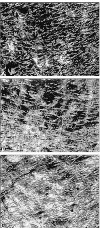
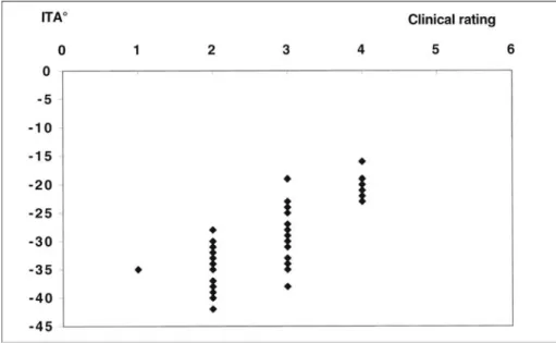
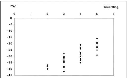
Xerosis of leg ashiness (Fig. 1) was recognized by dry dermoscopy grading (2.7 ± 0.8), ULEV assessment (3.3 ± 0.9) and CSSS xerosis grading (3.7 ± 0.9). Correla-tions were found between each of the 3 rating methods of xerosis and ITA° values (Table III, Figs. 2-4).
Discussion
Xerosis responsible for ashiness reduces the natural skin shine and presents as whitish areas in darker skinned
indi-viduals. Cross-polarized imaging combining image analy-sis and clinical pattern recognition has been suggested to quantify this skin condition [5]. Skin weathering, particu-larly during winter, results in some physiological and struc-tural changes [11, 15-25]. Exposure to a cold environment alters the activity of desquamatory enzymes [11, 26]. This effect is amplified when the relative humidity of air is reduced with ensuing water depletion in the outer stratum Table II. Patterns and severity scores of xerosis as assessed
by the ultraviolet light enhanced visualization (ULEV) method and by cyanoacrylate skin surface strippings
Grade Pattern of xerosis 0 Normal stratum corneum
1 Hyperkeratosis of the primary and secondary lines and/or the appendageal orifices 2 Hyperkeratosis covering less than 30% of the
plateaus
3 Hyperkeratosis covering more than 30% of the plateaus
4 Diffuse confluent scales
5 Thick, uneven scales covering the entire surface, obliterating the skin surface furrows
A
B
C
Figure 1. Ultraviolet light-enhanced visualization (ULEV) of skin ashiness.
corneum [19, 23]. The combination of cold and dry threat can be expressed by dew point variations [15, 20]. The resulting effect of such a process is the development of a peculiar type of xerosis [11, 21, 27] due to a defect in
corneodesmolysis and desquamation [28-30]. The altered specific enzymes are proteases, particularly serine and cathepsin-like enzymes.
The present study was performed combining colorimetric assessments and xerosis ratings by visual inspection, and by the ULEV and CSSS methods. It shows that skin ashi-ness is similarly objectivated by assessing the stratum cor-neum texture than by measuring skin colour. The whitish, dull and opaque appearance results from the overall in-crease in diffuse light scattering and a reduction in Fresnel reflection (optical phenomenon responsible for skin glare) at the skin surface. This optical phenomenon is responsible for the variations in skin glare.
Erythema was not evidenced by measurements of parame-ter a* at the site of skin ashiness. This suggests the absence of clinically-relevant inflammation at the origin of this skin condition.
Table III. Coeffıcients of correlation r between the individual typology angle (ITA°) and xerosis ratings by dry dermoscopy, ultraviolet light enhanced visualization (ULEV) and by cyanoacrylate skin surface stripping (CSSS) examination
ULEV CSSS ITA° Dry dermoscopy 0.65 0.68 0.73 ULEV 0.83 0.76 CSSS 0.83
Figure 2. Correlation between ITA° values and visual rating of skin ashiness of the legs.
Figure 3. Correlation between ITA° values and xerosis rating by the ultraviolet light enhanced visualization (ULEV) of skin ashiness of the legs.
In conclusion, ashiness as a consequence of skin weather-ing does not appear to be related to inflammation. Xerosis with a reduction of Fresnel reflexion seems to be the major cause of this common skin condition in individuals with a dark skin complexion. j
Acknowledgements. This work was supported by the
“Fonds d’Investissement de la Recherche Scientifique” of the University Hospital of Liège.
References
1. Berardesca E, Maibach H. Ethnic skin: overview of structure and
function. J Am Acad Dermatol 2003; 48: S139-42.
2. Halder RM, Nootheti PK. Ethnic skin disorders overview. J Am
Acad Dermatol 2003; 48: S143-8.
3. Baranda L, Torres-Alvarez B, Cortes-Franco R, Moncada B,
Portalez-Perez DP, Gonzalez-Amaro R. Involvement of cell adhesion and activation of molecules in the pathogenesis of erythema dyschro-micum perstans (ashy dermatitis). The effect of clofazimine therapy. Arch dermatol 1997; 133: 325-9.
4. Jang KA, Choi JH, Sung KS, Moon KC, Koh JK. Idiopathic eruptive
macular pigmentation: report of 10 cases. J Am Acad Dermatol 2001; 44 (suppl): 351-3.
5. Santanastasio H, Zhang S, Krishnan S. Velthuizen R, Shah P, Tsaur
L. Quantifying skin ashing using cross-polarized imaging. Skin Res Technol 2003; 9: 194.
6. Piérard GE. EEMCO guidance for the assessment of skin colour. J
Eur Acad Dermatol Venereol 1998; 10: 1-11.
7. Chardon A, Cretois I, Hourseau C. Skin colour typology and
suntanning pathways. Int J Cosmet Sci 1991; 13: 191-201.
8. Serup J. EEMCO guidance for the assessment of dry skin (xerosis)
and ichthyosis: clinical scoring systems. Skin Res Technol 1995; 1: 109-14.
9. Piérard GE, Piérard-Franchimont C, Saint-Léger D, Kligman AM.
Squamometry: the assessment of xerosis by colorimetry of D-squame adhesive discs. J Soc Cosmet Chem 1992; 47: 297-305.
10. Piérard-Franchimont C, Petit L, Piérard GE. Skin surface patterns
of xerotic legs: the flexural and accretive types. Int J Cosmet Sci 2001; 23: 121-6.
11. Piérard-Franchimont C, Piérard GE. Beyond a glimpse at
seaso-nal dry skin. A review. Exog Dermatol 2002; 1: 3-6.
12. Piérard-Franchimont C, Piérard GE. Skin surface strippings in
diagnosing and monitoring inflammatory, xerotic and neoplastic diseases. Ped Dermatol 1985; 2: 180-4.
13. Piérard-Franchimont C, Piérard GE. Assessment of aging and
actinic damages by cyanoacrylate skin surface strippings. Am J Dermatopathol 1987; 9: 500-9.
14. Piérard GE. EEMCO guidance for the assessment of dry skin
(xerosis) and ichthyosis: evaluation by stratum corneum strippings. Skin Res Technol 1996; 2: 3-11.
15. Gaul LE, Underwood GB. Relation of dew point and barometric
pressure to chapping of normal skin. J Invest Dermatol 1952; 19: 9-19.
16. Rawling AV, Watkinson A, Rogers J, Mayo AM, Hopes J, Scott
IR. Abnormalities in stratum corneum structure, lipid composition, and desmosome degradation in soap-induced winter xerosis. J Soc Cosmet 1994; 45: 203-20.
17. Yoshikawa N, Imokawa G, Akimoto K, Jin K, Higaki Y,
Kawashima M. Regional analysis of ceramides within the stratum corneum in relation to seasonal changes. Dermatology 1994; 188: 207-14.
18. Rogers J, Harding C, Mayo A. Stratum corneum and lipids: The
effect of aging and the seasons. Arch Dermatol Res 1996; 288: 765-70.
19. Denda M, Sato J, Masuda Y, Tsuchiya T, Koyama J, Kuramoto M,
Elias PM, Feingold KR. Exposure to a dry enviroment enhances epidermal permeability barrier functions. J Invest Dermatol 1998; 111: 858-63.
20. Paquet F, Piérard-Franchimont C, Fumal I, Goffin V, Paye M,
Piérard GE. Sensitive skin at menopause; dew point and electrome-tric properties of the stratum corneum. Maturitas 1998; 28: 221-7.
21. Berry N, Charmeil C, Goujon C, Silvy A, Girard P, Corcuff P,
Montastier C. A clinical, biometrological and ultrastructural study of xerotic skin. Int J Cosmet Sci 1999; 21: 241-52.
22. Black D, Del Pozo A, Lagarde JM, Gall Y. Seasonal variability in
the biophysical proterties of stratum corneum from different anatomi-cal sites. Skin Res Tecnol 2000; 6: 70-6.
23. Watkinson A, Harding C, Moore A, Coan P. Water modulation
of statum corneum chymotryptic enzyme activity and desquamation. Arch Dermatol Res 2001; 293: 470-2.
24. Kikuchi K, Kobayashi H, Le Fur I, Tschachler E, Tagami H. The
winter season affects more severely the facial skin than the forearm skin. Comparative biophysical studies conducted in the same Japa-nese females in later summer and winter. Exog Dermatol 2002; 1: 32-8.
Figure 4. Correlation between ITA° values and xerosis rating on cyanoacrylate skin surface strippings (CSSS) harvested from skin ashiness of the legs.
25. Piérard GE. Skin weathering. The face at the interface.
Derma-tology, in press.
26. Piérard GE, Goffin V, Hermanns-Lê T, Piérard-Franchimont C.
Corneocyte desquamation. Int J Mol Med 2000; 6: 217-21.
27. Piérard GE. What do you mean by dry skin? Dermatologica
1989; 179: 1-2.
28. Sato J, Denda M, Nakanishi J, Koyama J. Dry condition affects
desquamation of stratum corneum in vivo. J Dermatol Sci 1998; 18: 163-9.
29. Harding CR, Watkinson A, Scott IR, Rawlings AV. Dry skin,
moisturization and corneodesmolysis. Int J Cosmet Sci 2000; 22: 21-52.
30. Rawlings AV. Trends in stratum corneum research and the
management of dry skin conditions. Int J Cosmet Sci 2003; 25: 63-95.
![Table I. Overall dry skin (ODS) score according to the EEMCO guidelines [from ref. 8]](https://thumb-eu.123doks.com/thumbv2/123doknet/6172479.158598/1.892.461.805.920.1107/table-overall-skin-ods-score-according-eemco-guidelines.webp)