i
Université de Montréal
THE INVOLVEMENT OF NITRIC OXIDE IN BOVINE
FOLLICULAR DEVELOPMENT AND OVULATION
Par
GUSTAVO DE OLIVEIRA ZAMBERLAM
Centre de recherche en reproduction animale (CRRA)
Département de biomédecine vétérinaire
Faculté de médecine vétérinaire
Thèse présentée à la Faculté de médecine vétérinaire
en vue de l’obtention du grade de
philosophiae doctor (Ph.D.)
en sciences vétérinaires
option reproduction
Août, 2013
© Gustavo Zamberlam, 2013
ii
Résumé
Comprendre les événements paracriniens qui régulent la fertilité chez la vache est nécessaire non seulement en raison de l'importance agricole de cette espèce, mais aussi pour son utilisation potentielle comme modèle chez l’humain. L'oxyde nitrique (NO), un gaz de radicaux libres, a été impliqué dans la croissance folliculaire et l'ovulation chez les rongeurs et d'autres espèces, mais chez la vache c’est une énigme fascinante : le NO est produit par les cellules de la granulosa bovine et est régulé par la FSH, mais la présence et le profil d'expression des enzymes responsables de la synthèse de NO (NOS) dans les cellules de la granulosa tout au long de la croissance folliculaire ne sont pas claires. Les objectifs de cette thèse sont: (1) élucider le mécanisme de contrôle des NOS et les conséquences de la production d'oxyde nitrique pour le fonctionnement des cellules de la granulosa au cours du développement folliculaire chez la vache et (2) déterminer la régulation des NOS pendant la cascade ovulatoire induite par LH chez les cellules de la granulosa bovine et si l'activité des NOS pour l’expression des gènes critiques dans la cascade ovulatoire chez cette espèce. Les résultats sont séparés en 2 articles. Dans le premier article, la régulation de NOS2 dans les cellules de la granulosa bovine a été explorée. L'abondance des ARNm codant pour NOS2 a été stimulée par la FSH et l’IGF1 en augmentant l’estradiol, et un blocage de l'action de l’estradiol a conséquemment réduit les niveaux d'ARNm codant pour NOS2. De plus, l'inhibition de l'activité des NOS a augmenté l'apoptose dans les cellules de la granulosa in vitro. Dans le second article, il a été démontré que le pic de LH induit une activation des NOS dans les cellules de la granulosa, et que l'activité de NOS induit la production de NO, ce qui est essentiel pour l’expression des gènes critiques dans la cascade ovulatoire induite par LH comme EREG/AREG/PTGS2. Ensemble, les résultats présentés dans ces 2 articles suggèrent que les niveaux physiologiques d'activité des NOS peuvent contribuer à la croissance et la survie des cellules de la granulosa et indiquent également que NO peut être essentiel pour l'ovulation chez les bovins.
Mots-clés: oxyde nitrique, NOS, ovaire, follicule, cellules de la granulosa, apoptose, ovulation, vache
iii
Abstract
Understanding the paracrine events that regulate fertility in the cow is necessary not only because of the agricultural importance of this species, but also its potential use as a model for humans. Nitric oxide (NO), a free-radical gas, has been implicated in follicular growth and ovulation in rodents and other species, but the cow is an intriguing enigma: NO is produced by bovine granulosa cells and is regulated by FSH, but the presence and the expression pattern in granulosa cells of the enzymes responsible for NO synthesis (NOS) throughout follicular growth are unclear. The objectives of the present thesis were (1) to elucidate the mechanism of control of NOS and the consequences of nitric oxide production for granulosa cell function during follicle development in cattle; and (2) to determine the regulation of NOS during the LH-induced ovulatory cascade in bovine granulosa cells and whether NOS activity is critical for the ovulatory cascade in this species. The results are separated in 2 articles. In the first article, the regulation of NOS2 in bovine granulosa cells was explored. Abundance of mRNA encoding NOS2 was stimulated by FSH and IGF1 through increased estradiol, and a blockade of estradiol action consequently lowered NOS2 mRNA levels. Further, inhibition of NOS activity increased apoptosis in granulosa cells in vitro. In the second article, it was demonstrated that the LH surge induces NOS activation in granulosa cells, and that NOS activity induces the production of NO, which is essential for EREG/AREG/PTGS2 expression, critical genes in the LH-induced ovulatory cascade. Together, the results presented in these 2 articles suggest that physiological levels of NOS activity may contribute to growth and survival of granulosa cells, and also indicate that NO may be essential for ovulation in cattle.
iv
Table of contents
Résumé...ii Abstract...iii Table of contents...iv List of figures...vii List of abbreviations...ix Acknowledgments...xii Introduction...1Chapter 1: Literature review...3
1. Ovarian follicular development and growth...4
1.1 Ovarian follicles...4
1.1.1 Primordial follicles...5
1.1.2 Primary follicles...6
1.1.3 Secondary follicles...6
1.1.4 Antral follicle formation...7
1.2 Follicle steroidogenesis...8
1.2.1 Roles of gonadotropins and insulin-like factor-1 (IGF1) in steroidogenesis...10
1.3 Follicular dynamics in the cow...10
1.3.1 Follicle wave emergence...11
1.3.2 Follicle selection and diameter deviation...12
1.3.3 Follicle dominance...13
1.3.4 Ovulatory follicles...14
1.4 Follicle atresia...15
1.4.1 Apoptosis in ovarian follicles...16
v
2. Ovulation...18
2.1 The preovulatory cascade...19
2.1.1 LH signaling pathways activation...19
2.1.2 ADAMs...20
2.1.3 EGF-like growth factors...21
2.1.4 Prostaglandins...23
2.1.5 Cumulus expansion and oocyte maturation...23
2.2 Follicle rupture...25
2.3 Corpus luteum...25
2.3.1 Corpus luteum functions...26
2.3.2 Luteolysis...26
3. Nitric oxide...27
3.1 Nitric oxide biosynthesis...27
3.1.1 Nitric oxide synthases...28
3.1.1.1 Transcriptional regulation of NOS...29
3.1.1.2 NOS protein structure...30
3.1.2 Enzymatic reaction...31
3.1.3 NOS activation...31
3.2 Nitric oxide chemistry...33
3.2.1 NO redox species and its interactions...34
3.3 Nitric oxide biology...35
3.3.1 Mechanisms of action of NO...35
3.3.2 Roles of nitric oxide in general physiology...36
3.3.3 Roles of nitric oxide in general physiopathology...37
3.4 Nitric oxide functions in the ovary...38
3.4.1 Nitric oxide and steroidogenesis...38
3.4.2 Nitric oxide production and follicle development and growth...39
3.4.3 Nitric oxide and apoptosis in granulosa cells...40
3.4.4 Nitric oxide and the ovulatory process...40
vi
3.4.6 Nitric oxide and corpus luteum formation and luteolysis...42
3.5 NOS expression in the ovary...43
3.5.1 Nitric oxide synthases identified in the ovary...43
3.5.2 NOS expression during follicle development...44
3.5.3 NOS expression during peri-ovulatory period...45
3.5.4 NOS expression during corpus luteum formation and luteolysis...45
Chapter 2: Hypotheses and objectives...46
Chapter 3: Articles...48
Article 1. Regulation of inducible nitric oxide synthase expression in bovine ovarian granulosa cells...49
Abstract...50
Introduction...51
Materials and methods...53
Results...59
Discussion...61
References...65
Figures...70
Article 2. Nitric oxide synthase activity is critical for the LH-induced ovulatory cascade in bovine granulosa cells...78
Abstract...79
Introduction...80
Materials and methods...81
Results...85
Discussion...87
References...91
Figures...95
Chapter 4: General discussion...109
Chapter 5: Final conclusions...119
vii
List of figures
Literature review
Figure 1: Ovarian follicle classification...5
Figure 2: Antral follicle structure...7
Figure 3: Major steroidogenic pathways in the follicle...9
Figure 4: Ovarian follicular dynamics during the oestrous cycle...15
Figure 5: Important signaling cascades in ovulation...20
Figure 6: LH-induced RAS/ERK1/2 signaling pathway...22
Figure 7: The general structure of the NOS enzymes...30
Figure 8: Nitric oxide synthesis from L-arginine...31
Figure 9: The regulation of NOS3 by phosphorylation...33
Article 1 Figure 1: Effect of hormones and growth factors on abundance of iNOS mRNA and oestradiol secretion from granulosa cells in vitro...72
Figure 2: iNOS mRNA abundance is stimulated by oestradiol...74
Figure 3: Effects of an iNOS inhibitor on IGF1-stimulated granulosa cells in vitro...76
Figure 4: Follicle size, oestradiol (E2) concentration and abundance of mRNA encoding aromatase (CYP19) and iNOS in six pairs of early dominant (D) and non-dominant (ND) follicles...78
viii Article 2
Figure 1: LH induces NOS mRNA abundance in a time-dependent manner...100
Figure 2: NOS3 mRNA levels are regulated in a dose-dependent manner by LH and EGF...102
Figure 3: Prostaglandins regulate NOS3 mRNA levels...104
Figure 4: LH and EGF increase nitric oxide production...106
Figure 5: Effect of NOS inhibitor on LH-induced preovulatory genes...108
Figure 6: Effect of NOS inhibitor on EGF-induced preovulatory genes...110
Figure 7: Nitric oxide is not essential for EGF-induced ERK1/2 and AKT phosphorylation...108
9
List of abbreviations
3β-HSD: 3β-hydroxysteroid dehydrogenase 17β-HSD: 17β-hydroxysteroid dehydrogenase A4: Androstenedione ADAM: A Disintegrin and Metalloproteinase
AREG: Amphiregulin AKT: Protein Kinase B
Bax: Bcl-2–associated X protein Bcl-2: B-cell lymphoma 2 BH4: Tetrahydrobiopterin BP: Base Pair
BTC: Betacellulin
cAMP: Cyclic Adenosine Monophosphate
CAMKII: Calcium/calmodulin-dependant Kinase II
cGMP: Guanosine 3’5’-Monophosphate CL: Corpus luteum
COC: Cumulus-Oocyte Complexes CYP11A1: Cytochrome P450 Cholesterol Side-Chain Cleavage, Family 11, Subfamily A, Polypeptide 1 CYP17: cytochrome P450 17α-
hydroxylase
CYP19A1: Cytochrome P450, Family 19, Subfamily A, Polypeptide 1 D: dominant follicle
DNA: Deoxyribonucleic Acid eCG: Equine chorionic gonadotropin eNOS: Endothelial Nitric Oxide Synthase
E2: Estradiol
EGF: Epidermal Growth Factor EGFR: Epidermal Growth Factor Receptor
EGR1: Early Growth Response 1 EREG: Epiregulin
10 ERK: Extracellular-Signal-Regulated
Kinase
FAD: flavin adenine dinucleotide Fas: Apoptosis Antigen 1
FasL: Apoptosis Antigen 1 Ligand FGF2: Fibroblast Growth Factor 2 FMN: Flavin Mononucleotide FSH: Follicle-Stimulating Hormone FSHR: Follicle-Stimulating Hormone Receptor GnRH: Gonadotropin-Releasing Hormone GPCR: G protein-coupled receptor GVBD: germinal vesicle breakdown H2AFZ: H2A histone family, member Z HAS2: Hyaluronan Synthase 2
hCG : Human Chorionic Gonadotropin HDL: High-density Lipoprotein
HSD17B1: Hydroxysteroid (17-beta) dehydrogenase 1
HSD3B2: Hydroxy-delta-5-steroid dehydrogenase, 3 beta- and steroid deltaisomerase 2
IGF1: Insulin-like Growth Factor-1 iNOS: Inducible Nitric Oxide Synthase LDL: Low-density Lipoprotein
LH: Luteinizing Hormone
LHR: Luteinizing Hormone Receptor LPS: Lipopolysaccharide
MAPK: mitogen-activated protein kinase
mRNA: Messenger RNA NADPH: nicotinamide adenine dinucleotide phosphate reduced form ND: non-dominant follicle
NOS: Nitric Oxide Synthase
NOS1: Neuronal Nitric Oxide Synthase NOS2: Inducible Nitric Oxide Synthase NOS3: Endothelial Nitric Oxide
Synthase
NOx: Nitrogen Oxide Species
11 P4: Progesterone
PG: Prostaglandin PGE2: Prostaglandin E2 PGF2α: Prostaglandin F2α
PI3K: Phosphatidylinositol 3-Kinase
PKA: Protein Kinase A PKG: Protein Kinase G
PMSG: pregnant mare's serum gonadotropin
PKC: Protein Kinase C PTGS2: Prostaglandin-Endoperoxide
Synthase 2
PTX3: Pentraxin related gene RAS: Rat Sarcoma
RNA: Ribonucleic Acid
RT-PCR: Reverse Transcription-Polymerase Chain Reaction SDS: Sodium Dodecyl Sulfate sGC: Soluble Guanylyl Cyclase
SNAP: S-nitroso-N-acetylpenicillamine SNP: Sodium Nitroprusside
StAR: Steroidogenic Acute Regulatory protein
TGF-β: Transforming Growth Factor Beta
TSG-6: Tumor Necrosis Factor-Inducible Gene 6 Protein ZP: Zona Pellucida
2
Acknowledgements
I want to express my deeply-felt thanks to Dr. Christopher Price for everything. Thank you for being more than an excellent supervisor, but also a great person. It was an honor to be your Ph.D. student.
I want to give special thanks to my special lab mates Hilda Guerrero, Atefeh Abedini and Fatiha Sahmi. Thanks for your collaboration and advices. Thanks for your friendship and for making the lab an excellent place to be.
My deepest gratitude to my collaborator and friend Dr. Valério Portela. He not only gave me the pleasure to know Dr. Price, but also directly and indirectly participated in my formation as a Ph.D. student.
I want to thank Alysson Macedo and Gustavo Zemke, for all the help during their training in our lab.
I am happy to acknowledge all the CRRA employees who helped me during this period. I want to thank Mira Dobias-Goff, Vickie Roussel and Jacinthe Therrien for their technical support and their kindness. My sincere gratitude to Eliane Auger, Geneviève Provost, Julie Blouin and Micheline Sicotte for helping me to solve all bureaucratic obstacles. Thank you ladies!
I want to thank all CRRA students who shared their experiences, knowledge and friendship during all these years.
I want to thank the Fonds québécois de la recherche sur la nature et les technologies (FQRNT) for the scholarship and the Natural Sciences and Engineering Research Council (NSERC) for the financial support for the experiments.
3
I would like to thank my mother Regina, my father Jurandir, my brother Alexandre and my sisters Cristina and Clarissa, for all the encouragement and emotional support sent from thousands kilometer from here. You are always on my mind. I love you guys! I want to express my deeply-felt thanks to my wife Lucilene Bernardi, my best friend and love. Thank you for always being there for me. Thank you for sharing this amazing experience with me.
I would like to thank God for these blessed years of my Ph.D.
1
Introduction
Ovarian follicles are the functional units of the ovary. Each follicle contains normally one oocyte, the female reproductive germ cell. The other cells that surround the oocyte to compose a mature ovarian follicle are somatic cells, and include cumulus and mural granulosa cells, and the cells of the theca layer [1]. The formation, development and maturation of an oocyte is defined as oogenesis, while the process that involves the proliferation and differentiation of somatic cells, and consequently, the maturation of the whole ovarian follicle is refered to as folliculogenesis. Both oogenesis and folliculogenesis are linked in an intimate and mutually dependent relationship [2, 3].
During the course of folliculogenesis, oocytes first acquire meiotic competence and then gradually acquire developmental competence, a biochemical and molecular state that allows a mature oocyte to undergo normal fertilization, support normal preimplantation embryo development and subsequent healthy growth of the implanted embryo to term. The support for the acquisition of oocyte competence is maybe the most important function of the follicle, with granulosa cells exerting an essential role [4, 5]. For this reason, it is crucial that granulosa cells are healthy and working properly. To guarantee the good functioning of granulosa cells, many endocrine factors such as gonadotropins, paracrine growth factors and intracrine modifiers of cell function modulate their development and function. Some, such as FSH and LH, have well defined roles in granulosa cells function, but the roles of others are less well defined. This is the case for nitric oxide (NO), a short-lived gas produced by the action of the enzyme nitric oxide synthase (NOS). This free-radical is produced in the ovary and has been implicated in different ovarian processes of several species. Although many studies have determined that NO modulates processes like steroidogenesis, follicular growth, oocyte maturation and ovulation [6-8], many questions about the regulation of the NO generation system as well the physiological effects of NO in granulosa cells still need to be answered.
2
This thesis contains results from studies using cell models carefully selected to represent granulosa cells at different stages of development in cattle. The regulation of NOS mRNA levels in granulosa cells by natural ligands, including gonadotropins, steroids and growth factors in conditions that mimic follicle growth and differentiation, as well in conditions that simulate the periovulatory period, have been determined. The roles exerted by NO on granulosa cells during these two distinct physiological moments have also been described. These findings may provide new and clinically relevant information on the physiological role of a highly potent free radical gas in the ovary. The data obtained will advance significantly our understanding of follicle development and ovulation and should lead to better clinical approaches to infertility.
3
Chapter 1:
Literature review
4
1. Ovarian follicular development and growth
Follicular development and growth can be driven by different regulators and involve complex interactions between the three main cell types within the follicle: theca cells, granulosa cells and the oocyte. The systemic endocrine regulation of folliculogenesis is related not only to the pituitary gonadotropins FSH and LH, but various locally produced hormones and growth factors. The oocyte has been confirmed as a major regulator of preantral and early antral follicular growth. On the other hand, late steps of antral follicle development and growth involve gonadotropins and growth factors, specially the insulin-like growth factor (IGF) system [3, 9].
The following sections will focus mainly on basic aspects of ovarian follicular development and growth in cattle.
1.1 Ovarian follicles
In ruminants, ovarian follicular formation is completed during fetal life. In cattle, follicular growth is initiated before the last primordial follicles are formed and then continues throughout fetal, neonatal and adult life [10]. Ovarian follicles can be classified as primordial, primary, secondary and tertiary or antral follicles (Figure 1). Some authors divide antral follicles in early or small antral follicles and late or large antral follicles. They not only present differences in their morphology, but also in their responsiveness to different regulators [3, 11].
5
Figure 1. Ovarian follicle classification (http://www.bme.umich.edu/labs/shikanov/).
1.1.1 Primordial follicles
In cattle, after day 90 of fetal life, the first follicles separate themselves by producing a basement membrane, forming the primordial follicles which are the largest population of follicles in the ovary. Each primordial follicle contains a small non-growing oocyte and a layer of non-dividing flattened pre-granulosa cells encapsulated by the follicular basal lamina. The ovary has a reservoir of primordial follicles that is depleted as follicles gradually and regularly leave this resting pool and initiate growth [11]. In these follicles, the oocyte and granulosa cells have receptors for some growth factors, but not LH or FSH. The primordial follicles, however, do not require gonadotropins for their survival and continued development [12]. Many of the proteins expressed in primordial follicles are associated with cell maintenance and preparation for growth. A primordial follicle expresses hundreds of genes that fulfill housekeeping and signalling functions, cytoskeletal events, DNA repair, mRNA processing, ribosomal function, protein synthesis and ubiquitination. The delay between the appearance of the first primordial and the first primary follicles is relatively long, at 50 days in cattle [13, 14].
6 1.1.2 Primary follicles
The first activated primary follicles do not appear in bovine fetal ovaries until Day 140 of pregnancy. Once follicles have left the pool of primordial follicles they undergo gonadotropin-independent growth, meaning that FSH and LH are not essential for their growth. These small pre-antral follicles present continuous growth that is mainly controlled by factors secreted by the oocyte [12]. The transformation of the flattened pre-granulosa cells of the primordial follicle into a single layer of cuboidal granulosa cell marks the transition to primary follicle [13]. As a follicle grows to the primary stage, the granulosa cells not only change shape but also divide and increase in number and the oocyte enlarges. The primary follicle is also characterized by the development of the zona pellucida (ZP), that was absent in primordial follicles. Several hundred genes not found in primordial follicles are activated during this stage of growth, including those related with synthesis of the ZP, as well some involved in mitochondrial function, cell signalling and communication [11].
1.1.3 Secondary follicles
The secondary follicles are a group of large preantral follicles. They gain multiple layers of granulosa cells, from two to six layers around the oocyte. They also present a well delimited zona pellucida and a theca interna. The secondary follicles are considered gonadotropin-responsive because these follicles present not only FSH-responsive granulosa cells but are also characterized by the development of LH-responsive theca interna. The acquisition of the enzymes required for thecal androgen production is essentially complete before antrum formation [14, 15].
7 1.1.4 Antral follicle formation
The antrum is a fluid-filled cavity that is formed in the follicles under the influence of FSH. A follicle with an antrum is named tertiary or antral follicle (Figure 2). As antral follicles form, the granulosa cells differentiate into two anatomically and functionally distinct lineages; the mural granulosa cells that line the wall of the follicle and that have principally a steroidogenic role; and the cumulus cells, that form an intimate life-support association with the oocyte [16, 17].
Figure 2. Antral follicle structure (http://www.studyblue.com/notes/note/n/week-7-female-reproductive-system-parysek/deck/5613015).
As follicle development progresses, follicles gradually become more and more reliant on gonadotropins, first as gonadotropin-responsive follicles and then as gonadotropin-dependent follicles [3].
8
Follicular growth is highly related to the secretion of steroids, especially estrogens. They are necessary for granulosa cell proliferation, growth of the oocyte and acquisition of LH receptors [18, 19].
1.2. Follicle steroidogenesis
One of the most important functions of the follicle is the production of steroids. Follicular steroidogenesis in ruminants, as in other species, starts usually with cholesterol and ends with the formation of several steroid metabolites [20]. This involves both theca and granulosa cells (Figure 3). Basically, cholesterol is imported into the cell through internalization of blood-borne lipoproteins. Within the cell, cholesterol is maintained within lipid droplets as cholesterol esters. The enzyme cholesterol ester hydrolase converts the cholesterol esters to free cholesterol. Within the cytoplasm the free cholesterol is mobilized to the mitochondria, and then internalized. This internalization of cholesterol by the mitochondria is the rate-limiting step for the general steroidogenic pathway, and is mediated by steroidogenic acute regulatory protein (StAR). Once inside the mitochondria, cholesterol is converted to pregnenolone by the enzyme cytochrome P450 cholesterol side-chain cleavage (CYP11A1 or P450scc). Pregnenolone can then be converted to progesterone by the enzyme 3β-hydroxysteroid dehydrogenase (3β-HSD or HSD3B2), or to 17α-hydroxypregnenolone by the enzyme cytochrome P450 17α- hydroxylase (CYP17 or P45017-OH). In ruminant luteal and granulosa cells, the enzyme CYP17 is not expressed, and so steroidogenesis goes through to progesterone; this progesterone is not metabolised further, and is secreted. In theca cells, however, there is abundant CYP17 activity, and so pregnenolone is converted to 17α-hydroxypregnenolone. This 17α-hydroxypregnenolone then undergoes sequential conversion to androstenedione by CYP17 and 3β-HSD activities. Ruminant theca cells convert limited amounts of androstenedione to testosterone with the enzyme 17β-hydroxysteroid dehydrogenase (17β-HSD or HSD17B1), and both androstenedione and testosterone are secreted. A good portion of these secreted
9
androgens are absorbed by the neighbouring granulosa cells and are further converted to estrogens. Ruminant granulosa cells prefer to metabolize androstenedione to estrone by the enzyme cytochrome P450 aromatase (CYP19A1), and then the estrone is metabolized to estradiol by 17β-HSD. Alternatively, testosterone can be metabolised to estradiol by CYP19A1 [21, 22].
10
1.2.1 Roles of gonadotropins and insulin-like growth factor-1 (IGF1) in steroidogenesis The production of both estradiol and progesterone is regulated within the follicle throughout follicle growth. Summarizing the steroidogenic pathway described above, in ruminants, granulosa cells convert theca-derived androgens to estrogens with the enzyme CYP19A1 and may convert androstenedione to testosterone and/or estrone to estradiol with 17β-HSD. These and other steroidogenic enzymes are under the regulation of gonadotropins and growth factors.
Theca and luteal cells express LH receptors and the steroidogenic enzymes present in these cells are normally up-regulated by LH. Consequently, LH induces androgen secretion from theca cells and stimulates progesterone secretion from luteal cells. In granulosa cells of smaller follicles, the only gonadatropin receptor expressed is FSHR; and FSH regulates both estradiol and progesterone secretion. In cattle as well as other species, FSH acts mainly through a cAMP pathway and can be considered one of the primary stimulators of granulosa CYP19A1 expression, but also regulates the expression of CYP11A1 [23, 24]. In larger follicles, LHR is also expressed in granulosa cells and LH modulates mainly progesterone secretion [25, 26].
A number of growth factors have also been shown to alter steroid production. A growth factor critical to follicular growth is IGF1. It stimulates estradiol and progesterone secretion in bovine follicles. IGF1 acts through PI3K and PKC pathways to increase expression of CYP19A1 [24, 27] and other steroidogenic enzymes in bovine granulosa cells and to stimulate progesterone and androstenedione secretion in theca cells [28-30].
1.3 Follicular dynamics in the cow
The cow, like women and mares, is a mono-ovulatory species, and generally ovulates one follicle per cycle. As a non seasonal polyestrus species, the cow continually has estrous cycles all year around. The entire estrous cycle averages 21 days
11
and studies using ultrasonic imaging to monitor follicle populations in different size categories or to monitor individually identified follicles have convincingly documented that follicular growth occurs in a wave-like fashion and that the majority of estrous cycles in cattle are comprised of two or three such waves. Two-wave cycles are consistently shorter (19–21 days) than three wave cycles (22–23 days) [31, 32].
In gonadotropin-responsive and gonadotropin-dependent follicles, inadequate support from gonadotropins leads to their regression. It is the developing dependence on gonadotropins that transforms folliculogenesis from a linear process in the preantral and early antral stages of development into a wave-like process during the terminal stages of folliculogenesis, as gonadotropin-dependent follicles grow and regress in a regular sequential pattern of waves [3].
1.3.1 Follicle wave emergence
During the antral growth stage, the most advanced follicles in the pool of gonadotropin-responsive follicles are those that emerge concomitantly with the increases in FSH to form what is commonly referred to as the cohort of gonadotropin-dependent follicles. In a more classic concept, follicular wave emergence is defined as the sudden growth of a group of small follicles that are initially detected by ultrasonography at a diameter of 3–5 mm. In cattle, in both two- and three-wave estrous cycles, the emergence of the first follicular wave occurs consistently on the day of ovulation [33]. Until recently, reference to a follicular wave in cattle was limited to follicles larger than 4 mm, based simply on the limit of resolution of existing ultrasound equipment. The accessibility to new ultrasound scanners capable of resolving structures as small as 1 mm permitted some authors to determine that, 1 or 2 days before conventionally defined wave emergence, 1–3 mm follicles have already developed in a wave-like manner in association with a FSH surge in plasma. During this phase, the follicles grow at an approximately similar rate and each follicle has the capacity for future dominance and no follicle exerts dominance over its cohort [34, 35].
12 1.3.2 Follicle selection and diameter deviation
The follicles emerging in the same wave present a similar growth rate for approximately 2 days, after which one follicle is selected for further growth. In cattle, as in other monovular species, this process is known as follicle selection [36, 37]. Selection of the dominant follicle is associated with decreasing blood FSH concentrations during the first 3 days of the wave. The nadir in FSH is reached 4 days after wave emergence, and concentrations remain low for the next 2–3 days. One of the reasons by which the selected follicle may continue its growth is related to the IGF system. IGF1 increases the sensitivity of small follicles (around 5 mm in cattle) to gonadotropins and simulates their transition from the gonadotropin-responsive to the gonadotropin-dependent stages [38].
The moment when the selected follicle continues its growth, while the remaining follicles cease growing, is known as diameter deviation [39]. At the beginning of deviation, the largest follicle in cattle is about 8.5 mm and second largest follicle is about 7.2 mm [40]. Although there is no significant difference in size, intrafollicular biochemical events ensure future dominance of the selected follicle. The intrafollicular factors responsible for these biochemical changes include those related to the IGF system, steroids, inhibin-A/activin-A peptides, gonadotropin receptors and angiogenic factors [40-42] . However, IGF1 and its associated system, estradiol secretion and the presence of LH receptors have been temporally and/or functionally well implicated with follicle deviation and may be useful markers. In cattle, it was shown that the concentrations of free IGF1 remains constant or increase in the largest follicle before the equivalent period at the beginning of deviation [43], which is also marked by an increase in estradiol levels. IGF1 not only induces estradiol secretion in granulosa cells, but also stimulates granulosa cell proliferation and synergizes with gonadotropins to promote differentiation of granulosa cells [29, 44]. All these data support the concept that the IGF-system via IGF1 is an initiator of the beginning of follicle deviation and therefore a good marker for selection.
13 1.3.3 Follicle dominance
Follicle dominance is defined as the emergence of one follicle as significantly larger than the rest of the cohort, and that is morphologically a functionally dominant [9]. Probably the most important characteristic of the dominant follicle is its greater capacity for estradiol production. After the wave emergence, estradiol content in the follicular fluid of the growing dominant follicle increases at least 20-fold by the day of selection [35, 37]. In cattle, follicular-fluid concentrations of estradiol begin to increase differentially in the largest versus second largest shortly before or at the expected beginning of deviation. This dominant follicle secretes sufficient estradiol and inhibin to suppress FSH, which as a consequence, promotes atresia in the remaining dependent follicles and preventing the emergence of a new cohort of gonadotropin-responsive follicles [39, 45, 46].
The increased estradiol secretory capacity of the dominant follicle is because it is molecularly distinct from the others even before the beginning of deviation. Estradiol synthesis is dependent upon gonadotropic stimulation of both androgen synthesis in theca cells and its aromatization to estradiol in granulosa cells. In cattle, the selected follicle presents higher expression of the gene for CYP19A1 near the beginning of deviation. Levels of mRNA for CYP17 in theca cells and for CYP19A1 in granulosa cells are higher in early dominant follicles than in recruited follicles, whereas mRNA for CYP11A1 is higher in granulosa, but not theca cells [47-49]. This explains the increased potential for estradiol production by the selected dominant follicle in comparison to the subordinate follicles.
Studies performed to determine changes that occur in granulosa cells when the most successful follicle of the cohort becomes dominant show that the majority of the transcripts up-regulated in granulosa cells of the dominant follicle are encoded by genes that regulate not only estradiol synthesis, but also cell proliferation and survival, signalling, organ development and extracellular tissue remodelling [50, 51].
14 1.3.4 Ovulatory follicles
In cattle, about three days after emergence, one or a few follicles achieve potentially ovulatory status. The dominant follicle shifts its gonadotropin dependence from FSH to LH during the FSH nadir, and is able to continue to grow while the subordinates regress. In bovine, LH receptor (LHR) mRNA in granulosa cells is detected in follicles greater than 8 mm, but not in follicles smaller than 8.0 mm or in subordinate follicles [26, 52].
The ovulation occurs, however, if the preovulatory follicle grows in the correct endocrine milieu that involves appropriate progesterone and estradiol levels and LH pulse frequency (Figure 4). Pulse frequency and amplitude of LH are influenced by circulating concentrations of both progesterone and estradiol. High levels of progesterone produced by a functional corpus luteum (CL) during diestrus or pregnancy suppress LH pulse frequency. The non-ovulatory wave is marked by the presence of a CL, and consequently, high levels of progesterone. In these conditions LH pulse frequency is suppressed and the gonadotropin-dependent dominant follicle undergoes atresia, secreting less estradiol and inhibin so that FSH concentration can increase and start a new wave [36, 53]. On the other hand, the dominant follicle present at the onset of luteolysis becomes the ovulatory follicle. The plasma progesterone concentrations decrease and the LH pulse frequency increases, permitting the dominant follicle grow larger and remain dominant. Increasing estradiol concentrations with decreasing progesterone after luteolysis increase LH pulse frequency, culminating in a large prevulatory LH surge and ovulation [54].
15
Figure 4. Ovarian follicular dynamics during the estrous cycle. Schematic of the pattern of secretion of FSH (blue line), LH (green lines), and P4 (orange line); and the pattern of growth of ovarian follicles during the estrous cycle in cattle (Taken from [55]).
1.4 Follicle atresia
The bovine ovary contains hundreds of thousands of follicles at birth, but very few follicles successfully ovulate and more than 99.9% undergo atresia, the process of degeneration of ovarian follicles. Atresia happens at various stages of follicular development [56], but the collective evidence suggests that the rate of follicular atresia is very low during the preantral stages of growth while the transition to an antral follicle is accompanied by a significant increase in the rate of atresia, indicating that it is a physiological challenge for the follicle to form an antrum and to maintain the granulosa– oocyte syncytium. It has been estimated that the incidence of atresia in bovine follicles is greatest after antrum formation, just before the final stages of follicular development [3, 57].
Both subordinate and dominant follicles may stop their growth and regress through the atretic process under different circumstances. General morphological signs
16
of atresia include decrease of follicle wall thickness characterized by the reduction of granulosa cells layer thickness, which becomes loose and disorganized. An advanced stage of atresia is characterized by follicle cell degeneration, initially in the granulosa cell layer. The death of granulosa cells leads to almost total destruction of the granulosa cells layer lining the inner follicular wall with the consequent destruction of follicular structure [57, 58]. All these morphological changes in an atretic follicle are preceded and/or accompanied by molecular and biochemical changes that include a marked decrease in concentrations of estradiol in follicular fluid and reduced expression of mRNA encoding FSHR, several steroidogenic enzymes and many survival genes [48, 59, 60].
1.4.1 Apoptosis in ovarian follicles
Apoptosis is a process of cell self-destruction and is an event associated with the initiation and progression of ovarian follicular atresia in all vertebrate species [56, 61]. Cell death is mediated through caspase activity. Caspases are cysteine proteases that cleave their substrate proteins specifically at an aspartate residue. They are constitutively expressed in an inactive proenzyme form and are activated after cleavage at specific aspartate residues. The activation of execution caspases, such as caspase-3, 8 and 9, indicates the point of no return in the apoptotic pathway. These proteins either directly or indirectly cleave a broad array of proteins necessary for cell survival, such as those involved in DNA maintenance and repair and organization of intermediate filaments. During the apoptosis, the nucleus breaks into several fragments, then the cell breaks up into several membrane bound smooth-surfaced apoptotic bodies [58].
Apoptosis of granulosa cells is an early feature of atresia in bovine follicles [58, 62, 63]. A number of mechanisms have been proposed to induce apoptosis and activation of caspases in granulosa cells. These include binding of specific ligands to their respective receptors, such as tumor necrosis factor-alpha [61], inhibition of cell– cell contact [64], presence or absence of specific growth factors [65], and altered levels
17
of hormones such as estrogens and androgens [66]. Some studies demonstrate that a high concentration of progesterone may play an important role in initiating the regression of non-ovulatory dominant follicles during the bovine estrous cycle [58]. In addition, follicular apoptosis may be induced by oxidative stress [67].
A particular and interesting trigger for apoptosis in granulosa cells is Fas antigen [68]. Fas is a cell surface receptor that induces apoptosis when bound by Fas ligand (FasL). The Fas system has been shown to mediate bovine granulosa cell apoptosis. In this species, granulosa and theca cells are susceptible to FasL-induced apoptosis to varying degrees. The expression of Fas mRNA and responsiveness of granulosa cells to FasL-induced apoptosis is higher in atretic subordinate follicles compared with healthy dominant follicles [65, 69]. The fact that both mRNA and protein for Fas and its ligand are high in follicles undergoing atresia, indicate that the Fas pathway is involved in the initiation and/or progression of apoptosis [69, 70]. Activation of the Fas pathway leads to cleavage and activation of caspases. Furthermore, cell death is inhibited by reagents that prevent binding of FasL to Fas, providing evidence that apoptosis is mediated, at least partially, by binding of endogenous Fas and FasL on granulosa cells [71].
1.4.2 Anti-apoptotic mechanisms in granulosa cells
Follicle cells are thought to initiate apoptosis in the presence of cytotoxic signals or in the absence of necessary survival signals [61]. Gonadotropins and growth factors have been reported to play critical roles in preventing apoptosis in granulosa cells of antral follicles [72]. The process of apoptosis in follicles is associated with decreased levels of FSHR and LHR mRNAs, and consequently, a consistent decreased response of granulosa cells to gonadotropins [59]. FSH binding to its receptor promotes ovarian follicle survival and growth not only by stimulating proliferation, but also inhibiting apoptosis by up-regulating the expression of intracellular anti-apoptotic proteins such as X-linked inhibitor of apoptosis protein [73, 74]. IGF1 also stimulates bovine granulosa
18
cell proliferation and survival [75]. In addition, IGF1 inhibits FasL-induced apoptosis of bovine granulosa cells [65].
The effects of FSH and IGF1 on follicle cell survival, however, are also related to their ability to stimulate estrogen synthesis. During follicular development, both FSH and IGF1 stimulate estrogen production in vivo and in vitro. Some studies have implicated estrogen as an inhibitor of apoptosis [66]. In cattle, follicles that are selected for continued growth and development to the ovulatory stage have increased capacity to secrete estradiol relative to follicles destined to undergo atresia [36]. The occurrence of apoptosis in individual atretic follicles is correlated with decreased levels of CYP19A1 mRNA and intrafollicular estrogen levels. One of the mechanisms used by estradiol to protect bovine granulosa cells from FasL-induced apoptosis in vitro is related to its effect on progression through the cell cycle [19].
2. Ovulation
Ovulation is the rupture of the follicle wall and release of the oocyte-cumulus complex. The ovulatory process depends on a coordinated activity of gonadotrophins, steroid hormones and mediators involved in an inflammatory reaction, such as prostaglandins. Some of the most significant changes that occur during the periovulatory period include meiotic maturation of the oocyte, follicular rupture and ovulation; and the shift in follicular steroidogenesis from androgen/estradiol to progesterone as the primary steroid product secreted by granulosa cells [76].
The following section will describe briefly the main aspects related to the preovulatory cascade in the cow and also in other species, especially rodents due to the large body of literature in mice and rats.
19 2.1 The preovulatory cascade
The main trigger of the preovulatory cascade is LH, which activates a cascade of signaling events that are propagated throughout the ovarian preovulatory follicle to promote ovulation of a mature egg. Although LH directly stimulates theca and granulosa cells, its effects on cumulus cells and oocytes are probably indirect, as both cell types express few or no LH receptors and fail to respond when directly stimulated by LH [77]. In minutes to hours post-LH, several genes are rapidly and transiently up-regulated, causing the required physiological and phenotypic changes in the follicular cells that culminate in ovulation and luteinization.
2.1.1 LH signaling pathways activation
LH activates a number of cellular signaling cascades within the preovulatory granulosa cell. The LHR, a classical G-protein-coupled receptor (GPCR), activates adenylate cyclase, resulting in a large intracellular cAMP increase that activates the cAMP-dependent serine kinase protein kinase A (PKA) [78]. Although LH rapidly induces in a PKA-dependent manner the expression of several genes in the preovulatory follicle, other important pathways are activated by LH for the induction of essential genes for ovulation (Figure 5), including extracellular regulated kinase (ERK1/2 or MAPK3/1), phosphoinositide 3-kinase/AKT (PI3K/AKT) and mitogen-activated protein kinase 14 (MAPK14 or p38) signaling pathways [77, 79, 80].
20
Figure 5. Important signaling cascades in ovulation (Modified from illustration on [81]).
Studies with porcine cumulus cells demonstrated that inhibition of MAPK14 or PKA activity resulted in significant inhibition of MAPK3/1 phosphorylation [82], suggesting that these pathways may converge on MAPK3/1. Moreover, mice in which ERK1 and ERK2 have been disrupted in granulosa cells exhibit normal follicle growth, but in response to LH, the COCs fail to expand, oocytes fail to re-enter meiosis, and follicles fail to either ovulate or luteinize [83]. The ERK1/2 pathway seems to be essential to LH effects during the preovulatory period.
2.1.2 ADAMs
ADAMs are type I transmembrane proteins with both metalloproteinase and disintegrin domains [84]. ADAMs are implicated in cell–cell and cell–matrix
21
interactions and shedding of membrane-bound precursors such as EGF family ligands [85, 86]. In terms of the preovulatory cascade, LH induces ADAMs expression/activity which in turn cleave and release the pre-formed EGF-like growth factors from the surface of mural granulosa cells [87, 88]. Increased expression of EGF family ligands accompanies the cascade of events resembling an inflammatory and/or tissue remodeling process during the preovulatory period, and shedding and action of such autocrine and paracrine signals is critical for LH actions [79, 89, 90].
2.1.3. EGF-like growth factors
The EGF-like factors include amphiregulin (AREG), epiregulin (EREG) and betacellulin (BTC). These factors were identified as genes rapidly induced by LH in granulosa cells and by EGF in cumulus cells as illustrated in the previous figures above [91]. Studies with mice in vivo demonstrate that AREG and EREG mRNA levels are increased within 1 h after hCG injection [87, 92-94]. In cattle, a recent in vitro study indicates that LH increased EREG mRNA levels within 1 h but did not alter AREG mRNA levels in mural granulosa cells until 6 h after challenge [95]. Once released, AREG/EREG then act in a paracrine manner to stimulate the EGF receptor of cumulus cells [89, 96]. EGF receptor activation results in AREG/EREG expression in the cumulus and increased prostaglandin-endoperoxide synthase 2 (PTGS2) expression. This same loop occurs in mural granulosa cells with EREG/AREG acting in autocrine manner to intensify the cascade [95].
Disruption of the EGF ligand/receptor signaling pathway in mice compromises ovulation, indicating that activation of this pathway is essential for LH-induced ovulation to occur [97]. The EGF-like factors bind their cognate receptors present on granulosa cells and cumulus cells, activate RAS, a small GTPase involved in transmitting signals, and induce expression of downstream target genes, including not only PTGS2 but also hyaluronan synthase 2, TNF-α– induced protein 6 and several other genes, each of which is a target of ERK1/2 in cultured cells [90, 98]. Thus,
22
ERK1/2 controls a master switch that mediates the global reprogramming of granulosa cells downstream of EGF-like–factor activation of the EGF receptor pathway. Several transcriptional regulators are known to affect ovulation and appear to help mediate the effects initiated by ERK1/2 (Figure 6).
Figure 6. LH-induced RAS/ERK1/2 signaling pathway. Activation of ERK1/2 is
essential to turn off the FSH regulated gene expression program that controls genes essential for preovulatory follicle growth and differentiation. Key transcription factors that are activated by ERK1/2 phosphorylation and affect ovulation and luteinisation (Taken from [90]).
23 2.1.4. Prostaglandins
A key element in the cascade that culminates in the rupture of the follicle wall is the synthesis of prostaglandins (PGs) by granulosa cells. Prostaglandins are produced from arachidonic acid by a LH-inducible enzyme, prostaglandin-endoperoxide synthase 2 (PTGS2; [99, 100]. In cattle, concentrations of prostaglandin E2 (PGE2) and prostaglandin F2alpha (PGF2α) in the follicular fluid remain low until late in the periovulatory period, increasing dramatically in the hours preceding ovulation [101]. In addition, studies in vitro using bovine granulosa cells collected from preovulatory follicles and challenged with LH show that PTGS2 mRNA levels significantly increase by 6 hours [95] or later [101]. In vivo studies with this species indicate that PTGS2 mRNA abundance increases around 12 hours post-hCG/GnRH challenge and suggest that PGs secretion do not commence until late in the periovulatory period, between 18 and 24 h after GnRH [102-104].
The essential participation of PGs to the ovulation has been demonstrated in different studies. PTGS2-null mice failed to ovulate [105] and the use of a broad-spectrum prostaglandin inhibitor (indomethacin) blocked ovulation in mice, rats, rabbits, sheep, pigs and primates [77]. In cattle, the intrafollicular injection of indomethacin [106] or a PTGS2-selective inhibitor also inhibited ovulation [107]. PGs promote a highly localized inflammatory response in the follicle prior to ovulation and act, at least in part, by stimulating the expression of proteases, including plasminogen activators, in the follicle wall to promote follicular rupture [108]. Prostaglandins have been also involved in a wide array of ovulatory and luteal events, including cumulus expansion, oocyte maturation, angiogenesis and progesterone production [109, 110].
2.1.5 Cumulus expansion and oocyte maturation
As a consequence of the ovulatory surge of LH, cumulus cells respond with a specific pattern of gene induction that leads the cumulus cells to produce a
hyaluronan-24
rich matrix that surrounds the oocyte prior to ovulation. This process, known as cumulus expansion or mucification, depends on a specific cascade of intracellular signals and extracellular matrix gene expression within the cumuls-oocyte complex (COC), and exerts a key role in the ovulatory process [111-113]. The set of genes expressed in periovulatory cumulus cells are critical for normal rates of ovulation and fertility. Studies of knockout mice demonstrated that EREG-null mice and PTGS2-null mice both exhibit defective cumulus expansion, reduced ovulation rate and infertility [97, 105]. Genes encoding COC matrix components are also induced in cumulus cells by FSH or EGF in conjunction with oocyte signals, including HAS-2, TSG-6 and pentraxin-3 [112-115].
Cumulus cell expansion and resumption of meiosis with germinal vesicle breakdown (GVBD) are major events in oocyte maturation. In mammalian follicles, primary oocytes enter meiosis but are arrested at the diplotene stage of prophase I. The oocytes stay in this dormant state for months and years until the preovulatory stage. In response to the preovulatory LH surge, the germinal vesicle of the oocytes in preovulatory follicles undergoes GVBD, which is then followed by chromatin condensation and the formation of meiotic spindles while the oocyte progresses through the maturation process. The transition from metaphase I (MI) to metaphase II (MII) is accompanied by extrusion of the first polar body. The maturing oocyte is the site of phosphorylation events that activate or deactivate the proteins involved in progression of the cell cycle [116]. Several kinases, including members of the mitogen-activated protein kinase (MAPK) family, are activated by a kinase pathway during this period [117].
Signals produced by cumulus cells appear to influence oocyte maturation [17]. The PTGS2-derived PGs produced by cumulus cells appear to constitute critical mediators not only of cumulus expansion, as described above, but also for oocyte maturation. PGE2 is the main PTGS2-related prostaglandin produced by cumulus cells both in vivo and in vitro and acts directly on the oocyte to activate the proteins involved
25
in meiosis progression. [118-120]. PGE2 acts as a direct enhancer of oocyte MAPK activity during the maturation process [110].
2.2. Follicule rupture
The follicle wall rupture that characterizes ovulation is one of the most important processes in female reproduction. The ovulation can be considered similar to an inflammatory process that is characterized by vascular changes and proteolytic degradation of the follicle wall [121-123].
As with all the other intrafollicular processes during the preovulatory period, follicle rupture is also linked to PGs action. In cattle, the inhibition of PGs synthesis by the intrafollicular administration of indomethacin blocks the LH surge-induced key mediators of extracellular matrix remodeling, consequently blocking ovulation [108]. PGE2 presents a key role in ovulation as a mediator of proteolytic degradation of the follicle wall. PGE2 alone or in combination with LH increased fibrinolytic activity in the medium of cultures of rat granulosa cells, whereas the PG synthesis inhibitor indomethacin blocked gonadotropin-induced fibrinolysis. Tissue-type PA (tPA) and urokinase-type PA (uPA) are serine proteases that convert plasminogen into the active proteolytic enzyme plasmin and studies demonstrate that PGs, especially PGE2, regulates PA-mediated proteolysis [124].
2.3 Corpus luteum
The LH surge causes ovulation and rapidly initiates a program of terminal differentiation of the ovulated follicle into a transient endocrine gland, the corpus luteum (CL) through a process termed luteinization. This essential process of luteal development is, as described previously, marked by the remodeling of extracellular matrix and by the differentiation and proliferation of cells derived from the
26
postovulatory follicle, such as granulosa, theca, and vascular endothelial cells [125]. In cattle, the CL rapidly develops within 2–3 days after ovulation, which is accompanied by active angiogenesis and vascularization from the preovulatory follicle [126].
2.3.1. Corpus luteum functions
The most important function of the CL is the production of progesterone, which is required for achievement and maintenance of pregnancy. In cattle, after CL formation, plasma progesterone concentrations progressively increase. Plasma progesterone concentrations peak between 10 and 14 days post-ovulation. The CL also produces many vasoactive factors such as nitric oxide [126], endothelin-1 [127], angiotensin II [128] and PGF2α [129]. In the cow, these factors are involved in the regulation of CL blood flow and progesterone secretion. If pregnancy does not occur successfully in this species, the CL is only functional for 17–18 days and it will regress in a process called luteolysis [126, 130].
2.3.2. Luteolysis
If there is no maternal recognition of pregnancy, PGF2α released from the endometrium of the nonpregnant cow induces luteolysis, characterized by hypoxic cell death resulting from hyalinization of blood vessels. In ruminants, it is well known that pulsatile PGF2α release from the uterus on days 17–18 of the estrous cycle is essential to induce regression of the CL [131]. Luteolytic PGF2α induces a drastic decrease in progesterone secretion from the CL as well as CL volume and blood flow in the non-pregnant cow [132, 133].
27
3. Nitric oxide system
For many years, nitric oxide (NO) was considered an atmospheric pollutant, formed as a product of nitrogen burn from industrial and automobile exhaust fumes. By 1987, it was confirmed that this labile molecule could be synthesized within cells of live organisms [134, 135]. In the following years many studies were published indicating that NO was a mediator of a variety of biological functions. In 1992, NO was named “Molecule of the Year” by Daniel E. Koshland, Editor for Science. In 1998, the researchers who discovered NO as a signal molecule in the cardiovascular system, Robert F. Furchgott, Louis J. Ignarro and Ferid Murad, were awarded with “The Nobel Prize in Physiology or Medicine” [136]. The signal transmission by a gas that could be produced with in a cell, penetrate through membranes and regulate the function of another cell, represented a new principle for signalling in biological systems.
3.1. Nitric oxide biosynthesis
Within the cell, NO is generated as a co-product of the enzymatic reaction that converts the amino acid L-arginine into the amino acid L-citrulline. The enzymes responsible for this reaction are called nitric oxide synthase (NOS). The amount of NO produced by each cell type depends not only on which NOS is present, but also the intensity of its activity in response to different stimulis. The proper amount of substrates and co-factors are also critical for NO synthesis. Availability of L-arginine is essential for NO generation because it is known that L-arginine is the only physiological nitrogen donor for NOS-catalyzed reactions [137-140].
28 3.1.1 Nitric oxide synthases
The different NOS enzymes are named according to the tissues from which the original cDNA and protein were isolated. The neuronal (nNOS) and endothelial (eNOS) enzymes, were first found in brain and vascular endothelial cells, respectively [141]. The inducible enzyme (iNOS) was first detected in macrophages, and expressed in response to inflammatory cytokines and lipopolysaccharides [137]. Some authors also consider a mitochondrial (mNOS) enzyme [142, 143], however, this is controversial and it is believed that the mNOS could be another NOS translocated to the mitochondria [144]. In this thesis, only information related to the neuronal, inducible and endothelial NOS will be reviewed. The current gene symbols for nNOS, iNOS and eNOS are NOS1, NOS2 and NOS3, respectively [145].
Molecular cloning has shown that different bovine NOS share around 60% homology in this species [146]. The mammalian NOS proteins show a very high level of conservation [146]. The first NOS to be purified and cloned was the rat NOS1, which is constitutively expressed at high levels in the brain [147]. The bovine NOS1 gene located in the chromossome 17 and consists of 25 exons and 24 introns. The transcript presents a length of 3975 bps and a protein of 1325 residues (Ensembl: ENSBTAG00000002023). In cattle, NOS1 can be detected in heart, kidney, intestin, spleen, brain, liver, uterus and testis [148].
The inducible NOS, NOS2, was first isolated from activated murine macrophages and characterized by a subunit of molecular mass of approximately 130-135 kDa [149, 150]. As the name suggests, the NOS2 is not generally expressed in unstimulated cells, although exceptions to this rule of course exist. Bovine NOS2 shows a high degree of similarity to NOS2 from other species, and also shares a common protein domain structure. In cattle, a 3471 bp transcript and a protein of 1156 amino acids have been identified [146]. The bovine NOS2 gene is located in chromosome 19 and contains 26 exons and 25 introns (Ensembl: ENSBTAT00000009062.5). This
29
enzyme is induced in a wide range of cell types and tissues. In bovine tissues, NOS2 can be detected in heart, kidney, intestin, spleen, brain, liver, uterus and testis [148].
The endothelial NOS gene, NOS3, is highly conserved among species [158] and is a membrane-bound protein of 135 kDa synthesized by endothelial cells and other cell types. In bovine tissues, NOS3 can be detected in heart, kidney, intestine, spleen, brain, liver, uterus and testis [148]. The NOS3 gene plus 2.9 kilobases of 5'-flanking sequence has been isolated and characterized in cattle. The gene (Ensembl: ENSBTAG00000017680) is located in chromosome 4 and spans 20 kilobases and contains 26 exons and 25 introns.
3.1.1.1 Transcriptional regulation of NOS
Analysis of the bovine NOS2 promoter, sequenced from a Holstein animal (GenBank: AF333248), identified a TATA box 30 bp upstream of the bovine transcription start and binding sites for the several transcription factors [151], including AP-1, IRF-1, Ets-1 and NF-kB [152, 153]. Increased expression of NOS2 can be correlated with a number of pathological situations and several studies indicate that NOS2 expression can be induced by immunostimulatory cytokines, oxidative stress and bacterial products [154]. On the other hand, there are many reports that show expression of NOS2 during normal physiology in response to signals that are noninflammatory or nonimmunologic. The induction of NOS2 in cattle has been shown following stimulation of cells with viruses, bacteria, LPS and cytokines [155-157], but also induced by hormones and different factors.
The 5'-flanking region of NOS3 lacks a typical TATA box but contains numerous putative transcription factor binding sites. These include consensus sequences for an AP-1 site, an NF-1 site, a tumor necrosis factor responsive element, two sterol regulatory elements, 3 acute-phase response element, two sterol regulatory elements, 3 acute-phase response elements, 6 GATA motifs, 16 CACCC boxes, 5 Sp1 sites, 15
30
estrogen half-palindromic motifs, and 9 fluid shear stress-responsive elements [158]. Although NOS3 is considered a constitutively expressed gene, it is known that NOS3 may be transcriptionally regulated [159].
3.1.1.2 NOS protein structure
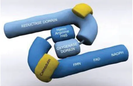
NOS proteins possess a bi-domain structure, consisting of two identical monomers, which are functionally divided into two major domains: a C-terminal reductase domain and an N-terminal oxygenase domain [160]. The reductase domain has binding sites for calmodulin, nicotinamide adenine dinucleotide phosphate reduced form (NADPH), flavin mononucleotide (FMN) and flavin adenine dinucleotide (FAD). The oxygenase domain has binding sites for the cofactors heme and tetrahydrobiopterin (BH4 or H4B) and the substrate L-arginine [161, 162] (Figure 7).
Figure 7. The general structure of the NOS enzymes (http://www.reading.ac.uk/nitricoxide/intro/no/synthesis.htm)
31 3.1.2 Enzymatic reaction
As mentioned previously, each NOS functions as a dimer consisting of two identical monomers. The heme is critical to the enzymatic reaction because it participates in dimerization, as NOS exists as monomers in its absence. Monomers of all the enzymes are unable to bind to BH4 or L-arginine [150, 163]. Each enzyme acts as a dimeric protein in catalysing the NADPH-dependant electron oxidation of L-arginine. Briefly, the reductase domain transfers electrons from NADPH along the flavins and calmodulin to the catalytic heme centre in the N-terminal portion of the protein [161]. L-arginine is then hydroxylated by NOS to form N-hydroxy-L-arginine (NHA) as an intermediate, which is subsequently oxidized to yield L-citrulline in addition to NO (Figure 8), in a 1:1 stoichiometry [160].
Figure 8. Nitric oxide synthesis from L-arginine (Taken from [164]).
3.1.3 NOS activation
An important molecule related to NOS activation is calcium. Increase in intracellular calcium triggers a cascade of events leading to NOS activation and NO synthesis. Intracellular calcium binds to calmodulin to form a calcium–calmodulin
32
complex and to regulate the binding of calmodulin to the ‘latch domain’, which permits electron transfer from NADPH via flavin groups within the reductase domain to a heme-containing active site, thereby facilitating the conversion of oxygen and arginine to L-citrulline and NO [165, 166].
The NOS1 and NOS3 enzymes are functionally similar and neither contain bound calmodulin. In the presence of calcium, however, when the high affinity association between calcium and calmodulin refered to above occurs, it results in the activation of the enzyme. For this reason NOS1 and NOS3 are commonly classified as calcium/calmodulin-dependent [162, 167], although NOS3 can also be activated in a calcium-independent manner [168]. These two NOS catalyse NO production within seconds in response to diverse stimuli and produce small quantities (at nM) of NO [150, 169]. On the other hand, NOS2 contains calmodulin so tightly bound that it is considered to be a subunit rather than a cofactor [170]. This synthase has the shortest sequence and binds calmodulin at all physiological concentrations of calcium and unlike the other two enzymes it is not regulated by calcium, therefore NOS2 activity is regarded as calcium-independent [141, 171]. There are only a few intracellular mechanisms that regulate NOS2 activity, which is generally considered to be at the transcriptional level. The NOS2 protein levels can be acutely induced [154] and this enzyme is characterised by release of large quantities (at μM) of NO even hours after exposure to inducing agents [172, 173].
Apart from calcium, several other factors can regulate NOS activity, especially NOS3 activation. NOS3 can be activated by certain stimuli without a sustained increase in calcium being necessary [169]. At the post-translational level, NOS3 activity is highly regulated by substrate and cofactor availability as well as endogenous inhibitors, lipid modification, direct protein-protein interactions, phosphorylation, O-linked glycosylation, and S-nitrosylation. The NOS3 signalosome is perhaps the best characterized of the three NOS isoforms since it has been clear for a few years that the association with calmodulin and caveolin has profound effects on the intracellular localization and activity of NOS3 [162, 169, 174]). This enzyme can be phosphorylated
33
on serine, threonine, and tyrosine residues, with numerous putative phosphorylation sites (Figure 9).
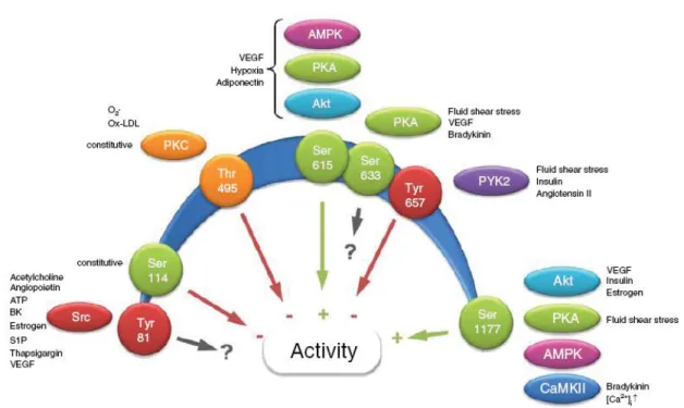
Figure 9. The regulation of NOS3 by phosphorylation. Schematic depiction of confirmed NOS3 phosphorylation sites, and their influence on enzyme activity (green arrows activation, red arrows inhibition, black arrow no direct effect on enzyme activity). The numbers refer to the human sequence (Taken from [169].
3.2 Nitric oxide chemistry
Nitric oxide is a simple, diatomic and non polar molecule. It is a colourless gas at room temperature and pressure. This inorganic free radical is also endogenously produced as a gas with a very short half-life from milliseconds to few minutes [141, 175]. Although NO has a very short half-life, due to its high solubility, NO can freely diffuse through biological membranes. Its chemistry and redox state nevertheless facilitate its interaction with various biomolecules to regulate different intracellular and
34
intercellular events. In this section a few illustrative examples of the reactivity of NO will be presented.
3.2.1 NO redox species and its interactions
Nitric oxide either diffuses directly to its target or it is converted to different derivates. One of the most unique and important chemical features of NO is that it is a paramagnetic species. Unlike other carbon, oxygen or nitrogen-centred radicals, NO does not have the tendency to dimerize at standard temperature and pressure, so it is capable of forming high-affinity-nitroso complexes with a variety of metal complexes [176]. In a general view, NO can be converted to a variety of nitrogen oxide species (NOx); to an organonitrosyl (E-NO) compound, where E is a sulfur-, nitrogen-, or carbon-containing moiety or to a metal-nitrosyl (M-NO) complex [175]. Some of these species are better suited for delivery of NO and others for longer-term storage.
From a biological point of view, some important reactions of NO are those with oxygen in its various redox forms. Nitric oxide gas reacts with O2 to form nitric dioxide gas (NO2), which dimerizes to dinitrogen tetroxide (N2O4). The N2O4 dismutates spontaneously in water and buffers at pH 7.4 to yield the stable end products nitrite (NO2−) and nitrate (NO3−). Estimation of NO2−, NO3− in aqueous biological samples is used to provide indirect means of estimating endogenous NO production [141, 177, 178]. Other important nitrogen oxide is peroxynitrite (ONOO-), that is formed in vivo by the diffusion-limited reaction between NO and superoxide [179]. This anion is highly oxidizing and can even effect tyrosine nitration, resulting in a variety of pathophysiological effects ranging from inflammation to cancer [180].
35 3.3 Nitric oxide biology
The NO synthesized within the cells freely diffuses through the membranes and acts as an intracellular and extracellular biological messenger, interacting with a variety of biomolecules such as enzymes, cytokines, membrane receptors, transcription factors and DNA to modulate several physiological and pathological processes in mammals and other living organisms [181]. The roles exerted by NO, however, may vary according to its concentration, when and where it is produced, and whether NO acts directly or via some of its redox species as cited above [175, 182].The effects and the mechanism of action of NO are strictly dependent on its concentration as well as on the presence of metals, proteins and low-molecular-weight thiols in a given cell. For this reason NO may exert dual effects on the same process in the same cell.
3.3.1 Mechanisms of action of NO
Nitric oxide has the capacity to modulate the activity of proteins through reversible reactions with available functional groups, notably with iron and thiols [175, 176]. NO can directly react with heme proteins such as cytochrome P450 [183-185], cyclooxygenase [186, 187] and guanylyl cyclase [182, 188]. This last one was one of the first targets identified for NO in biology. Soluble guanylyl cyclase (sGC) is a heterodimeric enzyme consisting of α- and β- subunits and a prosthetic heme group with ferrous iron [189]. It has been proposed that unique binding interactions of NO with the heme iron in guanylate cyclase allows the liberation of the transaxial ligand, histidine, which leads to enzyme activation that catalyzes the conversion of GTP into guanosine 3’5’-monophosphate (cGMP)[188]. The result is an increase in cGMP that represents an important intracellular second messenger that mediates many key biological actions of NO. The cGMP exerts its physiological actions through cGMP-dependent protein kinase (PKG), cGMP-regulated phosphodiesterases (PDE2, PDE3) and cGMP-gated cation channels, among which PKG might be the primary mediator. Importantly, the cGMP

![Figure 3. Major steroidogenic pathways in the follicle (Taken from [22]).](https://thumb-eu.123doks.com/thumbv2/123doknet/11401016.287783/22.918.181.713.344.942/figure-major-steroidogenic-pathways-follicle-taken.webp)

![Figure 5. Important signaling cascades in ovulation (Modified from illustration on [81])](https://thumb-eu.123doks.com/thumbv2/123doknet/11401016.287783/33.918.374.605.122.505/figure-important-signaling-cascades-ovulation-modified-illustration.webp)
![Figure 8. Nitric oxide synthesis from L-arginine (Taken from [164]).](https://thumb-eu.123doks.com/thumbv2/123doknet/11401016.287783/44.918.162.799.539.761/figure-nitric-oxide-synthesis-from-arginine-taken-from.webp)