Direction des bibliothèques
AVIS
Ce document a été numérisé par la Division de la gestion des documents et des archives de l’Université de Montréal.
L’auteur a autorisé l’Université de Montréal à reproduire et diffuser, en totalité ou en partie, par quelque moyen que ce soit et sur quelque support que ce soit, et exclusivement à des fins non lucratives d’enseignement et de recherche, des copies de ce mémoire ou de cette thèse.
L’auteur et les coauteurs le cas échéant conservent la propriété du droit d’auteur et des droits moraux qui protègent ce document. Ni la thèse ou le mémoire, ni des extraits substantiels de ce document, ne doivent être imprimés ou autrement reproduits sans l’autorisation de l’auteur.
Afin de se conformer à la Loi canadienne sur la protection des renseignements personnels, quelques formulaires secondaires, coordonnées ou signatures intégrées au texte ont pu être enlevés de ce document. Bien que cela ait pu affecter la pagination, il n’y a aucun contenu manquant.
NOTICE
This document was digitized by the Records Management & Archives Division of Université de Montréal.
The author of this thesis or dissertation has granted a nonexclusive license allowing Université de Montréal to reproduce and publish the document, in part or in whole, and in any format, solely for noncommercial educational and research purposes.
The author and co-authors if applicable retain copyright ownership and moral rights in this document. Neither the whole thesis or dissertation, nor substantial extracts from it, may be printed or otherwise reproduced without the author’s permission.
In compliance with the Canadian Privacy Act some supporting forms, contact information or signatures may have been removed from the document. While this may affect the document page count, it does not represent any loss of content from the document.
Université de Montréal
Microgels for oral delivery of therapeutic proteins
Par
ANTON BELOOUSSOV
Faculté de phannacie
Mémoire présenté
à
la Faculté des études supérieures en vue de
l'obtention du grade de
Maîtrise ès sciences (M. Sc)
Septembre, 2008
© Anton Belooussov
'''-~~:~-;;:::~'' -~Université de Montréal
Faculté des études supérieures
Ce mémoire intitulé :
Microgels for oral delivery of therapeutic proteins
présenté par:
Anton Belooussov
a été évaluée e) par un jury composé des personnes suivantes:
Professeure Françoise Winnik
Professeur Jean Norbert Mc Mullen
Professeur Grzegorz Pietrzynski
Professeur Kishore Pate!
président-rapporteur
directeur de recherche
co-directeur de recherche
membre du jury
ACKNOWLEDGMENTS
1 would like to thank my co-directors, Dr. Jean Norbert McMullen and Dr. Grzegorz Pietrzynski, for their advice, contributions and help. 1 would also like to thank Dr. Kishore Patel for invaluable help. 1 would like to thank Dr. Françoise Winnik for providing her vital contribution to my work as the president ofmy jury.
1 would also like to thank Dr. Valery Alakhov and the Supratek Pharma team for the support of my project.
More thanks go to Dr. Fahima Nekka for her evaluation ofmy project results, Dr. Patrice Hildgen who helped me with photo-imaging and Dr. Tomasz Popek who provided an important insight on spectroscopy.
Special thanks go to my friend Antoine Trottier, who helped me during my lengthy experiments.
SOMMAIRE
Le sujet de cette étude est la libération contrôlée de substances actives de grandes masses moléculaires: protéines et peptides. L'inconvénient commun de ces médicaments est leur administration par injection. Des micro gels ont été synthétisés en tant qu'excipients potentiels administrés par voie orale afin de protéger les protéines thérapeutiques contre les enzymes digestives, la dénaturation provoquée par le suc gastrique et pour permettre la libération prolongée de ces grosses molécules peu perméables directement aux muqueuses intestinales.
Ces microgels sont formés d'un réseau de copolymères de poly(acide acrylique) entrecroisés de diméthacrylate d'éthylèneglycol et greffées de copolymères séquencés -poly( oxyde d'éthylène)- -poly( oxyde de propylène). Ils sont synthétisés par une copolymérisation entrecroisée radicalaire en une étape relativement simple.(ll) La formulation est simplement constituée d'un mélange à sec de microgels avec la protéine lyophilisée qui est ensuite directement comprimé. Les microgels répondent au changement du pH de l'environnement: ils restent intact en conditions acides et se gonflent en pH neutre. Ils possèdent des propriétés mucoadhesives et sont non irritants.(6, 8, 10) Ces caractéristiques font de ces microgels de excipients potentiels pour la livraison orale des protéines thérapeutiques.
La première section de cette thèse est un examen des différentes formes de libération contrôlée. La section expérimentale décrit la synthèse de trois micro gels et leur caractérisation par une variété de méthodes sous forme de particules sèches aussi bien
que dans un milieu aqueux. Dans cette partie, des micro gels sont formulés avec de l'albumine lyophilisée de sérum de boeuf utilisée comme protéine modèle. La capacité de chargement de ces micro gels a été évaluée. Des profils de libération de micro gels en conditions imitant le milieu gastro-intestinale sont comparés entre eux et aux deux excipients généralement utilisés: lactose et Carbopol. Les résultats de l'étude de libération de protéines ont été interprétés par la méthode de Ritger et Peppas.
SUMMARY
The subject of this study is controlled release of proteins and peptides. Common disadvantages of these medications are their administration by injection. Microgels were synthesized as potential excipients for oral delivery of therapeutic proteins for their protection from the digestive enzymes and the denaturation caused by the stomach acid and to provide prolonged release of the se large and poorly permeable molecules directly to the intestinal mucosa.
Microgels are composed of copolymers of ethylene glycol dimethacrylate crosslinked network of poly(acrylic acid) with grafted block-copolymer chains of poly(ethylene oxide)-poly(propylene oxide), which are synthesized by a relatively straightforward one-step free-radical crosslinking copolymerization.(ll) The microgels respond to environmental pH change: they collapse in acidic conditions and swell at neutral pH. They are proven to possess mucoadhesive properties and are non-irritating.(6, 8, 10) The drug product is formulated by simple blending of dry microgel with lyophilized protein and directly compressed.
The theoretic part of this thesis is a review of the different controlled-release forms, therapeutic proteins and their delivery systems. The experimental part describes the synthesis of three microgels and their characterization by a variety of methods alone as well as in formulations with a model protein, bovine serum albumin, in conditions mimicking the gastro-intestinal tract. The results of prote in release study were interpreted by the method of Ritger and Peppas.
TABLE OF CONTENTS
Acknowledgments
Summary and key words (French and English) List of Figures
List of Tables
List of Abbreviations THEORETIC PART
1. CONTROLLED DRUG RELEASE 1.1. Introduction 1.2. Controlled-Release Terminology 1.2.1. Controlled release 1.2.2. Extended release 1.2.3. Modified release 1.2.4. Delayed release 1.2.5. Prolonged release 1.2.6. Site-specifie release
2. FUNDAMENTAL ASPECTS OF CONTROLLED RELEASE 2.1. Diffusion
2.2. Solute diffusion mechanism 2.3. Three types of the transport
2.3.1. Fickian type transport or "case 1" 2.3.2. "Case II'' type diffusion
2.3.3. "Case III" type diffusion
3. CONTROLLED-RELEASE SYSTEMS CLASSIFICATION
PAGES 1 II IX XI XII 1 1 3 3 4 4 4 5 5 5 5 7 8 8 9
10
12
3.1. Diffusion-controlled systems 3.1.1. Reservoir systems 3.1.2. Matrix systems 3.1.2.1 Hydrophilic matrixes 3.1.2.2 Inert matrixes 3.1.2.3 Lipid matrixes 3.2. Solvent-controlled systems
3.2.1. Osmotically controlled systems 3.2.2. Swelling controlled systems 3.3. Chemically controlled systems
3.3.1. Biodegradable systems 3.3.2. Grafted chains systems 3.4. Systems activated by magnetic fields 3.5. Bioadhesive delivery systems
4. THERAPEUTIC PROTEINS AND THEIR DELIVERY 4.1. Marketed proteins based drug products
4.2. Alternative ways of protein and peptide administration
12 12
14
14
16 17 18 18 1920
20
22 23 23 24 24 25 5. MICROGELS, NOVEL EXCIPIENTS FOR PERORALDELIVERY AND CONTROLLED RELEASE OF THERAPEUTIC 28 PROTEINS
5.1. Grafted block copolymers
28 5.2. Microgel synthesis model and annotated structure
29
5.3. pH responsive properties of microgels
32 5.4. Mucoadhesion of micro gels
EXPERIMENTAL PART Objectives
1. MATERIALS AND METHODS 1.1. Materials
1.2. Equipment 1.3. Methods
1.3.1. Microgel synthesis
1.3.1.1. Preparation of block copolymer (Solution A)
1.3.1.2. Emulsion/dispersion
1.3.1.3. Filtration, washing and drying 1.3.2. Microgel composition determination
solution
1.3.2.1. Fourier transform infrared spectroscopy (FTIR) 1.3.2.2. Estimation of effective degree of boding
34 35 35 36 38 38 38 38 39
41
41
41
1.3.2.3. High-performance (HPLC) liquid chromatography 42 1.3.3. Microgel characterization1.3.3.1. Differentiai scanning calorimetry (DSC) 1.3.3.2. Solid 13C nuclear magnetic resonance (NMR)
1.3.3.3. Swelling of microgels and pH titration studies
42 42 42 43 1.3.3.4. Particle size determination and imaging of dry 44
microgel particles
1.3.3.5. Carr's compressibility index determination
44
1.3.4. Microgel preparation and tabletting45 1.3.5. Acidic fluids and phosphate buffer preparation
1.3.6. Dissolution method and protein assay
1.3.7. Method of estimation of release kinetics parameters 2. RESUL TS AND DISCUSSION
2.1. Microgel synthesis
2.2. Microgels composition determination 2.2.1. Block copolymer binding 2.2.2. Acrylic acid monomer binding
2.2.3. Efficacy of the micro gel washing procedure 2.2.4. Microgel structure elucidation by solid l3C NMR 2.2.5. Estimation of microgel composition
2.3. Characterization of microgel in aqueous milieu 2.3.1. Swelling of microgels
2.3.2. Titration of micro gels
2.4. Characterization of dry microgel particles 2.4.1. Particle size determination
2.4.2. Dry particles morphology 2.4.3. Carr's compressibility index 2.5. Microgelloading capacity
2.5.1. Effect of loading on protein release
2.6. Protein release from the microgels at 10% loading 2.7. Kinetics study of protein release
3. CONCLUSION References Appendix 1 Appendix II 45 47 48 48 48 51 52 53 54 56
58
58
5960
60
61
63 64 65 6769
71
7380
88List of Figures
Pages 1. Blood drug concentration levels after administrating multiple doses 2
of conventional dosage form (An) vs. a single dose of a long-'acting controlled-release drug delivery system (B)
2. Blood drug concentration profiles of different controlled-release 3 dosage forms
3. Swelling of a hydrophilic polymer 8
4. Demonstration of Fickian (a), anomalous (b) and "case II'' types of 11 diffusion (c).
5. Simple osmosis-controlled oral system (OROS~ 18 6. Simplified release mechanism of a limited swelling matrix system 19 7. Schematic representation of the OROS® Push-Pull™ system 20 8. Theoretic drug release profile for diffusion-controlled (a) and 22
diffusion/erosion controlled (b) systems
9. Schematic representation of grafted chains system 22 10. Poly(ethylene oxide)-poly(propylene oxide) block copolymers 29
(pluronics~
11. Proposed structure of microgel fragment 12. Microgel synthesis, washing and drying
13. FTIR results for seriai dilutions of Pluronic® F127 (a), Pluronic® L92 (b), PPG 3500 (c) and acrylic acid monomers (d) in
dichloromethane
14. Standard curves for Pluronic® F127 (a), Pluronic® L92 (b), PPG 3500 (c) and acrylic acid monomers (d) in dichloromethane
31 40 48-49
50-50
15. Block Copolymer incorporation into the microgels structure 52 16. Acrylic acid monomer HPLC chromatograms used for calibration 52 17. Acrylic acid monomer were not detected by HlPLC in F127 53
microgel wash-outs
19. Estimated compositions of F127 microgel (a), L92 micro gel (b) and 57 PPO microgel (c).
20. Swelling profiles of the microgels 58 21. Microgels relative Hydrophobic and Hydrophilic Blocks 59 composition
22. Microgels pH Titration 60
23. Microgel particles size distribution. 61 24. Carr's compressibility index for F127, L92 and PPO microgels vs. 64 two widely used excipients: lactose (immediate release) and Carbopol (modified release)
25. F127 (a), L92 (b) and PPO (c) Microgels Loading Capacity: protein 65-66 release from seriai microgel formulations
26. Microgels protein release profiles of vs. Carbopol and Lactose 68 formulations
27. BSA release from the microgels: first 2 hours samples were treated 68 with acidic fluids at pH 2.2, after that solution was changed for
0.5M phosphate buffer pH 6.8
28. BSA release from the microgels after 2 hours of treatment with 69 acidic fluids at pH 2.2
List of Tables Tables
1. Diffusion type and the kinetic parameter "n" 2. Chemical shifts and assignments for 13C NMR
3. Kinetic parameters of protein release from the microgels
Pages 11
56
70
List of abbreviations:
Abbreviation Name
AAM Acrylic Acid monomers
API Active pharmaceutical ingredient
BSA Bovine Serum Albumin
Carbopol Carbopol@ 971 NF
DCM Dichloromethane
DSC DifferentiaI scanning calorimetry
EGDMA Ethylene Glycol Dimethacrylate
FDA US Food and Drug Administration
FTIR Fourier transformed infrared
GI tract Gastro-intestinal tract
HCI Hydrochloric Acid
Microgel F127 Pluronic® FI 27-based microgel
Microgel L92 Pluronic® L92-based microgel
Microgel PPO Poly(propylene glycol) 3500-based
microgel
NaOH Sodium Hydroxide
NMR Nuc1ear magnetic resonance
PAA Poly( acrylic acid)
PEO Poly( ethylene oxide)
PPG 3500 Poly(propylene glycol) 3500
PPO Poly(propylene oxide)
1. CONTROLLED DRUG RELEASE
1.1. Introduction
Controlled-release products are formulated to release drug's active ingredient gradually and predictably over a I2-hour to 24-hour period. These formulations compared with the immediate-release drugs might provide potentially greater effectiveness in the treatment of chronic conditions through a more consistent delivery of the medication: reduced side effects, greater convenience and higher levels of patient compliance due to a simplified dosage schedule.
The administration of a conventional or immediate-release dosage form provides rapid absorption initially with drug concentration in patient's plasma reaching peak maximum rapidly followed by a decrease in drug level due to progressive elimination of active pharmaceutical ingredient (urinary or other excretion, metabolism,
degradation, etc).
With conventional fast release drug products, in order to maintain plasmatic drug concentration within therapeutic range in order to provide effective therapy it was recommended to multiply the drug administrations. This type of medication has demonstrated numerous disadvantages such as fluctuation "peak and valley" of
plasmatic concentration of the drug, adverse side effects (if maximum plasma
concentration reaches toxic level) or lost of therapeutic effect, (plasma concentration decreasing rapidly, falling lower then the minimum effective concentration if the active pharmaceutical ingredient has a short half-life)
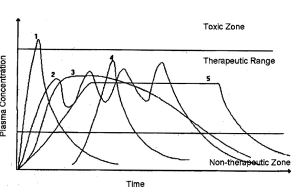
1 2
3
4Frequency of Usage
Figure 1: Blood drug concentration levels after administrating multiple doses of
conventional dosage form (An) vs. a single dose of a long-acting
controlled-release drug delivery system (B) (courtesy ofNoveon)
Thus once-daily dosing carries significant benefits in terms of convenience and compliance. The potential also exists to improve the side-effect profile and enhance the overall efficacy of existing drugs.
It is important to control drug release in such a way that drug concentration in
plasma is constant and the intervals between drug administrations are increased. The kinetics of dissolution of the active pharmaceutical ingredient might be modified by alteration of physical properties such as particle shape or type of polymorph. Drug release might be also controlled by adjustment of the dosage form to the requirements of the particular active pharmaceutical ingredient delivery.
c o
~
8
c o Ü co E CI) coa..
ToxicZone Therapeutic Range TimeFigure 2: Blood drug concentration profiles of different controlled-release dosage forms
1 - Conventional form 3 Extended release form 5 - Sustained release form
1.2. Controlled-Release Terminology
1.2.1. Controlled release
. 2 Pulsatile release form 4 - Delayed release form
Controlled drug delivery systems usually have the same route of administration as a conventional form. However, it delivers the drug at a predetermined rate and/or to a specifie location according to the need of the body and the disease states over a definite time period.(38)
1.2.2. Extended release
Extended-release systems include any dosage form that maintains effective therapeutic blood or tissue levels of the drug for a prolonged period. If the system can provide sorne actual therapeutic control of drug release in the body, whether it is temporal, spatial or both, it is considered a controlled delivery system. Extended release is not equivalent to controlled-release.(23)
1.2.3. Modified-release
This term is defined in the European Pharmacopoeia as a modification of the rate or place at which the active substance is released. Modified-release products coyer a wide range of release models, the principal types of which would include "delayed release" and "prolonged release" products.(3)
1.2.4. Delayed release
Delayed-release systems are either repetitive, intermittent dosing of a drug form one or more immediate-release units incorporated into a single dosage form, or an enteric delayed-release system. Examples of delayed-release systems include repeat-action tablets and capsules, and enteric-coated tablets where timed release is achieved by a barrier coating.(23) The European Pharmacopoeia de fines delayed release form as a modified release product, in which the release of active substance is delayed for a
resistant" (Ph.Eur.) oral tablets or capsules which remain intact in the stomach and only disintegrate in the higher pH of the small intestine]
1.2.5. Prolonged release
Prolonged-release form is a product in which the rate of release of active substance from the formulation has been reduced, in order to maintain therapeutic activity over a prolonged period of time, to reduce toxic effects or for sorne other therapeutic purpose.(52, 18, 21)
1.2.6. Site-specifie release
Site-specific release refers to targeting of a drug to a certain biologicallocation. The drug is adjacent to or in the diseased organ or tissue. This system satisfies the spatial aspect of drug delivery requirement and is also considered controlled drug delivery system.(23)
2. FUNDAMENT AL ASPECTS OF CONTROLLED RELEASE
2.1. Diffusion
". ". ".
··,'.·;>}M()1~êuhif@ffu,si6n:phèlfôm~rigl1:,~Q*ld:p(
âêfi#e:d:#s:a
:fnassJi#s,f~~:pf§sË~~'. ',' '. ' .. '. ::".\ _ ',.': .:. ' :':_ /.,;' .... _-:. <:, .. -:.. . " .' .,' _. " " '.1. '_', ~:: .•. ' :,',:/:'_" ~
'~~~s,~f~W~Io~,~rn;!~ri~~~}~l@;T?l~~":~'~d€~be;eaS~~:b~.:til19~~11~f
.COnceritration,"::graâièht:(4):··:J1i~':'prp:çéss,<·cif·,:'diffusion;":thetèfot~;,'~:.miilÎriii?:,es
'. :: ... ::- ":
"~':,"; ", :-:" :» :.":
~:::.; :;:.:-::":'(:',::-:" -,', .
/:.:-.:.:-:~:.:
:-:,:<:,;.-,:>. >:<'7:: ,,:-::-::: .:: ':::: ::~:'~" " <. ',' '. ':: ::':-:'" ,::,> '. ...' -'::
::'~:'",,:':X .·::>~\·::;.:\~:.>:~:.~;:~<;)Y :~~(.; . ; - . : • • • ; • . ; ' . ",,~ ' ' ' ' . ' , , ' , ' : ' , " " ' " ".;'. ' ' ' ' , ' , , - , : . ' : : ' ",:':":'.~'. ! . ."<.",:<, , . ' • , , ",' ~',' , , . .' ' . • · , 1 . . ; : . ' .. /:",'" -:"'!-::i'.~.·;" . . .
rf~;Jrg~i~,~:~S;;~j':gri~[~'i,(01U~
••
itiis;~oi
•• ,.
çhem.i~;~rf~ctiô,rj!,/~~i1;~~
's'pôntàne,olJsprÔcesso('~;passive':';:fornioftra1isport.<
" .. ,. ' ... ',:.: .... :/.
:<' .. : .':,", >,>:,:.,,"'i::/C1:,;: '.'In this equation
J
S
D
dc/dx
is the flux of drug in the direction of decreasing concentration
(amountlarea-time; mol/cm2 .sec)
is the mass of the drug released at the time t (mol)
is the surface of diffusion (cm2)
is the diffusion coefficient of the drug (cm2/sec)
is the concentration gradient (mol/cm4)
The negative sign of the drug flux simply demonstrates that the vector of J is
opposite to the vector of the concentration gradient.
This first law of diffusion is used to derive the equations applièable to diffusion-controlled reservoir drug systems, which are characterized by the constant concentration gradient (steady state).
In matrix systems the concentration gradient on contrary varies with time. In this model it is assumed that the solid drug dissolves from the surface layer of the dosage form first; when this layer becomes exhausted of drug, the next layer begins to be depleted by dissolution and diffusion through the matrix to the bulk solution. The
interface between the region containing dissolved drug and that with dispersed drug moves into the interior as a front.
Fick's second law of diffusion is applied to non-steady or continually changing diffusion state:
(eq.2)
Where···
It is important to stipulate that both Fick's laws in the forms of equation 1 and 2 respectively are not applicable if diffusion coefficient D is not constant at unvarying temperature.
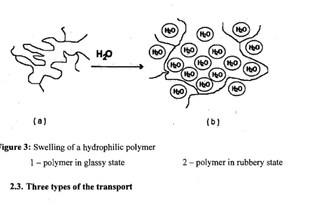
2.2. Solute diffusion mechanism
Considering that distribution of an active pharmaceutical ingredient within the pol ymer matrix is uniform, the release mechanism can be explained as follows: water or another biological fluid, while in contact with a pol ymer, penetrates into the material progressively dissolving the active pharmaceutical ingredient, which diffuses toward the exterior via a porous network or through intermolecular space. The type of the
transfer largely depends on a polymer state (glassy or rubbery).(61) According to the temperature, the polymers are in glassy state if T < Tg or are in rubbery state if T > Tg, where Tg is the temperature of glass transition and T is the temperature of the polymer.
The macromolecular chains of the pol ymer, when in rubbery state, move constantly creating large spaces or "pockets", which are permeable for solvent molecules. (Fig. 3)
"
(a)
Figure 3: Swelling of a hydrophilic pol ymer
1 polymer in glass y state
2.3. Three types of the transport
2.3.1. Fickian type transport or "case 1"
(bl
2 - polymer in rubbery state
This type of the transport is applicable to rubbery state polymers, where macromolecular chains are very flexible and adjust rapidly to the presence of solvent molecules(61) when the diffusion velocity of solvent molecules is significantly lower than the relaxation of macromolecular chains. This is a Fickian type also known as "case 1" diffusion.
The quantity of diffused substance Mt at the time t might be expressed as following:
Where
(eq.3)
The diffusion velocity, derived from the previous equation is:
dM/dt= 112. k.
fOS (eq.4)k
is a constant, which depends on form of the polymer and the diffusion coefficient., kthethrit:
(sec)
i'2.3.2. "Case II'' type diffusion
This type of transport is characteristic to polymers in glassy state where the relaxation of macromolecular chains is significantly lower then the velocity of solvent molecules diffusion.(61)
The quantity of adsorbed or desorbed solvent is expressed as follows:
(eq.5)
And the velocity of diffusion is respectively:
2.3.3. "Case III" type diffusion
Non-Fickian or "anomalous" diffusion is observed when the velo city of solvent molecules diffusion and the relaxation of the macromolecular -chains are of the same order. This type of the transport is in between the "case 1" and the "case II''. The quantity of adsorbed or desorbed solvent is given as:
(eq.7)
And the velocity of diffusion is respectively:
dM/dt=n.k.f-
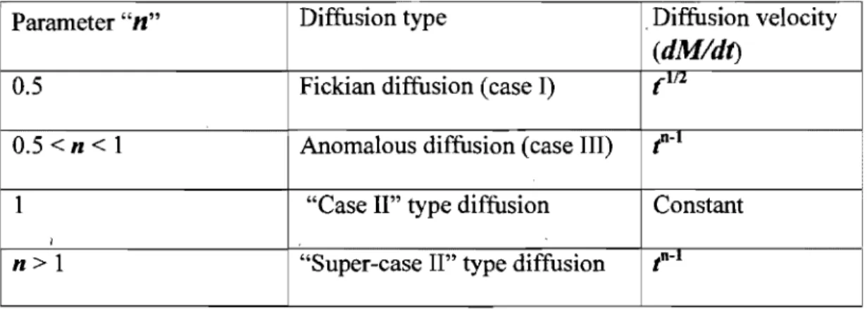
1 (eq.8)It has to be noted that the diffusion follows the Fick's law (case 1) if n=0.5; if the value of the parameter
n
is in between 0.5 and 1, then the process is anomalous (case III). However, when this parameter is' close to one, the diffusion of the solute within the polymers approaches the zero order kinetics (casé II). Table 1 demonstrates the three cases.( 48)Graphie presentation of the theory of solute diffusion (release) from the polymers for three types of diffusion is shown in the Figure 4.(33)
Parameter
"n"
Diffusion type , Diffusion velocity(dM/dt)
0.5 Fickian diffusion (case I) (Ifl.
0.5 < n < 1 Anomalous diffusion (case III)
f-I
i
1 "Case II'' type diffusion Constant
1 1
n>l "Super-case II'' type diffusion tn-l
Table 1: Diffusion type and the kinetic parameter
"n"
Figure 4: Demonstration of Fickian (a), anomalous (h) and "case II'' (c) types of
3. CONTROLLED-RELEASE SYSTEMS CLASSIFICATION
Systems of the release might be sorted in four major classes according to the nature of the drug transport.(34)
3.1. Diffusion-controlled systems
These systems are more frequently studied and utilized. There are two diffusion modes: the active pharmaceutical ingredient might diffuse through the polymer structure (within the intermolecular space) as well as from a porous network filled with the solvent. There are two major types of formulation: reservoir and matrix.
3.1.1. Reservoir systems
In reservoir systems, the active pharmaceutical ingredient is coated with swellable or non-swellable polymerie film. The drug might be in solid form, in solution or . concentrated suspension as weIl as mixed with another solid excipient. The polymerie membrane controls the drug release. In general, these systems include coated tablets and granules, macro and microcapsules, liposomes and hollow fibers. Fick's equation characterizes the diffusion of the drug:
dQ/dt
=D.S.A.(CrCJJ/X
(eq.9) WheredQ/dt
is amount of active pharmaceutical ingredient (g) diffused per unit oftime (sec)s
IS partition coefficient of the substance between the coateddosage form and the aqueous phase of the gastro-intestinal tract is a membrane surface (cm2)
IS a concentration of solution saturated with active
. pharmaceutical ingredient (g/cm3)
is concentration of APIon GI tract side (g/cm3)
x
is a membrane thickness (cm)In this case, the released drug is characterized by "zero order" kinetics as long as API within the film coated compartment is highly saturated since the diffusion coefficient, the partition coefficient, the membrane thickness and the surface area are constant.(32) The rate of release may be modified by changing the thickness of membrane, the surface area or the diffusion coefficient of API. For example, permeability of a membrane might be modified by adding more hydrophobie or hydrophilic polymers, like poly-ethylene oxide and poly-propylene oxide block copolymers or Pluronics®.
A major advantage of these systems is the possibility to obtain "zero order" release, which is problematic with other dosage forms.
There are a few disadvantages to the reservoir systems such as: accidentaI damage of a dosage form coating might cause dumping of the API contained in the reservoir.(19) "Zero order" is obtained only for the period of time when API inside the
coating is at high saturation, this varies with solubility. The manufacturing of reservoir dosage forrns is still relatively expensive due to the technology requirements.
3.1.2. Matrix systems
In the matrix systems the drug is uniforrnly distributed within the solid insoluble polymer excipient. The API embedded in this excipient network provides extended release.(32) The matrix may also be constituted of hydrophilic substances, which, when is in contact with water, forrn a gel.
The solvent penetrates into the system and progressively dissolves the API, which then diffused to the exterior through the porous network or interrnolecular space.
A matrix may be forrned by the evaporation of the solvent from a polymer solution with dispersed or solubilized API. The polymer should be hydrate d, dissolved or disintegrated before the drug is dissolved and diffused from the polymeric matrix. These systems are characterized by slow release. The API is not chemically bound to the polymer. The drug is active and is not modified.
Based on the nature of the pol ymer, matrix system may be classified as hydrophilic, inert or lipid.
3.1.2.1 Hydrophilic matrixes
These matrixes include a cellulose-derived polymers (hydroxyl-propyl methylcellulose and carboxyrnethylcellulose sodium salt) and vegetable-derived polysaccharides (alginates, agar-agar and modified amidons) or polyrners of animal
origin (gelatin and chitosane) as weIl as synthetic materials (polyviny 1 acetate, polyethylene glycol and polyacrylic acid).
Polysaccharide hydrophilic polymers are often used in oral extended-release formulations. Compared to inert and lipoid matrixes they are simple to formulate, might be loaded with API up to 80% w/w, are relatively inexpensive and versatile as they can be formulated with varied types of API.( 40)
At the releasing contact area of a hydrophilic matrix, sorne amount of the drug is released instantly (burst effect), then hydratation of the gel-forming agent leads to the fast formation of progressively increasing gelated barrier on the surface of the dosage form. The API diffuses through this gelated barrier in to the aqueous surrounding. The release velocity progressively decreases as a function of the square root of the time.(36) For example, drug release from a flat disk-shaped matrix might be estimated by this form of the Hugichi equation (29):
Where
M
=-VIC •.
(2.A-CJ.D.tj
(eq.lO)M
amount of drug released from the unit of surface during the timet (g/cm2)
solubility of the drug in the matrix (g/cm3)
t
time(sec)D
diffusion coefficient (cm2/sec)3.1.2.2 Inert matrixes
Polymers used for inert matrixes are of a porous structure, non-toxic and chemically neutral to the API (polyvinyl' chloride, polyethylene, ethylcellulose, silicone etc.).
In these dosage forms formulated drug dissolves and diffuses through the pores. The release is thus controlled by the porous structure. The rate of release may be modified by the alteration of a network pores' diameter,(53) shape and the dimensions of the dosage form.(3)
The amount of released API is proportional to the square root of time. This matrix system release might be characterized by the solubility of the API, diffusion coefficient, porosity and tortuosity of the dosage form.
Higuchi proposed a following mathematical model for a plane surface (29):
Mt
=
S .
...JID.é.C~(2Cu-é.CJ.t/rJ
(eq.l1)Where
Mt
amount of API released at the time t (g)D
API diffusion coefficient (cm2/sec)li
matrix porosity (sec/cm)CS
API solubility in water (g/cm3)matrix tortuosity (cm)
Co
concentration of API at the time zero (g/cm3)Inert matrixes are relatively simple to formulate and manufacture by dry blending and direct compression. The release of API from these matrix systems is independent of the exterior conditions.
3.1.2.3 Lipid matrixes
A variety of biodegradable excipients is used to formulate these matrixes: natural and synthetic waxes, fatty acids and their esters, fatty alcohols, hydrogenised oils, etc. The API may be released through diffusion and through erosion of the matrix by enzymatic hydrolysis of the lipids.(13)
Drug release from lipid matrixes depends on digestive enzyme composition of the stomach fluids.(20) The amount of released drug substance is proportional to the quantity of the excipient hydrolyzed by the enzymes.(S1)
3.2. Solvent-controlled systems
3.2.1. Osmotically controlled systems
The majority of the se systems were developed by ALZA Corporation. In the relatively simple dosage form, OROS®,(59) an osmotically active API in a core (drug substance blended with sodium or potassium chloride as osmosis agent) is surrounded by semi-permeable membrane which is permeable only to water. The drug substance is pumped out of the system through laser-drilled orifice at the same rate as the volume of water entering into core multiplied by drug concentration. The "zero order" drug release is controlled by the osmotic properties of the dosage form, surface, thickness and permeability of a membrane.
Drug solution < Laser-drilled
'.' . l.L,Orffice. '.,
i~'I)'~IoiIIoiII~~'" .,.. ... ~ . . . IIooW~ ... ' ...
':. Water
, ,'::':-,,',
•
'. Osmotically active drug
Rigid semi-permeable membrane
Figure 5: Simple osmosis-controlled oral system (OROS®) (courtesy of ALZA
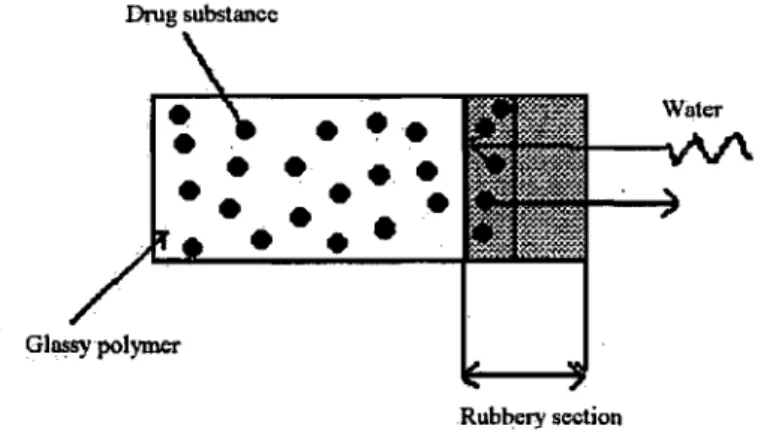
3.2.2. Swelling controlled systems
Swelling controlled systems are composed of a slowly hydrating polymer loaded with a drug substance that allows controlled permeation of the solvent.( 46) Limited swelling depends in general on the nature of a pol ymer and in particular on the degree of cross-linking. At the initial phase, the polymer is in glassy form when API diffusion is nearly zero due to very low diffusion coefficient. Solvent permeation initiates phase transition to the rubbery state at the swelling interface. The drug substance diffuses through the rubbery portion toward the aqueous exterior.(35) Drug release is controlled by the progressive move of the permeation front.
Drug substance
•••
••
••
••
••••
•
••
•
•
Glossypolymer Rubbery sectionFigure 6: Simplified release mechanism of a limited swelling matrix system
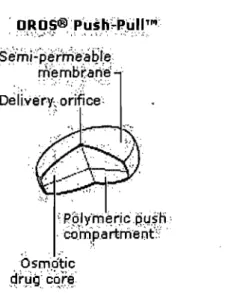
Developed by ALZA Corporation, the OROS® Push-Pull™ system IS
controlled by both: osmosis and swelling. A push compartment containing swellable polymer material is placed inside semi-permeable membrane provides delivery of a low solubility drug substance in a form of fine suspension.( 43)
Osmdtic
drûg'c6ré
'. '< '~, " ,',,,~.' / ~
Figure 7: Schematic representation of the OROS® Push-Pull™ system.
(Courtesy of ALZA Corporation)
3.3. Chemically controlled systems
3.3.1. Biodegradable systems
Matrix technology is more often applied to biodegradable systems rather than to reservoir. However, the latter exists mostly as skin patches and microcapsules. In the case of matrix technology the system erodes gradually releasing the APL In. reservoir systems the polymer coating may de grade after complete drug substance release.
Heller categorized biodegradable systems in three major groups based on their bio-erosion mechanism (1,27):
a. Insoluble polymers with biodegradable main chain, which breaks into smaller soluble molecules when hydrolyzed
1
X
b. Hydrolysis of cross-linking bonds
1 x 1 x x x
1
c. Insoluble polymer, which IS dissolved after their side chains are
hydrolyzed, ionized or protonated
1 1 1
A B
c
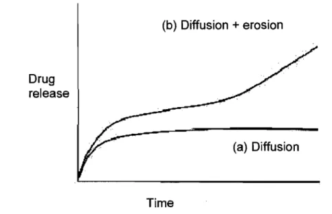
B--C = hydrolysis, ionization or protonation
For the majority of the biodegradable systems release, kinetics is the result of a combination ofboth - diffusion of drug and erosion of the polymer.(56)
J (b) Diffusion + erosion Drug release Time (a) Diffusion
Figure 8: Theoretic drug release profile for diffusion-controlled (a) and controlled by
diffusionlerosion (b) systems
3.3.2. Grafted chains systems
In these systems pharmaceutically active substance that chemically bound to a polymer structure is released by hydrolysis or enzymatic cleavage. The polymer backbone itself might be biodegradable. Drug substance might be attached directly to the polymer backbone or through a linking agent, which would affect the hydrophilic properties of the system and eventually drug release rate.( 47)
Covalent bond Drug substance
·rrrI···
HYdr~IYSiS
or' / .Enzyme activity
)
3.4. Systems activated by magnetic fields
These systems are characterized by having a drug substance and small magnetic particles uniformly distributed in the polymer matrix. The dosage form, when placed into aqueous milieu, releases active pharmaceutical ingredient in accordance with c1assic mechanism of diffusion described in respective section. However, drug substance is released at significantly higher rate when exterior oscillating magnetic fields are applied.
This phenomenon might be explained by increase of the matrix pores due to attraction and repulsion between magnetic particles. The latter allows the drug substance to be released faster.
3.5. Bioadhesive delivery systems
Bioadhesion is a complex phenomenon related to the ability of sorne natural and synthetic macromolecules to adhere to biological tissues. If a biological substrate is a mucus membrane, bioadhesive interactions occur primarily with the mucus layer and this process is referred to as mucoadhesion. The bonds involved are more likely to be of secondary chemical nature, combined with physical entanglement of polymer chains. The process is a reversible one, where the mucoadhesive detachment is caused either by the breakage of low energy bonds or by the physiological process of mucus turnover. (58)
Mucoadhesive controlled release dosage formulations have gained considerable attention due to their ability to adhere to the mucus layer and release the loaded drug in
1
a sustained manner. By using these dosage forms, contact time with the mucus surface increases, which results in an increased drug retenti on at the site leading to improved therapeutic efficacy, especially for large molecules like proteins. Diverse classes of polymers have been investigated for potential use as mucoadhesives. These include well known mucoadhesive polymers such as poly(acrylic acid), hydroxypropyl methylcellulose and poly(methylacrylate) derivatives, as weIl as naturally occurring polymers such as hyaluronic acid and chitosane.( 60)
4. THERAPEUTIC PROTEINS AND THEIR DELIVERY
Therapeutic proteins are promising alternative to drug small molecules. Protein based drugs have demonstrated extraordinary selectivity and lower risk of undesired side effects with the administration of micro gram amounts per daily dose. Unlike other medicines, they are not synthetically manufactured, and are usually produced through microbial fermentation or by mammalian cell culture. They are also more complicated, time-consuming and expensive to produce than synthetic drugs.(60)
4.1. Marketed protein based drug products
Currently therapeutic proteins are used to relieve patients suffering from
<i various cancers (monoclonal antibodies, interferons), heart attacks, strokes, cystic
fibrosis (enzymes, blood factors), diabetes (insulin), anemia (erythropoietin) and hemophilia (blood clotting factors). Leading protein based drug products on the market
Amgen respectively, Intron A ® and Peg-Intron® for hepatitis from Schering-Plough and few others.(60)
The delivery of these protein based drug products is currently performed through intravenous or subcutaneous injections, which is inconvenient for patients, increases treatment cost, and bears aU the risks related to this mode of drug administration.
4.2. Alternative ways of protein and peptide administration
Varietyof approaches has been developed as alternative delivery systems for therapeutic proteins and large peptides in order to avoid injections. Oral and nasal administration, pulmonary inhalation, implants and other pathways have been studied by different research groups and pharmaceutical companies for the deli very of large drug molecules.
Pulmonary inhalation, which does not require deep lung delivery was applied by Syntonix Pharmaceuticals to develop a novel formulation for Interferon-beta therapy for multiple sclerosis.(60)
Nasal formulation of leuprolide (ChiSys-Leuprolide™), a gonadotropin-releasing hormone agoni st that is used in the treatment of hormone-responsive cancers and control of ovarian stimulation in in vitro fertilization is undergoing phase II clinical
A number of the research groups work currently on implant delivery systems for prolonged release oftherapeutic proteins and peptides. Kajihara et al. has developed subcutaneous silicone implant covered with human serum albumin or interferon in order to achieve a zero-order release of protein drug for 30-100 days without significant initial burst.(30) In another implant system, bovine serum albumin was loaded into poly(D,L-lactic-co-glycolic acid) micropartic1es coated with poly(vinyl alcohol) and incorporated into PLGA tissue-engineered scaffolds. These tissue-engineered implants have provided long-term controlled release of the protein.(28)
The main barriers to oral administration of therapeutic proteins are the delicate physiochemical properties of protein-based drugs, which are sensitive to digestive enzymes and may be denatured by stomach acid before absorption. Another issue is the size of therapeutic proteins.(41) Being large and bulky molecules and characterized by poor permeability through intestinal mucosa, they require long time to be adsorbed with direct contact to the mucosa in order to traverse the intestinal tissue.
Orally administered enzyme products are being developed by Altus Pharmaceuticals. In these protein based drug products an active ingredient is cross-linked and crystallized in order to protect the substance from degradation in the stomach.(60)
Marschutz M.K. et al. prepared matrices of the mucoadhesive pol ymer sodium carboxymethylcellulose with the covalently bound Bowman-Birk enzyme inhibitors and elastatinal enzyme inhibitors for the protection of embedded insulin from degradation by the luminally secreted serine-proteases. These oral drug-carrier matrices
have provided up to 80% of insulin recovery after one hour of treatment in artificial intestinal fluid.(39)
Peppas N.A. and Foss A.C. et al. have developed nanospheres of crosslinked networks of methacrylic and acrylic acids grafted with poly( ethylene glycol) for use as oral insulin delivery devices.( 49) It was determined using photon correlation spectroscopy that nanospheres increased ten times in diameters in response to pH change from 2.0 to 6.0. Loading of insulin up to 7% w/w into the copolymers was achieved by partitioning from concentrated insulin solutions. In vitro studies have confirmed that insulin was protected at pH lower then 3.0 and released at pH 5.5. Animal studies were performed to investigate the abilities of insulin-Ioaded copolymer samples to influence the serum glucose levels of rats. In studies with diabetic rats, the serum glucose level was lower than control values for the animaIs that received the insulin-Ioaded copolymers and lasted for at least 6 h. The insulin loaded copolymer nanospheres caused a significant reduction of serum glucose with respect to that of control animaIs.
Therefore oral administration of therapeutic proteins is considered to be an advantageous alternative to injections and may be applied to novel therapeutic proteins and peptides as well as to already marketed products.
5. MICROGELS, NOVEL EXCIPIENTS FOR PERORAL DELIVERY AND CONTROLLED RELEASE OF THERAPEUTIC PROTEINS
In the past thirty years, work on hydrogels based· oncrosslinked copolymers has led to the establishment of a number of controlled release products for pharmaceutical agents.(24, 25, 62) Poly(acrylic acid)-based resins have been a subject of numerous drug master files with the U.S. FDA.(15)
In this study microgels as copolymers of poly(acrylic acid) network crosslinked by ethylene glycol dimethacrylate with grafted block-copolymers chains
(poly(ethylene oxide)-poly(propylene oxide) or poly(propylene glycol)) were
synthesized in order to be studied as a potential system for oral delivery of therapeutic proteins.
5.1. Grafted block copolymers
Three block copolymers with different hydrophile-lipophile characteristics were selected to be cross-linked to microgels. Pluronic® is a trade name for group of poly(ethylene oxide)-poly(propylene oxide) (PEO-PPO-PEO) block copolymers approved by the FDA as food additives and pharmaceutical ingredients. (2) Pluronic® L92 is hydrophobic with a molecular weight of 3650 Da, Pluronic®F127 is relatively hydrophilic and has a molecular weight about three times higher. The hydrophile-lipophile properties of Pluronics® depend on their composition (Fig.10): Pluronic® F127 has a PPO:PEO ratio of 30:70 when L92 is 80:20. The third copolymer of choice
was poly(propylene glycol), a hydrophobic material with a molecular weight of 3500 Da.(54) ~~---..~~---~
.
.--.
XY
XPEO-PPO-PEO
MW lkD-13kD
x
= 0 - 300 y = 15 - 80Figure 10: Poly( ethylene oxide )-poly(propylene oxide) block copolymers or Pluronics®
5.2. Microgel synthesis model and annotated structure
The synthesis model has been studied extensively.(7, 9, 11) The synthesis sequence for micro gels involved free-radical polymerization of acrylic acid with chain transfer to block copolymer resulting in C-C bonding between P AA and Pluronic® .
At 70°C, initiators form free radicals (R -) that trigger propagation of acrylic acid (AA) homo-polymer chains and abstraction ofhydrogen (H) from the block copolymers
(XmH). Latter activation of block copolymers (Pluronics® and PPG 3500) leads to the
chain transfer that is followed by grafting of P AA chains on block copolymers:
R-AAn-Introduction of di-vinyl crosslinker leads to formation of a PAA network, crosslinked by EGDMA, with grafted block copolymer chains. (Fig.ll)
5.3. pH responsive properties of microgels
The microgels are responsive to environmental pH changes, which is crucial for protection of API from the acidity and proteolytic enzymes of gastric fluids. At low pH of the human stomach, the microgels stay collapsed and drug is not released, however, at pH closer to neutral, in the intestine, these materials start to swell and release active pharmaceutical ingredient.(11 )
5.4. Mucoadhesion of micro gels
Among various possible bioadhesive polymeric hydrogels, P AA has been considered as a good mucoadhesive. It has been intensively studied and a number of mucoadhesive formulations was developed based on this polymer.(26, 37, 42) Polyether modified PAA formulations demonstrated mucoadhesive properties and are proven to be non-irritating.(6, 8, 10)
Objectives:
The general objective of this project is to synthesize and study micro gels of crosslinked poly(acrylic acid) network with grafted block-copolymers chains (poly(ethylene oxide)-poly(propylene oxide) or oxide)-poly(propylene glycol)) as a potential matrix for oral administration of therapeutic proteins and peptides. These microgels are synthesized in order to protect large
,
1. MATERIALS AND METHODS
1.1. Materials
Name Lot Number Supplier
Acetonitrile A25627 Aldrich
Acrylic Acid mono mers (AAM) 10630LB Aldrich 4' -4-azobis-( 4-cyanovaleric acid) 12688/1 Fluka Bovine Serum Albumin A 7906 113K0669 Sigma Carbopofw 971 NF CC9DAAJ085 Noveon Dichloromethane 99.9% LI00262KI Aldrich
Dodecane DA027360A Aldrich
Ethylene Glycol Dimethacrylate 07711LO Aldrich
Ganex V-216 03200068154 ISP
technologies
Hexane 10655HC Aldrich
Hydrochloric Acid IN standard 00507HC Aldrich
Lactose 03912MC Sigma
Lauroyl Peroxide 03727PO Sigma
Magnesium Stearate 18724MA Aldrich
Pluronic® F127 WPNX60C BASF
Pluronic® L92 WPDX5335B BASF
Poly(propylene glycol) 3500 08831AO Aldrich
RS Protein Assay 81495A Bio-Rad
Sodium Hydroxide 91040 MAT
Sodium Hydroxide IN standard 03427DD Aldrich Sodium Phosphate dibasic anhydrous 85H0425 Sigma Sodium Phosphate monobasic mono hydrate 67H1245 Sigma Trifluoroacetic Acid 001210-50209- PSIG
1.2. Equipment
• Mechanical stirrer BDC 1850, Caframo
• Hot plate with magnetic stirrer, Fisher Scientific
• Filter paper 10Jlm, Whatman
• Soxhlet extractor
• Market grade test sieves mesh 20(0.86mm), 40(0.36mm) and 100(0. 14mm), Dual Manufacturing Co.
• Optical microscope Labophot-2, Nikon
• Tap density tester, Vanderkamp
• Differentiai scanning calorimeter DSC-30, Mettler
• Nuclear magnetic resonance spectrometer Avance 600 MHz, Bruker
• Fourier transformed infrared spectrometer FTS800, Varian
• Reversed-phase HPLC column 218TP54, Vydac
• DuaI À absorbance detector 2487, Waters
• HPLC sampler manager 2700, Waters
• pH meter Orion 525A+, Thermo Electron
• Tube rotator, Scientific Equipment Products
• Hydraulic press, Carver
• Punch and Die 0 1 cm, custom made
• Tablet hardness tester PTB 302, Pharma Test
• Balance BP211D, Sartorius
• Incubator, Power Scientific
• Variable-rate flask shaker, St. John Associates
1.3. Methods
1.3.1. Microgel synthesis
The microgels were synthesized on laboratory scale according to the method (11) originally described by Dr. Lev Bromberg (Fig. 12) with sorne modifications.
1.3.1.1. Preparation of Block Copolymer Solution (Solution A)
39mL of AAM was neutralized by 0.5mL of 5M NaOH, charged into a flat-bottom flask and mixed. 24 g ofblock copolymer (Pluronic® or PPG 3500) and 1.1mL ofEGDMA was charged into the flask and ~ixed vigorously. The mixture of initiators: 100mg of Lauroyl peroxide and 100mg of 4' -4-azobis-( 4-cyanovaleric acid) in 2mL of AAM was added. The solution was stirred at 200rpm by magnetic stirrer for 10 minutes. AlI these operation were performed under a constant flow of dry nitrogen at room temperature. The mixture was deoxygenated for 30 minutes.
1.3.1.2. EmulsionlDispersion
A 500-mL three-necked round-bottom flask was charged with 250mL of 1 % w/v Ganex solution in dodecane and deoxygenated ovemight with constant stirring at 100rpm with a purge of dry nitrogen at room temperature. Next day, the flask was placed into an oil bath with temperature controller. The flask was equipped with a mechanical stirrer and a thermometer.
Solution A was charged into the round-bottom flask. Dry nitrogen purge was initiated and kept
until the end of the reaction. The mixture was stirred for 30 min at 200rpm at room temperature. Stirring rate was raised to 300rpm and maintained at this intensity until the end of
the reaction. Step-wise heating was started at the rate of 1-2°C per minute up to 70°C. The temperature was maintained at 70°C until the end of the reaction, for 4 hours.
1.3.1.3. Filtration, Washing and Drying
The reaction slurry was cooled to room temperature and filtered using Whatman filter paper (10Ilm). The wet filtrate was repeatedly washed with hexane and dried under vacuum
10-3 Torr at room temperature for 36 hours. The dried microgel was then washed in a Soxhlet
extractor with dichloromethane for 72 hours. AH the wash-out from both steps were collected and evaporated under vacuum to be used in the estimation of the effective degree of bonding between block copolymers and poly(acrylic acid). The washed micro gel was dried under a vacuum of 10-3 Torr at room temperature for 48 hours.
2SO!n1.. 1%GanelC indodecane O.$mL 5MNaOH
~
~
(j
""
~,;
stlITlng(100rpm) deoxygenallon , 0 .... ~ (OIN) \ 1--'
filtratlon (10 IJm Whalman..
filter paper) washing (hexane), evaporatlon (vaCUum 10-3 TorrFigure 12: Microgel synthesis, washing and drying
lOOnlg Lauruy! Peroxlde
1
100mg 4',.4 ~obis-{4-cyanovaleric acid) 1 1.1 ml. EGOMA 249 Pluronic L 92 stining (200rpm.10 min) deoxygenation (aO min)...
Slifring 300rpm OilBath heallng 10 70°C .\::===::::::==} (1 ·2 OC/min) washingin soxhlel extractor•
(dichloromethane) 3days stirrlng 300rpm, dry nitrogen flow. temperature 70°C. 4 heursdrying under vacuum (10-3 Torr)
1.3.2. Microgel composition determination
1.3.2.1. Fourier transform infrared spectroscopy (FTIR)
FT IR analysis was done using a Varian FTS800. Block copolymer samples were prepared in dichioromethane in seriaI dilutions and analyzed for characteristic peaks at a wavenumber of 1100. Dried synthesis washouts from aH the batches, reconstituted in dichloromethane were analysed by FTIR for the peak intensity at the respective wavenumber, and quantities of unbound block copolymers for each batch were estimated from standard curves.
1.3.2.2. Estimation of effective degree of boding
An effective degree ofbonding between block copolymers and poly(acrylic acid) was
calculated for aIl three microgels according to the following equation:
Where:
. DoB
DoB
=
(P
ch -P,).100% / P
ch (eq.12)effective degree ofbonding (%)
initial weight ofblock copolymer charged into the reactor (mg)
weight of block copolymer In washouts, not· hound during the
1.3.2.3. High-performance liquid chromatography (HPLC)
An analytical HPLC system with reversed-phase column was equilibrated using 0.1 % (v/v) aqueous trifluoroacetic acid at a flow rate of 1mLlmin. A linear gradient of water (0.1 % TF A)/ Acetonitrile (0.1 % TF A) from 9: 1 (v/vI) to 3:7 (v/v) was used. SeriaI dilutions of AAMs were prepared in water. Samples were injected by an automatic autosampler and monitored at 225nm and 280nm by means of a dual  Absorbance Detector.
1.3.3. Microgel characterization
1.3.3.1. DifferentiaI scanning calorimetry (DSC)
DSC evaluation was performed on each batch of dry micro gel particles and aU starting materials: AAMs, Pluronic® F127, Pluronic® L92 and PPG 3500. AH samples were tested utilizing Mettler DSC 30in standard aluminum crucibles at heating rate of lOoK/min was applied to aH DSC samples. Scanning was performed under constant nitrogen flow and with the starting temperature not higher them -1 OooK.
1.3.3.2. Solid 13C nuclear magnetic resonance (NMR)
Samples from each batch ofF127, L92 and PPO Microgels and Pluronic® F127 were prepared in tubes for solid state NMR. 13C spectra were acquired on a Bruker Avance 600 MHz spectrometer at 600 MHz by staff of the Department of Chemistry of the University of Montreal.
1.3.3.3. Swelling of microgels and pH titration studies
Swelling experiments were performed on each batch of micro gel in duplicate. Samples of lcm3 of dry material, independent of weight, were placed into 50ml plastic measuring flat bottom cylinders. The cylinders were sealed with rubber stoppers thoroughly at all time. The study was done at 37°C. Samples were pre-swelled in 30ml of distilled water for 2 days. After pre-swelling, the pH of the slurry was lowered to 2.0. Then the pH was increased gradually adding O.4mL of NaOH standard solution. These steps were repeated until the pH reached 12.0 for each microgel except micro gel PPO, which swelled to· the maximum volume at pH 6.0. IN HCI and IN NaOH standard solutions were used t6 adjust the pH. AlI samples were alIowed to stand for a minimum of 3 ho urs after adding NaOH or HCI and then the height was measured
The swelling ratio (S) was calculated as folIows:
Where:
Ho
(eq. 13)
height ofthe swelled microgel column during the experiment
height of the microgel column at time "0" (1 ml of dry micro gel pre-swelled in distilled water)
Microgel PPO titration continued after the point of maximum swelling at pH 6.0 up to pH 12.0. These results were normalized per gram of dry micro gel and plotted.
1.3.3.4. Particle size determination and imaging of dry microgel particles
Particle size analysis was performed for dry micro gel particles using market grade test sieves with mesh 20(0.86mm), 40(0.36mm) and 100(0.14mm). The fractions were weighted by means of an electronic balance (±0.01 mg). Images of the dry particles were obtained using an optical microscope Labophot-2.
1.3.3.5. Carr's compressibility index determination
A Vanderkamp Tap density tester with a tapping frequency 150 taps per minute was used to determine Carr's compressibility index.(16, 17) The tared 250-mL measuring cylinder was filled with pre-weighted test material to 100 mL, then the initial (or bulk) density was estimated. With minimal disturbance of the measuring cylinder, it was
transferred to the tap density apparatus, tapping was done 150 times then the final (or tap) density was estimated considering the reduced volume of the sample after tapping.
The following equation was used to calculate Carr' s compressibility index (x):
x=
(Do-Dt) .100% / Do
(eq.14)Where:
Do
initial (bulk) density (glcm3)1.3.4. Microgel preparation and tabletting
Dry blending was used to fonnulate both test and reference products. Series of dry blends were prepared for each batch of microgel in duplicate. F127 microgel, L92 microgel, PPO microgel, Carbopol and Lactose were dry blended with 2%, 10%, 20% or 50% w/w of protein (BSA) and 0.5% w/w of Magnesium Stearate as a compression lubricant. BSA was sieved through a series of market grade test sieves before blending and the fraction between meshes 40 (0.36mm) and 100(0.14mm) was selected. Blank blends without BSA were prepared for each batch as a negative control. Blending was carried out at room temperature in glass vials on vertical tube rotator at 10 rpm for twenty minutes.
Flat-faced plain tablets weighting 300mg, having a thickness of 3mm and with a diameter of 10mm, were compressed on a Carver press with 5000kg force applied.
1.3.5. Acidic fluids and phosphate buffer preparation
Acidic fluids were prepared by lowering the pH of distilled water to 2.2 by adding
Hel (lN) volumetric standard solution. The 0.5M phosphate buffer pH 6.8 was prepared as
follow: 14.08g of sodium phosphate monobasic monohydrate and 13.92g of sodium
phosphate dibasic anhydrous were dissolved in four litres of distilled water. The final pH was
aqjusted to 6.8 if required.
1.3.6. Dissolution method and protein assay
The limited sensitivity of the protein assay did not allow using the standard USP dissolution apparatus. The dissolution tests were perfonned on synchronised flask shakers
placed into a temperature-controIled environment of an incubator. Sample tablets were placed into SOmL sealed plastic Falcon tubes. Tablets were tested at 37°C for 48 hours: two hours in SOmL of acidic fluids at pH2.2, then the solution was changed using the same volume of O.SM phosphate buffer pH 6.8 to mimic the GI tract conditions. During the acidic treatment samples and blanks were shaken at 15 rpm then, after the change to phosphate
buffer shaking was increased to 100 rpm. Aliquots of O.3mL were taken at 0, 1, 2, 2.5, 3, 4,
5,8,24 and 48 hours. The aliquots were compensated with the same volume of the phosphate buffer at the same temperature (37°C) at each measurement. Acidic aliquots were neutralized using NaOH IN standard solution as required for the protein assay taking in consideration the dilution factor.
Bio-Rad RC protein assay was used to determine the concentration of BSA. AIl the aliquots were centrifuged using a microcentrifuge at 10,000 rpm for 10min prior the protein assay. Sampling of the supematant was done in order to avoid presence of microgel particles in the assay. Samples with a concentration higher then the assay range were diluted
respectively. AlI aliquots were assayed in triplicates. The protein assay was carried out in
accordance with the following standard operational protocol:
1 Prepare 6 dilutions of a protein standard (BSA) containing from 0.2 mg/mL to 1.2 mg/mL. A standard curve is prepared each time the assay is performed in triplicate and in the same buffer as the sample.
2 Pipette 5 J.lL of standards or samples into a clean, dry microtiter plate.
4 Add 200 flL of reagent B into each well. Place the plate on a microplate reader and let the plate mix for 5 seconds.
5 After 15 minutes, read the absorbance at 750 nm. The absorbance is stable for about 1 hour.
1.3.7. Method of estimation of release kinetics parameters
The Peppas equation was applied to determine the kinetic parameters of BSA
dissolution.( 45)
MIMex;
=k.1' (eq.15)Where:
degree of BSA release
kandn
kinetic parameterst time(hours)
The estimation of the kinetic parameters "k" and "n" was performed by linear regression of the logarithmic values of the time and degree of BSA release using the linear form of Peppas equation:
2. RESULTS AND DISCUSSION
2.1. Microgel synthesis
Two consecutive batches of each micro gel were synthesized in accordance with the method described in the section 1.3.1 at laboratory scale in adiabatic mode by a one-step free-radical crosslinking copolymerization of acrylic acid monomers and block copolymers with the covalent crosslinker ethylene glycol dimethacrylate. Both batches of each material were used in subsequent experiments.
2.2. Microgels composition determination
Given that resulting microgels are solid and absolutely insoluble whereas key starting compounds are mostly liquid, it was not feasible to use the same analytical technique to determine the degree of bonding for block copolymers. Thus washouts from the synthesis were collected and analysed by FTIR and HPLC to determine the composition of the solid fraction. FTIR results for seriaI dilutions of block copolymers obtained by the method described in section l.3.2.1, are presented in Figure13 a. b and c
Pluronie F127 1. 1. 1101.510 12.470 O. fl "
..
o. -e..
1099.756 5.679 ;f o. o. O . .(J. c 1200 Il 1100 1050 1000 WW'I8numberi
1 ~ a _ _ _ _ _ ~1 225 2085---
--
---
--
---
-
---~
"~ . / ' 1100.827 S.oo1 ,. Pluronic L92 , /"oo.67~ 3.141 -1---~~---=---1,'02.363 1.773-2-____
---~l--- ---1.~ ._ 1100.975 0.861 1220 b 1100.979 24.770/ poly(propylene glycol) WlY8numb.r ---=-~----
--
--
--
---1.~ 1C~ -l100.1sa 6.027 ~---~,~œ;.7~499î2ii926--- ---0.8-1
O.E 0.4 '-0.2 ---c 1097.712 1.341\
1097.836 0.630 \Figure 13: FT IR results for seriaI dilutions of Piuronic® FI27 (a), Piuronic® L92 (b)