HAL Id: inserm-01845093
https://www.hal.inserm.fr/inserm-01845093
Submitted on 20 Jul 2018
HAL is a multi-disciplinary open access
archive for the deposit and dissemination of sci-entific research documents, whether they are pub-lished or not. The documents may come from teaching and research institutions in France or abroad, or from public or private research centers.
L’archive ouverte pluridisciplinaire HAL, est destinée au dépôt et à la diffusion de documents scientifiques de niveau recherche, publiés ou non, émanant des établissements d’enseignement et de recherche français ou étrangers, des laboratoires publics ou privés.
irradiated bone
Guillaume Michel, Pauline Bléry, Michaël Henoux, Jérôme Guicheux, Pierre
Weiss, Sophie Brouard, Olivier Malard, Florent Espitalier
To cite this version:
Guillaume Michel, Pauline Bléry, Michaël Henoux, Jérôme Guicheux, Pierre Weiss, et al.. Bone marrow cell extract promotes the regeneration of irradiated bone. PLoS ONE, Public Library of Science, 2017, 12 (5), pp.e0178060. �10.1371/journal.pone.0178060�. �inserm-01845093�
Bone marrow cell extract promotes the
regeneration of irradiated bone
Guillaume Michel1,2
*, Pauline Blery2,3, Michae¨l Henoux1,2, Je´roˆ me Guicheux2,
Pierre Weiss2,3, Sophie Brouard4, Olivier Malard1,2, Florent Espitalier1,2
1 Service d’O.R.L. et de chirurgie cervico-faciale, Centre Hospitalier Universitaire, Nantes, France, 2 INSERM, UMRS 791, LIOAD, Universite´ de Nantes, Nantes, France, 3 Service d’Odontologie
Restauratrice et Chirurgicale, Centre Hospitalier Universitaire, Nantes, France, 4 INSERM UMR 1064, ITUN, Universite´ de Nantes, Nantes, France
*guillaumemichel@live.fr
Abstract
Mandibular osteoradionecrosis is a severe side effect of radiotherapy after the treatment of squamous cell carcinomas of the upper aerodigestive tract. As an alternative to its treatment by micro-anastomosed free-flaps, preclinical tissular engineering studies have been developed. Total bone marrow (TBM) associated with biphasic calcium phosphate (BCP) significantly enhanced bone formation in irradiated bone. One mechanism, explaining how bone marrow cells can help regenerate tissues like this, is the paracrine effect. The bone marrow cell extract (BMCE) makes use of this paracrine mechanism by keeping only the soluble factors such as growth factors and cytokines. It has provided sig-nificant results in repairing various tissues, but has not yet been studied in irradiated bone reconstruction. The purpose of this study was to evaluate the effect of BMCE via an intraosseous or intravenous delivery, with a calcium phosphate scaffold, in irradiated bone reconstruction.
Twenty rats were irradiated on their hind limbs with a single 80-Gy dose. Three weeks later, surgery was performed to create osseous defects. The intraosseous group (n = 12) studied the effect of BMCE in situ, with six combinations (empty defect, BCP, TBM, BCP-TBM, lysate only, BCP-lysate). After four different combinations of implantation (empty defect, BCP, TBM, BCP-TBM), the intravenous group (n = 8) received four intravenous injections of BMCE for 2 weeks. Five weeks after implantation, samples were explanted for histological and scanning electron microscopy analysis. Lysate immunogenicity was studied with various mixed lymphocyte reactions.
Intravenous injections of BMCE led to a significant new bone formation compared to the intraosseous group. The BCP-TBM mixture remained the most effective in the intraosseous group. However, intravenous injections were more effective, with TBM placed in the defect, with or without biomaterials. Histologically, highly cellularized bone marrow was observed in the defects after intravenous injections, and not after an in situ use of the lysate. The mixed lymphocyte reactions did not show any proliferation after 3, 5, or 7 days of lysate incubation with lymphocytes from another species.
a1111111111 a1111111111 a1111111111 a1111111111 a1111111111 OPEN ACCESS
Citation: Michel G, Blery P, Henoux M, Guicheux J,
Weiss P, Brouard S, et al. (2017) Bone marrow cell extract promotes the regeneration of irradiated bone. PLoS ONE 12(5): e0178060.https://doi.org/ 10.1371/journal.pone.0178060
Editor: Maria Cristina Vinci, Centro Cardiologico
Monzino, ITALY
Received: September 28, 2016 Accepted: May 8, 2017 Published: May 18, 2017
Copyright:© 2017 Michel et al. This is an open access article distributed under the terms of the
Creative Commons Attribution License, which permits unrestricted use, distribution, and reproduction in any medium, provided the original author and source are credited.
Data Availability Statement: All relevant data are
within the paper and its Supporting Information files.
Funding: The authors received grants from "Ligue
contre le cancer" foundation. For the whole project, we received 20 000 euros ( https://www.ligue-cancer.net/).
Competing interests: The authors have declared
This study evaluated the role of BMCE in irradiated bone reconstruction. There were sig-nificant results arguing in favor of BMCE intravenous injections. This could open new per-spectives to irradiated bone reconstruction.
Introduction
Treatment of squamous cell carcinomas of the upper aerodigestive tract remains a major health challenge today, with more than 263,000 new cases per year in the world [1]. Their treatment often requires surgery and postoperative high-dose external radiotherapy. Both have major side effects. Surgery requires large removal and induces long-term esthetic and func-tional disorders. Radiotherapy reduces healing capacities because of the decrease of bone vas-cularization [2]. Mandibular osteoradionecrosis (ORN) is a severe side effect of radiotherapy that affects 5% of treated patients [3] despite preventive actions. It leads to mandibular frac-tures and deglutition and phonation disorders [4]. The standard reconstruction procedure for extended ORN is the micro-anastomosed free-flap [5]. However, this procedure requires pro-longed general anesthesia, with a higher rate of complications on postradiation patients [6].
In this context, preclinical studies have been developed using calcium phosphate biomateri-als. Total bone marrow (TBM) associated with biphasic calcium phosphate (BCP) significantly enhanced bone formation in irradiated bone [7][8]. One of the mechanisms explaining how bone marrow cells can help regenerate such tissues is the paracrine effect [9]. Bone marrow cells release soluble factors such as cytokines and growth factors that induce neovasculariza-tion, cytoprotection and tissue regeneration [10]. To study this paracrine effect, a number of recent studies have developed a bone marrow cell extract (BMCE) containing intracellular fac-tors. Thus, in infarcted heart [11][12] as in irradiated salivary glands [13], injection of BMCE leads to an improvement comparable to intact cell therapy. Moreover, both intravenous and intraglandular injections are as effective in repairing irradiated salivary glands [13].
BMCE has not been studied in irradiated bone reconstruction. However, it could repair the tissues damaged by radiotherapy by increasing neovascularization and tissue remodeling [10]. The purpose of this study was to evaluate the role of BMCE and the calcium phosphate scaffold to promote bone formation after irradiation. Intravenous and intraosseous injections with dif-ferent combinations were compared to the BCP-TBM association, currently the most efficient material available.
Materials and methods
1. Biphasic calcium phosphate
The biomaterial used for this study was granules of a macroporous biphasic calcium phosphate (MBCP™, Biomatlante, Vigneux de Bretagne, France). The granules were about 800 μm in diameter and were composed of hydroxyapatite andβ tricalcium phosphate in a 60/40 ratio corresponding to a 1:60 ratio of Ca:P. The measured mean porosity was 40± 10%. Eppendorf tubes (Costar, Corning, NY, USA), each containing 0.015 g of MBCP™, were steam-sterilized at 121˚C for 20 min before implantation.
2. Animals
The study was conducted on 8-week-old inbred male Lewis 1A-haplotype RT1arats (n = 27)
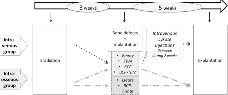
g. Animal care was provided in accordance with European directive number DE2010/063EU, after the “Ethics Committee for Animal Experimentation of Pays de la Loire” approval and Ministry of Research validation n˚1315. Animal euthanasia was performed with an intracar-diac injection of 50 mg of thiopenthal. Animals were examined clinically every day, including food intake and walking ability. A weighing was carried out once a week. Pain was managed by immediate post operative subcutaneous injection of meloxicam (1 mg/kg) then mixed with water during five days. The outline of the whole protocol is presented inFig 1. Seven rats were specially designated as donors: two TBM donors and five BMCE donors. Twenty rats under-went external irradiation. Bone defects and implantations were performed 3 weeks after irradi-ation. The “intraosseous group” (n = 12) received injections of BMCE in the bone defect, with
different combinations. The “intravenous group” (n = 8) received intravenous injections of
BMCE as an adjuvant to the bone implantations. Five weeks after implantations, i.e., 8 weeks after irradiation, implanted bones were removed immediately after euthanasia.
3. Radiation delivery procedure
The radiation delivery was performed at the Institut de Cance´rologie de l’Ouest (Centre de Lutte Contre le Cancer Rene´ Gauducheau, Saint Herblain, France). Twenty rats were irradi-ated under general anesthesia induced by an isoflurane inhalation (Forène1, Abbott France, Rungis, France). Then the animals were placed in the feet-first prone position in the XRAD225Cx irradiator (Precision X-Ray Inc., North Branford, CT, USA). A single 80-Gy dose was administered on their hind limbs. The purpose was to reproduce the histological and mechanical effects of irradiation, appearing beyond 70 Gy [14]. The target was the patella, with half of the femoral height and half of the tibial height in the radiation field. The field diameter was 2.8 cm at the isocenter. A total of 572 s per foot were needed to obtain the required dose of 80 Gy on the cortical bone, at 225 kVp and an intensity of 13 mA. The absorbed dose rate on the cortical bone was 8.39 Gy/min, and 3.3 Gy/min on the surrounding soft tissues.
An absorbed dose calculation was made using Monte Carlo simulation [15], based on CT images of the first irradiated rat (voxels, 0.2×0.2×0.2 mm3), with a statistical dose variation below 4% in the targeted bone and below 8% in the adjacent soft tissue.
4. Surgical procedure
All the surgical procedures were performed under general anesthesia using 4% isoflurane inha-lation for induction and 2% for preservation.
4.1. Isolation of TBM. The TBM was collected the day of the implantations from femoral, tibial, and humeral bones of two nonirradiated donors. The extremities of each bone were cut and 6 mL of physiological saline PBS (Gibco, 1X, pH 7.2) was infused through the medullar cavity. The TBM was collected and centrifuged (5 min at 600g). The cell pellet was
resus-pended in physiological saline and a nucleated cell count was performed using both the trypan blue exclusion test and cytological myelographic analysis. The TBM was immediately placed in tubes containing anticoagulant (Vacuette Premium1, Greiner bio-one, Germany). An average of 107cells were implanted in each defect filled with TBM.
4.2. BMCE preparation. Five days before implantation, the tibial, femoral, and humeral bones of five nonirradiated donors were collected to prepare the BMCE according to a proto-col previously used in other studies [11][12][13]. The TBM collection technique was the same as described in section 4.1. Then the suspension was strained through a 70-μm nylon filter and the cell concentration was adjusted to 107viable cells/μL. The cells were subjected to three freeze—thaw cycles, using a−80˚C freezer and thawing in a 37˚C bath, followed by microcen-trifugation at 14,000g at 26˚C for 30 min (Centrifuge 5804R, Eppendorf, Germany) to remove
insoluble materials. The total number of nucleated cells was 493× 107. Forty-eight injections of BMCE from 107cells were performed.
4.3. Implantations. Surgery was performed as previously described [16]: 3-mm-diameter osseous defects were surgically created in bilateral tibial and femoral metaphysis (four defects per animal). The defects were then totally filled with either BCP only, TBM only, a mixture of BCP and TBM, or no filling.
The “intravenous group” received intravenous injections of extract obtained from 107cells in 0.5 mL physiological saline. The injections were performed in the dorsal vein of the penis, twice a week over 2 weeks under general anesthesia.
The defects of the “intraosseous group” were filled with two added mixtures: BMCE only (50μL cell extract from 107cells) and a mixture of BCP and BMCE.
Postoperative analgesia was immediately supported by a subcutaneous injection of bupre-norphine (0.1 mg, Bupre´care1, Animalcare, York, Great Britain), repeated at day 3 and day 5, and by a subcutaneous injection of meloxicam (1 mg/kg, Metacam1, Boehringer, Germany), mixed with water over the following 5 days. An antibiotic treatment of marbofloxacin (3 mg/ kg, Marbocyl1, Ve´toquinol, Paris, France) was started the day of the surgery and repeated for 5 days.
4.4. Histological examination. Five weeks after implantation, all the animals were sacri-ficed. Femurs and tibiae were then dissected and fixed for 5 days in a 4% paraformaldehyde phosphate-buffered saline. After dehydration through a graded series of ethanol and acetone, nondecalcified bone specimens were infiltrated and embedded in methyl-methacrylate resin, which hardens at low temperature (Technovit 9100, Kulzer, Wehrheim, Germany). After dif-ferent preinfiltration and infiltration steps, the polymerization mixture was combined with bone samples and placed at−20˚C for 10 days. After polymerization, serial 5-μm sections were cut perpendicular to the osseous defects and the surrounding bone using a diamond saw made for nondecalcified tissues (Polycut SM2500, Leica, Wetzlar, Germany). The bone sections were then stained with Movat pentachrome using an automated slide stainer (Shandon Gemini ES,
Fig 1. Outline of the protocol. Events concerning all animals (n = 20): white; events for the IV group (n = 8): dotted; events for the intraosseous group
(n = 12): dashed.
ThermoFisher Scientific, Waltham, USA) and observed with a digital slide scanner (NanoZoo-mer 2.0 HT, Hamamatsu Photonics, Hamamatsu, Japan).
4.5. Scanning electron microscopy and quantitative image analysis. Each bone sample was sanded then gold-palladium-coated. Scanning electron microscopy (SEM) studies of implanted defects were conducted with backscattered electrons (Leo 1450 VP, Zeiss, Oberko-chen, Germany) in conjunction with image analysis. The quantity of newly formed bone and ceramic degradation was determined using a semi-automatic image analyzer procedure from SEM observations (Leica Qwin Pro v3.5.1) as previously described [17]. The quantity of newly formed bone, soft tissue, and remaining BCP was automatically calculated within the total area.
4.6. Immunogenicity. Mixed lymphocyte reactions (MLR) were performed in the Research Center in Transplantation and Immunology INSERM UMR 1064, Nantes, France. We collected total splenocytes and performed a cellular marking with CellTrace Violet (1μL CTV for 106cells). A cell culture with a gradation of BMCE from 0 to 50μL and 200 to 150 μL of total splenocytes in a culture medium (RPMI- 1% PS– 1% GLU– 10% SVF; Gibco) was per-formed at 37˚C. BMCE from a Lewis 1A rat was incubated with total splenocytes from a Wistar rat, and vice versa. After 3, 5, and 7 days of incubation, a viability marking was made using 3.5μL of Fixable Viable eFluor 660, then a flow cytometry analysis. For each condition, viable lymphocytes were selected and their CTV expression was measured to observe any cell proliferation.
4.7. Statistical analysis. Bone ingrowth and BCP resorption were compared using the Mann-Whitney and Kruskal-Wallis tests with statistical significance set at 5% (p<0.05).
Results
1. Radiation delivery and surgical procedures
No death occurred during this study. Both the radiation delivery and surgical procedure were well tolerated with daily care. Eight weeks after irradiation, 51 samples were included in this study. The other samples could not be interpreted, particularly because of fractures.
2. SEM and image analysis
2.1. Qualitative SEM study. A highly necrotic bone with wide gaps [18] was observed in every sample (Fig 2). In the “intraosseous group,” no bone formation was observed except with the BCP-TBM mixture implantation (Fig 3). In the “intravenous group,” there was visible new bone formation on every sample filled with TBM or BCP-TBM (Fig 4).
2.2. Quantitative SEM study. The ratio of bone ingrowth was significantly higher: • in the “intravenous group” (BCP, TBM, empty defect, BCP-TBM) compared to the
“intraosseous group” (BCP, TBM, empty defect, BCP-TBM) (p = 0.001) (Fig 5). • after implantation of TBM followed by intravenous BMCE injections compared to the
empty defect (p = 0.002), BCP alone (p = 0.007), TBM alone (p = 0.012), BMCE alone
(p = 0.021), or with BCP (p = 0.008), BCP-TBM (p = 0.019), and BCP followed by
intrave-nous BMCE injections (p = 0.044).
• after implantation of BCP-TBM followed by intravenous BMCE injections compared to the empty defect (p = 0.005), BCP alone (p = 0.018), TBM alone (p = 0.027), BMCE alone
Finally, the ratio of bone ingrowth was significantly higher after TBM or BCP-TBM fol-lowed by intravenous BMCE injections than every other mixture (respectively,p = 0.003 and p = 0.012,Fig 5).
3. Histological examination
The aspect of newly formed bone, the connection between BCP and surrounding tissues and the formation of blood vessels in the osseous defects were evaluated. The histological examina-tion was correlated with the qualitative SEM study (Fig 6).
After TBM or BCP-TBM implantation followed by intravenous BMCE injections, there was new bone formation towards the center of the defect. In both conditions, osteoblasts were arranged in rows to synthesize the osteoid matrix (data not shown). Newly formed bone was surrounded by many medullar cells. This highly cellularized bone marrow was observed in the defects after intravenous injections and not after an in situ use of the lysate. In the other associ-ations, there was either no bone formation or new bone formation at the periphery of the defects. A noncellular tissue filled the defects, consistent with fibrosis.
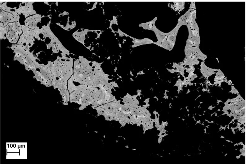
Fig 2. SEM backscattered electron image of irradiated bone (×60 magnification). There is destruction of the bone microarchitecture and presence of
poly-lobed gaps, suggesting Dambrain periosteocytic lysis.
Fig 3. SEM backscattered electron images of samples explanted 8 weeks after irradiation from the intraosseous group (×20 magnification). Only the BCP-TBM mixture induces significant new bone formation. White arrow indicates new bone formation.
4. Immunogenicity study
MLR analysis after 3, 5, and 7 incubation days did not show any lymphocyte proliferation, regardless of the condition used. The ratio of proliferative cells was always below 2%, and most of the time below 1%. Statistical analysis confirmed no difference between the MLR. Adding a BMCE from another species did not lead to a proliferation reaction.
Discussion
Histological effects on bone were observed from single 20-Gy doses [19], with osteopenia. Osteopenia appeared as a consequence of a primary reduction of the number and the activity of osteoblasts [20], then an osteocyte reduction. Bone morphology and its biomechanical prop-erties were degraded because of an alteration of the cellular pool and the vasculature, then pro-gressive fibrosis and local necrosis [2]. We used an animal model that had been previously
Fig 4. SEM backscattered electron images of samples from the IV group (×20). Defects filled with BCP-TBM and TBM only and followed by IV
injections induced significant new bone formation. White arrows indicate new bone formation.
developed [16] to mimic the side effects of irradiation on bone. After histological examination, wide fibrosis areas and a decrease of medullar cells were observed. Numerous polycyclic gaps consistent with periosteocytic lysis, described by Dambrain as pathognomonic of ORN [18], were observed on SEM analysis. Furthermore, our radiation delivery procedure was reproduc-ible and not deadly.
The BCP-TBM mixture was the most effective combination to induce new bone formation after irradiation [16]. Among the available biomaterials, BCP was chosen because of its biocompatibility, biodegradability, and osteoconductivity properties. TBM was included because of its regenerative capacity, due to various mechanisms: differentiation into osteoblas-tic lineage [21], vasculogenesis from endothelial progenitor cells contained in the bone mar-row [22], and release of soluble factors acting in a paracrine fashion [10]. This is the
mechanism we studied by lysing medullar cells to keep only the soluble factors. BMCE has
Fig 5. Box plots representing the percentage of newly formed bone according to the different combinations. The circles are outliers, and the
asterisks are extreme outliers. The rate of bone ingrowth is significantly higher after lysate IV injection, particularly with TBM in the defect, with or without BCP (ns).
previously been used in infarcted heart with a significant improvement of the left ventricular ejection fraction and a reduced infarct size [11]. The BMCE was as effective as whole live BM cells in repairing infarcted heart, suggesting a paracrine effect of MB cells. Other studies used the same protocol to prepare the BMCE and tested it in various tissues. On irradiated salivary glands [13], intraglandular or intravenous (IV) injections were both effective in restoring sali-vary flow rates to normal levels and increasing progenitor cells. Both BMCE delivery pathways were as effective as whole live BM cells. For salivary glands with Sjo¨gren disease [23], as for erectile function recovery after cavernous nerve injury [24], significant results were obtained with BMCE injections.
This is, to our knowledge, the first study evaluating BMCE in repairing irradiated bone. We did not identify significant results on bone formation with in situ injections, unlike other pre-viously studied tissues [11][13]. It is possible that the soluble factors contained in the lysate injected in bone did not find enough substrate to act, considering cellular depletion and hypo-vascularization [25]. A dog study did not find significant bone formation after in situ associa-tion of MSC lysate andβ-TCP [26]. The fibrosis subsequent to ORN [27] could have limited the lysate circulation.
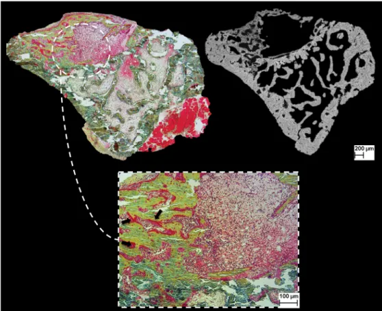
However, IV injections of BMCE enhanced bone formation with osseous implantation of BCP-TBM or TBM only. IV injections of BMCE, including all combinations, gave better results than the condition without IV injections. These injections were histologically associated with highly cellular bone marrow in bone defects. These results were observed despite the high
Fig 6. Image of histological analysis (Movat pentachrome staining) and correlation with SEM examination. Defect is filled with TBM, followed by IV injections. There is substantial bone formation, with
numerous osteoid areas (black arrows) and rich bone marrow.
levels of radiation used, far beyond the 15 Gy delivered to salivary glands [13], and despite the 3-week delay after irradiation to perform injections. This prolonged time frame has been cho-sen so as to be closer to clinical reality. In comparison, Tran et al [13] tested IV injections 5 days after irradiation, while repair capacity is not totally degraded. We highlighted new bone formation in an established ORN. Finally, considering intraosseous injections only in the pres-ent study, the BCP-TBM mixture still is, as published elsewhere [16], the best mixture to improve bone formation. However, IV injections led to a greater gain, in association with TBM in situ, with or without biomaterial.
To understand these results, proteins contained or stimulated by the lysate must be identi-fied. A recent study [28] characterized the angiogenesis-related growth factors in BMCE, using a microarray approach. They identified nine angiogenesis-related proteins, like HGF and MMP8-9, and four cytokines. After deactivation of these factors, injections of deactivated BMCE was no better than injection of saline, while injections of native BMCE restored func-tions and vascularization of irradiated salivary glands. This study is a first step to understand results of BMCE injections, involving native proteins, but not the nucleic acids, lipids or carbohydrates.
This mechanism, using the secretome of TBM cells, is already known for MSCs. It has been proved that intracoronary injections of the MSC culture medium only led to a reduced infarct size in a pig model [29]. But BMCE includes all cell types of whole BM and consequently numerous proteins, cytokines, and paracrine factors [11]. For example, MMP8-9 and osteo-pontin are highly expressed in BMCE [28], and are known as factors degrading the matrix sur-rounding endothelial cells and promoting their migration, proliferation, then angiogenesis [30–32].
The highest factor expressed in BMCE was the Platelet Factor 4 (PF4) [28]. This factor is known to protect MSCs and bone marrow niches from irradiation, by reducing DNA damage and increasing survival of MSCs [33]. BMCE injections could counter the effects of radiother-apy on MSCs and their microenvironment.
Furthermore, we found that histologically, defects filled with TBM presented a high density of cells only after IV injections. It may be an action of SDF-1 and IL-1ra, found in BMCE [28]. SDF-1 is a factor inducing stem cells to migrate from the bone marrow to the damage site [34]. IL-1ra was reported to stimulate macrophages into a wound healing phenotype and promoting endothelial cell migration [35].
The BMCE is easy and quick to obtain. It does not involve the injection of live cells, which carry the risk of differentiating into tumorigenic cells [13], but it does involve potential sys-temic effects by IV injections, and therefore it must be considered only in the remission period, never with cancerous cells in place. We evaluated the possible immunogenicity of the BMCE. We performed several MLRs to determine whether lymphocyte proliferation appeared in the presence of a BMCE from another species. The lack of any proliferation argues in favor of a lack of immunogenicity. These results are concordant with the literature that has pre-sented the BMCE as nonimmunogenic [12] [13] [23]. Studying the lack of immunogenicity, Angeli et al. [12] injected a human BMCE in a rat infarcted heart model. It improved heart function, without using immunosuppressed animals or immunosuppressive treatment, and without any sign of a graft-versus-host reaction. Fang et al [28] had similar findings, with a protein-mediated action of BMCE less tumorigenic and immunogenic than cell therapies. With no immune consequences, the lysate could be produced in advance from a human bone marrow bank and kept frozen until injections in patients.
This study evaluated the use of BMCE via intraosseous or IV delivery, with a calcium phos-phate scaffold, in irradiated bone reconstruction. The TBM-BCP mixture remains the gold standard for new bone formation without BMCE IV injections. However, IV injections of
BMCE significantly enhance new bone formation when the defect is filled with TBM, with or without BCP, acting like an adjuvant to boost the bone formation.
BMCE opens new perspectives but raises many questions. Provided that these results are validated and the mechanism of action clarified, an application to humans may be considered.
Supporting information
S1 Table. Quantitative SEM study: Datas after semi-automatic image analyzer procedure. (XLSX)
Acknowledgments
We would like to thank S.Sourice and P.Pilet for their help in this work.
Author Contributions
Conceptualization: JG PW OM FE. Data curation: GM PB MH FE. Formal analysis: GM PB MH FE. Funding acquisition: FE. Investigation: GM PB MH FE. Methodology: JG PW SB OM FE. Project administration: JG PW. Resources: OM FE. Supervision: PB SB FE. Validation: JG SB OM. Writing – original draft: GM.
Writing – review & editing: GM JG SB OM FE.
References
1. Jemal A, Bray F, Center MM, Ferlay J, Ward E, Forman D. Global cancer statistics. CA Cancer J Clin. avr 2011; 61(2):69–90.https://doi.org/10.3322/caac.20107PMID:21296855
2. Fenner M, Park J, Schulz N, Amann K, Grabenbauer GG, Fahrig A, et al. Validation of histologic changes induced by external irradiation in mandibular bone. An experimental animal model. J Cranio-maxillofac Surg. janv 2010; 38(1):47–53.https://doi.org/10.1016/j.jcms.2009.07.011PMID:19951841
3. Epstein JB, Thariat J, Bensadoun R-J, Barasch A, Murphy BA, Kolnick L, et al. Oral complications of cancer and cancer therapy: from cancer treatment to survivorship. CA Cancer J Clin. de´c 2012; 62 (6):400–22.https://doi.org/10.3322/caac.21157PMID:22972543
4. Chang EI, Leon P, Hoffman WY, Schmidt BL. Quality of life for patients requiring surgical resection and reconstruction for mandibular osteoradionecrosis: 10-year experience at the University of California San Francisco. Head Neck. fe´vr 2012; 34(2):207–12.https://doi.org/10.1002/hed.21715PMID: 21584893
5. Goh BT, Lee S, Tideman H, Stoelinga PJW. Mandibular reconstruction in adults: a review. Int J Oral Maxillofac Surg. juill 2008; 37(7):597–605.https://doi.org/10.1016/j.ijom.2008.03.002PMID:18450424
6. Mueller CK, Schultze-Mosgau S. Radiation-induced microenvironments—the molecular basis for free flap complications in the pre-irradiated field? Radiother Oncol. de´c 2009; 93(3):581–5.https://doi.org/ 10.1016/j.radonc.2009.08.009PMID:19733409
7. Malard O, Guicheux J, Bouler J-M, Gauthier O, de Montreuil CB, Aguado E, et al. Calcium phosphate scaffold and bone marrow for bone reconstruction in irradiated area: a dog study. Bone. fe´vr 2005; 36 (2):323–30.https://doi.org/10.1016/j.bone.2004.07.018PMID:15780959
8. Lerouxel E, Weiss P, Giumelli B, Moreau A, Pilet P, Guicheux J, et al. Injectable calcium phosphate scaffold and bone marrow graft for bone reconstruction in irradiated areas: an experimental study in rats. Biomaterials. sept 2006; 27(26):4566–72.https://doi.org/10.1016/j.biomaterials.2006.04.027 PMID:16698077
9. Prockop DJ. Repair of Tissues by Adult Stem/Progenitor Cells (MSCs): Controversies, Myths, and Changing Paradigms. Mol Ther. juin 2009; 17(6):939–46.https://doi.org/10.1038/mt.2009.62PMID: 19337235
10. Gnecchi M, Zhang Z, Ni A, Dzau VJ. Paracrine mechanisms in adult stem cell signaling and therapy. Circ Res. 21 nov 2008; 103(11):1204–19.https://doi.org/10.1161/CIRCRESAHA.108.176826PMID: 19028920
11. Yeghiazarians Y, Zhang Y, Prasad M, Shih H, Saini SA, Takagawa J, et al. Injection of bone marrow cell extract into infarcted hearts results in functional improvement comparable to intact cell therapy. Mol Ther. juill 2009; 17(7):1250–6.https://doi.org/10.1038/mt.2009.85PMID:19384293
12. Angeli FS, Zhang Y, Sievers R, Jun K, Yim S, Boyle A, et al. Injection of human bone marrow and mono-nuclear cell extract into infarcted mouse hearts results in functional improvement. Open Cardiovasc Med J. 2012; 6:38–43.https://doi.org/10.2174/1874192401206010038PMID:22550548
13. Tran SD, Liu Y, Xia D, Maria OM, Khalili S, Wang RW-J, et al. Paracrine effects of bone marrow soup restore organ function, regeneration, and repair in salivary glands damaged by irradiation. PLoS ONE. 2013; 8(4):e61632.https://doi.org/10.1371/journal.pone.0061632PMID:23637870
14. Barth HD, Zimmermann EA, Schaible E, Tang SY, Alliston T, Ritchie RO. Characterization of the effects of x-ray irradiation on the hierarchical structure and mechanical properties of human cortical bone. Bio-materials. de´c 2011; 32(34):8892–904.https://doi.org/10.1016/j.biomaterials.2011.08.013PMID: 21885114
15. Sarrut D, Bardiès M, Boussion N, Freud N, Jan S, Le´tang J-M, et al. A review of the use and potential of the GATE Monte Carlo simulation code for radiation therapy and dosimetry applications. Med Phys. juin 2014; 41(6):064301.https://doi.org/10.1118/1.4871617PMID:24877844
16. Espitalier F, Vinatier C, Lerouxel E, Guicheux J, Pilet P, Moreau F, et al. A comparison between bone reconstruction following the use of mesenchymal stem cells and total bone marrow in association with calcium phosphate scaffold in irradiated bone. Biomaterials. fe´vr 2009; 30(5):763–9.https://doi.org/10. 1016/j.biomaterials.2008.10.051PMID:19036434
17. Skedros JG, Bloebaum RD, Bachus KN, Boyce TM, Constantz B. Influence of mineral content and com-position on graylevels in backscattered electron images of bone. J Biomed Mater Res. janv 1993; 27 (1):57–64.https://doi.org/10.1002/jbm.820270108PMID:8420999
18. Dambrain R. [The pathogenesis of osteoradionecrosis]. Rev Stomatol Chir Maxillofac. 1993; 94 (3):140–7. PMID:8337586
19. Hopewell JW. Radiation-therapy effects on bone density. Med Pediatr Oncol. sept 2003; 41(3):208–11. https://doi.org/10.1002/mpo.10338PMID:12868120
20. Dudziak ME, Saadeh PB, Mehrara BJ, Steinbrech DS, Greenwald JA, Gittes GK, et al. The effects of ionizing radiation on osteoblast-like cells in vitro. Plast Reconstr Surg. oct 2000; 106(5):1049–61. PMID:11039376
21. Grove JE, Bruscia E, Krause DS. Plasticity of bone marrow-derived stem cells. Stem Cells. 2004; 22 (4):487–500.https://doi.org/10.1634/stemcells.22-4-487PMID:15277695
22. Rajantie I, Ilmonen M, Alminaite A, Ozerdem U, Alitalo K, Salven P. Adult bone marrow-derived cells recruited during angiogenesis comprise precursors for periendothelial vascular mural cells. Blood. 1 oct 2004; 104(7):2084–6.https://doi.org/10.1182/blood-2004-01-0336PMID:15191949
23. Misuno K, Tran SD, Khalili S, Huang J, Liu Y, Hu S. Quantitative analysis of protein and gene expres-sion in salivary glands of Sjogren’s-like disease NOD mice treated by bone marrow soup. PLoS ONE. 2014; 9(1):e87158.https://doi.org/10.1371/journal.pone.0087158PMID:24489858
24. Albersen M, Fandel TM, Lin G, Wang G, Banie L, Lin C-S, et al. Injections of adipose tissue-derived stem cells and stem cell lysate improve recovery of erectile function in a rat model of cavernous nerve injury. J Sex Med. oct 2010; 7(10):3331–40.https://doi.org/10.1111/j.1743-6109.2010.01875.xPMID: 20561166
25. Marx RE. A new concept in the treatment of osteoradionecrosis. J Oral Maxillofac Surg. juin 1983; 41 (6):351–7. PMID:6574217
26. Byeon Y-E, Ryu H-H, Park SS, Koyama Y, Kikuchi M, Kim WH, et al. Paracrine effect of canine allo-genic umbilical cord blood-derived mesenchymal stromal cells mixed with beta-tricalcium phosphate on
bone regeneration in ectopic implantations. Cytotherapy. sept 2010; 12(5):626–36.https://doi.org/10. 3109/14653249.2010.481665PMID:20438297
27. Delanian S, Depondt J, Lefaix J-L. Major healing of refractory mandible osteoradionecrosis after treat-ment combining pentoxifylline and tocopherol: a phase II trial. Head Neck. fe´vr 2005; 27(2):114–23. https://doi.org/10.1002/hed.20121PMID:15641107
28. Fang D, Hu S, Liu Y, Quan V-H, Seuntjens J, Tran SD. Identification of the active components in Bone Marrow Soup: a mitigator against irradiation-injury to salivary glands. Sci Rep. 2015; 5:16017.https:// doi.org/10.1038/srep16017PMID:26526154
29. Timmers L, Lim SK, Arslan F, Armstrong JS, Hoefer IE, Doevendans PA, et al. Reduction of myocardial infarct size by human mesenchymal stem cell conditioned medium. Stem Cell Res. nov 2007; 1(2):129– 37.https://doi.org/10.1016/j.scr.2008.02.002PMID:19383393
30. Fang C, Wen G, Zhang L, Lin L, Moore A, Wu S, et al. An important role of matrix metalloproteinase-8 in angiogenesis in vitro and in vivo. Cardiovasc Res. 1 juill 2013; 99(1):146–55.https://doi.org/10.1093/ cvr/cvt060PMID:23512982
31. Bendeck MP. Macrophage matrix metalloproteinase-9 regulates angiogenesis in ischemic muscle. Circ Res. 6 fe´vr 2004; 94(2):138–9.https://doi.org/10.1161/01.RES.0000117525.23089.1APMID: 14764648
32. Wang Y, Yan W, Lu X, Qian C, Zhang J, Li P, et al. Overexpression of osteopontin induces angiogene-sis of endothelial progenitor cells via the avβ3/PI3K/AKT/eNOS/NO signaling pathway in glioma cells. Eur J Cell Biol. aouˆt 2011; 90(8):642–8.https://doi.org/10.1016/j.ejcb.2011.03.005PMID:21616556
33. Chen J-J, Gao Y, Tian Q, Liang Y-M, Yang L. Platelet factor 4 protects bone marrow mesenchymal stem cells from acute radiation injury. Br J Radiol. aouˆt 2014; 87(1040):20140184.https://doi.org/10. 1259/bjr.20140184PMID:24922360
34. Li S, Wei M, Zhou Z, Wang B, Zhao X, Zhang J. SDF-1αinduces angiogenesis after traumatic brain injury. Brain Res. 20 mars 2012; 1444:76–86.https://doi.org/10.1016/j.brainres.2011.12.055PMID: 22330724
35. Richmond J, Tuzova M, Cruikshank W, Center D. Regulation of cellular processes by interleukin-16 in homeostasis and cancer. J Cell Physiol. fe´vr 2014; 229(2):139–47.https://doi.org/10.1002/jcp.24441 PMID:23893766