HAL Id: hal-02481829
https://hal.archives-ouvertes.fr/hal-02481829
Submitted on 17 Feb 2020
HAL is a multi-disciplinary open access
archive for the deposit and dissemination of
sci-entific research documents, whether they are
pub-lished or not. The documents may come from
teaching and research institutions in France or
abroad, or from public or private research centers.
L’archive ouverte pluridisciplinaire HAL, est
destinée au dépôt et à la diffusion de documents
scientifiques de niveau recherche, publiés ou non,
émanant des établissements d’enseignement et de
recherche français ou étrangers, des laboratoires
publics ou privés.
Ultrafast imaging of in vivo muscle contraction using
ultrasound
Thomas Deffieux, Jean-Luc Gennisson, Mickaël Tanter, Mathias Fink,
Antoine Nordez
To cite this version:
Thomas Deffieux, Jean-Luc Gennisson, Mickaël Tanter, Mathias Fink, Antoine Nordez. Ultrafast
imaging of in vivo muscle contraction using ultrasound. Applied Physics Letters, American Institute
of Physics, 2006, 89 (18), pp.184107. �10.1063/1.2378616�. �hal-02481829�
Appl. Phys. Lett. 89, 184107 (2006); https://doi.org/10.1063/1.2378616 89, 184107 © 2006 American Institute of Physics.
Ultrafast imaging of in vivo muscle
contraction using ultrasound
Cite as: Appl. Phys. Lett. 89, 184107 (2006); https://doi.org/10.1063/1.2378616
Submitted: 07 July 2006 . Accepted: 21 September 2006 . Published Online: 03 November 2006 Thomas Deffieux, Jean-Luc Gennisson, Mickaël Tanter, Mathias Fink, and Antoine Nordez
ARTICLES YOU MAY BE INTERESTED IN
Explososcan: A parallel processing technique for high speed ultrasound imaging with linear phased arrays
The Journal of the Acoustical Society of America 75, 1273 (1984); https:// doi.org/10.1121/1.390734
Sono-activated ultrasound localization microscopy
Applied Physics Letters 103, 174107 (2013); https://doi.org/10.1063/1.4826597
Adaptive focusing in scattering media through sound-speed inhomogeneities: The van Cittert Zernike approach and focusing criterion
The Journal of the Acoustical Society of America 96, 3721 (1994); https:// doi.org/10.1121/1.410562
Ultrafast imaging of in vivo muscle contraction using ultrasound
Thomas Deffieux,a兲 Jean-Luc Gennisson, Mickaël Tanter, and Mathias FinkLaboratoire Ondes et Acoustique, ESPCI, Université Paris VII, CNRS UMR 7587, 10 Rue Vauquelin 75005 Paris, France
Antoine Nordez
Laboratoire “Motricité, Interactions, Performance,” JE 2438, UFR STAPS, Université de Nantes, Nantes Atlantique Universités, 25 bis Bd. Guy Mollet BP 72206, Nantes F-44000, France
共Received 7 July 2006; accepted 21 September 2006; published online 3 November 2006兲 In this letter, an innovative way of imaging transient and local shear vibrations of an in vivo contracting muscle is proposed. The principle is to use an ultrafast ultrasound scanner 共up to 5000 frames s−1兲 able to follow with a submillimeter resolution the motion of the muscle tissue in a two dimensional plane. This ultrafast echographic imaging technique leads to both local and transient in vivo studies of the contraction of a muscle as reported by these first experiments done on the biceps brachii. © 2006 American Institute of Physics. 关DOI:10.1063/1.2378616兴
Numerous monitoring techniques are commonly used to study muscular or neuromuscular function. Electromyogra-phy 共EMG兲 is used to record the electrical activity of the muscle. It can be reported as the sum of action potentials propagating in a muscle’s fibers.1Mechanomyography is the recording of the muscular vibrations produced by the active muscle. It can be used as a monitor of muscle stiffness and could be related to the muscle force production.2 Unfortu-nately, all of these methods have a poor accuracy to assess local measurements and are thus not suitable for fully under-standing the underlying structure and mechanical behavior of the muscle. In order to create maps of the local response of the muscle, a few techniques have been applied to recon-struct the local velocity distributions of the muscle in three or two dimensions 共3D or 2D兲: phase-contrast magnetic-resonance imaging can reconstruct full 3D images of the muscle motion in a stroboscopic way. From these images local strains are calculated.3Doppler tissue imaging gives the tissues’ velocity distribution in a 2D plane and allows axial strain assessments.4 Recently, ultrasound image correlations at low frame rates have also been used to track the muscle motion.5 While very promising, these techniques can only image the muscle up to a few tens of frames per second. These low frame rates cannot be considered high enough to fully visualize the transient phenomena occurring during muscle activation.
Recently, ultrafast ultrasound scanners were designed by our group. Our last generation of echographic devices gives access to 2D radio frequency共rf兲 images at a few thousand hertz using a modified imaging sequence, i.e., a hundred times faster than any conventional ultrasound scanner. From these rf images, B-mode images are constructed. The cross correlation between two successive images permits us to as-sess the local axial particle velocity, and a complete movie of the axial velocity maps can be finally deduced. Such an ap-proach allows us to provide both very high spatial 共submil-limetric兲 and temporal accuracy 共less than a millisecond sam-pling兲, overcoming all the respective drawbacks of the previously cited techniques. This scanner6 is a 128 multi-channel fully programmable system driving any kind of
con-ventional ultrasonic probe. The sequence consists in emitting a single plane wave pulse. Ultrasonic echoes backscattered by tissue heterogeneities are then stored in memories. These ultrasonic raw data are processed in a posttreatment to create images with submillimetric resolution by applying a conven-tional beam formation共time delay and sum operations兲 in the receive mode. Finally, consecutive echographic images are compared using one dimensional cross correlation along the ultrasound beam axis in order to compute their axial relative displacements7 共note that “axial” terminology refers to the ultrasonic beam z direction and not to the fibers’ main axis, see Fig.1兲. This method enables us to measure relative
dis-placements as low as 1m between two consecutive frames and thus gives an estimation of tissue local velocity.
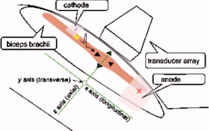
To trigger the muscle contraction with the imaging sys-tem, surface neuromuscular electrostimulation was achieved. A homemade electrostimulation device composed of a func-tion generator 共Agilent, model 33220A, Palo Alto, CA, USA兲, a power amplifier 共Brüel & Kjær, type 2718, Nærum, Denmark兲, and two noninvasive surface electrodes 共Compex, Ecublens, Switzerland兲 were used. The cathode was placed on the motor point, localized by qualitatively looking for the maximum muscle excitation, and the anode was placed on the distal portion of the biceps. A 30 V amplitude, 400s
a兲Electronic mail: thomas.deffieux@espci.fr
FIG. 1. 共Color online兲 Measurement protocol. Muscle fibers were excited with neuromuscular electrostimulation on the motor point, and the axial displacements were acquired with the 2D ultrasound ultrafast scanner. Both the arm and the transducer array were strongly fixed to avoid movements.
APPLIED PHYSICS LETTERS 89, 184107共2006兲
wide rectangular pulse was used to fire an action potential in the fibers just below the motor point共Fig.1兲.
In our first experiments, a conventional 8 MHz trans-ducer array was placed either parallel共longitudinal position兲 or perpendicular共transverse position兲 to the fibers’ main di-rection in order to follow the contraction in different planes 共Fig.1兲. This means that the displacements seen were never
those corresponding to the shortening of the fibers but rather those corresponding to their lateral widening.
After the acquisition of a conventional gray-scale echo-graphic image 共Fig. 2兲, the scanner took 500 images at
1500 Hz, and a movie of the axial velocity 共Fig. 3兲 was
computed. This movie was used to retrieve contraction and relaxation times and create maps of the maximum axial dis-placement. The average tissue velocity共cm/s兲 in the in situ region of interest A共Fig.2兲 is plotted as a function of time
共Fig.4兲. Contraction time is taken as the positive part of the
axial velocity 共fiber bundles contracting and thus moving toward the probe兲, while relaxation time is the negative part 共fiber bundles moving back to its original position兲. Contrac-tion time is thus found to be around 58 ms and relaxaContrac-tion time around 120 ms, values that are consistent with the literature.8The axial共z direction兲 displacements can be com-puted from the integration of the axial tissue velocity.
De-tecting the maximum local displacements over time enables us to create maps of the contracting fiber bundles共Fig.5兲.
To illustrate the temporal accuracy of the scanner, peri-odic excitations with different pulse repetition rates 共from 5 to 20 Hz with 5 Hz steps兲 have been used and displace-ments computed inside a region of interest A共Fig.2兲.
De-pending on the frequency of the excitation and as expected from surface EMG measurements but now for each point inside the muscle, temporal summation and tetany can be observed 共Fig. 6兲. As the frequency of excitation rises,
muscle fibers do not have the time to come back to their initial position and temporal summation occurs, leading to a stronger contraction.1 When the sum of the contraction and relaxation times is less than the period of stimulation pulse, no temporal summation occurs共5 Hz兲. When the contraction time is less but the sum of the contraction and relaxation times is greater, oscillations can be seen 共10 and 15 Hz兲. Finally, when the contraction time is greater than the period of excitation, the displacement is an increasing curve, reach-ing a maximum共20 Hz兲.
The ultrafast echographic imaging approach enables the assessment of the transient mechanical behavior of entire muscles with a millimetric resolution. From the acquisition of local axial tissue velocities of tissues over the entire or-gan, one can compute not only axial displacements but also axial strains by simply deriving the displacement field along
FIG. 2. 共Color online兲 B-mode images of the muscle. Probe was either parallel共a兲 or perpendicular 共b兲 to the fibers. The aligned fibers of the biceps brachii are clearly visible.
FIG. 3.共Color online兲 Movie of the axial velocity 共cm/s兲. A 30 V amplitude, 400s wide square pulse excitation on the motor point triggers the contrac-tion. The contracting area can easily be visualized. The ultrasonic probe is placed parallel to the fibers; the imaging frame rate is set to 1500 Hz.
FIG. 4.共Color online兲 Axial velocity profile 共cm/s兲 vs time 共s兲 in region of interest A共Fig.2兲.
FIG. 5.共Color online兲 Maximum axial displacement map 共mm兲.
the z direction in a posttreatment step. It allows us to get rid of the global translational movements of the muscle. How-ever, this operation is always sensitive to noise and do not discriminate active and passive movements.
Much beyond the visualization of strains, the spatial and temporal accuracies of the system will allow us to retrieve the exact localization of the contracting bundles. This re-quires us to recover the active displacement sources by solv-ing the inverse source problem of the elastic wave propaga-tion. Assuming the medium to be linear, homogeneous, isotropic, incompressible, and piecewise homogeneous, and considering only the null divergence terms 共shear compo-nents兲, the elastodynamics equation9
describing the elastic wave propagation can be described as follows:
2u
t2共r,t兲 =⌬u共r,t兲 + s共r,t兲, 共1兲
where , , u共r,t兲, and s共r,t兲 represent density, Young’s modulus, displacement fields, and the volumetric force source terms, respectively. Equation共1兲requires the knowl-edge of Young’s modulus共stiffness兲 as well as the longitudi-nal共x axis, along the fibers兲 and transverse 共y axis, perpen-dicular to the imaging plane兲 displacements. Shear modulus has already been measured in the biceps brachii10,11and also
shown to be transverse isotropic along fibers.10 Transverse displacements can thus be assumed to have the same order of magnitude as the measured axial 共z axis兲 displacements. Moreover, longitudinal displacements along fibers could be computed using a synthetic aperture technique12 already implemented on ultrafast echographic devices. Using this complementary information, the only remaining unknown is
s共r,t兲. The inverse source problem can be solved, permitting
us to retrieve the localization of the transient sources of con-traction inducing the global concon-traction of the muscle.
Using this ultrafast device, a high space and time reso-lution imaging of a transient in vivo muscle contraction was achieved. We were able to follow the contraction of the muscle and to extract both local and transient properties such as the contraction time, the relaxation time, or maps of the contracting areas. The ultrafast echographic scanner, coupled with an EMG device, will provide a powerful tool to under-stand the link between electrical and mechanical activities of the muscle and to study the spatial recruitment of fibers and its associated disorders such as fatigue and neuromuscular diseases.
1J. V. Basmajian and C. De Luca, Muscles Alive共Williams and Wilkins,
Baltimore, 1983兲, Vol. 1.
2T. W. Beck, T. J. Housh, J. T. Cramer, J. P. Weir, G. O. Johnson, J. W.
Coburn, M. H. Malek, and M. Mielke, Biomed. Eng. Online 4, 67共2005兲.
3J. E. Drace and N. J. Pelc, Radiology 193, 423共1994兲.
4N. R. Grubb, A. Fleming, G. R. Sutherland, and K. A. Fox, Radiology
194, 837共1995兲.
5I. D. Loram, C. N. Maganaris, and M. Lakie, J. Appl. Physiol. 100, 1311
共2006兲.
6L. Sandrin, M. Tanter, S. Catheline, and M. Fink, IEEE Trans. Ultrason.
Ferroelectr. Freq. Control 49, 426共2002兲.
7J. Ophir, I. Céspedes, H. Ponnekanti, Y. Yasdi, and X. Li, Ultrason.
Imaging 13, 111共1991兲.
8F. Buchtal and H. Schmalbruch, Acta Physiol. Scand. 79, 435共1970兲. 9L. M. Brekhovskikh and V. Goncharov, Mechanics of Continua and Wave
Dynamics共Springer, Berlin, 1993兲, Vol. 1.
10J. L. Gennisson, S. Catheline, S. Chaffaï, and M. Fink, J. Acoust. Soc. Am.
114, 536共2003兲.
11J. Bercoff, M. Tanter, and M. Fink, IEEE Trans. Ultrason. Ferroelectr.
Freq. Control 51, 396共2004兲.
12M. Tanter, J. Bercoff, L. Sandrin, and M. Fink, IEEE Trans. Ultrason.
Ferroelectr. Freq. Control 49, 1363共2002兲. FIG. 6. 共Color online兲 Displacement 共mm兲 vs time 共ms兲 in a region of
interest for different excitation frequencies.