A critical role for dendritic cells in the evolution of IL-1
β
-mediated murine airway disease
Mitsuo Hashimoto*, Haruhiko Yanagisawa*, Shunsuke Minagawa*, Debasish Sen*, Amanda
Goodsell†, Royce Ma*, Catherine Moermans*, Kate J. McKnelly*, Jody L. Baron†, Matthew
F. Krummel*, and Stephen L. Nishimura*,‡
*Department of Pathology, University of California, San Francisco, CA †Department of Medicine, University of California, San Francisco, CA
Abstract
Chronic airway inflammation and fibrosis, known as airway remodeling, are defining features of chronic obstructive pulmonary disease (COPD) and are refractory to current treatments. How and if chronic inflammation contributes to airway fibrosis remains controversial. Here, we use a model of COPD airway disease utilizing adenoviral (Ad) delivery of IL-1β to determine that adaptive T-cell immunity is required for airway remodeling since mice deficient in α/β T-T-cells (tcra −/−) are protected. Dendritic cells (DCs) accumulate around COPD airways and are critical to prime adaptive immunity, but have not been shown to directly influence airway remodeling. We show that DC depletion or deficiency in the crucial DC chemokine receptor, ccr6, both protect from Ad-IL-1β-induced airway adaptive T-cell immune responses, and fibrosis in mice. These results provide evidence that chronic airway inflammation, mediated by accumulation of α/β T-cells and driven by DCs, is critical to airway fibrosis.
Introduction
Chronic obstructive lung disease (COPD) is the third leading cause of death in the United States (1). Peribronchial chronic inflammation and fibrotic scarring of small airways, known as airway remodeling, is refractory to steroid-based therapies and causes airflow obstruction in COPD (2, 3). The mechanisms leading to steroid-refractory chronic inflammation and whether that inflammation causes fibrotic scarring in COPD remain areas of active investigation (4).
Cigarette smoke (CS) is the major cause of airway remodeling by inducing cellular injury and increasing the susceptibility to respiratory pathogens, in particular viruses (5). Rodents when exposed to CS, viruses or both together demonstrate exaggerated airway remodeling (6, 7). CS and viruses engage similar host danger pathways leading to inflammasome activation and enhanced interleukin-1 (IL-1β) secretion (6, 8, 9). IL-1β protein levels are increased in human COPD biospecimens (10–13), delivery of IL-1β to the airways causes
HHS Public Access
Author manuscript
J Immunol. Author manuscript; available in PMC 2016 April 15.
Published in final edited form as:
J Immunol. 2015 April 15; 194(8): 3962–3969. doi:10.4049/jimmunol.1403043.
Author Manuscript
Author Manuscript
Author Manuscript
experimental airway remodeling (14, 15) and perturbation of IL-1 signaling protects against experimental CS-induced airway remodeling (16, 17).
Primary human or mouse lung fibroblasts upon IL-1β stimulation become pro-synthetic, increase TGF-β activation and highly express the potent dendritic cell (DC) chemokine, CCL20 (15). Proteomic cytokine analysis of lungs from IT-Ad-IL-1β- treated mice reveal elevated levels of CCL20. Increased CCL20 levels are associated with airway remodeling induced by CS in combination with the viral mimetic polyinosinic-polycytidylic acid [poly(I:C)] (15).
CCL20 is increased in COPD samples and is the only known ligand for chemokine receptor, CCR6 (18). Thus, ccr6 deficient mice can be used to interrogate the functions of CCL20, in vivo. CCR6, which is expressed by dendritic cells (DCs), has been shown to be required for DC recruitment and CS-induced emphysema in mice (19). DCs are critical antigen
presenting cells that have been implicated in the pathogenesis of COPD through priming pathologic adaptive T-cell immune responses (20). DC accumulation surrounding airways correlate with COPD disease severity (18). In mice, IL-1β or CS-exposure induces CCL20 expression, which correlates with lung (DC) numbers, adaptive Th1 and Th17 immune responses, and expression of the profibrotic cytokines IL-4, IL-13, and IL-17 (15, 21). The causal role of the immune response in airway remodeling remains to be fully elucidated. Recent evidence suggests that the adaptive immune response may be important for CS-induced lung pathology as mice deficient in the IL17 Receptor A (IL-17RA) are protected from CS-induced emphysema (22). Clodronate depletion of both macrophages and DCs has recently been found to protect against CS-induced airway remodeling (23). As critical players in innate and adaptive inflammation, the roles and mechanisms of DC in the pathogenesis of airway remodeling important to define.
Here we elucidate a causal linkage between immune and fibrotic pathology by demonstrating: 1) DCs are required for airway inflammation and fibrosis; 2) the DC chemokine receptor CCR6 is required for pathology adaptive T-cell immunity and airway remodeling; 3) α/β T-cells are required for airway remodeling. Taken together these data suggest that therapeutically targeting lung DCs may represent a strategy to prevent or treat airway remodeling in COPD.
Materials and Methods
Mice
All mice were bred and housed in specific pathogen–free housing under an IRB approved protocol and in accordance with the guidelines of the Laboratory Animal Resource Center of the University of California, San Francisco (San Francisco, California). Cd11c-dtr (B6.FVB-Tg Itgax-DTR/GFP 57Drl), ccr6 −/− (B6.129P2-ccr6tm1Dgen/J), tcra −/−
(B6.129S2-tcratm1Mom/J) and WT mice, all in the C57BL/6 background were obtained from The Jackson Laboratory (Bar Harbor, ME). Mouse CD11c-EYFP (24) transgenic reporter mice were provided by M. Nussenzweig (The Rockefeller University, New York, NY). Approval
Author Manuscript
Author Manuscript
Author Manuscript
for the use of mice in this study was obtained from the Institutional Animal Care and Use Committee of the University of California, San Francisco.
Recombinant Adenovirus
The recombinant E1-E3 deleted type 5 adenovirus, either empty (Ad-C) or expressing human active IL-1β (Ad-IL-1β), has been described in detail elsewhere (25). The replication-deficient virus was commercially amplified and purified by cesium chloride gradient centrifugation and PD-10 Sephadex chromatography, plaque titered on 293 cells and checked for wild-type contamination (ViraQuest Inc., North Liberty, IA). Recombinant type 5 Adenoviral vectors expressing Cre-eGFP fusion protein, eGFP, or LacZ were obtained from the Gene Transfer Vector Core (University of Iowa, Iowa City, IA)
Intratracheal injections
Mice were anesthetized with IP injection of Avertin (250 mg/kg, IP). Then Ad-hIL-1β or Ad-LacZ (2.5 ×10 8 pfu in 75 µ l sterile PBS) was instilled intratracheally with a needle (Popper® 24G-1’ Straight 1.25mm ball) using the direct visualized instillation (DVI) technique (26). The control was Ad-LacZ.
Mouse organ harvests and bronchoalveolar lavage (BAL)
For BAL, and organ harvest the trachea was cannulated and the lungs were lavaged 5 times using 0.8 ml of sterile PBS with 5mM EDTA, and the BAL or organs harvested as described (27).
Airway morphometry
Measurements of airway inflammation were estimated using Hematoxylin and Eosin (H&E) stained slides and wall fibrosis was assessed by the presence of thick collagen bundles stained by the trichrome method essentially as described by Hogg (28), which estimates wall fibrosis as a measure of the trichrome stained area from the the airway lumen to the outer edge of adventitial connective tissue and expressed as wall area/basement membrane length. The wall area and basement membrane length is determined using image analysis software (Image J, v1.36b). Microtome sections from H&E or trichrome stained sections of paraffin embedded mouse lungs were digitally imaged at 200× magnification (QCapture v2.68.2, Surry, BC, Canada). The slides were coded and an investigator blinded to the experimental groups acquired 5 digital images representing each lung lobe (and two images from the largest lobe) and the images coded and catalogued. Airway inflammation was defined as the inflammatory infiltrate extending from the airway basement membrane towards the lung parenchyma. Airway fibrosis was defined as thick collagen bundles (stained blue in trichrome stains). A minimum of 12 airways was examined/mouse.
Preparation of lung sections for live cell imaging in the lung
Mice were given a lethal overdose of Avertin and exsanguinated by cutting the renal artery. The lungs and trachea were exposed by cutting through the diaphragm and chest wall. The mice were intubated by tracheotomy with the sheath from an 18-gauge i.v. catheter. Lungs were inflated with 1 ml of 2% low melting temperature agarose in sterile PBS maintained at
Author Manuscript
Author Manuscript
Author Manuscript
37°C, and the solution was solidified by briefly rinsing the inflated lungs with PBS at 4°C. Inflated lungs were then excised from the mouse and placed in a sterile 50-ml conical containing RT RPMI without phenol red (Invitrogen, Life Technologies). The left lobe was isolated, cut into 360-µm sections using a vibratome filled with cool PBS, mounted on plastic slides with Vectabond (3M), and placed in a dish containing RPMI without phenol red before imaging.
Real-time two-photon imaging
A custom resonant-scanning two-photon instrument (29) contains a four-photomultiplier tube detector and collects data at video rate. Where indicated, lung sections were stained with Hoechst for 10 min at a concentration of 10 µg/ml and then maintained at 36°C in RPMI medium bubbled with 95% O2 and 5% CO2 for up to 8 h. The health of lung sections
was assessed by ciliary movement in large airways. Samples were excited with a 10-W Mai Tai Ti:Sapphire laser (Spectra-Physics) tuned to a wavelength of 910 nm, and emission wavelengths of 440/40 nm (for Hoechst), and 542/27 nm (for YFP), were collected. Micromanager (Vale Laboratory, UCSF) was used for image acquisition. Each lung section was first surveyed in a raster scan spanning 1567 µm × 1300 µm × 175 µm in xyz.
Imaris-based analysis of morphology
Images were analyzed with Imaris software (Bitplane) using isosurface with masking and spot tracker applications. Three-dimensional images were rendered by Imaris or MetaMorph software (Molecular Devices), and sphericity was calculated by Imaris using the ratio of the surface area of a sphere (with the same volume as the given particle) to the surface area of the particle (29).
Flow cytometry
Lung cell collection, staining and gating was performed essentially as described, with the following modifications (15). Cells were stained without stimulation and cytokine capture assays for IL-17A and IFN-γ were performed using cytokine secretion assay kits, as per the manufacturer’s instructions (Miltenyi Biotech, Auburn, CA).
ELISA assays
Lung specimens were uniformly homogenized with stainless steel ball bearings and the TissueLyserII (Qiagen, Valencia, CA) homogenizer. Homogenates were obtained in phosphate buffered saline with 1% triton-X 100 and protease inhibitors; protease inhibitor cocktail set I (Calbiochem, Billerica, MA), 1mM Na3VO4 and 2 mM
phenylmethanesulfonylfluoride (PMSF) final concentrations. Homogenates were thoroughly standardised to a working concentration of 1mg/ml total protein using the BCA assay (Thermo Scientific, Waltham, MA). IL-17A sandwich ELISAs were performed using the human ELISA kits (R & D systems, Minneapolis, MN) according to the manufacturer’s instructions.
Author Manuscript
Author Manuscript
Author Manuscript
Statistical Analysis
All data are reported as means ± S.E. Comparisons between two different groups were determined using Student's t test for parametric data or Mann-Whitney for non-parametric data. One-way analysis of variance was used for multiple comparisons and Tukey's, Dunn’s or Bonferroni’s post hoc tests used to test for statistical significance. Significance was defined as p<0.05. Logistic regression analysis was performed using Stata (v12.1). All other statistical analyses were performed using the software package Prism 4.0b (GraphPad Software, San Diego, CA).
Results
Depletion of DCs protects mice from IL-1β induced airway inflammation and fibrosis
As dendritic cells (DCs) are crucial for adaptive T-cell immune responses, we sought to test the hypothesis that DCs were important for Ad-IL-1β-mediated airway inflammation and fibrosis. To deplete DCs we used transgenic mice expressing the simian diphtheria toxin receptor (DTR) under the control of the murine CD11c promoter (30). In this system, the DTR is mostly confined to the DC compartment, the majority of murine lung DCs express the transgene and in the presence of diphtheria toxin (DT) rapidly become apoptotic within 1 day due to inhibition of protein synthesis (30). The complete depletion lasts two days before DCs gradually repopulate (30). Murine cells do not possess a high-affinity receptor for DT and are thus insensitive (31).
We treated CD11c-DTR mice with intratracheal (IT) injection of Ad-IL-1β, an airway remodeling system that recapitulates key features of human airway remodeling in COPD, including increased numbers of neutrophils, macrophages, DCs, CD4+ Th1 and Th17 cells, accompanied by increased localization of inflammatory cells surrounding the airways with accompanying fibrosis (15). In this system, one week after IT-Ad-IL-1β instillation, IL-1β levels and lung inflammation peak (15). We therefore treated mice with DT on the 5th day
after Ad-IL-1β so that depletion would coincide with peak IL-1β-levels.
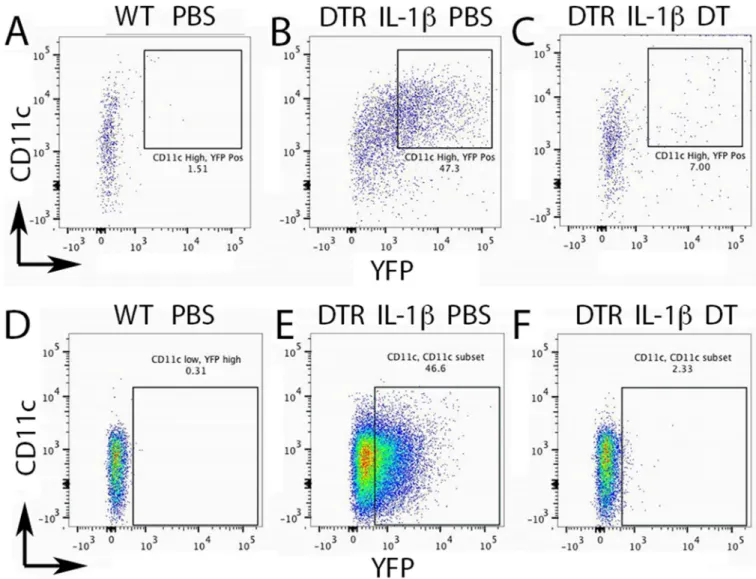
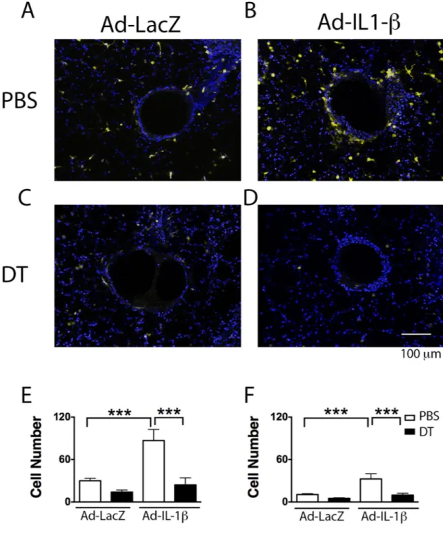
We confirmed the depletion of CD11c+ DCs cells by crossing the CD11c-DTR mice to transgenic CD11c-yellow fluorescent protein (YFP) mice, which express YFP in DCs and alveolar macrophages. We used a size and scatter gating strategy to discriminate DCs (CD11c high, CD11b high, MHCII high, Ly6C positive, F480 negative) from alveolar macrophages (CD11b high, Gr1 negative, Ly6c high, MHCII high, CD11c low) (Fig. S1, 2). Two days after diptheria toxin lung DCs were depleted (Fig. 1A–C), as were AMs, as previously reported (Fig. 1D–F) (32). To confirm the airway localization of DCs and their depletion by DT, we used 2-photon microscopy of living lung sections of compound transgenic mice (CD11c-DTR; CD11c-YFP). We found that Ad-IL-1β treatment caused a dramatic increase in the localization of DCs within 100 µm of the airways; DT significantly reduced DCs surrounding the airways (Fig. 2).
We next measured the effect of depletion of CD11c-DTR positive cells on Ad-IL-1β-induced airway inflammation and fibrosis. CD11c-DTR mice were treated with DT 5 days after IT-Ad-IL-1β and euthanized 4 days later. IT-Ad-IL-1β caused a significant increase in total bronchoalveolar lavage (BAL) cell count, macrophages, neutrophils and lymphocytes
Author Manuscript
Author Manuscript
Author Manuscript
(Fig. 3), airway inflammation and airway wall fibrosis (Fig. 4); these increases were all significantly reduced by DT treatment (Fig. 3,4). The fibrotic response was confined to airways and was not significantly increased around vessels (p=0.99). These data demonstrate that CD11c positive cells, which consist of both DCs and AMs, are required for pathologic airway inflammation and fibrosis.
Deficiency of ccr6, the chemokine receptor for CCL20, protects mice from IL-1β induced
airway inflammation and fibrosis
Mice deficient in ccr6 are expected to have defective DC influx, since ccr6 is expressed by DCs and is the only receptor for CCL20 (33). IT-Ad-IL-1β caused significantly less airway wall inflammation (Fig. 5) and fibrosis (Fig. 5) compared with wild type (WT), suggesting that a CCR6/CCL20-dependent inflammatory response was coupled to the subsequent fibrotic response.
α/β T-cells are required for airway fibrosis
We next sought to determine whether T-cells, which have been shown to be involved in the remodeling response (15) were required for airway fibrosis in the Ad-IL-1β model. T-cells, of which >90% in mice and humans express the α/β heterodimeric T-cell receptor, have been implicated in fibrotic responses through the elaboration of cytokines such as IL-17 (34). Thus, we used tcra −/− mice, to test the role of T-cells in IL-1β-mediated airway fibrosis. Compared to WT mice (Fig. 5), tcra−/− mice failed to develop either airway inflammation (Fig. 5) or fibrosis (Fig. 5) 14 days after IT-Ad-IL-1β. These data support a role for adaptive immunity, specifically α/β T-cells in fibrotic airway disease.
CCR6-expressing cell types in the lungs of Ad-IL-1β treated mice
The cell types reported to express CCR6 include DCs, alveolar macrophages, CD4+ Th17
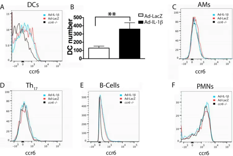
cells, B-cells and neutrophils (35–38). We detected obvious surface staining of CCR6 on CD11b+ DCs and B-cells, only trace expression on alveolar macrophages and CD4+ Th17
cells and no expression on neutrophils from WT compared with ccr6 −/− mice (Fig. 6). There were significant increases in numbers of ccr6+ DCs in WT mice treated with Ad-IL-1β compared with Ad-LacZ (Fig. 6B). There was a slight non-significant increase in ccr6+ B-cells (18%, p=0.11), and no increase in ccr6 + alveolar macrophages or CD4+ Th17
cells in WT mice treated with Ad-IL-1β compared with Ad-LacZ. These data suggest that DCs are the most likely ccr6-expressing cell type that are involved in Ad-IL-1β-mediated airway fibroinflammatory responses.
DC expression of ccr6 is required for IT-Ad-IL-1β induced increases in innate and adaptive
immune effectors
We sought to understand how CCR6 expressing DCs and adaptive α/β T-cell immunity are coupled to a fibrotic airway response. DCs, as well as the profibrotic CD4-T-cell subsets, Th1 and Th17 cells, are characteristically increased in COPD samples as well as in the
Ad-IL-1β model (15). Resident DCs in the murine lung are comprised of two major subsets, the most numerous characterized by high expression of CD11c, CD11b and absence of CD103 (CD11b+), while the minor subset expresses CD11c and CD103, but not CD11b (CD103+)
Author Manuscript
Author Manuscript
Author Manuscript
(39). It is the former DC subset, CD11b+ that predominates in the lung in the Ad-IL-1β model (15). This subset expresses intermediate levels of Ly6c, suggesting a monocytic lineage (20). We found that CD11b+ DC numbers were significantly increased 3 days after Ad-IL-1β treatment and this increase was sustained (Fig. 7A). Significant increases in numbers of Th1 and Th17 cells actively secreting IFN-γ or IL-17, respectively, were not
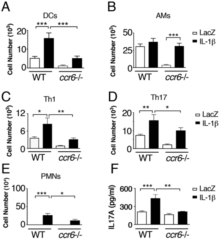
significantly increased until day 9 (Fig. 7B). These day 9 increases in CD11b+ DCs, Th1 and
Th17 cells were significantly attenuated in ccr6 −/− mice (Fig. 8A, C, D). Interestingly,
alveolar macrophage (AM) numbers were also significantly increased in IT-Ad-IL-1β treated ccr6 −/− mice to comparable levels as seen in IT-Ad-IL-1β treated WT mice (Fig. 8B). However, steady state AM numbers were significantly decreased in ccr6 −/− compared with WT Ad-LacZ control mice (Fig. 8B). This decrease was related to the Ad-LacZ virus as the decrease was not seen in untreated ccr6 −/− mice (in BAL, AM numbers: WT 7.1×10⁵ ± 5.8; ccr6 −/− 7.1 × 10⁵ ± 6.8, p=N.S.). Evaluation of BAL revealed a significant increase in neutrophils in IT-Ad-IL-1β treated compared with Ad-LacZ treated WT mice, and this increase was significantly attenuated in ccr6−/− mice (Fig. 8E). Analysis of whole lung lysates revealed a significant increase in IL-17A protein in IT-Ad-IL-1β treated WT but not ccr6−/− mice (Fig. 8F). These data suggest that CCL20/CCR6 is essential for increasing DC but not AM numbers in response to IL-1β Furthermore, in the Ad-IL-1β model, DCs are critical for Th1 and Th17 adaptive immunity.
Discussion
This study has addressed the mechanistic connection between innate and adaptive immunity with airway wall fibrosis in COPD. Here we have determined that CD11c+ cells that accumulate around airways in response to IL-1β are required for airway inflammation and fibrosis. The IL-1β-induced DC accumulation is ccr6-dependent and required for enhanced adaptive immune responses, of which α/β T-cells are critical for airway remodeling. The temporal relationship between DC accumulation, adaptive immunity and airway fibrosis suggest that DC accumulation is critical for establishing profibrotic adaptive immune responses (18, 28).
How or if immune responses contribute to the development of fibrosis remains hotly debated. Clodronate depletion of both macrophages and DCs has recently been found to protect against CS-induced airway remodeling (23). Since DCs and macrophages express CD11c and are depleted in the CD11c-DTR model, we cannot definitively rule out a role for macrophages in airway fibrosis. However, in Ad-IL-1β-treated mice, of the CCR6
expressing cells types (35–38) alveolar macrophages were not decreased by ccr6 deficiency and DCs were the only CCR6+ expressing cells that were increased by Ad-IL-1β. These findings suggest that DCs and not macrophages are required for the fibrotic airway responses.
In Ad-IL-1β-treated ccr6 −/− mice, alveolar macrophages were increased nearly to WT levels in contrast to DCs which were markedly reduced in numbers. These data confirm that Ad-IL-1β-dependent DC accumulation in mice is mainly dependent on ccr6 while alveolar macrophage accumulation is ccr6-independent. We have not yet determined the
compensatory mechanism explaining increased macrophage influx in Ad-IL-1β-treated ccr6
Author Manuscript
Author Manuscript
Author Manuscript
−/− mice, but speculate that the CCL2/ccr2 axis is involved since Ad-IL-1β-treated mice express high levels of CCL2 in the lung (15). In addition, AMs in ccr6-deficient mice have other notable differences from WT mice as ccr6 −/− AMs in Ad-LacZ treated mice were decreased compared with untreated WT mice, suggesting that ccr6-deficient AM survival was influenced by the Ad-LacZ virus. Whether or not this difference in survival is linked to compensatory increases in other chemokine receptors such as ccr2 remains to be determined. DCs have the capacity to directly produce profibrogenic cytokines, and thus could directly contribute to airway fibrosis. DCs express the integrin αvβ8, which is a potent and critical activator of latent-TGF-β, a profibrotic cytokine that is expressed in a latent form that must be activated to function. Genetic deletion of αvβ8 on DCs has been shown to block airway smooth muscle contraction, but not airway remodeling (21). In contrast, conditional deletion of αvβ8 on fibroblasts or global inhibition of αvβ8 using neutralizing antibodies efficiently blocks airway remodeling suggesting that TGF-β activated by cell types other than DCs contribute to airway fibrosis (7, 15).
Our present and past studies support an indirect role for ccr6-expressing DCs in airway fibrosis. Here we have begun to address the components downstream of a DC-mediated profibrotic adaptive immune response in airway remodeling. We find a temporal increase in DCs precedes an adaptive CD4+ Th1 and Th17 response. These temporal relationships are
likely to not only apply to the IL-1β airway remodeling system but also to other airway remodeling induced by CS, since IL-17A is also increased during CS-induced airway remodeling (7). IL-17A is mainly secreted by α/β CD4+ T-cells (i.e. Th17 cells) (34) and
deficiency of all α/β T-cells protects against IL-1β-induced airway fibrosis. IL-17A is increased in expression in COPD and mediates bleomycin-induced pulmonary fibrosis in mice (40). Therefore, we speculate that IL-17A is critical in the role that DCs play in airway fibrosis. However, we cannot exclude that other α/β T-cells, such as CD4+ Th1, or
neutrophils also contribute independently to airway fibrosis.
The mechanisms driving DC accumulation around airways are important to define to understand the genesis of the subsequent profibrotic inflammatory responses. We have recently determined that airway fibroblasts are an important source of the chemokine ligand for CCR6, CCL20 (Brand, et al, manuscript submitted). Furthermore, we have determined that an important mechanism driving CCL20 expression from fibroblasts involving integrin αvβ8-mediated activation of TGF-β. TGF-β activated by an αvβ8-dependent mechanism initiates signaling through the canonical TGF-β signaling pathway (i.e. SMADs); SMAD4 binds to an upstream enhancer element on the CCL20 promoter that forms a complex with a proximal NFκB subunit that is required for CCL20 expression (Brand, et al, manuscript submitted). We have recently used 2-photon imaging of living lung tissue to determine that αvβ8- or ccr6-dependent DC chemokinesis in response to Ad-IL-1β or cigarette smoke drives the microanatomic localization of DCs around large and small airways (Hashimoto, et al, manuscript submitted).
2-photon microscopy has provided a resolution not afforded by static immunohistochemical studies and has provided additional novel insights into DC airway localization, in addition to confirming that DCs are depleted in the CD11c-DTR model. In the Ad-IL-1β model, DCs
Author Manuscript
Author Manuscript
Author Manuscript
preferentially localize within 100 µm of the airway in close proximity to airway fibroblasts and airway epithelial cells, CCL20 expressing cell types, and very few, if any, DCs were observed interdigitating with the airway epithelium. Of the two major resident lung DC subsets (CD103+ or CD11b+ DCs,) intraepithelial DCs have been suggested to represent the CD103+ subset. (20). While we cannot exclude a role for CD103+ DCs in the
fibroinflamamtory responses in the Ad-IL-1β model, the paucity of intraepithelial DCs and the larger relative increase in numbers of the CD11b+ DC subset suggests that CD11b+ DCs are more likely to play a role in pathologic airway remodeling. In Ad-IL-1β-treated mice the CD11b+ subset expresses Ly6c which provides evidence of a hematogenous origin and monocytic lineage and suggests that these cells are newly recruited and replace the steady-state resident CD11b+ DCs. Such newly recruited CD11b+ DCs are also the dominant lung DC population following influenza infection and at the peak of infection are the major DC subset involved in antigen presentation and T-cell cross-priming in the draining lymph node (41). In the Ad-IL-1β model, the CD11b+ DC population is the major ccr6 expressing lung DC subset, is efficiently deleted in DT-treated DTR mice, appears in the lung before CD4+ Th1 and Th17 cells and is the most numerous lung DC subset that migrates to the draining
lymph node (15, 41). For these reasons, we hypothesize that the CD11b+ DC subset is the airway adjacent DC subset seen on 2-photon microscopy and is also the subset most likely to play a major role in driving ccr6-dependent pathologic adaptive CD4+ Th1 and Th17 cell
responses.
In summary, our data provide compelling evidence that CD11b+ DCs play a role in the evolution of murine airway disease.
Supplementary Material
Refer to Web version on PubMed Central for supplementary material.
Acknowledgments
We thank M. Nussenzweig for CD11c-EYFP transgenic reporter mice.
This work was supported by the grants from the NIH HL113032, HL063993, HL090662, NS044155, UCTRDRP, UCSF Academic Senate, UCOP POC award (S.L.N.), UCSF/NHLBI CAI award, UCSF Liver Center
(P30DK026743) to (S.L.N. and J.L.B.).
References
1. Miniño AM, Xu JQ, Kochanek KD. Deaths: Preliminary data for 2008. National Vital Statistics Reports. 2010; 59
2. Ito K, Ito M, Elliott WM, Cosio B, Caramori G, Kon OM, Barczyk A, Hayashi S, Adcock IM, Hogg JC, Barnes PJ. Decreased histone deacetylase activity in chronic obstructive pulmonary disease. N Engl J Med. 2005; 352:1967–1976. [PubMed: 15888697]
3. Postma DS, Timens W. Remodeling in asthma and chronic obstructive pulmonary disease. Proc Am Thorac Soc. 2006; 3:434–439. [PubMed: 16799088]
4. Araya J, Cambier S, Markovics JA, Wolters P, Jablons D, Hill A, Finkbeiner W, Jones K, Broaddus VC, Sheppard D, Barzcak A, Xiao Y, Erle DJ, Nishimura SL. Squamous metaplasia amplifies pathologic epithelial-mesenchymal interactions in COPD patients. J Clin Invest. 2007; 117:3551– 3562. [PubMed: 17965775]
Author Manuscript
Author Manuscript
Author Manuscript
5. Papi A, Bellettato CM, Braccioni F, Romagnoli M, Casolari P, Caramori G, Fabbri LM, Johnston SL. Infections and airway inflammation in chronic obstructive pulmonary disease severe exacerbations. Am J Respir Crit Care Med. 2006; 173:1114–1121. [PubMed: 16484677] 6. Kang MJ, Lee CG, Lee JY, Dela Cruz CS, Chen ZJ, Enelow R, Elias JA. Cigarette smoke
selectively enhances viral PAMP- and virus-induced pulmonary innate immune and remodeling responses in mice. J Clin Invest. 2008; 118:2771–2784. [PubMed: 18654661]
7. Minagawa S, Lou J, Seed RI, Cormier A, Wu S, Cheng Y, Murray L, Tsui P, Connor J, Herbst R, Govaerts C, Barker T, Cambier S, Yanagisawa H, Goodsell A, Hashimoto M, Brand OJ, Cheng R, Ma R, McKnelly KJ, Wen W, Hill A, Jablons D, Wolters P, Kitamura H, Araya J, Barczak AJ, Erle DJ, Reichardt LF, Marks JD, Baron JL, Nishimura SL. Selective targeting of TGF-beta activation to treat fibroinflammatory airway disease. Science translational medicine. 2014; 6:241ra279.
8. Lucattelli M, Cicko S, Muller T, Lommatzsch M, De Cunto G, Cardini S, Sundas W, Grimm M, Zeiser R, Durk T, Zissel G, Sorichter S, Ferrari D, Di Virgilio F, Virchow JC, Lungarella G, Idzko M. P2X7 receptor signaling in the pathogenesis of smoke-induced lung inflammation and emphysema. Am J Respir Cell Mol Biol. 2011; 44:423–429. [PubMed: 20508069]
9. Geraghty P, Dabo AJ, D'Armiento J. TLR4 protein contributes to cigarette smoke-induced matrix metalloproteinase-1 (MMP-1) expression in chronic obstructive pulmonary disease. J Biol Chem. 2011; 286:30211–30218. [PubMed: 21730072]
10. Singh B, Arora S, Khanna V. Association of severity of COPD with IgE and interleukin-1 beta. Monaldi archives for chest disease = Archivio Monaldi per le malattie del torace / Fondazione clinica del lavoro, IRCCS [and] Istituto di clinica tisiologica e malattie apparato respiratorio, Universita di Napoli, Secondo ateneo. 2010; 73:86–87.
11. Gessner C, Scheibe R, Wotzel M, Hammerschmidt S, Kuhn H, Engelmann L, Hoheisel G, Gillissen A, Sack U, Wirtz H. Exhaled breath condensate cytokine patterns in chronic obstructive pulmonary disease. Respiratory medicine. 2005; 99:1229–1240. [PubMed: 16140223]
12. Pauwels NS, Bracke KR, Dupont LL, Van Pottelberge GR, Provoost S, Vanden Berghe T, Vandenabeele P, Lambrecht BN, Joos GF, Brusselle GG. Role of IL-1alpha and the Nlrp3/ caspase-1/IL-1beta axis in cigarette smoke-induced pulmonary inflammation and COPD. The European respiratory journal. 2011; 38:1019–1028. [PubMed: 21622588]
13. Sapey E, Ahmad A, Bayley D, Newbold P, Snell N, Rugman P, Stockley RA. Imbalances between interleukin-1 and tumor necrosis factor agonists and antagonists in stable COPD. J Clin Immunol. 2009; 29:508–516. [PubMed: 19291375]
14. Lappalainen U, Whitsett JA, Wert SE, Tichelaar JW, Bry K. Interleukin-1beta causes pulmonary inflammation, emphysema, and airway remodeling in the adult murine lung. Am J Respir Cell Mol Biol. 2005; 32:311–318. [PubMed: 15668323]
15. Kitamura H, Cambier S, Somanath S, Barker T, Minagawa S, Markovics J, Goodsell A, Publicover J, Reichardt L, Jablons D, Wolters P, Hill A, Marks JD, Lou J, Pittet JF, Gauldie J, Baron JL, Nishimura SL. Mouse and human lung fibroblasts regulate dendritic cell trafficking, airway inflammation, and fibrosis through integrin alphavbeta8-mediated activation of TGF-beta. J Clin Invest. 2011; 121:2863–2875. [PubMed: 21646718]
16. Churg A, Zhou S, Wang X, Wang R, Wright JL. The role of interleukin-1beta in murine cigarette smoke-induced emphysema and small airway remodeling. Am J Respir Cell Mol Biol. 2009; 40:482–490. [PubMed: 18931327]
17. Doz E, Noulin N, Boichot E, Guenon I, Fick L, Le Bert M, Lagente V, Ryffel B, Schnyder B, Quesniaux VF, Couillin I. Cigarette smoke-induced pulmonary inflammation is TLR4/MyD88 and IL-1R1/MyD88 signaling dependent. J Immunol. 2008; 180:1169–1178. [PubMed: 18178857] 18. Demedts IK, Bracke KR, Van Pottelberge G, Testelmans D, Verleden GM, Vermassen FE, Joos
GF, Brusselle GG. Accumulation of dendritic cells and increased CCL20 levels in the airways of patients with chronic obstructive pulmonary disease. Am J Respir Crit Care Med. 2007; 175:998– 1005. [PubMed: 17332482]
19. Bracke KR, D'Hulst A I, Maes T, Moerloose KB, Demedts IK, Lebecque S, Joos GF, Brusselle GG. Cigarette smoke-induced pulmonary inflammation and emphysema are attenuated in CCR6-deficient mice. J Immunol. 2006; 177:4350–4359. [PubMed: 16982869]
20. Lambrecht BN, Hammad H. Biology of lung dendritic cells at the origin of asthma. Immunity. 2009; 31:412–424. [PubMed: 19766084]
Author Manuscript
Author Manuscript
Author Manuscript
21. Kudo M, Melton AC, Chen C, Engler MB, Huang KE, Ren X, Wang Y, Bernstein X, Li JT, Atabai K, Huang X, Sheppard D. IL-17A produced by alphabeta T cells drives airway
hyper-responsiveness in mice and enhances mouse and human airway smooth muscle contraction. Nat Med. 2012; 18:547–554. [PubMed: 22388091]
22. Chen K, Pociask DA, McAleer JP, Chan YR, Alcorn JF, Kreindler JL, Keyser MR, Shapiro SD, Houghton AM, Kolls JK, Zheng M. IL-17RA is required for CCL2 expression, macrophage recruitment, and emphysema in response to cigarette smoke. PLoS One. 2011; 6:e20333. [PubMed: 21647421]
23. Beckett EL, Stevens RL, Jarnicki AG, Kim RY, Hanish I, Hansbro NG, Deane A, Keely S, Horvat JC, Yang M, Oliver BG, van Rooijen N, Inman MD, Adachi R, Soberman RJ, Hamadi S, Wark PA, Foster PS, Hansbro PM. A new short-term mouse model of chronic obstructive pulmonary disease identifies a role for mast cell tryptase in pathogenesis. The Journal of allergy and clinical immunology. 2013; 131:752–762. [PubMed: 23380220]
24. Lindquist RL, Shakhar G, Dudziak D, Wardemann H, Eisenreich T, Dustin ML, Nussenzweig MC. Visualizing dendritic cell networks in vivo. Nat Immunol. 2004; 5:1243–1250. [PubMed:
15543150]
25. Kolb M, Margetts PJ, Anthony DC, Pitossi F, Gauldie J. Transient expression of IL-1beta induces acute lung injury and chronic repair leading to pulmonary fibrosis. J Clin Invest. 2001; 107:1529– 1536. [PubMed: 11413160]
26. Ganter MT, Roux J, Miyazawa B, Howard M, Frank JA, Su G, Sheppard D, Violette SM, Weinreb PH, Horan GS, Matthay MA, Pittet JF. Interleukin-1beta causes acute lung injury via alphavbeta5 and alphavbeta6 integrin-dependent mechanisms. Circ Res. 2008; 102:804–812. [PubMed: 18276918]
27. Mu D, Cambier S, Fjellbirkeland L, Baron JL, Munger JS, Kawakatsu H, Sheppard D, Broaddus VC, Nishimura SL. The integrin alpha(v)beta8 mediates epithelial homeostasis through MT1-MMP-dependent activation of TGF-beta1. J Cell Biol. 2002; 157:493–507. [PubMed: 11970960] 28. Hogg JC, Chu F, Utokaparch S, Woods R, Elliott WM, Buzatu L, Cherniack RM, Rogers RM,
Sciurba FC, Coxson HO, Pare PD. The nature of small-airway obstruction in chronic obstructive pulmonary disease. N Engl J Med. 2004; 350:2645–2653. [PubMed: 15215480]
29. Thornton EE, Looney MR, Bose O, Sen D, Sheppard D, Locksley R, Huang X, Krummel MF. Spatiotemporally separated antigen uptake by alveolar dendritic cells and airway presentation to T cells in the lung. The Journal of experimental medicine. 2012; 209:1183–1199. [PubMed: 22585735]
30. Jung S, Unutmaz D, Wong P, Sano G, De los Santos K, Sparwasser T, Wu S, Vuthoori S, Ko K, Zavala F, Pamer EG, Littman DR, Lang RA. In vivo depletion of CD11c+ dendritic cells abrogates priming of CD8+ T cells by exogenous cell-associated antigens. Immunity. 2002; 17:211–220. [PubMed: 12196292]
31. Pappenheimer AM Jr, Harper AA, Moynihan M, Brockes JP. Diphtheria toxin and related proteins: effect of route of injection on toxicity and the determination of cytotoxicity for various cultured cells. The Journal of infectious diseases. 1982; 145:94–102. [PubMed: 6798133]
32. van Rijt LS, Jung S, Kleinjan A, Vos N, Willart M, Duez C, Hoogsteden HC, Lambrecht BN. In vivo depletion of lung CD11c+ dendritic cells during allergen challenge abrogates the
characteristic features of asthma. The Journal of experimental medicine. 2005; 201:981–991. [PubMed: 15781587]
33. Baba M, Imai T, Nishimura M, Kakizaki M, Takagi S, Hieshima K, Nomiyama H, Yoshie O. Identification of CCR6, the specific receptor for a novel lymphocyte-directed CC chemokine LARC. J Biol Chem. 1997; 272:14893–14898. [PubMed: 9169459]
34. Kolls JK, Linden A. Interleukin-17 family members and inflammation. Immunity. 2004; 21:467– 476. [PubMed: 15485625]
35. Ito T, t. Carson WF, Cavassani KA, Connett JM, Kunkel SL. CCR6 as a mediator of immunity in the lung and gut. Experimental cell research. 2011; 317:613–619. [PubMed: 21376174]
36. Krzysiek R, Lefevre EA, Bernard J, Foussat A, Galanaud P, Louache F, Richard Y. Regulation of CCR6 chemokine receptor expression and responsiveness to macrophage inflammatory
protein-3alpha/CCL20 in human B cells. Blood. 2000; 96:2338–2345. [PubMed: 11001880]
Author Manuscript
Author Manuscript
Author Manuscript
37. Yamashiro S, Wang JM, Yang D, Gong WH, Kamohara H, Yoshimura T. Expression of CCR6 and CD83 by cytokine-activated human neutrophils. Blood. 2000; 96:3958–3963. [PubMed:
11090084]
38. Yamazaki T, Yang XO, Chung Y, Fukunaga A, Nurieva R, Pappu B, Martin-Orozco N, Kang HS, Ma L, Panopoulos AD, Craig S, Watowich SS, Jetten AM, Tian Q, Dong C. CCR6 regulates the migration of inflammatory and regulatory T cells. J Immunol. 2008; 181:8391–8401. [PubMed: 19050256]
39. Lambrecht BN, Hammad H. Lung dendritic cells in respiratory viral infection and asthma: from protection to immunopathology. Annual review of immunology. 2012; 30:243–270.
40. Wilson MS, Madala SK, Ramalingam TR, Gochuico BR, Rosas IO, Cheever AW, Wynn TA. Bleomycin and IL-1beta-mediated pulmonary fibrosis is IL-17A dependent. The Journal of experimental medicine. 2010; 207:535–552. [PubMed: 20176803]
41. Ballesteros-Tato A, Leon B, Lund FE, Randall TD. Temporal changes in dendritic cell subsets, cross-priming and costimulation via CD70 control CD8(+) T cell responses to influenza. Nat Immunol. 2010; 11:216–224. [PubMed: 20098442]
Author Manuscript
Author Manuscript
Author Manuscript
Fig. 1. Dendritic cells (DCs) and alveolar macrophages (AMs) are depleted in intratracheal (IT) Ad-IL-1β treated CD11c-DTR mice
Percoll enriched lung immune cell populations were analyzed by multicolor flow cytometry multicolor staining of lung immune cell populations from wild-type (WT), untreated (A, D), or compound transgenic (CD11c-DTR;CD11c-YFP) mice (B, C, E, F) treated with IT- Ad-IL-1β Five days after infection, CD11c-DTR;CD11c-YFP mice were treated with PBS or diphtheria toxin (DT). At day seven, at the peak of IL-1β expression, mice were euthanized and lung cell suspensions stained for CD11c, CD11b, MHCII, F480, Ly6c and Gr1. Staining for CD11c on the y-axis and YFP fluorescence on the x-axis are indicated for lung DC populations (A–C) and alveolar macrophages (D–F). The gates used to determine depletion of DCs or AMs are shown and the gating strategy shown in Fig. S1 and S2.
Author Manuscript
Author Manuscript
Author Manuscript
Fig. 2. DC airway localization is increased by IT-Ad-IL-1β treatment of CD11c-DTR;CD11c-YFP mice and completely blocked by DT
CD11c-DTR;CD11c-YFP mice were treated with Ad-LacZ (A, C) intratracheal (IT)-Ad-IL-1β (B, D). Five days after infection, CD11c-DTR;CD11c-YFP mice were treated with PBS (A, B) or diphtheria toxin (DT) (C, D). At day seven, at the peak of IL-1β expression, mice were euthanized and vibratome living lung sections analyzed using 2-photon
microscopy. The YFP positive cells are shown as yellow cells against a Hoechst (blue) counterstained background. The number of DCs within 100 µm (E), or outside of 100 µm
Author Manuscript
Author Manuscript
Author Manuscript
(F) from the airway basement membrane was assessed using automated cell analysis (IMARIS). Bar = 100 µm.
Author Manuscript
Author Manuscript
Author Manuscript
Author Manuscript
Fig. 3. Lung inflammation is blocked by depletion of CD11c-expressing cells
Bronchoalveolar lavage (BAL) of CD11c-DTR;CD11c-YFP mice that were treated with Ad-LacZ or intratracheal (IT)-Ad-IL-1β. Five days after infection, CD11c-DTR mice were treated with PBS (white bars) or diphtheria toxin (DT, black bars). Cytospin preparations were assessed for total cells count (A), alveolar macrophages (AMs, B), neutrophils (C), or lymphocytes (D). N=4 in PBS and 8 in Ad-IL-1β groups, *p<0.05, **p<0.01, ***p<0.001 by ANOVA and Bonferroni’s post-test.
Author Manuscript
Author Manuscript
Author Manuscript
Fig. 4. Depletion of CD11c-expressing cells protects against Ad-IL-1β-mediated airway remodeling
Representative histologic images from CD11c-DTR mice treated with either Ad-LacZ (A, D) or Ad-IL-1β (B, C, E, F). 5 days after adenoviral administration mice were treated with PBS (A–C), or DT (4 ng/g body weight, D–F) I.P. and mice were euthanized on day 9. Bar=100 µm. Airway morphometry of H&E stained sections was performed to determine the area of inflammation around the airways (A, B, D, E, G) or the extent of fibrosis around the airway wall (C, F, H), as determined by trichrome staining and airway morphometry as described by Hogg, et al (28) and adapted to mice in Kitamura, et al (15). Open bars = PBS; closed bars = DT. A=Airway lumen. Error bars are S.E.M; ***p<0.001 as determined by ANOVA and Bonferroni’s post-test.
Author Manuscript
Author Manuscript
Author Manuscript
Fig. 5. α/β T-cells and ccr6 are required for IL-1β-induced airway fibrosis
Wild type (WT) (A, B, E, F), tcra −/− (C, G), or ccr6−/− (D, H) mice were treated with intratracheal-Ad-IL-1β (B–D, F–H) or Ad-LacZ (A, E) as a control. After 14 days, inflammation (A–D, Haemotoxylin and Eosin (H&E)) and fibrosis (E–H, Trichrome) were assessed by airway morphometry, as above (I, J). ***p<0.001 by one-way ANOVA and Bonferroni’s post-test. N≥8 for every group.
Author Manuscript
Author Manuscript
Author Manuscript
Fig. 6. DCs are the major ccr6 expressing lung immune cell type in Ad-IL-1β-treated mice
WT or ccr6 −/− (A–F) mice (N ≥ 7) were treated with intratracheal-Ad-IL-1β or Ad-LacZ as a control. After 9 days, Percoll enriched lung immune cell populations were analyzed by multicolor flow cytometry for: A–B) CD11b+ DCs (CD11c positive, CD11b positive, Ia high, ly6c intermediate, non-autofluorescent) C) AMs (CD11c positive, Ia low,
autofluorescent), D) CD4+Th17-cells (TCRβ positive, Ia neg, IL-17A surface positive,
Surface cytokine expression was determined using unstimulated cells stained using a cytokine secretion assay (Miltenyi Biotech, Auburn, CA). E) B-cells (CD19, Ia positive, Nk1.1 negative, TCRβ negative) (F) PMNs (Gr1 high, Ly6c high, CD11b high, Ia negative). Represenative histograms overlays (A, C–F) of CCR6 expression of lung immune
populations from WT mice treated with Ad-IL-1β (blue) or Ad-LacZ (red), compared with ccr6 −/− mice (black) as a negative control. In panel B, the number of CCR6+ lung DCs in Ad-IL-1β (filled) or Ad-LacZ (open) treated mice are shown. N=7 **p<0.01 by Student’s t-test.
Author Manuscript
Author Manuscript
Author Manuscript
Fig. 7. CD11b+ DC numbers increase prior to CD4+ Th1 and Th17 cells after Ad-IL-1β treatment
WT mice (N ≥ 3 at each time point) were treated with intratracheal-Ad-IL-1β and lung immune cell populations were assessed by multicolor flow cytometry 3, 5 and 9 days after Ad-IL-1β treatment. Day 0 mice were WT untreated mice. Percoll enriched lung immune cell populations were analyzed for: A, B) CD11b+ DCs (CD11c positive, CD11b positive, Ia high, ly6c intermediate, non-autofluorescent) and compared to A) CD4+Th1 (TCRβ positive,
Ia neg, IFNγ surface positive), or B) CD4+Th17 cells (TCRβ positive, Ia neg, IL-17A
surface positive). Surface cytokine expression was determined using unstimulated cells stained using a cytokine secretion assay (Miltenyi Biotech, Auburn, CA). *<0.05 by ANOVA and Dunn’s multiple comparisons test.
Author Manuscript
Author Manuscript
Author Manuscript
Fig. 8. ccr6 is required for innate and Th1 and Th17 adaptive immunity in Ad-IL-1β-treated mice
WT or ccr6 −/− (A–F) mice (N ≥ 7) were treated with intratracheal-Ad-IL-1β or Ad-LacZ as a control. After 9 days, Percoll enriched lung immune cell populations were analyzed by multicolor flow cytometry for: A) DCs (CD11c positive, CD11b positive, Ia high, ly6c intermediate, non-autofluorescent), B) AMs-CD11c positive, Ia low, autofluorescent), C) CD4+Th1 (TCRβ positive, Ia neg, IFNγ surface positive), or D) CD4+Th17-cells (TCRβ
positive, Ia neg, IL-17A surface positive). Surface cytokine expression was determined using unstimulated cells stained using a cytokine secretion assay (Miltenyi Biotech, Auburn,
Author Manuscript
Author Manuscript
Author Manuscript
CA). Bronchoalveolar lavage (E) was performed to assess neutrophil numbers (N=5 per group). IL-17A ELISA (F) was performed using whole lung lysates to assess total IL-17A levels. N=6 per group repeated 3 times with similar results *p<0.05, **p<0.01, ***p<0.001 by ANOVA with Bonferroni’s post-test.