HAL Id: dumas-03087038
https://dumas.ccsd.cnrs.fr/dumas-03087038
Submitted on 23 Dec 2020
HAL is a multi-disciplinary open access archive for the deposit and dissemination of sci-entific research documents, whether they are pub-lished or not. The documents may come from teaching and research institutions in France or abroad, or from public or private research centers.
L’archive ouverte pluridisciplinaire HAL, est destinée au dépôt et à la diffusion de documents scientifiques de niveau recherche, publiés ou non, émanant des établissements d’enseignement et de recherche français ou étrangers, des laboratoires publics ou privés.
J’ai un DSA, c’est grave docteur ? De novo donor
specific antibody initial and evolutive characteristics
predictive of graft survival in kidney transplant
recipients at Grenoble university hospital (the
DYNAMIK study)
Catherine Casiez
To cite this version:
Catherine Casiez. J’ai un DSA, c’est grave docteur ? De novo donor specific antibody initial and evo-lutive characteristics predictive of graft survival in kidney transplant recipients at Grenoble university hospital (the DYNAMIK study). Human health and pathology. 2020. �dumas-03087038�
AVERTISSEMENT
Ce document est le fruit d'un long travail approuvé par le
jury de soutenance.
La propriété intellectuelle du document reste entièrement
celle du ou des auteurs. Les utilisateurs doivent respecter le
droit d’auteur selon la législation en vigueur, et sont soumis
aux règles habituelles du bon usage, comme pour les
publications sur papier : respect des travaux originaux,
citation, interdiction du pillage intellectuel, etc.
Il est mis à disposition de toute personne intéressée par
l’intermédiaire de
l’archive ouverte DUMAS
(Dépôt
Universitaire de Mémoires Après Soutenance).
Si vous désirez contacter son ou ses auteurs, nous vous
invitons à consulter la page de DUMAS présentant le
document. Si l’auteur l’a autorisé, son adresse mail
apparaîtra lorsque vous cliquerez sur le bouton « Détails »
(à droite du nom).
Dans le cas contraire, vous pouvez consulter en ligne les
annuaires de l’ordre des médecins, des pharmaciens et des
sages-femmes.
Contact à la Bibliothèque universitaire de Médecine
Pharmacie de Grenoble :
1
UNIVERSITÉ GRENOBLE ALPES UFR DE MÉDECINE DE GRENOBLE
Année 2020
J’AI UN DSA, C’EST GRAVE DOCTEUR ?
DE NOVO DONOR SPECIFIC ANTIBODY INITIAL AND EVOLUTIVE CHARACTERISTICS PREDICTIVE OF GRAFT SURVIVAL IN KIDNEY TRANSPLANT RECIPIENTS AT GRENOBLE
UNIVERSITY HOSPITAL (THE DYNAMIK STUDY) THÈSE
PRÉSENTÉE POUR L’OBTENTION DU TITRE DE DOCTEUR EN MÉDECINE DIPLÔME D’ÉTAT
Catherine Casiez
THÈSE SOUTENUE PUBLIQUEMENT À LA FACULTÉ DE MÉDECINE DE GRENOBLE Le 21/09/2020
DEVANT LE JURY COMPOSÉ DE : Président du jury :
M. Rostaing Lionel Membres :
M. Jouve Thomas (directeur de thèse) M. Malvezzi Paolo
Mme Masson Dominique M. Noble Johan
M. Thaunat Olivier
L’UFR de Médecine de Grenoble n’entend donner aucune approbation ni improbation aux opinions émises dans les thèses ; ces opinions sont considérées comme propres à leurs auteurs.
6
Remerciements
C’est tout un ensemble de choix et de rencontres qui font qu’aujourd’hui je fais un métier qui m’épanouit, entourée de gens incroyables, dans une région qui m’émerveille sans cesse. Pour tout ce bonheur, MERCI.
Professeur Rostaing, vous avez toujours été bienveillant dans votre encadrement, vous avez réussi le défi de transformer le service dans lequel je suis ravie d’avoir trouvé une petite place pour un an. Merci beaucoup pour votre confiance.
Professeur Thaunat, c’est un honneur de vous accueillir aujourd’hui, et de bénéficier de votre expertise dans l’immunologie.
Thomas, tu me guides depuis mes premiers pas dans le service, et maintenant pour ces grandes étapes que sont la thèse et le mémoire. Merci pour ton enseignement et ton écoute.
Paolo, c’est un vrai plaisir de travailler avec toi, entre écoute, bienveillance, humour et générosité.
Johan, beaucoup de bons moments passés ensembles, j’ai hâte de travailler à nouveau avec toi en novembre.
Docteur Masson, merci pour votre accueil au sein du laboratoire de l’EFS, et d’avoir partagé votre expertise en matière de DSA.
Merci à tous d’avoir accepté d’être membres de mon jury de thèse.
A Max, tu étais là avant même que je me lance dans cette aventure, présent à mes côtés à chaque étape. Je n’y serais jamais arrivée seule, sans ton soutien et ton écoute. Notre petite histoire entre lycéens s’est transformée en grande histoire, qui a traversé la France pour atterrir au milieu des montagnes, où la suite reste à écrire.
A mes parents, qui m’ont toujours apporté un soutien infaillible. Le chemin de la confiance en soi aura été long mais aujourd’hui grâce à votre persévérance il m’aura amené à l’épanouissement, même si le départ pour Grenoble n’a pas été facile pour vous. Vivement l’heure de la retraite pour que vous veniez passer plus de temps ici !
A mes frères Benoit et Alain, mes belles-sœurs Sandra et Juliette, pour tout votre soutien, humour et amour, mes neveux et nièces Léanne, Emilie, Jean, Noé et Laurette, vous pourrez venir passer des vacances à la montagne chez tatie autant que vous voudrez !
A Mamie et Papi, j’aurais aimé que vous soyez là aujourd’hui, vous me manquez. Au Dr Vannelle, merci d’avoir cru en moi pour que je me lance dans cette aventure, vous aviez vu si juste.
7 A mon parrain et Isabelle, vous m’avez toujours encouragée, au point de faire le déplacement jusqu’ici pour me soutenir.
A mes cousins et cousines, que j’aimerais pouvoir voir plus souvent. Camille, hâte de te passer te revoir à Budapest, on a toujours tant de choses à se raconter.
A ma belle-famille également, pour leur grand soutien pendant toutes ces années. A Justine et Andrej, on s’est déjà dit beaucoup de choses le 27 aout, on va bientôt pouvoir reprendre notre rythme d’apéros, soirées jeux, randonnées, virées à ski… et faire cobaye pour gouter les pâtisseries.
Au reste de l’équipe de néphrologie de Grenoble, Hamza, Florian, Eloi, Bénédicte et Marc, ainsi que les infirmier(e)s, aide-soignant(e)s et secrétaires au top, Gaelle, Séverine, Méryll, Perrine, Quentin, Tom, Sylvie, Audrey, Sandie, etc, c’est un réel bonheur de tous vous retrouver en novembre ! Et Mélanie, merci de ton aide et de ton temps à l’EFS !
A l’équipe de néphrologie de Chambéry, Jean-Baptiste, Claire, Laure, Stéphane, Bertrand, Jacques, Dominique : merci beaucoup pour votre confiance, j’ai toujours du mal à réaliser la chance que j’ai de pouvoir vous rejoindre !
A Laurence et Sandrine, et le reste de l’équipe des explorations fonctionnelles rénales de Lyon, où les journées sont passionnantes !
A mes co-internes de néphro : Antoine mon co-interne des premières galères, Momo, Yann, Loulou (mon cousin éloigné avec qui j’ai le plus dormi), Valoche (hâte de reformer la team blonde du nord en novembre), Anne-So : beaucoup de bons moments passés tous ensembles, et de boite-mail piratées. Les plus jeunes, qui n’ont pas l’air d’être beaucoup plus sages, Carla (Carlô), Jules, Reda et Alexandra.
Aux autres co-internes, Thomas le plus sympa des internes de cardio (et c’est rare !), qui aura au moins essayé de comprendre à quoi servait la protéinurie. Les co-internes de la Mut, Salamèche, Malikou (pour rester polie) et Gui (ça se prononce comment ?), beaucoup de fous rires, c’est à cause de vous que j’ai hérité de mon surnom. Jessica, on se partageais notre placard à balais. Les co-internes de réa, Thomas (le plus sympa des pneumos, juste pour embêter Justin), Lucie, Natacha (un modèle inégalable), Elodie, Lisa, Théo et Mariano (les experts du café), et Manuella, c’est grâce à vous que le semestre en réa a été aussi sympa ! La dreamteam de covidologie-gonocoquologie, Justin (qui mérite à être connu), Tiphaine, Yanis (la force tranquille), Olivator et Rémi, avec des chefs au top, Marion, Isabelle, Fanny, Anne-Laure, Patricia, Olivier. Et enfin, les co-internes de Lyon, Luc, Amélie, Paule et Arthur, avec qui j’ai hâte de retourner aux quais du Rhône quand tout ça sera fini.
8
I. Table des matières
II. Abstract ... 9
III. Résumé ... 11
IV. Contextualisation ... 13
A. Le système HLA ... 13
B. Les techniques d’identification des anticorps anti-HLA ... 16
V. Abréviations ... 19
VI. Introduction ... 20
VII. Materials and Methods ... 22
VIII. Results ... 25
General population characteristics ... 26
dnDSA panorama ... 29
Graft survival: associated covariates ... 34
IX. Discussion ... 42
9
II. Abstract
Introduction: Occurrence of de-novo donor-specific antibodies (dnDSA) can reduce graft survival in kidney-transplant recipients (KTR). However, development kinetic has been poorly described, we assessed the initial and developmental characteristics of dnDSA to predict death-censored graft survival (DCGS).
Materials and methods: All KTR at Grenoble University Hospital between 2008 and 2017 with no preformed DSAs were included. Most patients received antithymocyte globulins as induction therapy and maintenance immunosuppression of tacrolimus and mycophenolic acid, plus early steroid-withdrawal.
Results: A total of 967 KTR were included: a dnDSA occurred in 73 (7.5%), of which 65 were anti-class-2 and eight were exclusively anti-class-1. Class-2 dnDSA occurred, on average, after 2.22 years post transplantation. This resulted in antibody-mediated rejection (ABMR) in 30% of cases and graft loss in 40%. Compared to dnDSA-free patients, KTR that developed a class-2 dnDSA had significantly lower DCGS (HR=4.24, p<0.001). Univariate prognostic factors associated with DCGS in patients that developed class 2 dnDSA were i) co-occurrence of class-1 dnDSA (p=0.007), ii) donor's age (p=0.02class-1), iii) a high (>class-10,000) initial mean fluorescence intensity (MFI) (p=0.085), and iv) persisting dnDSA (>50% of Luminex searches; p=0.091). In multivariate analyses, persisting dnDSA and co-occurrence of class 1 dnDSA remained associated with DCGS (p=0.01 and p=0.001, respectively), independently of donor age (p=0.002). Overall, 49% of KTR that developed a class-2 dnDSA received a specific treatment; however, this had no effect on DCGS (p=0.37) or raw survival (p=0.25). Selection bias probably caused the most severe cases of ABMR to be within the treated group. Overall, persisting
10 dnDSA with a high initial MFI were the most detrimental, whereas transitory, low initial MFI dnDSA had a survival rate close to those of dnDSA-free KTR.
Conclusion: In our cohort, dnDSA were infrequent but had poor outcomes. Nonetheless, dnDSA with a low initial MFI and infrequent detection over time were far less detrimental: these factors could allow KTR to be risk stratified at when developing a dnDSA.
11
III. Résumé
Introduction : le développement des anticorps spécifiques du greffon formés de-novo (dnDSA) réduit la survie du greffon chez les patients transplantés rénaux (TxR). Cependant, leur cinétique a été faiblement décrite : dans cette étude, nous évaluons les caractéristiques initiales et évolutives des dnDSA prédictives de la survie du greffon censurée sur le décès (SGCD).
Matériel et méthodes : Tous les TxR entre 2008 et 2017 à Grenoble, sans DSA préformé, ont été inclus. La majorité a reçu une induction par anti-thymoglobulines, le traitement d’entretien consistant en une association de tacrolimus, mycophenolate-mofetyl et un arrêt précoce des corticoïdes.
Résultats : 967 TxR ont été inclus : des dnDSA se sont développé chez 73 (7.5%) TxR, dont 65 en classe 2 et 8 exclusivement en classe 1. Les dnDSA de classe 2 apparaissaient après 2.22 ans en moyenne, induisant 30% de rejets humoraux et 40% de pertes de greffon. En comparaison aux TxR ne développant pas de dnDSA, les patients développant des dnDSA de classe 2 présentaient une Survie du Greffon Censurée par le Décès (SGCD) significativement plus faible (RR=4.24, p<0.001). Les facteurs pronostics associés à la SGCD en analyse univariée chez les patients développant un dnDSA de classe 2 étaient i) le co-développement d’un dnDSA de classe 1 (p=0.007), ii) l’âge du donneur (p=0.021), iii) une MFI initiale >10000 (p=0.085) et iv) la persistance du dnDSA (>50% des déterminations ; p=0.091). En multivarié, la persistance du dnDSA et le co-développement d’une classe 1 demeuraient associés avec la SGCD (p=0.01 et p=0.001 respectivement), indépendamment de l’âge du donneur (p=0.002). Au total, 49% des TxR ont reçu un traitement spécifique ; cependant, sans effet sur la SGCD (p=0.37) ou la survie brute (p=0.25). Les cas de rejet humoraux les plus sévères étant parmi
12 les patients traités, probablement par un biais de sélection. Sur toute la cohorte, les dnDSA persistants avec une MFI initiale élevée étaient les plus péjoratifs, alors que ceux qui étaient transitoires, avec une MFI initiale basse présentaient une survie proche des TxR sans dnDSA. Conclusion : Dans notre cohorte, le dnDSA ne sont pas fréquents mais péjoratifs. Nonobstant, les dnDSA avec une MFI initiale faible et peu persistants sont de loin les moins nuisibles : ces facteurs pourraient permettre de stratifier les TxR développant un dnDSA.
13
IV. Contextualisation
A. Le système HLA
Le système HLA (Human Leucocyte Antigen), en français Complexe Majeur d'Histocompatibilité (CMH), fait partie de l’immunité adaptative. Il est constitué d’antigènes situés sur la membrane des cellules présentatrices d’antigènes, qui assurent la présentation du non-soi au récepteur des lymphocytes T (TCR).
Le système HLA comporte deux classes. La classe I est présente à la surface de toutes les cellules à l’exception des hématies. Elle comporte une chaîne lourde alpha liée à la protéine bêta-2-microglobuline. Les lymphocytes T qui expriment la protéine CD8 interagissent avec ces molécules de classe I ; ce sont le plus souvent des lymphocytes T cytotoxiques. La classe II est exprimée sur les cellules présentatrices d’antigène (lymphocytes B, macrophages, cellules dendritiques, cellules de Langerhans) et les lymphocytes T activés ; la plupart des cellules nucléées (par exemple les cellules endothéliales) peuvent être amenées à exprimer les molécules du CMH de classe II. Elles sont composées de 2 chaînes (alpha et bêta). Les lymphocytes T stimulés par la classe II du CMH expriment la protéine CD4 et sont souvent des lymphocytes T Helper.
14
Structure du CMH de classe I et II
L’ensemble du système HLA est codé par plus de 10 000 allèles ; du fait de ce polymorphisme important, il est différent entre presque chaque individu. Il est codé par des gènes situés sur le chromosome 6 : la chaîne lourde de classe I est codée par des gènes des loci HLA-A, HLA-B ou HLA-C ; les deux chaînes protéiques de classe II sont codées par des gènes situés dans la région HLA-DP, -DQ ou -DR. Du fait d’une codominance, nous exprimons les deux allèles pour chaque gène (à la fois celui transmis par la mère et par le père). Chaque individu exprime donc 12 allèles.
15
Loci des gènes codants pour le système HLA
La nomenclature du système HLA a évolué en fonction de l’avancée des techniques ; initialement le typage était sérologique, déterminé par des anticorps spécifiques d’un antigène HLA. Ce typage est maintenant dit de basse résolution, avec 2 chiffres de description dans la nomenclature HLA moderne, ou « digits ». Depuis 2010, la nomenclature est dite allélique ou de biologie moléculaire, permettant un typage haute résolution à 4 chiffres ou plus. En effet, grâce aux avancées du séquençage des allèles par PCR, chaque allèle HLA identifié a sa nomenclature, au minimum en quatre digits.
16
Nomenclature du système HLA par biologie moléculaire
B. Les techniques d’identification des anticorps anti-HLA
Les anticorps anti-HLA sont synthétisés dans des situations de reconnaissance de non-soi : non-soit au cours d’une grossesse, non-soit lors d’une transfusion, ou une transplantation d’organe.
Historiquement, les anticorps anti-HLA étaient détectés en mettant en contact le sérum du receveur avec un panel de cellules de donneurs. En présence de complément, l’anticorps dirigé contre l’antigène HLA entrainait une mort cellulaire qui était alors quantifiée en pourcentage de réaction contre le panel, ou PRA (« panel reativity antibody »). Cette méthode avait pour avantage de déterminer la cytotoxicité des anticorps, mais la spécificité des anticorps était difficile à déterminer. D’autre part, la sensibilité était limitée par le pool de donneurs testés.
Depuis une dizaine d’années, les anticorps anti-HLA sont identifiés par des techniques dites « solid phase assays ». Le typage sérologique par méthode ELISA a été le premier
17 développé ; les antigènes HLA sont fixés sur une plaque, le sérum du receveur est ajouté : si des anticorps anti-HLA sont présents, ils se fixent et sont ensuite détectés par un chromogène. La méthode Luminex® ou “Micro-bead flow cytometry assay », actuellement utilisée, consiste en une mise en contact du sérum des receveurs avec des billes de polystyrène sur lesquelles des antigènes HLA purifiés sont attachés. Un marqueur fluorescent est ensuite ajouté, détecté en cytométrie de flux. Dans un premier temps un dépistage est réalisé par la présence de plusieurs antigènes HLA par bille (« screening beads »). En cas de positivité, et afin de permettre l’identification des anticorps anti-HLA présents, un seul antigène HLA est présent par bille (« single antigen beads »).
Détermination des anticorps anti-HLA par « solid phase assays » : méthode ELISA (A), méthode Luminex® (B)
Cette méthode est limitée par un résultat semi-quantitatif, sous la forme d’une intensité de fluorescence (« Mean Fluorescence Intensity » ou MFI), qui ne permet pas de déterminer le titre exact des anticorps. Du fait de la grande sensibilité de cette méthode1, le seuil de MFI au-delà duquel un anticorps est significatif est discuté. D’autre part, des
18 variabilités intra et inter-laboratoire des MFI sont présentes du fait de l’absence d’automatisation des tests (variabilité possible de 25% au sein d’un même laboratoire). Enfin, des faux-négatifs sont décrits par le biais d’une interférence avec les traitements par immunoglobulines intra-veineuses, ou par la présence d’un taux très élevé d’anticorps occultant le fluorochrome à l’origine d’un effet « pro-zone ».
19
V. Abréviations
ABMR : antibody mediated rejection DCGS: death-censored graft survival DSA : donor specific antibody
dnDSA: de novo donor specific antibody GFR: glomerular filtration rate
HLA: human leucocyte antigen MFI: mean fluorescence intensity MMF: mycofenolate mofetil SD: standard deviation
20
VI. Introduction
Kidney transplantation is the treatment of choice for end-stage kidney disease 2,3. In case of HLA incompatibility between the donor and the recipient, de novo donor specific HLA antibodies (dnDSA) may develop and reduce kidney transplant survival4,5. DSA pathogenicity is due to complement dependant6 or independent (such as NK-cells activation7) injuries, resulting in kidney endothelial damage, arteriosclerosis acceleration and interstitial fibrosis with tubular atrophy (IFTA) development6. Different ABMR types were defined: hyperacute, acute or chronic active Antibody-mediated rejection (ABMR)5,8–11, sometimes in the absence of any biological or clinical manifestation 12.
ABMR is histologically defined by the Banff classification. Since 2005, thanks to the understanding of DSA pathogenicity and to a better understanding of the multifactorial nature of graft loss, the notion of chronic allograft nephropathy was made obsolete and in part replaced by chronic active ABMR in the Banff classification13. The current definition of ABMR (according to the Banff classification), whether acute or chronic active, includes three criteria: tissue injuries, evidence of an interaction between DSA and the endothelium, and the detection of DSA in blood samples14. Since 2013, the presence of C4d deposition is no longer mandatory for the diagnosis of ABMR15. Since 2019, serologic evidence of circulating DSA may not be essential anymore16, recognizing the pathogeny of other non-HLA antibodies in the process of ABMR.
ABMR is currently the leading cause of kidney graft loss 6,17. Therapeutic options for the treatment of ABMR are scarce. They are based on optimization of the immunosuppressive treatment (eg. targeting a higher tacrolimus trough levels, associating tacrolimus, mycophenolate-mofetil (MMF), and corticosteroids), apheresis, intravenous
21 immunoglobulins and rituximab18,19. Other therapeutic avenues have been explored in recent years, including the use of terminal complement blockers20, IL-6 inhibitors21, and proteasome inhibitors22. Despite these advances, ABMR remains a clinical challenge and one of the main causes of graft loss: its prevention is therefore the best therapeutic option.
Many risk factors for dnDSA development have been elucidated5, mostly class II HLA mismatches between the recipient and the donor, low treatment adherence, TCMR occurrence.
At the time of dnDSA occurrence, many factors were shown to be associated with graft survival 12,23: dnDSA mean fluorescence intensity (MFI) sum score, biopsy data (mostly the cg score from the Banff classification), GFR decline, C1q/C3d binding activities of dnDSA, immunoglobulin subclass24. However, longitudinal characteristics of dnDSA, such as the MFI evolution, the potential early or late disappearance, the persistence over time, or the response to a treatment, remain poorly defined.
In this study, we investigate the initial and evolutive characteristics of dnDSA predictive of the kidney graft survival.
22
VII. Materials and Methods
Study population
All kidney transplant recipients at Grenoble University Hospital between January 1st of 2008 and December 31st of 2017 were included, provided DSA data were available. Recipients with historical DSA, including desensitized patients, were excluded, as well as patients with a graft survival below one year. The follow-up period extended from the day of transplantation to graft loss, death, or December 31st of 2019.
Consent
All patients provided written and informed consent to the anonymous use of their medical data. The electronic medical records of the hospital were used to collect demographic data on recipients and donors. All medical data were collected from our database [CNIL (French national committee for data protection) approval number 1987785v0].
Primary end point
The primary end point was to determine dnDSA initial and evolutive characteristics predictive of the kidney graft survival.
HLA antibodies
HLA antibodies were determined using a Luminex assay. Class 1 (A, B, C) and class 2 (DPB, DRB, DQB) dnDSA were considered. All dnDSA with a single occurrence and a MFI below 500 were excluded, considered as negligible.
For recipients developing dnDSA, all available HLA antibody determinations were collected (screening and single antigen beads), with the time post transplantation, the number
23 of dnDSA, the allelic identification (two- or four-digits precision depending on the available HLA type), and the MFI.
HLA typings
Donors and recipients’ HLA typings were determined by serology or molecular biology, depending on the available technique at the time of transplantation. When needed to settle the antibody specificity, high resolution HLA typing was performed in the donor and / or the recipient.
Immunosuppressive regimen
Induction therapy consisted in ATG or Basiliximab. Initial maintenance immunosuppression was an association of tacrolimus, mycophenolate-mofetyl (MMF), and steroids, with a target trough tacrolimus level of 5-8 µg/l beyond 1-month post-transplantation. Steroids were weaned off after three months post-transplantation, depending on the results of a concomitant systematic allograft biopsy.
Immunodominant dnDSA
For recipients developing several dnDSA in the same class (1 or 2), the one with the highest MFI over all samples was defined as immunodominant and considered as such for further analyses.
Major vs minor dnDSA
dnDSA were qualified as major, or respectively minor, if they were detected in > 50%, or respectively < 50% of the samples from their first positivity.
Ongoing vs non-ongoing dnDSA
A dnDSA was defined as ongoing if still positive on the last available sample. Other dnDSA were deemed non-ongoing.
24 Specific treatments administered
Data concerning specific therapeutic strategies targeting the dnDSA were collected for every recipient developing a dnDSA and classified according to the treatment intensity. Major treatments included any combination of plasmapheresis, intravenous immunoglobulins, anti-CD20 antibody or thymoglobulin. Minor treatments were either a change in the maintenance immunosuppression (whether molecule change or dose change), or the maintenance over time of the oral corticosteroid therapy.
Transplant biopsies
Data from protocol and for-cause biopsies performed after dnDSA occurrence were collected, with the latest Banff classification scoring available at the time of biopsy.
Statistical analysis
Death-censored graft survival (DCGS) analyses were performed. The log-rank test was used for general comparisons. Cox proportional hazard models were used to investigate individual covariates association with DCGS. A p-value threshold of 0.1 was used for inclusion in the multivariate model. A significance threshold of 0.05 was used for all other analyses. Time post-transplantation was used to provide comparison with dnDSA free patients, time post-dnDSA development was used to investigate dnDSA characteristics associated with graft survival.
Absolute graft survival (non-censored) was used similarly to evaluate the effect of the dnDSA treatment strategy.
25
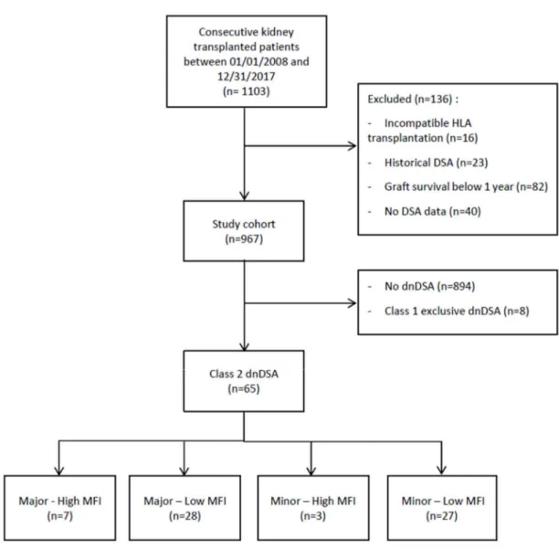
VIII. Results
A total of 1065 patients received a kidney transplant at Grenoble University Hospital between January 1st of 2008 and December 31st of 2017; of these, 967 were included (figure 1), after excluding patients without available HLA antibody data or patients with a graft survival < 1 year. Of note, no dnDSA-positive patient had a graft survival < 1 year. Among these patients, 65 (6.7%) recipients developed a class 2 dnDSA (exclusive or associated with a class 1), and 8 recipients developed a class 1 exclusive dnDSA, with a total of 7.5% of recipients developing a dnDSA. Recipients developing a class 2 dnDSA were grouped according to the dnDSA initial MFI (with a 10000 threshold), and the major/minor characteristic. Seven recipients developed a Major-HighMFI dnDSA, 28 patients developed a Major-LowMFI dnDSA, 3 patients developed a Minor-HighMFI dnDSA and 27 patients developed a Minor-LowMFI dnDSA.
26 Figure 1: Flow-chart
General population characteristics
The population general characteristics are reported on table 1, grouped according the intensity/persistence features defined above. Recipients and donors ages were different across groups, patients developing a Major-lowMFI antibody being the youngest. Among patients developing a dnDSA, 29.2% had a pre-transplantation HLA sensitization, comparable to 24.3% of patients who did not develop a dnDSA. Most patients received an ATG-based
27 induction (87.2%). All patients were treated by a combination of tacrolimus, mycophenolate-mofetyl and steroids.
Table 1: Population general characteristics
Legend: DCD: donor with circulatory death (with Maastricht classification); yrs: years; SD: standard deviation
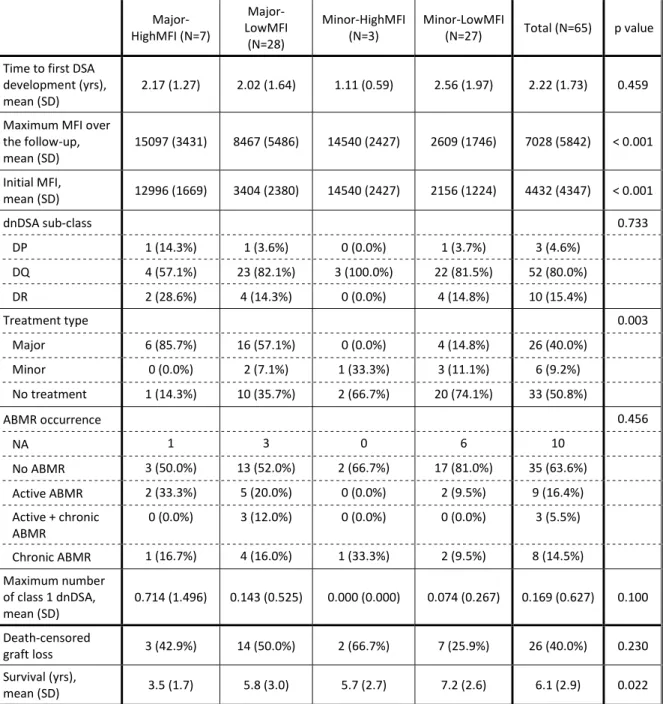
Among recipients developing a class 2 dnDSA (table 2), the time to first dnDSA occurrence was 2.22 years on average (SD 1.73), with no difference between groups (p=0.459). Most of the dnDSA were DQ (80%), with no difference between groups (p=0.733). Histological ABMR was proven in 30% of recipients developing a class 2 dnDSA, with an equal repartition of active and chronic active ABMR. Treatment administration was different
Class 2 dnDSA occurrence
No dnDSA (N=894) (N=959) Total value p Major-HighMFI (N=7) Major-LowMFI (N=28) Minor-HighMFI (N=3) Minor-LowMFI (N=27) Total (N=65) Donor type 0.763 DCD 1 / 2 0 (0.0%) 1 (3.6%) 0 (0.0%) 1 (3.7%) 2 (3.1%) 42 (4.7%) 44 (4.6%) DCD 3 1 (14.3%) 1 (3.6%) 0 (0.0%) 0 (0.0%) 2 (3.1%) 17 (1.9%) 19 (2.0%) Brain-death 5 (71.4%) (71.4%) 20 3 (100.0%) 22 (81.5%) 50 (76.9%) 683 (76.4%) 733 (76.4%) Living 1 (14.3%) 6 (21.4%) 0 (0.0%) 4 (14.8%) 11 (16.9%) 152 (17.0%) 163 (17.0%) Recipient age (yrs), mean (SD) 53.6 (20.5) 44.2 (12.1) 58.7 (13.5) 50.4 (16.0) 48.531 (15.116) 53.2 (14.4) 52.9 (14.5) 0.019
Donor age (yrs)
mean (SD) (21.9) 55.5 (15.4) 45.1 67.6 (7.7) 51.1 (14.4) (16.170) 49.815 53.0(15.8) 52.8(15.9) 0.044 Pre-transplantation HLA sensitization 1 (14.3%) 7 (25.0%) 1 (33.3%) 10 (37.0%) 19 (29.2%) 217 (24.3%) 236 (24.6%) 0.587 ATG induction therapy, n (%) 7 (100.0%) 22 (78.6%) 3 (100.0%) 21 (80.8%) 53 (81%) 750 (87.0%) 803 (86.7%) 0.405 Missing 0 0 0 1 1 32 33 Death-censored graft loss, n (%) 3 (42.9%) 14 (50.0%) 2 (66.7%) 7 (25.9%) 26 (40.0%) 74 (8.3%) 100 (10.4%) <0.00 1 Survival (yrs), mean (SD) 3.5 (1.7) 5.8 (3.0) 5.6 (2.7) 7.1 (2.6) 6.141 (2.921) 5.6 (2.8) 5.6 (2.8) 0.020
28 between groups (p=0.003), but not the histological ABMR confirmation (p=0.456), nor the maximum number of class 1 dnDSA (p=0.1).
Table 2: Characteristics of the class 2 dnDSA
Major-HighMFI (N=7) Major-LowMFI (N=28) Minor-HighMFI
(N=3) Minor-LowMFI (N=27) Total (N=65) p value Time to first DSA
development (yrs),
mean (SD) 2.17 (1.27) 2.02 (1.64) 1.11 (0.59) 2.56 (1.97) 2.22 (1.73) 0.459
Maximum MFI over the follow-up, mean (SD) 15097 (3431) 8467 (5486) 14540 (2427) 2609 (1746) 7028 (5842) < 0.001 Initial MFI, mean (SD) 12996 (1669) 3404 (2380) 14540 (2427) 2156 (1224) 4432 (4347) < 0.001 dnDSA sub-class 0.733 DP 1 (14.3%) 1 (3.6%) 0 (0.0%) 1 (3.7%) 3 (4.6%) DQ 4 (57.1%) 23 (82.1%) 3 (100.0%) 22 (81.5%) 52 (80.0%) DR 2 (28.6%) 4 (14.3%) 0 (0.0%) 4 (14.8%) 10 (15.4%) Treatment type 0.003 Major 6 (85.7%) 16 (57.1%) 0 (0.0%) 4 (14.8%) 26 (40.0%) Minor 0 (0.0%) 2 (7.1%) 1 (33.3%) 3 (11.1%) 6 (9.2%) No treatment 1 (14.3%) 10 (35.7%) 2 (66.7%) 20 (74.1%) 33 (50.8%) ABMR occurrence 0.456 NA 1 3 0 6 10 No ABMR 3 (50.0%) 13 (52.0%) 2 (66.7%) 17 (81.0%) 35 (63.6%) Active ABMR 2 (33.3%) 5 (20.0%) 0 (0.0%) 2 (9.5%) 9 (16.4%) Active + chronic ABMR 0 (0.0%) 3 (12.0%) 0 (0.0%) 0 (0.0%) 3 (5.5%) Chronic ABMR 1 (16.7%) 4 (16.0%) 1 (33.3%) 2 (9.5%) 8 (14.5%) Maximum number of class 1 dnDSA, mean (SD) 0.714 (1.496) 0.143 (0.525) 0.000 (0.000) 0.074 (0.267) 0.169 (0.627) 0.100 Death-censored graft loss 3 (42.9%) 14 (50.0%) 2 (66.7%) 7 (25.9%) 26 (40.0%) 0.230 Survival (yrs), mean (SD) 3.5 (1.7) 5.8 (3.0) 5.7 (2.7) 7.2 (2.6) 6.1 (2.9) 0.022
29
dnDSA panorama
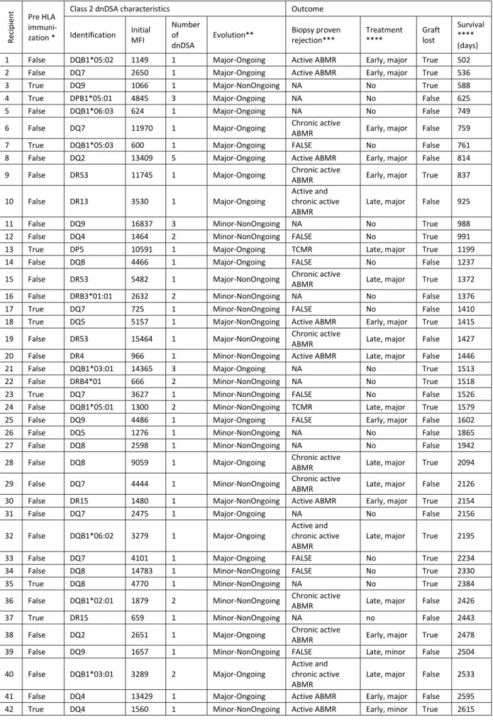
For each recipient developing a class 2 dnDSA, the immunodominant dnDSA and the graft outcome is described of table 3, ordered by the graft survival. The timelines (figure 2) illustrate graft outcomes: class 2 dnDSA timeline is observed according to the time post transplantation, among recipients presenting a death-censored-graft loss (above) and recipients without graft-loss (below). Some recipients experienced a short lasting dnDSA that however resulted in graft loss (eg recipient 12, 42), while others experienced a long lasting dnDSA without graft loss (eg 53, 45). In case of graft loss (above), this occurred either early after dnDSA occurrence (eg recipients 2, 3 or 21), or after several years (eg recipients 51, 51, 58 or 59).
When considering treatment options (figure 2.B), major treatments tended to be administered to recipients with the shortest survivals, and recipients with the longest survival did not receive any treatment, or a minor one.
30 Table 3: Characteristics of recipients developing a class 2 dnDSA
Re ci pi en t Pre HLA immuni-zation *
Class 2 dnDSA characteristics Outcome
Identification Initial MFI Number of
dnDSA Evolution**
Biopsy proven
rejection*** Treatment **** Graft lost
Survival **** (days) 1 False DQB1*05:02 1149 1 Major-Ongoing Active ABMR Early, major True 502 2 False DQ7 2650 1 Major-Ongoing Active ABMR Early, major True 536 3 True DQ9 1066 1 Major-NonOngoing NA No True 588 4 True DPB1*05:01 4845 3 Major-Ongoing NA No False 625 5 False DQB1*06:03 624 1 Major-Ongoing NA No False 749 6 False DQ7 11970 1 Major-Ongoing Chronic active ABMR Early, major False 759 7 True DQB1*05:03 600 1 Major-Ongoing FALSE No False 761 8 False DQ2 13409 5 Major-Ongoing Active ABMR Early, major False 814 9 False DR53 11745 1 Major-Ongoing Chronic active ABMR Early, major True 837
10 False DR13 3530 1 Major-Ongoing Active and chronic active
ABMR Late, major False 925 11 False DQ9 16837 3 Minor-NonOngoing NA No True 988 12 False DQ4 1464 2 Minor-NonOngoing FALSE No True 991 13 True DP5 10591 1 Major-Ongoing TCMR Late, major True 1199 14 False DQ8 4466 1 Major-Ongoing FALSE No False 1237 15 False DR53 5482 1 Major-NonOngoing Chronic active ABMR Late, major True 1372 16 False DRB3*01:01 2632 2 Minor-NonOngoing NA No False 1376 17 True DQ7 725 1 Minor-NonOngoing FALSE No False 1410 18 True DQ5 5157 1 Major-NonOngoing Active ABMR Early, major True 1415 19 False DR53 15464 1 Major-NonOngoing Chronic active ABMR Late, major False 1427 20 False DR4 966 1 Minor-NonOngoing Active ABMR Late, major False 1446 21 False DQB1*03:01 14365 3 Major-Ongoing NA No True 1513 22 False DRB4*01 666 2 Minor-NonOngoing NA No True 1518 23 True DQ7 3627 1 Minor-NonOngoing FALSE No False 1526 24 False DQB1*05:01 1300 2 Minor-NonOngoing TCMR Late, major True 1579 25 False DQ9 4486 1 Major-Ongoing FALSE Early, major False 1602 26 False DQ5 1276 1 Minor-NonOngoing NA No False 1865 27 False DQ8 2598 1 Minor-NonOngoing NA No False 1942 28 False DQ8 9059 1 Major-Ongoing Chronic active ABMR Late, major True 2094 29 False DQ7 4444 1 Minor-NonOngoing Chronic active ABMR Late, major False 2126 30 False DR15 1480 1 Major-NonOngoing Active ABMR Early, major True 2154 31 False DQ7 2475 1 Major-Ongoing NA No False 2156 32 False DQB1*06:02 3279 1 Major-Ongoing Active and chronic active
ABMR Late, major True 2195 33 False DQ7 4101 1 Major-Ongoing FALSE No True 2234 34 False DQ8 14783 1 Minor-NonOngoing FALSE No True 2330 35 True DQ8 4770 1 Minor-NonOngoing NA No True 2384 36 False DQB1*02:01 1879 2 Minor-NonOngoing Chronic active ABMR Late, major False 2426 37 True DR15 659 1 Minor-NonOngoing NA no False 2443 38 False DQ2 2651 1 Major-Ongoing Chronic active ABMR Early, major True 2478 39 False DQ9 1657 1 Minor-NonOngoing FALSE Late, minor False 2504 40 False DQB1*03:01 3289 2 Major-Ongoing Active and chronic active
ABMR Late, major False 2533 41 False DQ4 13429 1 Major-Ongoing Active ABMR Early, major False 2595 42 True DQ4 1560 1 Minor-NonOngoing Active ABMR Early, minor True 2615
31 43 True DQ5 864 1 Major-Ongoing NA No False 2679 44 False DQ3 3420 1 Minor-NonOngoing FALSE Early, minor False 2821 45 False DQ2 3166 2 Major-Ongoing TCMR Late, major False 2890 46 True DQB1*03:01 2275 1 Minor-NonOngoing NA No True 2895 47 True DQ6 12000 2 Minor-NonOngoing Chronic active ABMR Early, minor False 2912 48 False DQ7 9549 1 Major-Ongoing Active ABMR Early, major False 3019 49 True DR53 544 1 Major-Ongoing Chronic active ABMR Early, major False 3213 50 False DQ4 2508 3 Major-NonOngoing TCMR No True 3287 51 False DPB1*04:02 1304 1 Minor-Ongoing NA No True 3298 52 True DQ6 2919 1 Minor-NonOngoing NA No False 3340 53 False DQB1*02:01 2500 2 Major-Ongoing Borderline TCMR Late, major False 3428 54 True DQ6 1176 1 Major-Ongoing NA No False 3526 55 False DQB1*05:03 1969 1 Minor-NonOngoing NA No False 3584 56 True DQ2 4443 2 Minor-NonOngoing NA No False 3595 57 False DQB1*03:01 2120 2 Minor-NonOngoing FALSE No False 3628 58 False DQB1*06:03 7579 1 Major-NonOngoing Chronic active ABMR Early, major True 3703 59 False DQ2 3084 1 Major-Ongoing FALSE Early, minor True 3729 60 True DQB1*02:02 3000 1 Minor-NonOngoing FALSE No False 3844 61 False DQ7 3961 2 Major-Ongoing FALSE Late, minor True 3883 62 False DQ2 552 1 Minor-NonOngoing NA No False 3889 63 True DQ5 1314 1 Minor-NonOngoing NA No False 3929 64 False DQB1*02 1472 1 Minor-NonOngoing NA No False 3933 65 False DQ5 3204 1 Minor-NonOngoing NA No False 4002
Legend:
* pre HLA sensitization: no donor specific HLA antibody former to transplantation ** NA: no biopsy performed after dnDSA occurrence / FALSE: no ABMR or TCMR
*** early treatment: administered in the first three months post dnDSA development / No: no specific treatment administered
32
Figure 2: class 2 dnDSA timelines
A: According to the initial MFI and the graft loss
Upper panel: timeline until death-censored graft loss (red ring)
33
B: According to the treatment administered and the DSA sub-class (DP, DQ, DR)
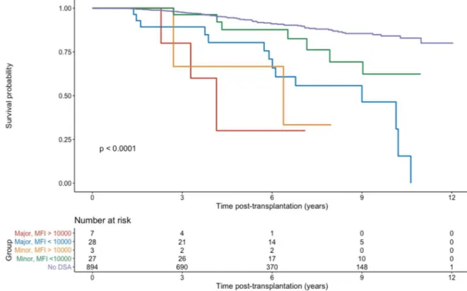
Outcome over all the study population (dnDSA occurrence or not)
In the whole study population, including dnDSA negative patients and the four previously defined groups (figure 3), there was a significant difference between groups (p<0.001). A total of 10.4% of recipients lost their graft, with a significant difference between groups (p<0.001): 40% of recipients developing a dnDSA lost their graft VS 8.3% of recipients without dnDSA.
34 Figure 3: Death-censored graft survival over the whole study population
Graft survival: associated covariates
Univariate and multivariate model
In univariate survival analyses, covariates significantly associated with DCGS were occurrence of a class 1 dnDSA (p=0.007), high initial MFI (>10000) (p=0.085) and major or minor dnDSA (p=0.091). On the other hand, recipient age (>65 years, p=0.191), donor age (>65 years, p=0.214), HLA pre-transplantation sensitization (non DSA, p=0.46), maximum MFI over the follow-up (>10000, p=0.559), major or minor treatment administration (p=0.498), and DP/DQ/DR sub-class (p=0.23) were not associated with DCGS. High initial MFI, major or minor dnDSA and occurrence of a class 1 dnDSA were therefore included in a multivariate Cox regression survival model: major or minor dnDSA (p=0.009) and class 1 dnDSA occurrence
35 (p=0.001) remained independently and significantly associated with death-censored graft-survival.
Outcome according to the treatment
Comparing recipients developing a class 2 dnDSA (figure 4), there was no difference in DCGS between major, minor or no treatment administered (p=0.37). There was a trend toward a worse outcome for recipients who received a major treatment. Concerning the raw graft survival (i.e. non death-censored, figure 5), there was no difference between the treatment groups either (p=0.25): treatment administration or intensity did not induce more death.
36 Figure 5: dnDSA raw-survival according to the treatment administered
When observing the class 2 dnDSA of recipients receiving a major treatment (table 4), most of them (65%) presented a major and ongoing dnDSA, among which 42% lost their graft; when developing a major and not ongoing dnDSA (20% of recipients treated with a major treatment), despite the disappearance of the dnDSA, all recipients lost their graft. For recipients treated with a minor treatment, the majority (66%) developed a minor and not ongoingdnDSA, with 25% of graft lost. For recipients who were not treated, most of them (64%) developed a minor and not ongoing dnDSA with 29% of graft lost. Most of the others developed a major and ongoing dnDSA (27%), without frequent graft loss (22%). In a nutshell, treatment options seem to be associated with the estimated severity of the clinical case.
37 Table 4: class 2 dnDSA evolution according to the treatment administered
Major treatment n= 26 (40%)
Type of dnDSA n (%) Graft loss n (%) Major and Ongoing 17 (65%) 7 (42%) Major and not Ongoing 5 (20%) 4 (80%) Minor and Ongoing 0 0 Minor and not Ongoing 4 (15%) 1 (25%) All dnDSA 26 12 (46%) Minor treatment
n= 6 (9%)
Type of dnDSA n (%) Graft loss n (%) Major and Ongoing 2 (33%) 2 (100%) Major and not Ongoing 0 0 Minor and Ongoing 0 0 Minor and not Ongoing 4 (66%) 1 (25%) All dnDSA 6 3 (50%) No treatment
n= 33 (51%)
Type of dnDSA n (%) Graft loss n (%) Major and Ongoing 9 (27%) 2 (22%) Major and not Ongoing 2 (6%) 2 (100%) Minor and Ongoing 1 (3%) 1 (100%) Minor and not Ongoing 21 (64%) 6 (29%) All dnDSA 33 11 (33%)
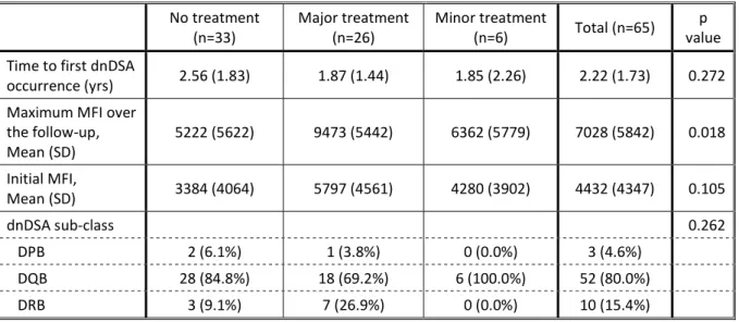
Time to first dnDSA occurrence was not different between the groups of treatment (p=0.272, table 5). The initial MFI was not different between groups either (p=0.105), but the mean initial MFI was numerically higher among recipients treated with a major treatment. On the other hand, the maximum MFI over the follow up was significantly higher for recipients treated with a major treatment, and lower among non-treated recipients. The dnDSA sub-class was not different between treatment groups (p=0.262).
38 Table 5: recipients characteristics according to the treatment administered
No treatment
(n=33) Major treatment (n=26) Minor treatment (n=6) Total (n=65) value p Time to first dnDSA
occurrence (yrs) 2.56 (1.83) 1.87 (1.44) 1.85 (2.26) 2.22 (1.73) 0.272 Maximum MFI over
the follow-up, Mean (SD) 5222 (5622) 9473 (5442) 6362 (5779) 7028 (5842) 0.018 Initial MFI, Mean (SD) 3384 (4064) 5797 (4561) 4280 (3902) 4432 (4347) 0.105 dnDSA sub-class 0.262 DPB 2 (6.1%) 1 (3.8%) 0 (0.0%) 3 (4.6%) DQB 28 (84.8%) 18 (69.2%) 6 (100.0%) 52 (80.0%) DRB 3 (9.1%) 7 (26.9%) 0 (0.0%) 10 (15.4%) Legend: yrs: years; SD: Stand Deviation
Outcome according to DP, DQ or DR type
Outcome according to dnDSA type is represented of table 6. Most of the class 2 dnDSA were DQ (80%). Among them, 29% developed an ABMR, with an initial MFI higher than 10000 in 20% of patients developing an ABMR VS 10% in those without ABMR (p=0.39). Most of the recipients receiving a major treatment developed an ABMR (72%) ; no patient with a biopsy-proven ABMR was left untreated, but 9 out of 24 treated patients had a non-biopsy related indication for treatment. The graft loss risk was similar with or without proven ABMR (50 VS 43%, p=0.779), possibly mitigated by our treatment strategies.
There was no DCGS difference between the DSA type (38% for DQ, 66% for DP and 40% for DR). There was also no difference between ABMR and DSA type. A very small number of recipients developed a DP dnDSA (n=3), limiting analysis. Two third of them lost their graft: one after TCMR occurrence, and one without biopsy performed post dnDSA occurence.
39 Table 6: Recipients outcome according to the class 2 dnDSA sub-class
DPB n= 3 n= 52 DQB n= 10 DRB Pre HLA immunization, n (%) 2 (66%) 15 (29%) 2 (20%) Major dnDSA, n (%) 3 (100%) 27 (52%) 6 (60%) Ongoing dnDSA, n (%) 3 (100%) 23 (44%) 3 (30%) Number of dnDSA, mean 1.4 1.4 1.3
Biopsy data ABMR ABMR No NA ABMR ABMR No NA ABMR ABMR No NA n (%) 0 (33%) 1 (66%) 2 (31%) 16 (35%) 18 (35%) 18 (70%) 7 0 (30%) 3 Initial MFI > 10000, n (%) 0 (100%) 1 0 (25%) 4 (5%) 1 (11%) 2 (28%) 2 0 0 Treatment Major n (%) 0 (100%) 1 0 (87%) 14 (22%) 4 0 (100%) 7 0 0 Minor n (%) 0 0 0 2 (12%) 4 (22%) 0 0 0 0 No treatment n (%) 0 0 2 (100%) 0 10 (56%) 18 (100%) 0 0 3 (100%) Graft loss, n (%) 0 1 (100%) (50%) 1 (50%) 8 (39%) 7 (28%) 5 (43%) 3 0 (33%) 1 2 (66%) 20 (38%) 4 (40%)
Legend: NA: no biopsy after dnDSA occurrence
40 Outcome according to the dnDSA persistence from dnDSA occurrence
Observing recipients according to the persistence of the dnDSA over the follow-up (defined as major or minor), there was no statistical DCGS difference (figure 7, p=0.085). However, there was a trend to a better outcome among recipients with a minor dnDSA.
Figure 7: death-censored graft survival according to the class 2 persistence, from time post DSA occurence
Outcome according to the initial MFI and the persistence of the dnDSA, from dnDSA occurrence Death-censored graft survival was compared between the 4 groups of recipients developing a dnDSA: Major-HighMFI, Major-LowMFI, Minor-HighMFI, and Minor-LowMFI, from dnDSA occurrence (figure 8). In this analysis, there was a nearly significant difference between the four groups. The association between the initial DSA MFI and its persistence over
41 time allows to stratify patients, with a high initial MFI and persistence over time pointing to the most toxic dnDSA.
42
IX. Discussion
In our cohort, 73 (7.5%) of kidney transplant recipients developed a dnDSA: 65 were class 2 dnDSA and 8 exclusively class 1 dnDSA. Class 2 dnDSA occured after 2.22 years on average, inducing 30% biopsy-proven ABMR and 40% graft loss. In univariate analysis, for class 2 dnDSA, the donor age, an associated class 1 dnDSA, a high initial MFI (>10000) and the persistence of the dnDSA over the follow-up were associated with death-censored graft survival. In a multivariate model, the persistence of the dnDSA over the follow-up and the associated class 1 dnDSA occurrence remained independent predictors of DCGS.
A total of 49% of KTR developing a class 2 dnDSA benefited from a treatment. The intensity of the treatment tended to be more important when the dnDSA had a high initial MFI, a high maximum MFI, a long lasting of the dnDSA over the follow-up or a persistence on the last sample, or an ABMR; inversely most of the recipients who did not receive any treatment developed a dnDSA without these pejorative initial or evolutive data. This suggests a selection bias for the treatment administered according to the initial characteristics of the dnDSA, its evolution and the biopsy data. Conforting this idea, the death-censored graft survival was not different between groups of treatments: treating the most pejorative evolutive dnDSA with a more intensive treatment may have avoided graft failures. There was also no significant difference in the raw graft survival between treatment groups: treatment administration and intensity did not induce more death.
Only 7.5% of recipients developed a dnDSA, most of them in class 2 (6.7%). Most recipients received an ATG-based induction and were treated by an association of tacrolimus and MMF, with a target trough tacrolimus level of 5-8 µg/l beyond one-month post-transplantation. In other studies with an equivalent immunological risk, 11 to 14%of recipients
43 developed dnDSA 5,12,25. One study assessed prospectively the dnDSA development according to the immunological risk; among the low immunological risk group (comparable to our cohort), 16% developed a dnDSA, 8% an ABMR and 24% a TCMR26. Our immunosuppressive strategy might have limited the risk of HLA sensitization.
DQ antibodies were the most frequent class 2 dnDSA, in line with previously described kidney transplant cohorts 27–29. However, we did not identify more ABMR or graft loss among DQ dnDSA compared to DR and DP dnDSA, and the death-censored graft survival was not different between DR, DQ and DP dnDSA. However, a limited recipients number developed no-DQ class 2 dnDSA.
Categorising class 2 dnDSA according to the initial MFI and its persistence over the follow-up allowed us to stratify patients according to death-censored graft survival. Unsurprisingly, dnDSA with a high initial MFI and persisting over the follow-up presented the most pejorative graft survival, and the ones with a low initial MFI and a short persistence over the follow-up had the longest survival, with a statistical difference between groups. The graft survival of recipients with a low initial MFI and a non-persistent class 2 dnDSA was close to recipients without dnDSA. The increased risk for recipients with a high initial MFI is probably related to the C1q and C3d binding activities of such dnDSA, which were previously shown to be associated with ABMR occurrence and graft survival23.
There are several limits to our study: the retrospective data collection, even if exhaustive data from dnDSA outcome were accessible; the lack of a standardized treatment strategy when a dnDSA developed, the lack of systematic biopsy in patients developing a dnDSA. However, hard outcomes such as DCGS and raw graft survival remain valid outcomes
44 of clinical importance. A limited number of patients developed a class 1 exclusive dnDSA, preventing further interpretation for this dnDSA class.
In conclusion, in our experience the development of a dnDSA is a rare but deleterious event. Close oversight of kidney transplant recipients developing a class 2 dnDSA is necessary to discuss a biopsy indication and a potential treatment, especially in case of dnDSA 1/ persisting over time , 2/ associated with a simultaneous class 1 dnDSA. Stratification of the risk associated with a dnDSA might be improved using these dynamic criteria.
45
X. Références
1. Tait BD. Detection of HLA antibodies in organ transplant recipients - triumphs and challenges of the solid phase bead assay. Frontiers in Immunology. 2016;7(DEC). doi:10.3389/fimmu.2016.00570
2. PORT, Friedrich K., WOLFE, Robert A., MAUGER, Elizabeth A. et al. Comparison of survival probabilities for dialysis patients vs cadaveric renal transplant recipients. Jama. 1993;270(11):1339-1343.
3. EVANS, Roger W., MANNINEN, Diane L., GARRISON JR, Louis P. et al. The quality of life of patients with end-stage renal disease. New England journal of medicine. 1985;312(9):553-559.
4. Hourmant M, Cesbron-Gautier A, Terasaki PI, et al. Frequency and clinical implications of development of donor-specific and non-donor-specific HLA antibodies after kidney transplantation. Journal of the American Society of Nephrology. 2005;16(9):2804-2812. doi:10.1681/ASN.2004121130
5. Wiebe C, Gibson IW, Blydt-Hansen TD, et al. Evolution and clinical pathologic correlations of de novo donor-specific HLA antibody post kidney transplant. American Journal of Transplantation. 2012;12(5):1157-1167. doi:10.1111/j.1600-6143.2012.04013.x
6. Loupy A, Hill GS, Jordan SC. The impact of donor-specific anti-HLA antibodies on late kidney allograft failure. Nature Reviews Nephrology. 2012;8(6):348-357. doi:10.1038/nrneph.2012.81
46 7. Legris T, Picard C, Todorova D, et al. Antibody-dependent NK cell activation is associated with late kidney allograft dysfunction and the complement-independent alloreactive potential of donor-specific antibodies. Frontiers in Immunology. 2016;7(AUG). doi:10.3389/fimmu.2016.00288
8. Terasaki PI, Cai J. Humoral theory of transplantation. Current Opinion in Immunology. 2005;17(5):541-545. doi:10.1016/j.coi.2005.07.018
9. Viglietti D, Loupy A, Vernerey D, et al. Value of donor-specific anti-HLA antibody monitoring and characterization for risk stratification of kidney allograft loss. Journal of the American Society of Nephrology. 2017;28(2):702-715. doi:10.1681/ASN.2016030368
10. Terasaki PI, Ozawa M. Predicting Kidney Graft Failure by HLA Antibodies: A Prospective Trial. American Journal of Transplantation. 2004;4(3):438-443. doi:10.1111/j.1600-6143.2004.00360.x
11. Lachmann N, Terasaki PI, Budde K, et al. Anti-human leukocyte antigen and donor-specific antibodies detected by luminex posttransplant serve as biomarkers for chronic rejection of renal allografts. Transplantation. 2009;87(10):1505-1513. doi:10.1097/TP.0b013e3181a44206
12. Wiebe C, Gibson IW, Blydt-Hansen TD, et al. Rates and determinants of progression to graft failure in kidney allograft recipients with de novo donor-specific antibody. American Journal of Transplantation. 2015;15(11):2921-2930. doi:10.1111/ajt.13347
47 13. Solez K, Colvin RB, Racusen LC, et al. Banff ’05 meeting report: Differential diagnosis of chronic allograft injury and elimination of chronic allograft nephropathy ('CAN’). American Journal of Transplantation. 2007;7(3):518-526. doi:10.1111/j.1600-6143.2006.01688.x
14. Haas M, Loupy A, Lefaucheur C, et al. The Banff 2017 Kidney Meeting Report: Revised diagnostic criteria for chronic active T cell–mediated rejection, antibody-mediated rejection, and prospects for integrative endpoints for next-generation clinical trials. American Journal of Transplantation. 2018;18(2):293-307. doi:10.1111/ajt.14625
15. Haas M. The Revised (2013) Banff Classification for Antibody-Mediated Rejection of Renal Allografts: Update, Difficulties, and Future Considerations. American Journal of Transplantation. 2016;16(5):1352-1357. doi:10.1111/ajt.13661
16. Loupy A, Haas M, Roufosse C, et al. The Banff 2019 Kidney Meeting Report (I): Updates on and clarification of criteria for T cell– and antibody-mediated rejection. American Journal of Transplantation. 2020;(February):2318-2331. doi:10.1111/ajt.15898
17. Sellarés J, de Freitas DG, Mengel M, et al. Understanding the causes of kidney transplant failure: The dominant role of antibody-mediated rejection and nonadherence. American Journal of Transplantation. 2012;12(2):388-399. doi:10.1111/j.1600-6143.2011.03840.x
18. Lefaucheur C, Nochy D, Andrade J, et al. Comparison of combination plasmapheresis/IVIg/Anti-CD20 versus high-dose ivig in the treatment of
48 antibody-mediated rejection. American Journal of Transplantation. 2009;9(5):1099-1107. doi:10.1111/j.1600-6143.2009.02591.x
19. BILLING, Heiko, RIEGER, Susanne, SÜSAL, Caner et al. IVIG and rituximab for treatment of chronic antibody-mediated rejection: a prospective study in paediatric renal transplantation with a 2-year follow-up. Transplant International. 2012;25(11):1165-1173.
20. KULKARNI, S., KIRKILES-SMITH, N. C., DENG, Y. H. et al. Eculizumab therapy for chronic antibody-mediated injury in kidney transplant recipients: A pilot randomized controlled trial. American Journal of Transplantation. 2017;17(3):682-691.
21. CHOI, J., AUBERT, O., VO, A. et al. Assessment of Tocilizumab (Anti–Interleukin-6 Receptor Monoclonal) as a Potential Treatment for Chronic Antibody-Mediated Rejection and Transplant Glomerulopathy in HLA-Sensitized Renal Allograft Recipients. American Journal of Transplantation. 2017;17(9):2381-2389.
22. ESKANDARY, Farsad, REGELE, Heinz, BAUMANN, Lukas et al. A randomized trial of bortezomib in late antibody-mediated kidney transplant rejection. Journal of the American Society of Nephrology. 2018;29(2):591-605.
23. Lee H, Han E, Choi AR, et al. Clinical impact of complement (C1q, C3d) binding de Novo donor-specific HLA antibody in kidney transplant recipients. PLoS ONE. 2018;13(11):1-18. doi:10.1371/journal.pone.0207434
24. Lefaucheur C, Viglietti D, Bentlejewski C, et al. IgG Donor-Specific Anti-Human HLA Antibody Subclasses and Kidney Allograft Antibody-Mediated Injury. Journal
49 of the American Society of Nephrology. 2016;27(1):293-304. doi:10.1681/ASN.2014111120
25. Wiebe C, Rush DN, Nevins TE, et al. Class II Eplet Mismatch Modulates Tacrolimus Trough Levels Required to Prevent Donor-Specific Antibody Development. Journal of the American Society of Nephrology. 2017;28(11):3353-3362. doi:10.1681/ASN.2017030287
26. Wan S. Development and Outcomes of De Novo Donor Specific Antibodies in Low, Moderate and High Immunological Risk Kidney Transplant Recipients. Journal of Substance Abuse Treatment. 2020;13(3):287-288. doi:10.1016/s0740-5472(96)90021-5
27. Willicombe M, Brookes P, Sergeant R, et al. De novo DQ donor-specific antibodies are associated with a significant risk of antibody-mediated rejection and transplant glomerulopathy. Transplantation. 2012;94(2):172-177. doi:10.1097/TP.0b013e3182543950
28. Devos JM, Gaber AO, Knight RJ, et al. Donor-specific HLA-DQ antibodies may contribute to poor graft outcome after renal transplantation. Kidney International. 2012;82(5):598-604. doi:10.1038/ki.2012.190
29. Lee H, Min JW, Kim J il, et al. Clinical significance of HLA-DQ antibodies in the development of chronic antibody-mediated rejection and allograft failure in kidney transplant recipients. Medicine (United States). 2016;95(11):1-9. doi:10.1097/MD.0000000000003094