Patients
with
Myocardial
Infarction
and
678 Myocardial Infarction and Normal Coronary Arteriogram (Legrand et a!)
Normal
Coronary
Arteriogram*
V. Legrand, M.D.; M. Deliege, M.D.; L. Henrard, M.D.;
1.
Boland, M.D.; and H. Kulbertus, M.D.Eighteen patients who survived an acute myocardial infarction were found to have a normal coronary arteri-ogram. Seven patients were younger than 35 years and
six were female. The myocardial infarction was
non-transmural in 1 1 cases. The mean follow-up was 21.6 months. Eleven patients developed residual chest pain at rest early after myocardial infarction. One, treated
by beta-blockers, suffered a recurrent myocardial
infarc-tion. Eight became asymptomatic, and two improved under antispastic therapy. Another patient developed a severe form of variant angina three m9nths after myo-cardial infarction; she died following plexectomy.
Final-M
yocardial infarction is generally associated withobstructive coronary artery disease and
thought to l)e the result of a permanent or transient
vascular O1)strUctiOfl leading to myocardial necrosis.1
The development of selective coronary arteriography
has revealed, however, that myocardial infarction
may occur, in the absence of atherosclerotic
obstruc-tive coronary lesions, in patients with patent
cor-onary arteries.2’4 In a recent prospective study,
Betriu et al’2 found normal coronary arteries in 3
percent of patients surviving an acute myocardial
infarction; it is thus a rare finding. Furthermore, the
identification of patients with myocardial infarction
and normal coronary arteries seems difficult as their
epidemiologic profile is similar to that of patients
with atherosclerotic OCCSS except for their
young-er age.4’9’4 As a consequence, the prognosis and
follow-up is not better known than the
physiopath-ologic process which leads to myocardial infarction
in this subset of patients. The purpose of the
pres-ent study was to evaluate the clinical profile of
pa-tients with myocardial infarction and normal
cor-onary arteries; to discuss the possible causes of
infarction in this group of patients; and to describe
their follow-up and long-term prognosis.
Patient Selection
METHODS
From January 1978 to July 1981, 1,727 patients were
ad-#{176}From the University of Liege School of Medicine,
De-partment of Cardiology, Liege, Belgium.
Manuscript received April 13; revision accepted July 13.
Reprint requests: Dr. Kulhertu.i, Cardiology, University Hospital, 66 Bd de la Constitution, Liege, Belgium B-4020
ly, two patients experienced rare episodes of angina at
rest. The stress ECG was negative in all cases.
Provoca-tive test for spasm was positive in three out of nine pa-tients. Diffuse narrowing associated with chest pain was demonstrated in two patients at angiography. Thus,
myocardial infarction and subsequent normal coronary
angiogram are mainly found in young female patients, and infarction is often nontransmural. Clinical evidence of vasospastic phenomena and increased vasomotor tone are found in most patients. Whenever residual chest pain is controlled by antispastic therapy, the follow-up course seems benign.
Illitte(l to the coronary care unit with a diagnosis of acute
niyocardial infarction. They met the fo1loving criteria: (1
history of prolonged chest pain; (2 ) typical EGG changes, and ( 3 ) characteristic rise and fall of serum enzyme values
(
creatine phosphokinase {CPK], glutamic oxaloacetictrans-aminase [SGOTJ, and lactic dehydrogenase [LDH] ). The
(liagnosis of subendocardial infarction ‘as made if the ECG
shoved persistent ischemic T waves or persistent ST
seg-ment depression lasting longer than 48 hours without
sub-sequent development of Q waves; the diagnosis of
trans-mural infarction included the first criteria but also
required the evolution of Q waves indicative of transmural
necrosis (0.04 s duration and amplitude greater than 25
percent of the succeeding R wave). A total of 236 patients (13M percent) died during hospitalization. Some 310 of
the survivors under 65 years of age ( 28.3 percent ) were
5ul)sequelltly submitte(l to coronary arteriograpliy.
Angiography was performed for one or more of the
fol-lowing reasons: (1) residual ischemia demonstrated by typ-ical angina pectoris, or positive exercise EGG and/or stress
thallium isehemia (280 patients ); (2) age under 35 (18
patients ); (3 ) subendocardial infarction (86 patients);
(
4) malignant arrhythmias in the posthospital phase ( yen-tricular tachycardia in three cases and frequent multifocalventricular beats or doublets at exercise in two cases).
A normal coronary angiogram was obtained in 18 patients
‘ho constitute the present study group. Their distribution
among the patients sul)mitted to angiography is given on Table 1.
Clinical Evaluation
On admission, each patient was interviewed on the circumstances of the event, past history of angina, smoking habits, and previous therapy. Serum glucose and lipids levels
were measured after a 12-hour fast, three months after
infarction. Patients with normal fasting glycemia were
fur-ther submitted to a 100 g sugar ingestion ‘with subsequent
dosage of both glycemia and insulinemia to evaluate their
glucose tolerance. Patients were considered hypertensive if
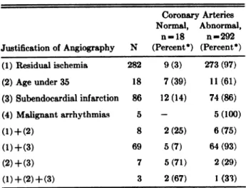
CHEST I 82 I 6 1 DECEMBER, 1982 679 Table 1-Patient Selection for Angiography
Justification of Angiography N Coronar Normal, n=18 (Percent ) v Arteries Abnormal, n=292 (Percent ) (1) Residual ischemia 282 9 (3) 273 (97) (2) Age under 35 18 7 (39) 11 (61) (3) Subendocardial infarction 86 12 (14) 74 (86) (4) Malignant arrhythmias 5 - 5 (100) (1)+(2) 8 2(25) 6(75) (1)+(3) 69 5(7) 64(93) (2)+(3) 7 5(71) 2(29) (1)+(2)+(3) 3 2(67) 1 (33)
4Pereentage of patients with normal or abnormal arteries
according to the justification of angiography.
was greater than 160/95 mm Hg. Patients were reviewed
at three months and every sixth month thereafter. The
mean duration of follow-up was 21.6 months with a range
of 6 to 42 months.
Angiography
Catheterization was performed using the Judkins
tech-nique at a mean delay of 70 days (range one to seven
months ) after infarction. The investigation included left
ventriculography in right anterior oblique projection,
selec-tive left coronary angiography ( six projections ), and
selec-tive right coronary angiography (four projections). Left
ventriculograms and coronary angiograms were read
mdc-pendently by at least two observers. Only patients with
normal or near normal angiograms ( irregularities of vessel wall with narrowings of less than 30 percent ) were included
in this study. Left ventricular ejection fractions were
calcu-lated using Simpson’s formula with the aid of an automated
system. Regional wall motion was assessed by comparison
of the left ventricular end-diastolic and end-systolic
silhou-ette according to the method of Ingels et al.’
Exercise Test
Three months after infarction, 16 patients were submitted
to a bicycle ergometer test. A progressive multistage
proto-col (increment of 10 W per minute) was applied. The
test was interrupted when fatigue, chest pain, shortness of
breath, fall in blood pressure, or malignant arrhythmias
de-veloped, or when the maximal heart rate for age was
reached. A standard 12-lead ECO was recorded before and
every minute throughout the exercise. The test was ended
five minutes after interruption of exercise. It was judged
positive in the presence of 1 mm or more horizontal or
downsloping ST depression for at least 0.08 s.
Provocative Test for Coronary Spasm
A provocative test for coronary spasm was performed in
the coronary care unit, according to the method described
by Waters et al,16 in seven patients (cases 5, 6, 8, 9, 10,
12, 13 ) and during catheterization according to the
tech-nique of Heupler,17 in two patients (cases 1 and 18). All
antispastic medications had been withdrawn for at least 12
hours with the exception of two patients ( cases 5 and 13)
who were receiving amiodarone, a drug recognized to have
a long half-life. The test was considered positive if the
patient developed typical chest pain relieved by
nitroglyc-erine and accompanied by ECG changes consisting of ST
elevation or depression of 1 mm or more from the control
tracing.
RESULTS Clinical Characteristics (Table 2)
Seven subjects
(
39 percent)
were younger than35 years and 6
(
33 percent) were female. Amongpa-tients submitted to angiography in whom
obstruc-tive coronary disease was found, only 11 out of 292
Table 2-Clinical Characteristics
Case
No. Age (yr)/Sex Hypertension
Diabetes (D) or Glucose Intolerance (Gi) Cholesterol, g/L Cigarettes Smoking, No/Day Previous Chest Pain and Duration Therapy Before Myocardial Infarction 1 43/F - Gi 2.03
So
+ 9 mu Pindolul nitrates 2 56/F + - 2.61 5 + 4 () Dihvdergotnmine 3 30/F - Gi 1.41 25 + 3 (laYs 4 28/M - Gi 2.21 25 + I (lay-5 49/M + Gi 1.65 60 + 4 (laVs A(’etvlahirvli( ari(l
6 19/M - - 1.59 0 + 1day -7 39/M - - 2.25 0 + 20 mo Nitrates, pr(’(lnioIon( 8 59/M - - 2.83 20 + 7 (laYs Nitrates 9 56/F - - 1.85 0 - - -10 30/M + - 1.95 25 + I (lay -I 1 47/M - Gi 2.04 35 + 4 mo Propranolol 12 22/M - - 2.53 0 + 3 mo Amphetamines 13 49/M - D 2.85 25 - - Glihencamide 14 60/M + - 1.87 25 + 2 (lays Chiorazepam 15 42/F - - 2.86 20 + 2 yr Dihydergotamine Nialamide 16 5l/M + Gi 2.51 5 + 5 yr -17 33/M - Gi 1.29 30 + I mo -18 34/M - - 2.19 25 - -
-680 Myocardial Infarction and Normal Coronary Arteriogram (Legrand et a!)
(4 percent) were younger than 35 (p <
0.001
), andonly 21
(
7 percent) were female (p < 0.001 ).Fif-teen patients described previous chest pain, often
atypical, occurring mainly at rest or after exercise.
In five instances, chest pain was first noted a few
hours or days before the infarct; it lasted for about
30 minutes in two cases
(
no 4 and 6). Patient 17 hadexperienced one episode of chest pain of 15-minute
duration one month before myocardial infarction.
One individual
(
case 1)
had chest pain at restassociated with ST depression in anterior leads
(
V2,v3,
V4)
and negative T wave in lateral leads(
V5,V6. D1, aVL) five days before the infarct.
Data Related to the Acute Phase of the Infarct
(Table 3)
In three instances, strenuous exercise preceded
the onset of the infarct. One further patient
(
case2) developed myocardial infarction during an
epi-sode of acute hypertension
(
270/140
mm Hg) ; laterinvestigations revealed a pheochromocytoma.
A high incidence of subendocardial infarcts (11
cases, 61 percent
)
was observed.The clinical course was not benign in all instances:
one patient
(
case 3)
was admitted a few minutesafter an episode of acute chest pain. She was found
in cardiorespiratory arrest with persistent
ventric-ular fibrillation and was resuscitated with success.
Three others suffered ventricular tachycardia or
fibrillation requiring electric countershock.
Eight patients described residual angina at rest 3
to 16 days after their myocardial infarction. The
ST changes during chest pain were noted in two
instances and consisted in ST depression in anterior
leads in patient 11 and ST elevation in inferior
leads in patient 14.
Follow-Up Data (Table 4)
Seven of the eight patients with early residual
angina received antispastic medications
(
nifedipine,verapamil, amiodarone, nitrates
)
. Five becameasymptomatic and two
(
8 and 11)
still describedchest pain at rest or exercise without demonstrable
EGG changes. The last patient
(
case 7)
with earlyresidual angina first received propranolol; 67 days
later, he developed a recurrent transmural anterior
myocardial infarction. Thereafter, he still had chest
pain despite nifedipine and amiodarone. This
pa-tient also had a hematologic disorder
(
Vaquezdis-ease
)
since the age of 20 years; he developedmyelo-Table 3-Characteristic Features of the Infarct *
Residual Case No. Circurn.stances of Infarct Location Peak CPK, III Peak SOOT, IU Shock Arrhvthmias Conduction Diturhanccs Ventricular Failure Angina (Delay from Infarction) 1 R(’St SE ant sept 627 195 No No No No No
2 Episode of SE inf hit 444 154 No VPBs No LVF K2 No
paroxysmal HTA
3 Mild stress TM ant lat 8866 1026 Yes
(CRA)
VF, VPBs No LVF K3 No
4 Rest TM ant 590 39 No No No No No
5 Mild stress TM ant lat 2229 396 No VPBs LBBB LVF K2 No
6 After stren- SE inf 1670 290 No No No No No
urnis stress
7 Rest SE ant lat 1040 136 No No No LVF K2 Yes 3 days
8 Rest TM inf 1046 199 No VF, VPBs No LVF K2 Yes 4 days
9 After stren- SE ant 196 55 No No No No No
uous stress
10 Sleep SE inf 533 101 No No No No Yes 16 days
11 Rest SE inf lat 1243 211 No VT, VPBs No No Yes 7 days
12 After stren- SE ant 700 73 No VPBs No LVF K2 No
UOUS stress
13 Rest TM inf 673 47 No No No No Yes 4 days
14 Sleep TM ant sept 3069 531 No VT, VPBs No LVF K2 Yes 3 days
15 Rest SE inf lat 312 51 No No No No No
16 Rest TM ant sept inf 1340 342 No VPBs No LVF K2 Yes 4 days
17 Rest SE sept 630 68 No No No No No
18 Rest SE inf 414 99 No VPBs No No Yes 7 days
4HTA indicates arterial hypertension; SE, subendocardial; TM, transmural; ant, anterior; sept, septal; lat, lateral; inf, inferior; CRA, cardiorespiratory arrest; VPB, ventricular premature beats; VF, ventricular fibrillation; VT, ventricular tachycardia;
LBBB, left bundle branch block; LVF, left ventricular failure; and Ki, K2, K3, functional class according to Killip’s classification
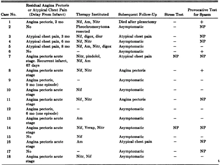
Case No.
Residual Angina Pectoris or Atypical Chest Pain
(Delay From Infarct) Therapy Instituted Subsequent Follow-Up Stress Test
Provocative lest for Spasm
1 Angina pectoris, 2 mo Nif, Am, Nitr Died after plexectomy - +
2 No Pheochromocytoma
resected
Asymptomatic - NP
3 Atypical chest pain, 3 mo Nif, digox, diur Atypical chest pain - NP
4 Atypical chest pain, 6 mo Nif, Nitr Asyniptomatic - NP
5 Atypical chest pain, 8 mo Nif, Am, Nitr, digox Asymptomatic -
-6 No - Asymptomatic - +
7 Angina pectoris acute Nitr, pindolol, Atypical chest pain NP NP
stage. Recurrent infarct, Nif, Am
67 days
8 Angina pectoris acute Nif, Nitr Angina pectoris - +
stage
9 Angina pectoris, - Asymptomatic -
-9 mo (one episode)
10 Angina pectoris acute Nif Asymptomatic -
-stage
11 Angina pectoris acute Nif, Nitr Angina pectoris - NP
stage
12 Angina pectoris, - Asymptoniatic -
-6 mo (one episode)
13 Angina pectoris acute Am Asymptomatic -
-stage
14 Angina pectoris acute Nif, Verap, Nitr Asymptomatic NP NP
stage
15 No Nif Asymptomatic -
-16 Angina pectoris acute Am Atypical chest pain - NP
stage
17 No - Asvrnptomatic - NP
18 Angina pectoris acute Nitr, Nif Asymptomatic -
-stage t
*Nif indicates nifedipine; Am, amiodarone; Nitr, nitrates; digox, digoxine; diur, diuretics; Verap, verapamil; and NP, n o
performed.
Table 4-Follow-Up and Results of Inves:igation
CHEST I 82 I 6 I DECEMBER, 1982 681
sclerosis and died 11 months after his first infarct.
Three other patients developed late recurrent
an-gina. In patient 1, it appeared at three months and
was associated with ST-segment elevation in the
inferior leads. The investigation showed a
hypo-kinetic anterior wall, normal coronary vessels but
spontaneous spasm of the right coronary artery.
Despite medical management
(
nifedipine,amio-darone, nitrates ), this patient remained
symptomat-ic, and 24 months later, a repeat catheterization was
performed. The coronary arteries were still normal,
and spasm of the right coronary artery was induced
by ergonovine. Plexectomy was performed, but the
patient died in intractable ventricular fibrillation.
Patient 9 experienced one episode of anginal pain at
rest with ST-depression at nine months. She
re-mained asymptomatic thereafter. Patient 12 was
re-admitted for typical anginal chest pain without ST
changes or enzyme elevation at six months.
Finally, patients 3, 4, and 5 described atypical
chest pain which improved or disappeared with
nifedipine.
Stress Test
A submaximal stress test was obtained in 16
pa-tients. It was negative in all instances; however, two
patients
(
8
and 11)
developed chest pain withoutST changes.
Provocative Test for Spasm
Seven patients had a provocative test for spasm in
the coronary care unit one day after catheterization.
Two
(
cases 6 and 8) had a positive response(
STdepression with chest pain
)
. Two tests wereper-formed during catheterization : one
(
case 1)
showedproximal obstruction of the right coronary artery
associated with angina and ST elevation and the
other
(
case 18) revealed severe, diffuse narrowingof the right coronary artery associated with chest
pain, without ST changes
(
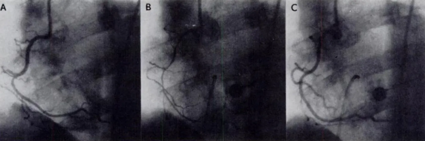
Fig 1).Angiography (Table 5)
FIvuuE 1. Angiography of the right coronary artery in left anterior oblique position obtained in
patient nr 18. A is before provocative test for spasm; B, after intravenous infusion of 0.1 mg
ergonovine; and C, after intracoronary injection of nitroglycerine. The injection of ergonovine produced a severe diffuse narrowing of the artery associated with chest pain. After injection of nitroglycerin, the artery dilated and chest pain disappeared.
682 Myocardial Infarction and Normal Coronary Arterlogram (Legrand et a!)
and showed minimal irregularities on the infarct
re-lated vessel in four cases (8, 11, 12, and 13), or on
another vessel in one case
(
16).The ventriculograms were normal in six cases (6,
9, 10, 13, 17, and 18) and showed contraction
ab-normalities corresponding to the infarct seen on the
EGG in the other cases.
No complication occurred during
catheteriza-tion. However, one patient
(
case 4)
developeddif-fuse narrowing of the left coronary artery with
angi-na pectoris and significant ST segment elevation
after the first intracoronary injection of contrast
me-dium, and patient 1 had at first catheterization
spon-taneous spasm of the right coronary artery with
ST-segment elevation. All signs of ischemia disappeared
after sublingual administration of nitroglycerin.
DiscussioN
Prevalence and Clinical Profile
The prevalence of patients with myocardial
in-farction who have normal or near normal coronary
arteries varies from 1 percent to 12 percent
accord-ing to the mode of investigation and the population
studied.214 Recent prospective studies12’18’19
indicate
that survivors of acute myocardial infarction have
normal coronary arteries in 1 percent to 3 percent
of cases. In our population, the prevalence of
pa-tients with normal vessels was 6 percent, but this
being a retrospective study, the patients submitted
to coronary arteriography were a selected subgroup
that was not necessarily representative of the whole
Table 5-Angiographic Findings*
Case
No. EGG
Tim MI
e from
(mo) LAD LCX RCA Ventriculogram EF, #{182}
1 SE anteroseptal 4 N N N Anterior hypokinesia 64
2 SE inferolateral 2 N N N Anterior hvpokinesia 58
3 TM anterolateral 6 N N N Anterior akinesia-apical dyskinesia 44
4 TM anterior 3 N N N Apical akinesia 56
5 TM anterolateral 7 N N N Anterolateral akinesia 36
6 SE inferior 1 N N N Normal 72
7 SE anterolateral 3 N N N Anterior akinesia 59
8 TM inferior 2 N N irreg <30% Inferior hypokinesia 60
9 SE anterior 1 N N N Normal 75
10 SE inferior 1 N N N Normal 55
1 1 SE inferolateral 3 irreg <30% N N Anteroapical hypokinesia 60
12 SE anterior 2 irreg <30% N N Anterior hypokinesia 48
13 TM inferior 1 N irreg <30% N Normal 70
14 TM anteroseptal 1 N N N Anterior akinesia 43
15 SE infero-lateral 1 N N N Anterolateral hypokinesia 61
16 TM anteroseptal 1 N irreg <30% N Anterior akinesia 50
17 SE septal 1 N N N Normal 63
18 SE inferior 1 N N N Normal 61
*MI indicates myocarclial infarction; LAD, left anterior descending coronary artery; RCA, right coronary artery; N, normal; SE, subendocardial; TM, transmural; LCX, left circumflex coronary artery; and EF, ejection fraction.
CHEST I 82 I 6 I DECEMBER, 1982 683
population recovering from acute myocardial
infarc-tion.
Our patients were characterized by their younger
age as compared to patients with myocardial
infarc-tion and obstructive coronary disease. This has also
been noted by others who found that 16 percent14
or even 45 percent12 of infarcts associated with
nor-mal coronary arteriogram occurred among patients
under 35 years of age. Another characteristic of the
studied subjects is the high proportion of women
(
33 percent), a feature which was not found inother series.9’10
Rosenblatt and Selzer9 pointed to the low
mci-dence of risk factors among patients with infarction
and normal angiogram, and Mc Kenna et al’#{176}
em-phasized the role of smoking. Among our patients,
the analysis of risk factors failed to show any salient
feature. Clinical, laboratory, and EGG features of
myocardial infarction were indistinguishable from
those of patients with obstructive atherosclerotic
coronary disease
(
Table 3)
. However, the incidenceof patients having nontransmural myocardial inf
arc-tion was higher than that in previously reported
similar studies9”2 and in the whole population of
patients hospitalized in our unit for acute
myocar-dial infarction
(
380/
1,727) (
p < 0.001). Limitations of Angiographic FindingsSince coronary angiographic findings obtained
one to seven months after the clinical episode of
in-farct may be different from those of the acute stage,
precise information concerning the anatomy of the
arteries at the time of infarction is lacking.
Regression of pre-existing obstructive
athero-sclerotic lesions has been demonstrated in animal
studies and might occur in man.2#{176}However, such
an evolution is slow and probably requires several
months as suggested by the study of Pichard et aP’
who found a significant reduction of obstructed
coronary vessels in the chronic phase of myocardial
infarction only after the sixth month. In our
popula-tion, with the exception of four patients, the
angio-gram was performed within the third month
follow-ing the myocardial infarction. Thus, regression of
pre-existing significant obstructive coronary lesions
seems unlikely.
Temporary occlusion of the coronary arteries has
been reported in cases of vasospastic angina, and
severe prolonged spasm may lead to myocardial
in-farction.m23 In such cases, angiography performed
after the acute event may reveal patent coronary
arteries.
Thromboembolic mechanisms are often associated
with acute myocardial infarction and obstruction or
narrowing of a coronary artery by a fresh thrombus
is frequently seen at autopsy of patients dying from
acute myocardial infarction.’ However,
recanaliza-tion of an initially obstructed vessel may be
ob-tamed by selective intracoronary thrombolysis with
streptokinase.24 Moreover, clot lysis may also occur
spontaneously as a result of physiologic
thronibo-lysis. Thus, the thrombus or emb()lus occluding a
coronary artery at the early stage of myocardial
in-farction may well disappear. In the absence of an
underlying atherosclerotic obstructive lesion , the
res-toration of the lumen may be complete.
Another limitation of the angiographic evaluation
is the misreading of coronary angiograins.
Occasion-ally, a coronary lesion may be masked or
underesti-mated, particularly if eccentric. In four patients, the
infarct-related vessel was not truly normal, and
minimal wall irregularities resulting in less than 30
percent obstruction were noted. Even if the lesion
was underestimated, it is unlikely that significant
narrowings were present and the use of multiple
projections including angulated views reduces the
possible pitfalls.
Evidence of Spasm or Abnormal Coronary Tone
Glinical angiographic and EGG evidence of
ex-cessive arterial coronary vasomotion or coronary
spasm were found in several of our patients. Six
individuals
(
33 percent)
had evidence of spasmeither spontaneous
(
cases 1, 9, 11, and 14)
orin-duced by ergonovine
(
cases 6 and 8)
, accompaniedby chest pain and ST changes. The relation between
coronary spasm and myocardial infarction has been
widely discussed,”3 and in a recent study,25
Fleupler observed that myocardial infarction
oc-curred in 7 percent of patients
(
two out of 30pa-tients
)
with normal coronary arteriograms andvaso-spastic angina.
Among the 12 other patients, focal coronary spasm
was not demonstrated in spite of residual chest pain.
Nevertheless, it is worthy of note that in eight of
11 patients in whom they were tried, calcium
an-tagonists suppressed or reduced residual chest pain,
while beta-blockers were ineffective or worsened
the symptoms. In addition, during coronary
angiog-raphy, two patients experienced chest pain
associ-ated with diffuse narrowing and slow filling of their
coronary arteries
(
spontaneously in one case andafter 0.1 mg ergonovine, in the other
)
. In bothcases, symptoms disappeared and coronary arteries
dilated after administration of nitrates suggesting a
diffuse increase in arteriolar resistances.
Factors known to increase vascular tone or to
in-duce spasm were noted in four patients before the
infarct. Three were receiving drugs such as
recog-684 Myocardial Infarction and Normal Coronary Arteriogram (Legrand et a!)
nized for their vasoconstrictive effects.25 The last
patient suffered from a pheochromocytoma and an
elevated vasomotor tone induced by
alpha-adrener-gic stimulation could also be expected. Indeed, after
tumor resection, the patient did not experience
chest pain on exercise even for a rate-pressure
prod-uct similar to that achieved during the paroxysmal
hypertensive episodes.
It has been demonstrated that increased
vaso-motor tone may have profound hydraulic effects by
increasing arteriolar resistances.26’27 If no fixed
stenoses or focal spasm are superimposed, a diffuse
reduction of coronary flow rather than a regional
ischemia may be induced. It can be hypothetized
that in some patients, the autoregulatory control of
coronary flow is disturbed with inadequate
de-crease in arterial resistances. In such patients, the
reduction of coronary flow might become critical
and induce a moderate diffuse ischemia, not
accom-panied by significant EGG alterations but associated
with chest pain improved by nitrates or calcium
antagonists.
As changes in vasomotor tone are often
spontane-ous and vary with time,27m it may be difficult to
demonstrate abnormal vasomotion. Moreover, the
response to the ergonovine test may be either
nega-tive or nonspecific in the absence of focal spasm.2lm
Determination of coronary blood flow and
resis-tances during cold pressure test3’ or after drug
ad-ministration32 is an interesting approach to study
this phenomenon. It should be envisaged in patients
with myocardial infarction and normal coronary
ar-teries.
Mechanisms of Infarction
The pathogenetic mechanism of acute myocardial
infarction remains largely speculative.L8lZ However,
recent studies in which coronary arteriography was
performed in the early stages of myocardial
infarc-tion have always disclosed severe narrowing or
corn-plete obstruction of at least one of the three major
arteries.33.M Later controls have, in some cases,
demonstrated near normal arteries with regression
of the initial narrowing, and angiograms
per-formed six hours after the onset of symptoms reveal
a significantly lower incidence of total occlusion
than those performed within the first six hours.3#{176}
In postmortem studies, thrombi are found in only
10 percent of patients with subendocardial infarcts,
while they are observed in more than 60 percent
of patients with transmural infarcts. As suggested
by Alpert and Braunwald,1 thrombosis may be a
secondary event in patients with subendocardial
necrosis. In such patients, a severe reduction of
cor-onary blood flow due to coronary stenosis or
coro-nary spasm or both probably constitutes the
mech-anisms of myocardial infarction. The high incidence
of subendocardial necrosis and the absence of
or-ganic coronary narrowing in our study group
sug-gest that coronary spasm might also play a major
role in the genesis of infarction among patients with
normal angiogram. Thus, it may be postulated that
the infarction is initiated either by a prolonged
vasospasm (or increased vasomotor tone) with
con-sequent blood stagnation, or by an aggregation of
platelets over an ulcerated intimal atherosclerotic
lesion. The consequent platelet aggregation is
ac-companied by thromboxane A2 release with
potenti-ation or initiation of vasoconstriction leading to
ischemia or necrosis.37 A thrombus may eventually
form, this last condition probably resulting in a
transmural necrosis. In a later phase, lysis and
re-canalization might occur, restoring an apparently
normal coronary tree. Some cases of myocardial
in-farction related to coronary embolism have been
reported,6 however, in the absence of an embolic
disease, this mechanism does not seem likely.
Clinical Implicatiorz
Acute myocardial infarction may occur in patients
with angiographically-normal coronary arteries.
These patients are generally younger and frequenfly
develop subendocardial necrosis. Goronary spasm
may be a significant factor in the pathogenesis of this
type of infarct, but is not observed in all subjects.
However, clinical data suggestive of increased
vaso-motor tone are often demonstrated before and after
the acute event. This may explain the high
fre-quency of residual chest pain and the efficacy of
antispastic medications on symptoms. The prognosis
after myocardial infarction seems good if the spastic
phenomenon is controlled by medication. In this
series, only one cardiac death and one recurrent
myocardial infarction were noted; the period of
follow-up
(
mean 21.6 months)
was, however, rathershort.
Angiographic evaluation of young patients
re-covering from acute myocardial infarction is
war-ranted in order to identify those without significant
obstructive disease. Ergonovine test may be
per-formed during catheterization in order to unmask
a focal spasm, but even if the test is negative,
anti-spastic therapy should be instituted at least among
patients with residual chest pain.
REFERENCES
1 Alpert JS, Braunwald E. Pathological and clinical mani-festations of acute myocardial infarction. In: Braunwald E, ed. Heart disease, Vol. 2. Philadelphia: WB Saunders,
CHEST I 82 I 6 I DECEMBER, 1982 685 2 Likoff W. Myocardial infarction in subjects with normal
coronary arteriograms. Am J Cardiol 1971; 28:742-43
3 Kimbiris D, Segal BL, Munir M, Katz M, Likoff W.
Myocardial infarction in patients with normal patent
cor-onary arteries as visualized by cinearteriography. Am J
Cardiol 1972; 29:724-28
4 Khan H, Haywood U. Myocardial infarction in nine
patients with radiographically patent coronary arteries. N Engl J Med 1974; 291:427-31
5 Ciraulo DA. Recurrent myocardial infarction and angina
in a woman with normal coronary angiograms. Am J
Cardiol 1975; 35:923-26
6 Arnett EN, Roberts WC. Acute myocardial infarction
and angiographically normal coronary arteries : an
un-proven combination. Circulation 1976; 53:395-400
7 Chesler E, Matison RE, Lakier JB, Popcock WA, Obel
IWP, Barlow JB. Acute myocardial infarction with
nor-mal coronary arteries. Circulation 1976; 54:203-09 8 Oliva PB, Breckinridge JC. Acute myocardial infarction
with normal or near normal coronary arteries. Am J
Cardiol 1977; 40: 1000-07
9 Rosenblatt A, Seizer A. The nature and clinical features
of myocardial infarction with normal coronary
arterio-grams. Circulation 1977; 55:578-80
10 McKenna WJ, Chew CYC, Oakley CM. Myocardial
in-farction with normal coronary angiogram. Br Heart J
1980; 43:493-98
11 Eliot RS, Baroldi G, Leone A. Necropsy studies in
myo-cardial infarction with minimal or no coronary luminal
reduction due to atherosclerosis. Circulation 1974; 49:
1127-31
12 Betriu A, Pare JC, Sanz GA, Casals F, Magrina J,
Castaner A, et al. Myocardial infarction with normal
coronary arteries: a prospective clinical-angiographic
study. Am J Cardiol 1981; 48:28-32
13 Strong JP. Myocardial infarction in patients with patent
coronary bed. In: Mason DJ, Neri Semen CC, Oliver
MF, eds. Myocardial infarction, Vol 2. Amsterdam:
Ex-cerpta Medica, 1979
14 Thompson SI, Vieweg WVR, Alpert JS, Hagan AD.
In-cidence and age distribution of patients with myocardial
infarction and normal coronary arteriograms. Catheter
Cardiovasc Diag 1977; 3: 1-7
15 Ingels NB, Daughters GT, Stinson EB, Alderman EL.
Evaluation of methods for quantitating left ventricular
segmental wall motion in man using myocardial
mark-ers as a standard. Circulation 1980; 61:966-72
16 Waters DD, Theroux P, Szlachcic J, Dauwe F, Crittin J,
Bonam R, et al. Ergonovine testing in a coronary care
unit environment. Am J Cardiol 1980; 46:922-30
17 Heupler FA. Provocative testing for coronary arterial
spasm risk method and rationale. Am J Cardiol 1980;
46:335-40
18 Turner JD, Rogers WJ, Mantle JA, Rackley CE, Russell
RO. Coronary angiography soon after myocardial in-farction. Chest 1980; 77:58-64
19 Bertrand ME, Lefebvre M, Laisne CL, Rousseau MF,
Carre AG, Lekieffre JP. Coronary arteriography in acute transmural myocardial infarction. Am Heart J 1979; 97:
61-69
20 Malinow MR. Regression of atherosclerosis in human:
fact or myth? Circulation 1981; 64: 1-3
21 Pichard A, Ziff C, Rentrop KP, Karsch K, Wiener I,
Teichholz LE, et al. Incidence of total coronary occlu-sion in the chronic phase of myocardial infarction.
Cir-culation 1981; 64(suppl IV ) :107
22 Maseri A, L’Abbate A, Baroldi G, Chierchia 5, Marzille
M, Ballestra AM, et al. Coronary vasospasm as a
pos-sible cause of myocardial infarction. N Engl J Med 1978; 299: 1271-77
23 Oliva PB, Breckinridge JC. Arteriographic evidence of
coronary arterial spasm in acute myocardial infarction. Circulation 1977; 56:366-74
24 Mason DT. International experience with percutaneous
transluminal coronary recanalization by streptokinase thrombolysis reperfusion in acute myocardial infarction:
new safe, landmark therapeutic approach salvaging
is-chemic muscle and improving ventricular function. Am
HeartJ 1981; 102:1126-33
25 Heupler BA. Syndrome of symptomatic coronary arterial
spasm with nearly normal coronary arteriograms. Am J
Cardiol 1980; 45:873-81
26 Gould KL. Dynamic coronary stenosis. Am J Cardiol
1980; 45:286-92
27 Epstein SE, Talbot TL. Dynamic coronary tone in
pre-cipitation, exacerbation and relief of angina pectoris. Am J Gardiol 1981; 48:797-803
28 Yasue HR, Omote S, Takizawa A, Nagao M, Miva K,
Tanaka S. Circadian variation of exercise capacity in
patients with Prinzmetal’s variant angina: the role of exercise-induced coronary arterial spasm. Circulation
1978; 59:938-48
29 Waters DD, Szlachcic J, Theroux P, Dauve F, Mizgala
HF. Ergonovine testing to detect spontaneous remissions
of variant angina during long term treatment with
cal-cium antagonist drug. Ai J Gardiol 1981; 47: 179-84
30 Magder SA, Johnstone DE, Huckell VF, Adelman AG.
Experience with ergonovine provocative testing for
cor-onary arterial spasm. Chest 1981; 79:638-46
31 ‘dudge GH, Grossman \V, Mills RN!, Lesch M,
Braun-wald E. Reflex increase in coronary vascular resistances
in patients with ischemic heart disease. N Engi J Med
1976; 295:1333-37
32 Gunther S, Green L, Muller JE, Mudge GH, Grossman
\v. Prevention by nifedipine of abnormal coronary
vaso-constriction in patients with coronary artery disease.
Circulation 1981 ; 63:849-55
33 Rentrop P, Blanke H, Karsch R, Kaiser H, K#{246}stering H,
Leitz K. Selective intracoronary thrombolysis in acute
myocardial infarction and unstable angina pectoris. Cir-culation 1981; 63:307-17
34 Mathey DG, Kuch KH, Tilsner V, Krebher HJ, Bleifeld
W. Nonsurgical coronary artery recanalization after acute
transmural myocardial infarction. Circulation 1981; 63:
489-97
35 Reduto LA, Smalling RW, Freund CC, Gould KL.
In-tracoronary infusion of streptokinase in patients with
acute myocardial infarction: effects of reperfusion on
left ventricular performance. Am J Cardiol 1981; 48:
403-09
36 De Wood MA, Spores J, Notske R, Mouser LT,
Bur-roughs R, Golden MS, et al. Prevalence of total
coro-nary occlusion during the early hours of transmural
infarction. N EngI J Med 1980; 303:897-901
37 Needleman P, Kulkarni PS, Raz A. Coronary tone
mod-ulation : formation and actions of prostaglandins
endo-perioxides and throml)oxanes. Science 1977; 195:409-12 38 Killip T, Kimball JT. Treatment of myocardial infarction
in a coronary care unit: a two years experience with