University of Montreal
The Advantages of Using Endoscopic Ultrasound in Adult Patients With
Early Stage Rectal Cancer, A Systematic Review
Rania Hashem
Department of Health Administration
School of public health
A thesis submitted
at the School of Public Health
in order to obtain a master
degree in health technology assessment.
FEB 2017
@Rania Hashem ,2017
Université de Montréal
The Advantages of Using Endoscopic Ultrasound in Adult Patients With
Early Stage Rectal Cancer, A Systematic Review
par
Rania Hashem
Département d’Administration de la Santé
École de Santé Publique
Mémoire présenté à l’École de santé publique en vue de l’obtention
du grade de M.Sc. en Évaluation des technologies de la santé
Feb, 2017
@Rania Hashem, 2017
Resume:
Contexte:
Le cancer colo-rectal est la deuxième cause de décès, par ordre de fréquence. L’utilisation de l’imagerie dans la stadification du cancer colo-rectal est un élément important de la prise en charge de la maladie. L’échographie endoscopique est une modalité qui permet de préciser la profondeur de l’atteinte néoplasique. Les données probantes concernant la performance diagnostique dans l’identification de cancers peu avancés sont variables.
Objectif :
Effectuer une revue systématique sur la performance diagnostique de l’échographie endoscopique dans l’identification de cancer de stade T1 et T2.
Devis :
Revue systématique.
Sources bibliographiques :
PubMed, EMBASE, Ovid and Cochrane library
Méthodes:
Dans un premier temps, une recherche de revue systématique publiée dans les 15 dernières années fût effectuée sur la précision diagnostique de l’échographie endoscopique dans les banques PubMed, Cochrne et trip database. Deux revues systématiques, publiées en 2008 et 2009 fûrent identifiées. Une deuxième recherche
mêmes banques bibliographiques. La qualité des études primaires a été évaluée à l’aide de la grille QUADAS2. Les mots clés utilisés étaient échographie endoscopique, EUS, cancer rectal, histo-pathologie, staging.
Sélection d’études :
Les critères d’inclusion : population adulte avec diagnostic de cancer du rectum pas avancé, articles complets publiés dans des revues avec comité de pairs, articles en anglais. Critères d’exclusion : population pédiatrique, cancers avancés avec atteinte métastatique, patients évalués avec d’autres modalités (CT ou IRM) sans échographie endoscopique, absence de confirmation histologique.
Résultats :
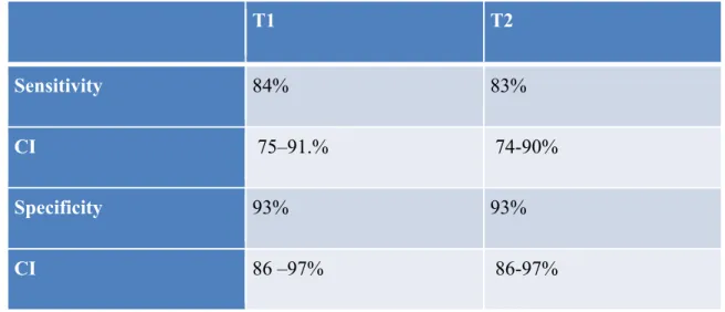
Dix articles, publiés depuis 2009, répondaient aux critères d’inclusion. Ces articles furent ajoutés aux articles retenus dans les revues systématiques déjà publiées. Au total,49 articles sont inclus dans cette revue systématique. La performance diagnostique de l’échographie endoscopique a été évaluée en calculant la sensitivité et la spécificité des études regroupées. Pour le stade T1, les valeurs de sensitivité et spécificité étaient 0.84 (CI 0.75-0.91) et 0.93 (CI 0.86–0.97), respectivement. Pour le stade T2 les valeurs de sensitivité et spécificité étaient 0.83 (CI 0.74–0.90) et 0.93 (CI 0.86–0.97), respectivement.
Conclusion:
L’échographie endoscopique présente une performance diagnostique pour l’identification de cancers de stade T1 et T2. Ceci permet d’orienter des patients vers des chirurgies moins invasives avec une survie égale et un taux de complications inférieures comparativement à des chirurgies plus invasives.
Abstract
Background:Colorectal cancer (CRC) is the second leading cause of death. The use of preoperative imaging in the staging of (CRC) plays a major role in the management.
Endorectal ultrasound (ERUS) is a precise imaging modality to determine the depth of penetration. The data on the precision of (ERUS) to predict early stage of rectal cancer has been variable
Objectives:
To conduct a systematic review, on the diagnostic performance of (ERUS) in the staging of T1 and T2 CRC.
Design:
Systematic review.
Data sources:
A literature search via PubMed, EMBASE, Ovid and Cochrane library.
METHODS:
An initial search for systematic review articles published in the last 15 years on the diagnostic accuracy of EUS in the staging of CRC using PubMed, Cochrane library, and trip database was conducted. After finding two systematic reviews that were published in 2008 and 2009, a second search of original studies published since the systematic reviews were conducted using the same databases from 2009 to 2016. The primary studies included in the systematic reviews and the primary studies published afterwards were included in the review.
diagnostic accuracy studies (QUADAS2) tool.
Terms used for search were endoscopic ultrasound, EUS, rectal cancer, histo-pathological finding, and staging.
Study selection:
Inclusion criteria includes adult people diagnosed with early stage CRC, all articles in english language and must be a full manuscripts published in peer-reviews journals. Exclusion criteria includes any recurrent or metastasis cancer and children with rectal cancer. Patients who were staged preoperatively by other imaging modality (MRI or CT) and no comparison with post operative pathology.
Results:
The search identified 420 articles, 97 articles were duplicate and excluded, and 232 refined articles were screened for title and abstract, reviewed. Thirty-two full text studies were assessed for eligibility, and ten published as full text and met the inclusion criteria; they were added to the articles identified in the earlier systematic reviews a total of 49 articles. Results of the evaluation of the accuracy of ERUS analyzed according to the diagnostic measures of sensitivities andspecificities calculated for each study.
The pooled sensitivity and specificity of EUS for stage T1 CRC was 0.84 (CI 0.75-0.91) and 0.93 (CI 0.86–0.97), and for T2 was 0.83 (CI 0.74–0.90) and 0.93(CI 0.86–0.97) respectively.
Conclusion:
The range of sensitivity and specificity values suggest that EUS performs well in accurately staging T1 and T2 cancers.
decision-making, and reduce the over staging drawback.
Keywords: Endorectal ultrasonography,rectal cancer,cancer staging,diagnostic accuracy
endorectal echography,sensitivity and specificity, rectum carcinoma,rectum tumor histopathology and early staging
Table of Contents
RESUME ………..iii-iv ABSTRACT………...v-vii TABLE OF CONTENTS……….viii-x
LIST OF TABLES ……… …....xi
LIST OF FIGURES………... …...xii
ABBREVIATIONS………. xiii
DEDICATION………...xiv
ACKNOWLEDGEMENTS ………xv
CHAPTER 1: INTRODUCTION 1.1 DESCRIPTION OF THE CONDITION………1
I.2 DESCRIPTION OF THE INTERVENTION……… 3
1.3 THE CONTRIBUTION OF SYSTEMATIC REVIEW……… 5
1.4 GAP OF THE EVIDENCE……… 6
1.5 HTA IN DIAGNOSTIC TEST ……….7
1.6 MEASURES OF DIAGNOSTIC ACCURACY………...9
CHAPTER2: RECTAL CANCER 2.1 EPIDEMIOLOGY………11
2.2 RISK FACTORS………..11
2.3 PRESENTATION……… 12
2.4 STAGING……… 13
2.5 SURGICAL OPTIONS FOR RECTAL CANCER TREATMENET………. 17
CHAPTER 3: RESEARCH OBJECTIVES
3.1 AIM OF THE STUDY REVIEW………22
3.2 RESEARCH QUESTION………22
3.3 PRIMARY OBJECTIVES………...22
3.4 SPECIFIC OBJECTIVES………23
CHAPTER 4: METHODOLOGY 4.1METHODS………...24
4.2 PICO FRAME WORK………...24
4.3 CRITERIA FOR STUDY INCLUSIN AND EXCLUSION………...26
4.4 SEARCH STRATEGY………29
4.5 THE KEY SEARCH TERM………30
4.6 STUDY SELECTION……….32
4.7 SEARCH METHODS FOR IDENTIFICATION OF STUDIES………33
4.8 DATA SOURCE………..33
4.9 DATA EXTRACTION AND QUALITY ASSESSMENT………. 34
4.10 ASSESSMENT OF METHODOLOGY QUALITY……….35
4.11 DATA SYNTHESIS AND ANALYSIS………...35
4.12 GRAPHIC REPRESENTATION………..36 CHAPTER 5: RESULTS 5.1 RESULT ANALYSIS……….38 5.2 METHODOLOGICAL QUALITY……….52 CHAPTER 6: DISCUSSION 6.1 MAIN FINDING……….54
6.2 STRENGHT AND LIMITAION……….60 6.4 POTENTIAL BIAS……….62
CAHPTER 7: CONCLUSION
7.1 IMPLICATION FOR PRACTICE………..63 7.2 IMPLICATION FOR RESEARCH……….64 REFRENCES……….65
APPENDICES
Appendix A, B: SEARCHSTRATEGY...i -vii
Appendix C: PRISMA-P……….………viii-ix
Appendix D: DATA EXTRACTION……….…x
Appendix E: PRISMA FLOW DIGARAM………...xi
Appendix F: QUALITY ASSESMENT TOOL QUADAS-2 ………xii-xxxiv Appendix G: QUALITY ASSESMNET TOOL FOR SYSTEMATIC REVIEWS
List of tables
Table 1: Rectal cancer staging.
Table 2: inclusion and exclusion criteria for selected paper. Table 3: characteristic of studies included in this analysis.
Table 4: The results of each included study and the author’s conclusion
Table 5: Critical appraisal results for included studies using the JBI critical appraisal checklist
Table 6:Diagnostic accuracy of EUS for T staging
Table 7: Sensitivity analysis including rectal cancer, results from 10-pooled studies Table 8: Pooled analysis from Pauli systematic review for T-stage
Table 9: The accuracy of EUS with Confidence interval from the combined analysis
List of figures
Figure 1: The 2x2 contingency table Figure 2: PRISMA Chart
Figure 3: Forrest plot showing sensitivity and specificity of EUS to diagnose T1 stage of rectal cancer
Figure 4: Forrest plot showing sensitivity and specificity of EUS to diagnose T2 stage of rectal cancer
Figure 5: Forrest plot showing sensitivity and specificity for T1 of our systematic review combined with the meta-analysis study.
Figure 6: Forrest plot showing sensitivity and specificity for T2 of our systematic review combined with the meta-analysis study.
List of abbreviation:
CRC: Colorectal cancer. ERUS : Endorectal ultrasound. CT: Computed tomography (CT) MRI: Magnetic resonance imaging PET: positron emission tomography
FAP: Familial adenomatous polyposis (FAP)
HNPCC: Hereditary non polyposis colorectal cancer IBD: Inflammatory bowel disease (IBD).
RCT: Randomized control study
TEM : Transanal endoscopic microsurgery TME : Total mesorectal excision.
LAR: Lower anterior resection. LN: Lymph nodes
QUADAS: Quality Assessment of Diagnostic Accuracy Studies.
PRISMA: Preferred Reporting Items for Systematic Reviews and Meta-Analysis CHUM: The Centre hospitalier de l'Université de Montréal
To my loving parents, my precious husband and to my adorable
children
ACKNOWLEDGMENT
First of all I’m grateful for the almighty GOD for guide me to complete this work. I would like to thank my supervisor Dr.Lugi Lepanto, for his support, constant availability, and help in setting achievable goals all through my master. His door was always open for my questions and concerns, and he kindly offered promote feedback during thesis writing. Words are not enough to express how privileged I’m to have him as a mentor.
A very special sincere thanks goes out to my mother and father (Souad and Mohammed) for the love they gave me, For their endless generosity and kindness, and for making my education a priority since I was a child .You were always there for me with unceasing support and prayers throughout my study. I cannot thank you enough for all the encouragement you provided.
For the most part I would like to express my deepest gratitude to my husband Raad for his great patience, which was invaluable in helping me take care of our kids. He has always believed in me, helped me stay positive, and motivated. This thesis would not have been possible without his love, encouragement, and moral support.
To my wonderful children Tuleen and Rakan, I express my love, blessing, and dedication. Thank you for giving me the happiness of life, and thank you for your respect and appreciation.
Special thanks go to my siblings Assad, Rabab and Ahmad who have always been very supporting and caring.
Reaching the goals of my research would not have been possible without the help of members of the health technology master; I am very grateful and thankful to all of them.
Chapter I
Introduction
1.1-Description of the condition:
Colorectal cancer (CRC) is a common cancer and is considered the second most diagnosed cancer in the world [1].
The incidence rate increases with age and peaks in the seventh decade of life (mean age 60-65 years). Approximately 30-40% of colorectal cancer begins in the rectum, which is defined as the distal margin of the tumor within 15 cm of the anal verge [2, 3].
Given the vitality of the difficult anatomy of the rectum within the pelvis and surrounding visceral structures, accurate preoperative staging by suitable imaging modality is essential to determine the consequent treatment.
Staging rectal cancer by a multidisciplinary team and accurate diagnostic imaging helps provide the best care for patients by offering treatment modalities, by guiding patients for either pre-operative chemo-radiotherapy or surgical management and assessment of prognosis of the tumor.
The multidisciplinary team includes colorectal surgeons, gastroenterologists and both radiation and medical oncologists.
Adequate surgical resection is considered the mainstay of treatment for rectal cancer; further, in the early stage of rectal cancer, the five-year survival rate is more than 90%, while for advanced-stage rectal cancer, the five-year survival rate is less than 10% [4]. Endorectal ultrasound (ERUS) has become the most prevalent diagnostic imaging modality for the local staging of rectal cancer; it is safe and less expensive [5, 6].
The muscularis propria of the rectum is a layer of muscle tissue considered the most important anatomic structure for physicians. It helps them decide whether the lesions are suitable for local excision if muscularis propria is not at the infiltrated stage (T1), or if extensive surgery is necessary if muscularis propria seems to be at the infiltrated stage (T2) or (T3). The accuracy and precision of muscularis propria involvement by the ERUS are superior compared to other existing imaging modalities [6].
Transanal local excision or endoscopic microsurgery is the modality of treatment for stage T1 or lower [7, 8], while a total mesorectal surgery would be used for stages T2 and T3.
Early detection in curable stages and accurate diagnosis can influence the therapeutic strategy and improve outcome. There is a consensus regarding the role of ERUS in local staging rectal cancer and how commonly it is used due to lower costs and patient accessibility to the equipment.
The focus of this review is to perform a systematic assessment that analyzes the accuracy and limits of the endorectal ultrasound (ERUS) method in early-stage CRC in comparison to the histopathology findings of the subsequent surgical specimen, and that highlights the impact of the ERUS value in staging CRC.
Developing systematic reviews that are applicable for clinical practice stances a major challenge, and requires strong idea about the possibility and purpose of the review.
1.2-Description of the intervention:
The endorectal ultrasound (ERUS) technique is the best diagnostic tool for the pre-treatment staging of rectal cancer; it examines the thickness of the rectal wall, assesses the depth of tumor invasion and helps in distinguishing between tumors localized to the rectal wall and tumors with transmural invasion [9]. A combination of imaging modalities such as computed tomography (CT), magnetic resonance imaging (MRI) and/or endorectal ultrasound (ERUS) are used to accurately measure the extent of rectal cancer. Endorectal ultrasound (ERUS) is considered the first choice for diagnostic modality; in ERUS, a tiny ultrasound transducer is installed on the tip of the endoscope to allow the transducer to get closer to the body’s organs to achieve high-quality ultrasound images, thus providing enhanced details about the organs inside the body. ERUS probes are available in different lengths, diameters, and frequencies. The higher the ultrasound frequency, the better the resolution, with an accuracy range from 85-95% for assessing the depthof rectal cancer invasion, published in many articles [10, 11, 12, 13, 14, 15].
It is the only method that has the capability to show images of the layers of the bowel wall, which can discriminate between T1 and T2 tumors [10]. ERUS indicated a better accuracy than other imaging modalities to assess the depth of invasion. The range for CT scans is between 65-75% and 75-85% for MRIs [16, 17, 18].
ERUS is a simple procedure usually done as an outpatient; most patients undergo ERUS simultaneously through the same colonoscopy session with conscious sedation.
Before the procedures start, a digital rectal examination will be performed to describe the tumor size, location, and distance from the anal verge. The patient will be in a left lateral position and the trans-anal probe covered in a water-filled balloon to allow for
visualization of the rectum and perirectal area and avoid compression of the tumor by the ultrasound probe, which leads to overstating
. This probe is inserted into the rectum above
the level of the tumor, then slowly withdrawn until it reaches the tumor level for complete evaluation of the tumor and lymph node and to assess the degree of rectal invasion [19].
The higher frequency (2, 12.5, 15, 20, 25 and 30 MHz) ultrasound allows for better resolution of the rectal wall layers and inspection of stenotic lesions, while the lower ultrasound frequencies are used for assessment of lymph nodes and perirectal tissue invasion [20, 21]. Digital ultrasound images are saved on a computer file.
The probe is inserted into the rectum above the level of the tumor, and then slowly withdrawn until it reaches the tumor level for complete evaluation of the tumor and lymph node and to assess the degree of rectal invasion [14]. Ultrasound can visualize the five layers of the rectum; usually they alternate between hyperechoic (white) and hypoechoic (dark) layers. Two are hypoechoic, three are hyperechoic and carcinomas are usually hypoechoic; the degree of penetration of the rectal wall layers suggests local or advanced-stage disease [22]. The first hyperechoic layer resembles a water-filled balloon; the mucosa represents the second hypoechoic layer while the third hyperechoic layer corresponds to submucosa. Muscularis propria, the fourth layer (hypoechoic) and the fifth layer (hyperechoic) relate to the interface between muscularis propria and perirectal fat
[23, 24]. EUS is the only method that has the capability of imaging the layers of the bowel wall, which can discriminate between a lesion on the sub mucosa and muscularis propria (T1-T2 rectal tumors) [10].
1.3-How systematic review will contribute to our understanding of the
problem addressed:
Systematic reviews are used to review a clearly formulated question with careful consideration of a review’s methodological approach. It identifies all evidence and analyzes data from the studies that would decide which research could be included or excluded based on inclusionary eligibility criteria to answer a specific research question.
[25,26]. In this thesis, the systematic review approach will be conducted to answer a certain question in relation to the advantage of ultrasound diagnostic imaging in early-stage rectal cancer.
The result of this thesis may improve the guidelines with respect to the added value of using an ultrasound in clinical practice for diagnosing early-stage rectal cancer and identifying further research needs byproviding and synthesizing more reliable findings from the included studies about this specific research topic so as to aid in decision-making.
In addition, systematic review can perform an assessment of the validity of the review’s outcomes, for example, evaluation of the risk of bias and confidence in cumulative estimates.
The PRISMA-P was followed in this review, which consists of checklist items
divided into three parts: administrative information, introduction, and methods
[27].
This checklist was designed to improve the conduct of systematic reviews, provide readers with a complete acceptance of evidence from existing studies, and
help in the evaluation of the effects of interventions in early-stage rectal cancer. See Appendix.
1.4-Gap of the evidence:
Early and accurate diagnosis can influence the therapeutic strategy and improve the outcome. Agreement exists regarding the role of ERUS in the local staging of rectal cancer and how commonly it is used due to lower costs and patient accessibility to the equipment. However, some studies showed inferior results from the EUS due to the experience of effects of the results.
A systematic review is necessary to highlight the value of the diagnostic test accuracy of ERUS imaging in staging preoperative rectal cancer (CRC) and to determine the efficacy
of evaluating the depth of colorectal cancer invasion.
In a review of the Cochrane database, Medline indicated two previous reviews performed in 2008-2009, which covered the diagnostic accuracy of EUS in the early staging of CRC.
1.5-HTA in diagnostic test:
Various details exist between diagnostic technologies evaluation and medical therapeutics. The most significant detail is that diagnostic test results influence outcomes but cannot determine health outcomes in patients. Tests performed on a person who has a symptom or sign of illness are usually termed diagnostic. Diagnostic tests are a critical component of health care; clinicians and patients usually have several questions regarding diagnostic tests, such as: What is the test used for? Does it improve the outcome? Is the test recommended in practice guidelines? How are the test results interpreted?
Imaging techniques allocate the generic features of all diagnostic tests; however, several issues are abnormal when imaging tests: 1-the test results are frequently multidimensional, 2-clear-cut points are rarely established, 3-images can reveal signs of different diseases, 4-imaging techniques may be associated with the risk of radiation-induced side effects, leading to a clinical tradeoff between benefit and harm, 5-image quality increases with improved resolution and 6-many emerging imaging tests are expensive.
Finally, an important feature of the evaluation of a diagnostic procedure is that different readers can assess images at different times, which allows us to analyze intra- and inter observer agreement.
There are six levels of diagnostic efficacy assessment: (a) technology, (b) diagnostic accuracy, (c) diagnostic thinking, (d) therapeutic planning, (e) patient outcomes and (f) society.
Level one is the domain of physicists and engineers who develop and refine an imaging technology before its clinical implementation and testing.
Level two is sensitivity and specificity, positive and negative predictive values or receiver operating characteristic (ROC) curves.
Level three, or “diagnostic thinking efficacy,” is used to measure the effect of diagnostic test results on the thinking of physicians.
The likelihood ratio represents the ratio of the frequency of a certain test result in patients with a disease to its frequency in patients without the disease. The likelihood ratio can be used to judge the usefulness of a particular test in a given clinical situation [28]. Advocates of evidence-based medicine have also recommended the use of likelihood
ratios in the evaluation of diagnostic technologies [29].
Level four is known as “therapeutic planning efficacy. Level five, or “patient outcome efficacy,” can really be assessed only in a prospective RCT, in which only some of the patients undergo the test and patient outcomes in the two groups (test vs. no test) are compared.
Level six, or “societal efficacy,” asks whether the societal benefit associated with the test is acceptable in relation to its cost [30].
Technology description of any diagnostic test is highly dependent on the population, disease and other features of the setting in which it are used.
In the absence of clinical data, diagnostic tests are estimated based on test accuracy – the ability of the test to correctly determine the disease status of an individual. Test accuracy is not a measure of clinical effectiveness and improved accuracy does not necessarily result in improvement.
Diagnostic tests are used to monitor therapeutic measures. Ideally, an evaluation should assess the clinical utility of a test. This evaluation was presented in two measures, namely sensitivity and specificity, to describe the characteristics of a diagnostic test.
A positive test result might lead to the induction of therapy when it otherwise might not have been considered. A negative test might lead to the decision not to initiate therapy when it otherwise would have been given [31].
1.6-Measures of diagnostic accuracy:
Sensitivity Specificity
Positive and negative predictive value
The area under a receiver operator characteristic (ROC) curve Likelihood ratios of positive and negative test results
Diagnostic odds ratios
Why are we doing diagnostic test accuracy?
Studies are easy to undertake; answers can be achieved quickly; required sample sizes are feasible and results do not depend much on human and health service factors.
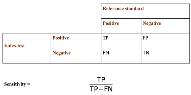
Figure 1: The 2x2 contingency table
Reference standard Positive Negative Index test Positive TP FP Negative FN TN Sensitivity =The proportion of people with the target condition who have a positive test result. How good is this test at identifying people with the condition?
Specificity =
The proportion of people without the target condition who have a negative test result. How good is this test at correctly excluding people without the condition?
Chapter 2
Rectal cancer
2.1-Epidemiology:
The incidence of colorectal cancer is 15 times higher in adults older than 50 years; it is 40% higher in men than in women and the mortality rates are highest in African American men and women [32]. About 72% of cases arise in the colon and about 28% in the rectum. It is uncommon to have CRC before the age of 40, except if there is a predisposing condition.
The incidence and mortality rate of CRC have been declining for the last several decades. This decrease in incidence has been influenced by the improvement of diagnostic techniques, screening programs, the removal of precancerous polyps and patient education [33-34].
2.2-Risk factors:
Many factors increase the risk of CRC; about 75% of colorectal cancers are sporadic and the etiological factors include physical inactivity, high fat and high consumption of red meat such as beef, lamb and processed meats. People who are overweight have a greater chance of developing colorectal cancer, cigarette smoking consider as an important risk. There is a link between colorectal cancer and heavy alcohol consumption [35,36,37].
The remaining 25% of cases occur in people who have a family history of CRC or adenomatous polyps or a personal history of chronic inflammatory bowel disease.
Other significant risk factors are genetic predispositions such as hereditary non-polyposis colorectal cancer (HNPCC), which is correlated with mutations in genes involved in the
repair pathway of DNA (MLH1 and MSH2) genes; responsible mutations in individuals with HNPCCcan also cause Lynch syndrome [38].
Additionally, inflammatory bowel disease (IBD), which includes ulcerative colitis, Crohn’s disease increases an individual's overall risk of developing colorectal cancer
[39], besides familial adenomatous polyposis (FAP), which is caused by changes (mutations) in the APC gene that a person receives from his or her parents.
Families with a history of adenomatous polyps in one or more first-degree relatives are at increased risk [40]. People with FAP are at a higher risk for other cancers, such as cancer of the stomach and small intestines.
People with a history of adenomatous polyps in one or more first-degree relatives are at an increased riskof developing rectal cancer [40].
2.3-Clinical presentation:
• Bleeding from the rectum
• Blood in the stool
• Dark- or black-colored stools
• Cramping
• Discomfort or an urge to have a bowel movement
• Constipation or diarrhea
• Other symptoms such as a change in bowel habits, weight loss, abdominal pain, and anemia [41,42].
The diagnosis is usually made by digital rectal examination to assess the rectal tone and detect penetration of the mass into the external and internal sphincters.
Some blood work (carcinoembryonic antigen measurement) is done, in addition to sigmoidoscopy and colonoscopy, double-contrast enema examination and histologic confirmation.
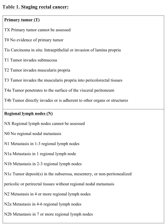
2.4-Staging:
The TNM staging system by the American Joint Committee (AJCC) is the recommended staging system for colorectal cancer [43].
Staging is the predictor of survival for patients with colorectal cancer [44].
The T refers to the extent of the primary tumors. The N refers to the involvement of regional lymph nodes and the lymphatic system. The M refers to metastatic disease.
Tis carcinoma in situ does not have any metastatic potential. T1 tumors invade the submucosa; without evidence of invasion into the muscularis propria, T2 tumors invade, but do not go through, the muscularis propria. T3 tumors invade the muscularis propria and infiltrate the perirectal fat. T4 tumors invade surrounding organs and structures.
Table 1. Staging rectal cancer:
Primary tumor (T)
TX Primary tumor cannot be assessed T0 No evidence of primary tumor
Tis Carcinoma in situ: Intraepithelial or invasion of lamina propria T1 Tumor invades submucosa
T2 Tumor invades muscularis propria
T3 Tumor invades the muscularis propria into pericolorectal tissues T4a Tumor penetrates to the surface of the visceral peritoneum
T4b Tumor directly invades or is adherent to other organs or structures
Regional lymph nodes (N)
NX Regional lymph nodes cannot be assessed N0 No regional nodal metastasis
N1 Metastasis in 1-3 regional lymph nodes N1a Metastasis in 1 regional lymph node N1b Metastasis in 2-3 regional lymph nodes
N1c Tumor deposit(s) in the subserosa, mesentery, or non-peritonealized pericolic or perirectal tissues without regional nodal metastasis
N2 Metastasis in 4 or more regional lymph nodes N2a Metastasis in 4-6 regional lymph nodes N2b Metastasis in 7 or more regional lymph nodes
Distant metastasis (M)
M0 No distant metastasis M1 Distant metastasis
M1a Metastasis confined to 1 organ or site (i.e., liver, lung, ovary,
non-regional node)
M1b Metastases in more than 1 organ/site or the peritoneum
AJCC stage
TNM stage
Stage 0
Tis N0 M0
Stage 1
T1 N0 M0
Stage 1
T2 N0 M0
Stage IIA
T3 N0 M0
Stage IIB
T4 N0 M0
Stage IIIA
T1-2 N1 M0
Stage IIIB
T3-4 N1 M0
Stage IIIC
AnyT, N2, M0
Stage IV
Any T, any N,
T1:
The tumor invades submucosa without invading the muscularis propria.
Accurate diagnosis of a T1 rectal cancer is essential in determining treatment options, which include either local excision or oncologic resection [45-46].
Local excision by transanal excision or transanal endoscopic microsurgery is performed only in certain patients if the rectal tumor is less than 3 cm, less than 8 cm from the anal verge, well to moderately differentiated and absent of lymphovascular or perineural invasion.
In the presence of lymphovascular invasion or poorly differentiated histology, T1 rectal cancer oncologic resection should still be endorsed [47].
T2:
The tumor invades the muscularis propria. It has always been difficult to stage T2 because the muscularis propria could appear thickened and irregular, which could be attributed to the presence of an inflammatory lymphocytic infiltration at the edge of the tumor.
This may lead to an over-staging problem, 22-24.
Local excision is not recommended in T2 due to a high local recurrence of more than 20% [48-49].
2.5-Surgical options for rectal cancer treatment:
In the early stages, rectal cancer surgery is the standard care for treatment, with the goal of optimizing oncologic control while reducing the effects of treatment on quality of life; therefore, different methods were recommended, including local excision and transanal endoscopic microsurgery (TEM).
Local excision is used to treat stage T1N0 rectal cancers, with strict selection criteria including a freely mobile lesion less than 3 cm in size and less than 30% of the bowel circumference, histology being well-to-moderately differentiated and a lack of involvement of lymphovascular or perineural invasion and negative node. Local excision is associated with a higher recurrence rate (40%), especially if it is used in higher risk stage I rectal cancer (T2) lesions [50].
The transanal endoscopic microsurgery (TEM) procedure was presented in 1984 as a minimally invasive procedure that served as an alternative to radical surgery and that provides better visualization of tumors and allows for the excision of lesions located in the rectum by endoscopic view of the rectum with decreased morbidity and mortality. The procedure is technically challenging due to the narrow operating field; the patient undergoes general anesthesia and a bowel preparation, then is placed supine or prone on the operating table to keep the lesion close to the 6 o’clock position. The TEM technique includes a laparoscopic camera, an operating proctoscope and modified laparoscopic instruments.
After a dilating digital exam, a proctoscope is inserted through the anus with a length of 20 cm and a 4-cm diameter. After insertion of the proctoscope, the lesion is identified and
the proctoscope is fixed to the operating table. The proctoscope has a port for high-flow carbon dioxide to maintain dilatation of the rectum [51].
The faceplate of the proctoscope has four ports for insertion of instruments, including a camera and three modified laparoscopic instruments to ease the full-thickness excision of rectal lesions; tumor excision is performed by monopolar hook cautery [51].
Several retrospective and prospective studies reported that TEM is highly successful, with the advantage of fast recovery [52], the absence of scars, a rapid return to regular activities [53], a decreased need for colostomies, a short hospital stay with less operating time, only brief use of analgesia, a decreased rate of complication of 2-12% [54,55] and recurrence rates of 0–19% [56,57].
TEM is an oncological procedure that is safe for early-stage rectal carcinomas and that has achieved low local recurrence and high survival rates. Complications after TEM are considerably rare, mainly urinary difficulty.
In case of positive resection margin or unfavorable histology, total mesorectal excision (TME) surgery will be performed [58].
If the pathology revealed a T2/T3 rectal cancer, patients’ cases will be presented at tumor board meetings for offering additional radical surgery, including low anterior resection (LAR) or abdominoperineal resection (APR).
LAR involves mobilization of the rectum, sigmoid colon, by performing mobilization of the rectum; a technique called total mesorectal excision (TME) is required.
TME is the therapeutic gold standard in patients as a part of low anterior resection for middle and lower third rectal cancers; it is a procedure defined as the excision of the rectum with the surrounding mesorectum at the level of the pelvic floor.
The patient is positioned in a lithotomy position. A Foley catheter is inserted and the rectum is irrigated with both saline and iodine. Ureteral stents are placed if indicated and a temporary colostomy or ileostomy is performed in case of LAR in addition to intensive postoperative monitoring. Most of the time, TME is performed laparoscopically in bloc resection of the rectal cancer with a complete pararectal lymph node dissection in addition to the resection of radial and circumferential margins, not breeching the fascia propria of the rectum [59]. Postoperative complications of TME include an increased risk of anastomotic bleeding, urinary leakage, urgency and the feeling of incomplete bladder emptying, while the lower anterior reaction side effects include sexual and urinary dysfunction, dehiscence, intestinal obstruction, anastomotic site stenosis, stoma problems and fistula [60].
An abdominal perineal resection (APR) is still done in selected patients with low-lying rectal adenocarcinomas or poor sphincter function.
APR includes the resection of the sigmoid colon, rectum, and anal sphincter using both anterior abdominal and perineal incisions and resulting in a permanent colostomy.
Patients who undergo APR must have one of these criteria: progressive rectal cancer, failure to achieve a negative distal margin by a sphincter-sparing procedure or local recurrent [61].
The importance of accurate staging of rectal cancer is essential for the clinician in helping patients select appropriate management, to identify patients who can undergo sphincter-preserving surgery or new adjuvant treatment and to foresee prognosis.
Many imaging modalities are utilized for staging rectal cancer, but the precision of EUS in the literature has been varied for T1 and T2 diagnosis; therefore, we conducted a
systematic review to summarize the accuracy of EUS and to predict the impact of EUS in the diagnostic workup by under diagnosing T1 or not recognizing it, and then performed a decision analysis to evaluate the added utility of EUS.
2.6-Diagnostic imaging:
Several imaging techniques’ modalities are used in the pre-operative staging of colorectal cancers, such as computed tomography (CT), magnetic resonance imaging (MRI), endorectal ultrasound (ERUS) and positron emission tomography (PET) with and without CT fusion.
Each modality has its own benefits and drawbacks; the benefits should be weighed against the drawbacks of using the modality. An important element in choosing an imaging modality for staging is the availability of that imaging modality.
MRI and ERUS are considered the two standard imaging techniques used for the primary staging of rectal cancer [10]
A computed tomography (CT) scan is considered in the staging of rectal cancer for the detection of metastatic disease spread to the liver and/or lungs; the accuracy of CT (T-stage) improves in more locally advanced tumors than in early staging [62].
A CT scan is not useful in evaluating the layers of the rectal wall due to inherent low-contrast resolution presented by CT imaging techniques. It can precisely detect lymph nodes staging in a range from 54 to 70% [19].
High-resolution magnetic resonance imaging (MRI) is considered an important component of rectal cancer staging. MRI is used to measure the extent of a tumor in the adjacent mesorectum and tumor proximity to the mesorectal fascia (MRF) to determine
the risk of local recurrence and identify nodal involvement [20].
If MRF was involved or proximal to the tumor, this would expand the risk of compromised radial, the circumferential resection margin CRM after radical surgery [21].
MRI fails to differentiate between T1 and T2 cancers because the submucosal layer is usually not visualized on MRI [63]; also, MRI cannot differentiate between T2 and T3 cancers because of the desmoplastic reaction seen near tumors [64,65]. A high-resolution MRI image appears to be superior to EUS for locally advanced disease and for the detection of lymph node metastases.
Nodal staging by MRI ranges between 39 and 95% [66,67,68,69]; it allows for visualization of nodes as small as 2 mm in the entire mesorectal part, as well as outside the mesorectum [67].
PET/CT scan with radioactive glucose is useful in displaying whether the cancer has spread to lymph nodes or nearby structures, or in the case of obstructing colorectal cancers; in addition, it will aid in the detection of a suspected recurrence of CRC or tumor response to therapy [69,70].
Few studies have suggested that preoperative PET combined with CT scan improves the staging of rectal cancer [71] and plays a role in changing preoperative management in about 17% of patients [72]. However, PET/CT is not used routinely for the staging of primary rectal cancer, as no specific evidence supports the routine clinical use of PET/CT.
CHAPTER 3:
Research Objective
3.1-Aim of the study review:
This study aims to perform a systematic review that analyzes studies to evaluate the diagnostic imaging accuracy of ERUS (endorectal ultrasound) in adult patients with local-stage rectal cancer as well as to summarize the diagnostic test accuracy (e.g., sensitivity, specificity) in comparison to the histopathology findings of the subsequent surgical specimen, thus highlighting the impact of EUS value in staging CRC. This study will also provide future practice recommendation.
3.2-Review question(s):
• What is the diagnostic accuracy of imaging techniques (ERUS)? (Measuring sensitivity and specificity)
• What is the impact of imaging techniques on the clinical outcome?
3.3-Primary objective:
The primary objective of this study is to systematically review the currently published articles that evaluate the diagnostic test accuracyof using endorectal ultrasound for the staging of primary rectal cancer. The review compares ERUS and histopathological findings for staging early rectal cancer patients.
3.4-Specific objectives:
To extract information about imaging diagnostic tests for the investigation of rectal cancer.
To identify all studies in the existing literature related to the diagnostic accuracy of ERUS used to detect early stages of rectal cancer.
To extract data on sensitivity and specificity for (T1, T2) rectal cancer. To synthesize and compare extracted data to pathology staging.
CHAPTER 4:
Methodology
4.1-Methods:
We will perform a literature search that inspects the studies identified and choose those that meet the eligibility criteria. The description of methods in this chapter will match the
diagnostic test accuracy methods at Cochrane Collaboration [73]. Next, the data will be extracted from the selected studies for an evaluation of their methodological quality. In this chapter, we follow the Preferred Reporting Items for Systematic Review Protocols
(PRISMA-P) recommendations [74] Appendix C.
4.2-PICO frameworks
Participants/PresentationInclusion: All adult patients ≥18 years, undergoing pre-operative ERUS staging and
diagnosed with primary rectal cancer stage 1 (T1/T2) will be eligible for inclusion.
Exclusion: People under 18 years, studies focusing on patients with advanced-stage rectal cancer, recurrent cancer, or metastasis and patients who received neoadjuvant treatment. Studies that did not meet all the inclusion criteria, Case reports, crossover studies and abstract material were not included in determining the accuracy of the study. Studies performing EUS after treatment, restaging imaging for follow-up or investigating local recurrence rates or responses to treatment were all excluded. So were studies not reporting an endoscopic ultrasound as a diagnostic measure, studies in which the restaging findings were not compared with pathological results, duplicate studies and other types of cancer.
Index test
The index test is endorectal ultrasound (ERUS)/endoscopic ultrasound (EUS).
All studies that evaluated the accuracy of ERUS as diagnostic imaging in primary colorectal cancer were included.
All studies involving humans were included. Studies performing this investigation after treatment or investigating recurrence rates or responses to treatment were excluded. Patients who were staged preoperatively by other imaging modality (MRI or CT) were excluded.
Comparator (reference standards)
The reference test is the “gold standard” test to which physicians use the results from the index test (ERUS) to reach a stage compared to the one founded by the reference standard. In this review, we included studies that used the histopathology of surgically resected specimens as a reference standard for the pretreatment staging of CRC.
We pursued comparative studies of ERUS test accuracy that evaluated the (ERUS) index test versus histopathology following either surgery or biopsy in staging early rectal cancer.
These comparisons will advise how many patients were staged correctly and allow us to calculate the test performance of sensitivity, specificity, and accuracy.
Outcome(s):
Primary outcome
• Diagnostic accuracy (sensitivity, specificity) of tumor staging by ERUS
• Test performance (understaging, overstaging) against a reference standard test (pathology examination)
• Changes in therapeutic management
• Impact of survival Secondary outcomes None
Time frame studies:
A well-defined period of time will be considered for research, starting form 2009- September 2016.
4.3. Criteria for study inclusion and exclusion:
Condition or domain being studied:The target conditions were colorectal cancer patients.
Study types include:
Diagnostic studies were included with any study design that evaluated test accuracy of EURS diagnosis, and compared with histopathology.
Findings:
Diagnostic case-control studies were excluded because clinically relevant estimates of specificity and sensitivity can be derived only from the clinical population and not healthy controls.
Exclusion:
Case reports because they lack sufficient diagnostic test accuracy data.
Case-control studies because they are prone to bias and estimates of specificity and sensitivity, which can be derived only from the clinical population and not healthy controls [75].
Conference abstracts because they do not include adequate details about experimental methods to permit an evaluation of study design and conduct.
Cross-over studies.
Setting:
Studies from the clinic or hospital were included.
Full-length articles:
Prospective study:
A prospective study collects data in the process, assesses outcomes, and is a good choice for rare exposure. The study usually involves taking a cohort of subjects and monitoring them over an extended period of time. Prospective studies usually have fewer potential sources of bias and confusion than do retrospective studies. The weaknesses of prospective studies include loss of follow up and difficulty selecting and maintaining a non-exposed group.
Retrospective study:
A retrospective study is one that collects data from the past; it looks backwards and examines exposures to investigate risk or protection factors in relation to an outcome, making this type of study appropriate for studying multiple outcomes. Most sources of error due to confusion and bias are more common in retrospective studies than in prospective studies, but timeframes for completion are usually short in retrospective studies. With respect to weaknesses, retrospective studies cannot demonstrate temporality; an investigator has no control over exposure and requires a large sample for rare exposure.
In the assessment of the diagnostic test, both retrospective and prospective studies are used to assess and compare accuracy. A retrospective study is usually recruited based on whether patients have the disease. In contrast, for a prospective study patients are selected based on their symptoms [75].
Observational study designs are admissible in our study; they include retrospective and prospective cohorts. Diagnostic study designs division of observational studies which evaluate the accuracy of diagnostic procedures and tests as compared to other diagnostic
measures, including diagnostic accuracy designs and diagnostic cohort designs [76]. The crossover study design was excluded from our study because a crossover design is a repeated measurements design in which patients receive different treatments during different time periods; it is a controlled trial in which each participant receives both therapies in a random order and is relevant only if the outcome, such as symptoms, is reversible with time.
The main disadvantage of the crossover study is the carryover effects, defined as the effects of the treatment from the previous time period on the current time period’s response; these effects cannot be estimated separately [77].
4.4-Search strategy:
Early scoping research:An initial systematic review of the literature was performed for articles published in the last 16 years about the diagnostic accuracy of ERUS in the staging of CRC by searching the databases:
1-PubMed 2-EMBASE 3-Ovid
4-Cochrane Library, which includes Cochrane Reviews, DARE and the Central Register of Controlled Clinical Trials
Search strategy:
In consultation with a librarian specialist in electronic bibliographic databases, information was identified for scoping the research. The guidelines for building a search strategy in each database were consulted. The search strategy used text words to identify articles discussing the accuracy of endorectal ultrasound in staging patients diagnosed with early rectal cancer.
The following electronic databases were used: MEDLIN (Ovid, PubMed), EMBASE and Cochrane Database; with modifications to search terms as necessary (see appendix A,B)
A limitation with respect to the publication year (2009 to 2016) was applied.
The search strategy used text words and relevant indexing to identify articles discussing the diagnostic accuracy of endorectal sonography for early detection of rectal. Results were limited to articles published in English between 2009 and 2016 in a peer-reviewed journal, terms relating to the intervention, text in the English language and text applying only to humans. Conference abstracts, comments, case reports, editorials, and letters were excluded.
Relevant studies of the diagnostic accuracy of ERUS in the staging of rectal cancer were identified.
4.5-The key search term:
To guarantee an efficient search, medical subject headings (Mesh) words, were used to define relevant articles for our study. For each database, keywords and index terms were used to identify other index terms to determine the relevance of each term.
search more precise.
The full MEDLINE strategy was applied to all databases, with modifications to search terms as necessary.
The MEDLINE database was searched using these keywords: (a)
“rectal neoplasm” (medical subject heading, or Mesh), (b) “EUS” (Mesh) or “ultrasonography” (Mesh) and (c) “specificity”, “sensitivity” or “accuracy”.
The EMBASE, Cochrane databases were also checked for significant articles by applying (a) “rectal cancer” and (b) “ultrasonography”, “accuracy” or “histopathology”.
We used terms such as “sensitivity and specificity” or “accuracy”, called methodological search filters; the search terms used are described in detail in the Appendix A-B.
4.6-Study selection:
Initially, one reviewer screened titles and abstracts for relevant studies. At this stage, studies were excluded if the condition was not early rectal cancer, if the study performed on patients received neoadjuvant treatment, if the study included recurrent cancer or metastases, or if the study did not use ERUS. Full texts of identified articles were then obtained and assessed for study eligibility using the full set of inclusion and exclusion criteria. The study selection process was illustrated using a PRISMA (Preferred
Reporting Items for Systematic Reviews and Meta-Analyses) flow diagram. See Appendix E.
4.7-Search methods for identification of studies:
Systematic electronic searches were customized to each of the following databases: MEDLINE via Ovid SP (2009 to September 2016; Appendix A.1)
Medline via PUBMED (2009 to September 2016; Appendix A.2) EMBASE via Ovid SP (2009 to September 2016; Appendix A.3) The Cochrane Library (2009 to September 2016; Appendix A.4)
In addition, reference lists for all included papers were searched for relevant studies. All results were collected and duplicate studies removed and exported to EndNote version 7.0.
The selection of qualified studies was performed in a stepwise approach (titles and abstracts, then full texts).
Research Ethics Board review was not required for this systematic review.
Studies eligible for our systematic reviews were required to compare ERUS assessment of T stage with histopathology T stage for early rectal cancer.
4.8-Data source:
Studies were classified by searching a range of electronic databases and reference lists of relevant studies.
We looked in the systematic review at the diagnostic accuracy of ERUS and compared it with the pathology result after the surgery.
ERUS studies were selected based on surgical histology. Standard criteria were used to determine the T stage:
T1: Hypoechoic mass in the lamina propria or submucosa, without evidence of invasion into the muscularis propria.
T2: Hypoechoic mass invading the muscularis propria.
Definitions:
Accuracy: the percentage of patients in whom the diagnostic imaging ERUS for staging
rectal cancer matched the pathological stage.
Under-staging: the percentage of patients in whom the ERUS stage was less than the
pathological stage.
Over-staging: the percentage of patients in whom the ERUS stage was greater than the
pathological stage.
4.9-Data extraction:
Data was collected according to the PICO framework, as explained above. One reviewer extracted data using the search strategy to identify studies that potentially met the inclusion criteria outlined above. This data was then verified by a second reviewer; both these reviewers, worked independently. All studies that met inclusion criteria were selected for full-text review. Relevant findings were summarized and presented in an evidence table.
Data extraction will include the following variables:
Study characteristicsTitle
Year of publication Study design
Confirmatory procedure
Population characteristics: inclusion/exclusion criteria for individual studies
Setting
Intervention test Outcome measures
2x2 table for diagnostic studies presenting the sensitivity and specificity of T1-T2 rectal cancer
4.10-Assessment of methodological quality (risk of bias):
Each full paper was assessed for risk of bias by one reviewer and proven for accuracy
with respect to all the assessed studies by another.
This checklist is a modified version of the quality assessment of the diagnostic accuracy studies’ (QUADAS-2) assessment tool [78, 79], which will be used to assess the quality of the included diagnostic accuracy studies.
QUADAS-2 consists of 4 key domains:
These domains include patient selection, index test, reference standard, and flow of patients through the study and timing of the index test(s) and a reference standard to evaluate each article. The items were rated as yes, no or unclear.
Each domain was assessed in terms of risk of bias; each of these domains also summarizes the review question, tailors the tool, produces review-specific guidance, constructs a flow diagram for the primary study and judges risk of bias and concern
regarding applicability [78].
The results of the quality assessment will be presented later to provide an evaluation of the quality of the selected papers.
For the systematic reviews, methodological quality will be assessed using the AMSTAR tool [80],
it’s an 11-item questionnaire that can be used to assess the methodological quality of systematic reviews by assessing the presence of:
An a priori design; duplicate study selection and data extraction; a complete literature search; and the use of status of publication as an inclusion criteria; a list of included/excluded studies; characteristics of included studies; documented assessment of the scientific quality of included studies; appropriate use of the scientific quality in forming conclusions; the appropriate use of methods to combine findings of studies; assessment of the likelihood of publication bias; and documentation of conflict of interest.
4.11-Strategy for data synthesis:
Developing a philosophy of how, why and for whom the intervention works, we will create an initial synthesis of findings of included studies and explore the relationships within and between studies. Results of the evaluation of the accuracy of ERUS will be analyzed according to the diagnostic measures of sensitivities andspecificities calculated for each study. We will produce the 2 X 2 contingency table for each study, so as to be able to determine the pooled estimates’ of sensitivity and specificity.
4.12-Graphic representation
Results of diagnostic test accuracy systematic reviews can be represented as Forrest plots graph, one for sensitivity, and the other for specificity for each of the selected primary studies.
Table 2. Inclusion and exclusion criteria of selected papers
Inclusion criteria Exclusion criteria
Population Adult patients with early rectal cancer
-Children with rectal cancer
-Adult patients with recurrent rectal cancer, metastasis or another type of cancer
-Neoadjuvant/adjuvant therapy
Intervention Endoscopic ultrasound (EUS)
Comparator Endoscopic ultrasound with pathological staging
No endoscopic ultrasound, no histopathology
Outcomes -Diagnostic accuracy -Survival
- Changes in therapeutic management
Study design - Diagnostic studies with any study design.
-Crossover -Case report
Publication type -Original research manuscripts published in a peer-reviewed journal
-Conference abstract -Abstract
Language All studies published in the English language
Languages other than English
Chapter 5:
Result
5.1-Result analysis:
Two systematic reviews published 2008-2009 were identified [81-82]; a second search looking for other articles was then conducted using the same databases from 2009 to September 2016. The terms used for search were endoscopic ultrasound, EUS, rectal cancer, histopathology, and staging.
Our search strategy identified 323 relevant articles of which 173 were excluded as titles articles and 118 as abstracts. Thirty-two studies were satisfied the eligibility criteria and retrieved for full-texts reviewed. After full-text review, 22 articles were excluded for various reasons. Thus, 10 articles published in peer reviews Journal as full text and met the inclusion criteria’s were included in this systematic review [Table 3]. Details of the selected papers are outlined in APPENDIX E in accordance with PRISMA guidelines for reporting of systematic reviews [93] figure 2.
All patients in the 10 studies were assessed clinically and then underwent pre-operative ERUS examination, then they compared the result to asses the staging accuracy with the histopathology reports, which were obtained for all patients. The results of each included study and the author’s conclusion were described in [Table 4]. The methodological quality (QUADAS-2) presents the percentage of included studies for which the item was rated “low,” “high” or “unclear,” for each quality assessment domain. (QUADAS-2) for each article are described in APPENDEX F.
Table 3. Characteristics of studies included in this analysis
Year First author Study design & confirmatory procedure Intervention & setting Population characteristics Outcome measure 1 2011 [83] Gloria Fernande z Esparrac h Prospective Cohort Surgery EUS and MRI Tertiary Center Mean age = 68 Gender = 54 males, 36 females Overstage Understage Sensitivity Specificity 2 2010 [84] Jimmy C.M. Li Prospective Cohort Surgery ERUS Hospital Mean age = 67 Gender = 36 males, 31 females Overstage Understage Sensitivity Specificity 3 2011 [85] Shiyong Lin Retrospective Surgery ERUS Hospital Mean age = 59 (23-83) Overstage Understage Sensitivity Specificity 4 2014 [86] D.Mond el Prospective Surgery ERUS Hospital Mean age = 66 Gender = 30 males, 23 females Overstage Understage Sensitivity Specificity 5 2014 [87] Rikesh Kumar Retrospective Analysis ERUS Tertiary Mean age = 58 52 patients Overstage UnderstagePatel Surgery Colorectal Center Sensitivity Specificity 6 2011 [88] Davide Ravizza Retrospective Observational Study Surgery ERUS Division of Endoscopy Clinic Mean age = 63 Gender = 56 males, 36 females Sensitivity Specificity 7 2014 [89] Alessand ra Surace Retrospective Surgery ERUS Hospital
Mean age =63 Sensitivity Specificity Overstage Understage 8 2010 [90] Belk›s ÜNSAL 1 Retrospective Study Surgery ERUS Hospital Mean age = 63 Gender = 22 males, 9 females Sensitivity Specificity 9 2012 [91] J. Yimei Prospective Study Surgery ERUS and MRI Hospital Mean age = 62 Gender = 77 males, 52 females Sensitivity Specificity Overstage Understage 1 0 2009 [92] Luigi Zorcolo Retrospective Analysis Surgery ERUS Hospital Mean age = 66 Gender = 46 males, 35 females Overstage Understage Sensitivity Specificity
Table 4: The results of each included study and the author’s conclusion.
Year First author Follow up Result Conclusion 1 2011 [83] Gloria Fernandez Esparrach2 years No significant differences between EUS and MRI in terms of sensitivity, specificity, positive and negative predictive value, and accuracy in T staging of rectal tumors EUS is good techniques for T and N staging of rectal cancer. 2 2010 [84] Jimmy C.M Li
2 years The overall accuracy rates of uT and uN staging were 86 and 66%. High accuracy rate for uT staging.
ERUS was accurate in preoperative staging of rectal cancer. 3 2011 [85] Shiyong Lin
6 years The overall accuracy
of ERUS T staging was 86.5%. When counted separately,
the accuracy rates were 86.7% for sonographic
stage T1, 94.0% for T2, 86.2%
Endoscopic sonography is safe and effective for preoperative staging of rectal cancer and should be a routine
examination before surgery
4 2014
[86]
D.Mondel 2 years Our study showed that in our institution, ERUS staging of early rectal tumours correlated with histopathological
staging in 90 % of patients
ERUS has a high accuracy in predicting the Tstage of early rectal cancers. 5 2014 [87] Rikesh Kumar Patel 25 months
ERUS. T-staging was accurate in 73.1% with identification of T1 lesions having a sensitivity of 70.8% and a specificity 100 % ERUS has 100% specificity in determining that a lesion is limited to the mucosa or submucosa, useful in determining suitability for local excision.
6 2011
[88]
Davide Ravizza
9 years The sensitivity, specificity, overall accuracy rate, PPV , and NPV of ERUS for pT0–1 were 86%, 95.6%, 91.3%, 94.9% and 88.7%.
ERUS has proved to be a reliable tool to identify rectal neoplasia suitable for local treatment
7 2014 [89]
Alessandra Surace
4 years Specificity for T1 88.2% .
Sensitivity and specificity for pT2 stage are respectively 83% and 91%. ERU represents very important radiological staging methods to evaluate T1 and T2 rectal cancer 8 2010 [90] Belk›s ÜNSAL1
1 year Endoscopic rectal
ultrasonography had 80.6% accuracy, 93.4% sensitivity, and 96.5% specificity in T stage.
ERUS is an effective and reliable method in the detection of the (T staging)
9 2012
[91]
J. YIMEI 1 year EUS had higher sensitivity in T1 and specificity in T2
than MRI.
Reference values for surgery EUS was. 76. 7%, The T Accuracy of ERUS was 83 %.
ERUS is good for early-stage patients Combined TN staging 10 2009 [92] Luigi Zorcolo
12 years ERUS enabled distinction
between early and advanced rectal lesion with 96%
sensitivity and 85% specificity, giving accuracy of 94% ERUS is useful to predict depth of mural invasion in early rectal cancer.
![Table 3. Characteristics of studies included in this analysis Year First author Study design & confirmatory procedure Intervention & setting Population characteristics Outcome measure 1 2011 [83] Gloria Fernande z Esparrac h](https://thumb-eu.123doks.com/thumbv2/123doknet/7211855.201465/54.918.147.787.154.1063/characteristics-confirmatory-procedure-intervention-population-characteristics-fernande-esparrac.webp)
![Table 4: The results of each included study and the author’s conclusion. Year First author Follow up Result Conclusion 1 2011 [83] Gloria Fernandez Esparrach](https://thumb-eu.123doks.com/thumbv2/123doknet/7211855.201465/56.918.123.815.214.1060/results-included-conclusion-follow-result-conclusion-fernandez-esparrach.webp)