THE SIS SUBMITIED TO
THE INSTITUT ARMAND-FRAPPIER
IN PARTIAL FULFlLLMENT OF THE REQUIRE.MENTS FOR THE DEGREE OF MASTER OF SCIENCE
II\ VIROLOGY AI\TD IMMUNOLOGY
BY
AURELIO BONA VIA
INFECTABILITY OF PRIMARY HUMAN NEURAL CELLS BY 1-1Utv1AN CORONA VJRUSES
229E AND OC43
To Herb (Dr. Eling) and Solveig (Dr. Turpin) that made this happen .. . To Dr. Makino who made me discover the field .. .
TABLE OF CONIENTS ... iü TABLES ...
vü
FIGURES ... ix ABBREVIA TIONS ... xi SOMMAIRE ... xiv SUMMARY ... xix IN'fRODUCfiON ... l LITERA TURE REVŒ\V ... 31. Coronaviruses ... 4
1.1 Isolation of coronaviruses from humans ... .4
1.2 Physical description ... 6 1.2.1 Surface protein ($) ... ? 1.2.2 Membrane protein (M) ... 8 1.2.3 Nucleocapsid protein (N) ... 8 1.2.4 Hemagglutinin-esterase (HE) ... 9 1.2.5 Non-structural proteins ... 9 1.3 Cellular receptor. ... 10 1.4 Genome sequence ... 11 1.5 Replication ... 12 1.6 Pathogenesis ... 14 2. Multiple sclerosis ... 16 2.1 Pathology ... 16
2.2 Demographie and geographical data ... 18
2.3 Gene tics ... 18
2.4 Environmental hypothesis ... 19
2.5 Other viruses implicated in MS ... 20
2.5.1 Measles virus ... 21
2.5.2 Epstein-Barr virus (EBV) ... 21
IV
2.5.4 Cytomegalovirus (CMV) ... 22
2.5.5 Human lymphotropic virus type 1 (HTL V -1) ... 23
2.5.6 Retroviruses ... 23
2.5.7 Herpes simplex (HSV) ... 24
2.5.8 Other viruses ... 24
3. Coronaviruses and multiple sclerosis ... 24
3.1 HCV found in MS patients ... 24
3.2 Infection of mice with HCV ... 27
3.3 Infection of primates with HCV ... 28
3.4 Infection of human neural continuous celllines with HCV ... 28
3.5 Infection of primary CNS cells with HCV ... 29
3.6 Infection of primary human CNS with other viruses ... 29
MA T'ERIALS Al\TD tv1ETHODS ... 31
1. Primary cultures of cells from human brain ... 32
2. Continuous cell lines ... 34
3. Virus ... 35
4. Production and purification of HCV -OC43 ... 35
5. Infection of ce lis ... 37
6. Purification of an ti body ... 38
7. Preparation of F(ab')2 fragments of antibody ... 40
8. Production of polyclonal antibody to HCV-OC43 ... .41
9. Production of monoclonal an ti body to HCV -OC43 ... .42
10. Simple immunofluorescence (SIF) ... .45
11. Double immunofluorescence (DIF) ... 46
12. Antibodies used in immunofluorescence ... .48
13. RNA extraction ... 50
14. Reverse transcription (RT) and polymerase chain reaction (PCR) ... 51
16. Micro protein assay ... 54
17. Electron microscopy (EM) ... .55
18. Indirect immunoperoxidase ... 56
19. Double immunodiffusion ... 57
RESULTS ... 58
1.
Virus production ... 592. Production of F(ab'h fragments of monoclonal antibodies ... 61
2.1 5-11H.6 ... 61
2.2 4-E 1 1.3 ... 64
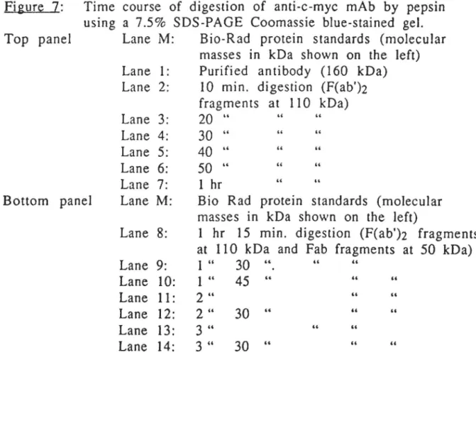
2.3 Anti-c-myc ... 67
3. Immunofluorescence ... 70
3.1 Biological activity of F(ab')2 fragments ... 70
3.1.1 5-11H.6 and anti-c-myc ... 70
3.1.2 4-Ell.3 ... 70
3.2 Op ti mization of fluorochrome-conjugated an ti bodies. 73 3.2.1 FITC guinea pig ... 73
3.2.2 TRITC rabbit. ... 73
3.2.3 TRITC mouse ... 73
3.3 Optimization of guinea pig polyclonal antiviral antibody ... 77
3.4 Characterizalion of antibodies to cell markers ... 77
3.5 Cross-reactivity of species-specific fi u or oc hrome-conj uga ted anti-immu noglobu lins ... 80
3.5.1 FITCgp vs. mouse Ab ... 80
3.5.2 FITCm vs. guinea pig Ab ... 80
3.5.3 TRITCr vs. mouse Ab ... 80
3.5.4 FITCgp vs. rabbit Ab and FITCr vs. guinea pig Ab ... 84
3.6 Detection of viral antigens ... 84
3.6.1 HCV -229E ... 84
3.6.2 HCV -OC43 ... 84
4. Genome detection ... 86
4.1 Adsorption test. ... 86
4.2 Detection of HCV -229E ... 94
VI
5. Electron microscopy ... 97 6. Detection of infectious virus ... 97 7. Hybridomas ... l02 DISCUSSION ... 106 CONCLUSION ... liS ACKNOWLEDGMEJ'\TTS ... ll8 BIDLIOGRAPHY ... ll9
Table 1 Hosts and associated pathogenesis of coronaviruses.
1 5
Table 2 Immunofluorescence in Ll32 cells inoculated 7 1 with HCV -229E detected with FITC mo use
at 1/100 dilution, to verify biological activity of mAb 5-11 H.6 F(ab')2 fragments, and to verify lack of Fe receptor activity of anti-c-myc
F(ab')2 fragments.
Table 3 lmmunofluorescence in HRT-18 cells 7 2 inoculated with HCV -OC43 detected with
FITC mouse at 11100 dilution, to verify biological activity of mAb 4-El1.3 F(ab')2 fragments.
Table 4 Immunofluorescence in HRT-18 cells 7 4 inoculated \Vith HCV -OC43 to determine
the optimal dilution of FITC guinea pig to be used in the IF tests.
Table 5 Immunofluorescence In L132 cells 7 5 inoculated with HCV-229E to determine
the optimal dilution of TRITC rabbit to be used in the IF tests.
Table 6 Immunofluorescence in HRT-18 cells 7 6 inoculated with HCV -OC43 to determine
the optimal dilution of TRITC mouse to be used in the IF tests.
Table 7 Immunofluorescence in HRT-18 cells 78 inoculated with HCV -OC43 to determine
the optimal dilution of guinea pig polyclonal antibody to be used in the IF tests.
Table 8 Immunofluorescence in HRT-18 cells 7 9 inoculated with HCV -229E to characterize
antibodies to cell markers.
Table 9 Immunofluorescence to verify any 8 1 cross-reactivity between FITC guinea
pig and mou se A bs. c;
Table
10
Immunofluorescence to verify any8
2 cross-reactivity between FITC mouseand guinea pig Abs.
Table 11 Immunofluorescence to verify any 8 3 cross-reactivity between TRITC rabbit
and mouse Abs.
Table 12 Immunofluorescence to verify any 8 5 cross-reactivity between FITC guinea pig
and rabbit Abs, and FITC rabbit and guinea ptg Abs. Table 13 Infectious viral particles in extracellular 1
0
1medium of cultures of cells inoculated with HCY -229E.
Table 14 Infectious viral partiel es in extracellular 1 0 3 medium of cultures of cells inoculated with
HCV-OC43.
Table 15 List of isotypes of the obtained mou se 1 0 4 anti-OC43 monoclonal antibodies.
Figure 1 Profile of the refractive index of fractions 6 0 collected from a continuous Nycodenz®
gradient used for the purification of HCV -OC43.
Figure 2 Coomassie blue-stained 7.5% SDS-PAGE gel 6 2 to detect pepsin digestion products of mAb
5-11H.6.
Figure 3 Ti me course of digestion of mAb 5-11 H.6 6 3 by pepsin, using a 12% SDS-PAGE Coomassie
stained gel.
Figure 4 Coomassie blue-stained 12% SDS-PAGE gel to 6 5 detect pepsin digestion products of
mAb 4-E11.3.
Figure 5 Time course of digestion of mAb 4-Ell.3 6 6 by pepsin, using a 12% SDS-PAGE
Coomassie blue-stained gel.
Figure 6 Coomassie blue-stained 7.5% SDS-PAGE gel 6 8 to detect pepsin digestion products of
anti-c-myc mAb.
Figure 7 Time course of digestion of anti-c-myc mAb 6 9 by pepsin, using a 7.5% SOS-PAGE Coomassie
blue-stained gel.
Figure 8 Human fetal astrocytes at 2 days 8 7 post-inoculation with HCV-OC43.
Figure 9 Human fetal astrocytes at 4 days 8 8 post-inoculation with HCV -OC43.
Figure 10 Hum an fetal astrocytes at 7 da ys post-inoculation with HCV -OC43. Figure 11 Human adult microglia at 2 days
89
post-inoculation with HCV -OC43. Figure 12 Human adult microglia at 4 days
post-inoculation with HCV -OC43. Figure 13 Human adult microglia at 7 days
post-inoculation with HCV -OC43.
9 1
92
Figure 14 Mix of human adult oligodendrocytes 9 3 and astrocytes at 7 days post-inoculation
with HCV-OC43.
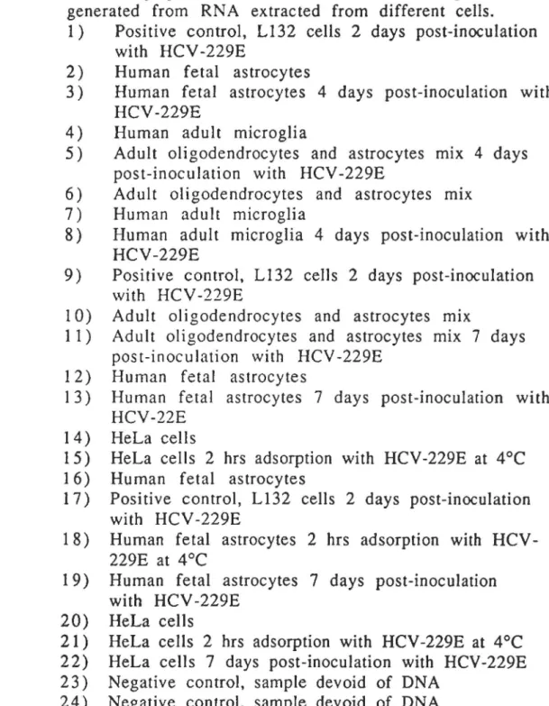
Figure 15 Autoradiograph of a Southern blot of 9 5 RT-PCR products generated from RNA
extracted from different cells.
Figure 16 Ethidium bromide stained 1.5% (w/v)
9
6 agarose gel showing RT-PCR amplificationproducts using different amounts (in )lg) of RNA from L 132 ce ils infected with HCV -229E.
Figure 17 Autoradiograph of a Southern blotting with products of a RT-PCR using different amounts (in llg) of RNA from Ll32
cells infected with HCV-229E.
Figure 18 Electron microscopy of ultra-thin cuts of human fetal astrocytes inoculated with HCV -OC43 at 7 da ys post-inoculation. Figure 19 Electron microscopy of ultra-thin cuts of
human ferai astrocytes inoculated with HCV -229E at 7 da ys post-inoculation.
98
99
100
Ab
ATCC
BCV
bp eDNACMV
CNPCNS
DEPC
DIF DNA EBV EDTA EMFBS
FIPV FITC GFAP gpHCV
HEVHEP ES
HIV An ti bodyAmerican Type Culture Collection Bovine coronavirus
Base pairs
Complementary DNA Cytomegalovirus
Cyclic nucleotide phosphodiesterase Central nervous system
Die thylpyrocarbon a te Double immunofluorescence Deoxyribonucleic acid Epstein-Barr virus Ethylenediaminetetraacetic acid Electron microscopy
Fetal bovine serum
Feline infectious peritonitis virus Fluorescein-conjugated antibody Glial fibrillary acidic protein Guinea ptg
Human coronavirus
Hemagglutinating encephalomyelitis vtrus
N -2-Hydroxyethylpiperazine-N'-2-ethanesulfonic a cid
HTLV-1 HSV IBV IF Ig ISH LFA
rn
mAb mRNA:MHV
MSNSE
NY ORF PBS PCR PFU r RERRNA
RTSDS
SDS-PAGE SIF TCIDsoHuman lymphotropic virus type 1 Herpes simplex virus
Infectious bronchitis virus Immunofl uorescence
Immunoglobulin
In situ
hybridizationLymphocyte function antigen Mou se
Monoclonal antibody Messenger RNA
Murine hepatitis virus Multiple sclerosis
Neuron specifie enolase Nuclear yellow
Open reading frame
Phosphate buffered saline Polymerase chain reaction Piaque-forming unit
Rabbit
Rough endoplasmic reticulum Ribonucleic acid
Reverse transcription Sodium dodecyl sulfate
SDS-Polyacrylamide gel electrophoresis Simple immunofluorescence
Lethal tissue culture infective dose
1EMED
TGEV
TRITC
N ,N,N' ,N' -tetramethyl-ethylenediamine
Porcine transmissible gastroenteritis virus
SOM!\'IAIRE
Les coronavirus humains se regroupent en deux souches sérotypiques, 229E et OC43, et causent des maladies respiratoires (Myint, 1994), gastro-intestinales (Resta
et al.,
1985), et ont été associés à des myocardites (Riski and Hovi, 1980). Plusieurs observations suggérent que les coronavirus humains pourraient aussi être neurotropes. Des particules coronavirales furent observées dans des échantillons pro\·cnan t de cerveaux de patients atteints de sclérose en plaques (Tanaka et al .. 1976). Des taux d'anticorps anti-coronavirus humains élevés ont aussi été trouvés dans le liquide céphalo-rachidien de patients atteints de sclérose en plaques, ce qut suggère une implication possible de ce vHus dans l'étiologie de la maladie (Salmi et al., 1982). Deux souches de coronavirus ont été isolées a partir d'échantillons pro\'enant du cerveau de patient atteints de sclérose en plaques (B urks et al., 1980) et 1 'inoculation intracérebrale de ces \'Jrus à des primates a provoqué une maladie démyélinisante (1\1urray et al.. 1992). En plus, le génome du coronavirus humain 229E (HCV-229E) a été amplifié à partir d'échantillons de patients atteints de sclérose en plaque mais pas de témoins (Stewart et al .. 1992).Ces études sont en accord avec l'hypothèse que les coronavuus humains pourraient être i mpl iq ués dans 1 'étiologie de maladies neurologiques comme la sclérose en plaques. Par contre, le neurotropisme comme tel n'a pas été prouvé jusqu'à date. Des études
avec des lignées continues ont démontré que les coronavirus humains peuvent infecter des cellules nerveuses murines et humaines, et que l'infection est productive (Pearson and Mims, 1985; Talbot et al., 1994; Talbot, communication personnelle). Toutefois, ces lignées cellulaires étant immortalisées, il est possible que leur infection ne reflète pas correctement le contexte naturel. Le but du projet de maîtrise était, donc de déterminer 1 'i nfectabi li té des cellules nerveuses humaines en culture primaire par les coronavirus humains 229E et OC43, une étape importante dans la démonstration du neurotropisme des coronavuus humains, les cultures primaires se rapprochant le plus de la situation
in vivo.
Pour détecter les :11Higènes viTaux exprimés dans les cellules infectées, nous a von s ut i 1 i sé 1 a méthode d' immunofl uorescence
indirecte. Pour dé tee ter les antigènes produits par HCV -229E, nous avons utilisé 1 'anticorps monoclonal 5-11 H.6 et un anticorps polyclonal de lapin, les deux dirigés contre 229E. Nous n'avons pas pu détecter la présence d'antigènes viraux, 111 clans les cellules humaines neuronales
et astrocytaires fœtales, ni clans les microglies, oligodendrocytes ou astrocytes adultes humains. Pour détecter les antigènes d'HCV-OC43, nous avons utilisé 1 'anticorps monoclonal 4-Ell.3 anti-HEV et un anticorps polyclonal cie cobaye anti-OC43. Ce dernier anticorps fut produit expressément pour les expériences présentées ici. Des antigènes de HCV -OC43 ont été détectés dans des cellules humaines astrocytaires fœtales, microgl ies et astrocytes adultes, mais n'ont pas
été détectés dans les neurones fœtaux m dans les oligodendrocytes adultes.
Étant donné les résultats négatifs en immunofluorescence dans le cas de HCV-229E, nous avons utilisé une technique beaucoup plus sensible, le RT -PCR/Southern blotti ng, qui permet de détecter 1 'ARN viral dans des cellules infectées. Avec cette technique, nous avons pu détecter l' ARN de 229E dans des cellules humaines astrocytaires fœtales, microglies adultes et dans un mélange d' oligodendrocytes et astrocytes adultes. Les échantillons de neurones étant très difficiles à obtenir, ils n'ont pas pu être testés avec cette technique.
Nous avons aussi pu détecter par microscopie électronique la présence d'HCV -OC43 dans des astrocytes fœtaux infectées en utilisant la méthode des coupes ultra-minces.
Nous avons clétectl~ ct quantifié les particules virales infectieuses produites après l'infection cles cultures primaires par la méthode d 'immunoperoxydase indirecte. Des particules infectieuses d 'HCV-229E et d'HCV-OC43 ont ~llnSJ été détectées dans des cultures d'astrocytes fœtaux infcct~s. Egalement, un taux très bas de particules infectieuses d 'HCV -OC4~ a été détecté dans les cultures de microglies et d'oligodendrocytes/astrocytes adultes. La différence dans les titres de vuus produits pourrait être due à la différence dans la concentration de cellules clans chaque échantillon. Étant donné que
nous avons travaillé avec des cultures pnmaues, nous ne pouvions pas contrôler ce paramètre.
Finalement, nous avons produit cmq hybridomes sécrétant des anticorps monoclonaux an ti -OC43.
Les résultats de ce projet de recherche montrent 1 'infectabilité de certains types de cellules gliales humaines par les deux souches prototypes du coronavirus humain.
La différence clans les degrés d'infectabilité par les deux souches du coronavuus humain n'est pas surprenante car ce phénomène a été montré dans le modèle de souris (Dubois-Dalcq et al., 1982; Gagneten et al., 1995). Le type de cellules qui sont infectables est similaire à celui observé dans l'étude fait avec HCV-OC43 et les cellules nerveuses primaires de souris et clans celui fait avec des cultures pnmaues foetales humaines (Pearson et !\1ims, 1985). Il est intéressant de constater que les résultats clans le modèle de souris concordent avec les résultats chez 1 'humain. Par contre, les résultats obtenus en culture pnmaue ne correspondent pas tout à fait avec ceux en lignées continues où les deux souches virales, 229E et OC43 montrent une haute infectabilité. un grand wux de production virale et où tous les types de cellules nerveuses sont infectables (Talbot, communication personnelle). Même si notre étude présente de l'information plus détaillée dans la caractérisation de 1 'infection des cellules nerveuses humaines par les coronavirus humains, il y encore beaucoup à faire
xviii
dans le domaine pour comprend re le neurotropisme des corona virus, et faire le lien avec des maladies neurologiques. Finalement, il est intéressant de noter que les coronavirus humains semblent infecter les mêmes types de cellules nerveuses que le virus de l'immunodéficience humaine, soit les astrocytes et les microglies (Sharpless et al., 1992)
The goal of this project was to determine the infectability of human primary cultured neural cells by human coronaviruses (HCV) 229E and OC43.
To detect viral antigens expressed m infected cells we used an indirect immunofluorescence technique. To detect antigens of HCV-229E we used monoclonal antibody 5-IIH.6 and a rabbit polyclonal antiserum, both anti-229E. \Ve could not detect the presence of viral antigens in infected human fetal neurons, fetal astrocytes, adult microglia, adult oligodendrocytes or adult astrocytes. To detect antigens of HCY -OC43 we used monoclonal an ti body 4-E 11.3 anti-HEV and a guinea pig polyclonal antibody to OC43. The latter reagent was produced for these experiments. Viral antigens were detected in infected human ferai astrocytes, a du lt microglia and adult astrocytes but not in fetal neurons and adult oligodendrocytes.
To verify the negative results obtained with HCV -229E we used a more sensitive technique, RT -PCR/Southern-blotting, to detect viral RNA in infected cells. With this method, we could detect HCV -229E RNA in fetal astrocytes, adult microglia and in a mtx of adult oligodendrocytes and astrocytes. The neuron samples were scarce so we could not test them with this technique.
Also, we have detected the presence of HCV-OC43 in infected cells by electron microscopy of ultra-thin sections.
possible for HCV -229E.
This was not
We have detected and quantified the infectious particles
produced after infection of the primary cultures using the indirect
immunoperoxidase method. Infectious HCV -229E and HCV -OC43
particles were detected in cultures of infected fetal astrocytes. Low
amounts of infectious OC43 were also detected in infected adult astrocytes, oligodendrocytes and microglial cultures.
Finally, five hybridomas secreting monoclonal antibodies
directed against HCV -OC43 were produced.
Human coronaviruses are known to cause respiratory (Myint, 1994) and gastrointestinal (Resta
et al.,
1985) diseases, and have been associated with myocarditis (Riski and Hovi, 1980). Human coronaviruses may also be neurotropic. The study of neurotropic human coronaviruses arase when coronavirus-like particles were observed in samples from the brain of a multiple sclerosis patient (Tanakaet al.,
1976). Since then, high titers of anti-human coronaviral antibodies were found in the cerebrospinal fluid of multiple sclerosis patients, suggesting a possible implication of this virus in the etiology of the disease (Salmi etal.,
1982). Two corona viral strains were isolated from brain samples of MS patients (Burks etal.,
1980) and their intracerebral inoculation in primates caused a demyelinating disease (Murray et al., 1992a). Furthermore, HCV -229E genome was amplified from brain samples of MS patients (Stewart et al., 1992).These studies are consistent with the hypothesis implicating coronaviruses in neurological diseases. However, the neurotropism of human coronaviruses has not yet been proven. Studies in continuous cell tines have shown that human coronaviruses can infect mouse and human neural cells and that the infection can be productive and persistent (Pearson and Mims, 1985; Talbot et
al.,
1994; Talbot, persona! communication). The project described here was designed to study the infectability of primary human neural cells by the two known strains of human coronaviruses, 229E and OC43. To support the hypothesis of a possible neurotropism, we wanted to determine which cell type was infectable and if the infection was productive.1.
Coronayjruses1.1 Isolation of coronaviruses from humans
Human coronavuuses (HCV) belong to the family Coronaviridae
(Tyrrell et al., 1975). Two serogroups are known, and only one strain per group has been weil characterized (Myint, 1994 ). The se strains are HCV -229E for group 1 and HCV -OC43 for group 2. Other hu man strains can be found in the literature. Among them are B814, EVS, LP, 692, and other OC strains (Kendall et al., 1962; Kapikian et al., 1973; Mclntosh et al., 1967b).
In 1962, a study was carried out to isolate vuuses from patients with common colds (Kendall et al., 1962). This study yielded the first isolation of the B814 strain. It was not until 1965 that it was reported that B814 was an ether labile virus that failed to be maintained in tissue culture and eggs. It could only be propagated in organ cultures of human fetal tracheal epithelium (Tyrrell and Bynoe, 1965). The same year, strain 229E was isolated from a patient with an upper respiratory tract infection. The vtrus was characterized as an ether-labile particle with a diameter of 89 nm. The virus was unrelated to the known human respiratory myxoviruses and had an RNA genome (Hamre and Procknow, 1966). In 1967, strains 229E and B814 were detected by electron microscopy and were shown to be related to avian infectious bronchitis virus (Almeida and Tyrrell, 1967). Other studies determined that it was possible to identify infectious bronchitis virus (lB V)-Iike or mou se hepatitis virus
like particles in specimens obtained from patients with a common cold (Mclntosh et al., 1967a). La ter in 1967, the previously isolated strains OC38 and OC43 (OC for organ culture) were successfully grown in suckling-mouse brains (Mcintosh et al., 1967b). In 1968, more viral isolations from patients with upper respiratory infections were reported: of these, nine samples were characterized and matched to 229E (Kapikian et al., 1969). For the first time, sorne epidemiological data was provided, including incidence of viral infection in the population according to age and also sorne serological studies.
In 1969 the Coronaviruses were classified as a new group by an informai group of virologists composed of J.D. Almeida, D.M. Berry, C.H. Cunningham, D. Hamre, M.S. Hofstad, L. Mallucci, K. Mclntosh, and D.A.J. Tyrrell (Almeida et al., 1969). La ter in 1969, it was reported th at 229E could be grown m L 132 ce lis (Bradburne, 1969). More epidemiological studies for OC strains and for 229E followed that report. A strong correlation was found between coronavuus infection and LRTD (lower respiratory tract disease) in infants and no correlation was found with upper respiratory infections. A difference in serum titers of 229E was also found in adults and in children (Mclntosh et al., 1970a). In 1970, strains OC38 and OC43 were successfully adapted to grow in cell monolayers of monkey origin (Bruckova et al., 1970). In 1972, strain 692 was detected by immune electron microscopy, and serological studies revealed th at is was not related to either 229E or OC43 (Kapikian et al., 1973 ).
Currently more than a dozen coronaviruses have been isolated from patients and the existence of HCV is weil accepted among the
scientific community. Recent studies focus on the characterization of
the viruses and understanding their replication mechanisms and
pathogenesis. The two serologically different strains, HCV -229E and
HCV -OC43, are adapted to cell culture, and were chosen as the prototypes for further studies.
1.2 Physical description
Human coronaviruses are large enveloped vuuses. They can be
detected by electron microscopy and show pleomorphic forms of about
80 to 200 nm in diameter. Their non-segmented genome is composed
of an infectious single-stranded positive RNA of 27-30 kb. The
buoyant density in sucrose is 1.18 g/ml (Pokorny et al., 1975). Human
coronaviruses are heat labile: they can be inactivated at 33°C or at
37°C (Bucknall et al., 1972). Strain 229E is more labile but retains
more infectability after freeze-thawing. Both strains are susceptible to
pH changes, but OC43 is more stable (Bucknall et al., 1972). Strain
229E requires a pH of about 6 for optimal growth (Lamarre and
Talbot, 1989). Both strains are also sensitive to liposolvents. Growth
of 229E in L132 cells can be inhibited by actinomycin D (Kennedy and
Johnson-Lussenburg, 1978). Strain OC43 can hemagglutinate human
group
0
erythrocytes, while 229E cannat (Kapikian et al., 1972). StrainOC43 may induce fusion of susceptible cells (Bruckova et al., 1970).
Each coronavirus particle contains approximately 200 peplomers
of 10 to 20 nm embedded in its lipid bi-layered surface which gives
them the appearance of a crown ("corona"). Human coronaviruses
have at least three structural proteins: S, M, and N (spike, membrane
associated protein and nucleocapsid respectively), with HE
(hemagglutinin esterase) being an additional structural protein in
HCV -OC43. The corona virus genome also codes for se veral
non-structural proteins that appear to be essential for viral genome
replication.
1.2.1 Surface protein (S)
The peplomers on the surface of coronaviruses are formed by
dimers or trimers of the S glycoprotein, previously named E2, (Myint,
1994 ). The S prote in has a molecular mass of 160-200 kDa and is
formed by two dissimilar portions called S 1 and S2 that can be
separated by digestion with trypsin (Sturman
et al.,
1985). The Sprotein has been implicated in the recognition of the cellular receptor,
cell mediated cytotoxicity, and cell fusion (Sturman
et al.,
1985), pH dependent thermolability, inhibition of hemagglutination and alsoneutralization (Collins et
al.,
1982; Schmidt and Kenny, 1982; Daniel and Talbot, 1990). The S protein bears different post-translational modifications such as acylation and glycosylation (Ricard etal .•
1985; Cavanagh, 1983). These modifications may be responsible for the1.2.2 Membrane protein (M)
The M prote in, previously named E 1, is the most abundant protein in the membrane and has a molecular mass of 25-26 kDa. Proteins of approxima tel y 20 , 24 , 27, and 40 kDa, bearing different levels of glycosylation, are observed and are probably linked by disulfide bonds (louvenne
et al.,
1994). The M protein varies in the extent of glycosylation, the type of linkage (N-link and 0-link), and the degree to which N-linked high-mannose glycans have been converted to complex glycans (Spaan etal.,
1990). The M protein is highly hydrophobie and bears threea
-helical trans membrane demains (louvenne etal.,
1990; Mounir and Talbot, 1992). The N-terminal half is exposed at the outer surface of the membrane (Rottieret al.,
1986),while the C-terminal half is located in the interior of the virus (Spaan
et al.,
1990). The protein was also found to interact with the nucleocapsid (Sturman et al., 1980; Spaan et al., 1988). The M protein apparently supports the structure of the envelope and is essential for virus budding (Mounir and Talbot, 1992). An ti bodies against the M protein require complement to neutralize viral infectivity (Collins etal.,
1982).1.2.3 Nucleocapsid protein (N)
The N protein has a molecular mass of 47-55 kDa. It is a basic non- glycosylated protein th at encapsidates the RN A genome in a flexible nucleocapsid with helical symmetry (Kennedy and
Johnson-Lussenburg, 1975). It combines three structural, and most
likely functional, domains, designated 1, II and III. Domains 1 and II
contribute to the basic character of the protein, having a large amount of positively charged residues while domain III has negatively
charged residues (Masters et al., 1990). This protein is thought to
have an important role in the replication cycle of the virus. Variation in the concentration of the N protein might be responsible for the
switch from transcription to replication (Stohlman and Lai, 1979).
It
has been reported that this protein may be involved in the immune
response against coronaviruses (Korner et al., 1991; Lecomte et al.,
1987).
1.2.4 Hemagglutinin-esterase (HE)
The HE protein, previously called E3, is only found m the OC43
strain of HCV. It has a molecular mass of 130-140 kDa and is
composed of two subunits of 65 kDa linked by disulfide bonds (King and Brian, 1982; Hogue and Brian, 1986), The HE protein recognizes sialic acid-containing receptors similar to those for influenza C viruses
(Vlasak et al., 1988).
1.2.5 Non-structural proteins
The non-structural proteins are proteins that are not packaged m the virion and are only synthesized and found in an infected host
proteins. The gene coding for the RNA dependent RNA polymerase
occupies the 5' two thirds of the viral genome and is called mRNA 1.
Strain 229E contains two ORFs in mRNA 4 and one ORF in mRNA 5
(louvenne et al., 1992). Strain OC43 contains two ORFs between the
genes of the S protein and the M protein (Mounir and Talbot, 1992). The functions of these non-structural proteins are still unknown but they are probably implicated in the synthesis of RN A or in the encapsidation process.
1.3 Cellular receptor
In the munne model, the S protein has been implicated m the
recognition of the cellular receptor (Sturman et al., 1985). Two
glycoproteins of 110 kDa and 58 kDa have be en fou nd to bi nd MHV
(Boyle et al., 1987; Holmes et al., 1994 ). The se glycoproteins belong to
the carcinoembryonic antigen family and are present on the surface of liver, intestinal epithelium, respiratory epithelium, brain and spleen
cells. Holmes et al., have shown that different strains of MHV shared
this cellular receptor (Holmes et al., 1989).
In hu mans, the cellular receptor for HCV -229E is aminopeptidase N, a cell-surface metalloprotease present on intestinal, Jung and
kidney epithelial cells (Yeager et al., 1992). The cellular receptor for
HCV -OC43 is stiJl unknown.
1.4 Genome sequence
The complete nucleotide sequence of the HCV -229E RNA is now known: mRNA 1 (Herold
et al.,
1993), mRNA 3 (S protein) (Raabeet al.,
1990), mRNA 4 and mRNA 5 (Raabe and Siddell, 1989b; louvenne
et
al.,
1992), mRNA 6 (M protein) (Raabe and Siddell, 1989a; louvenneet
al.,
1990), mRNA 7 (N protein) (Schreiberet al.,
1989; Myintet al.,
1990). For OC43, sequences are known for the N protein (Kamahora
et
al.,
1989), M protein (Mounir and Talbot, 1992), S protein (Mounir and Talbot, 1993b; Künkel and Herrler, 1993 ), HE (Zhanget al.,
1992), and mRNA 4 (Mounir and Talbot, 1993a).For HCV -229E, it has been reported th at the gene sequence for the N protein is highly homologous to the N protein of porcine transmissible gastroenteritis virus, TGEV, (Schreiber
et al.,
1989), and that the gene for the S protein is highly homologous to the S protein of IBV, feline infectious peritonitis virus (FIPV), TGEV, and MHV-JHM (Raabeet al.,
1990). For HCV -OC43, it has been determined that there is high leve! of identity between the gene for the N protein with the homologous one of bovine coronavirus (BCV). Similarly, there is relatedness between the leader sequence with MHV (Kamahoraet al.,
1989), the M protein with BCV (Mounir and Talbot, 1992), HE with BCV (Zhang
et al.,
1992), and the S protein with BCV (Mouniret al.,
1994 ). A phylogenetic relation has also been established between OC43 and influenza C virus, due to high homology between their HE protein (Zhang
et al.,
1992).1.5 Replication
The steps of adsorption, penetration and uncoating of coronaviruses are not weil understood. It is thought that virus attaches to the cellular receptor through the S protein, or the HE protein if present (Boyle et al., 1987; Talbot et al., 1984). It is not clear if penetration occurs by fusion of the virus envelope with the plasma membrane or with endosomal membranes (Krzystyniak and Du puy, 1984; Mizzen et al., 1985; Kooi et al., 1991 ). Once the virus uncoats, its genomic RNA, which is capped and poly-adenylated, attaches to ribosomes Ieading to the synthesis of the virus-specifie RNA-dependent RNA polymerase (Strauss and Strauss, 1983). This polymerase may be distinct from the polymerase responsible for the production of subgenomic mRNAs. The newly synthesized polymerase transcribes the positive RNA into a full length negative strand RNA (Lai et al., 1982a). White the exact mechanism is not understood, the minus strand then serves as the template for transcription of a nested set of 5 to 7 positive stranded subgenomic RN As which are 3' coterminal (Lai et al., 1982a; Stern and Kennedy, 1980). These mRNA are capped and polyadenylated (Siddell et al., 1983; Spaan et al.,
1988; Stern and Kennedy, 1980). The translated ORF in each mRNA is situated at the non-common 5' end (Siddell et al., 1982; Sturm an and Holmes, 1983 ).
The 5' end of the plus-strand genomic and subgenomic RNAs
con tains an identical leader sequence of 60-70 bases (Barie et al.,
1983; Brown et al., 1984; Lai et al., 1982b). This leader sequence is important in the replication of coronaviruses, although how the leader
functions is not clear. One model for coronavirus RNA transcription is
the discontinuous, leader-primed transcription mechanism. The leader
sequence, after being transcribed from the 3' end of the minus strand,
disassociates from its template and moves downstream, reattaching to
the template by means of a recognition signal which is complementary
to the leader. Transcription then restarts. This process is repeated
several times at the initiation site for each subgenomic RNA (Barie et al., 1985; Budzilowicz et al., 1985; Lai, 1986; Shieh et al., 1987). This leader primed transcription may explain the high frequency of
recombination found in coronaviruses and also the existence of
defective interfering particles (Makino et al., 1988). Because of their genome, coronaviruses are sensitive to a high frequency of mutation.
Most coronavirus proteins are translated in polysomes either
attached to the RER or in the cytoplasmic matrix. Sorne proteins (S,
HE) are co-translationally N -glycosylated whereas the M protein of
HCV -OC43 ts post-translationally 0-glycosylated. Virion assembly
takes place m the cytoplasm. The genome is encapsidated by the N
protein which starts the process by binding to the leader sequence
(Stohlman et al., 1988). The virion then buds from the RER or Golgi apparatus (Tooze et al., 1984), where the cellular proteins are excluded. Coronavirus budding only occurs where there is a high
concentration of the M protein (Holmes
et al.,
1981; Holmeset al.,
1984 ). The li fe cycle is therefore entirely. cytoplasmic.1.6 Pathogenesis
Coronaviruses are in general host specifie. Various strains infect
a vast spectrum of animais including cat, cattle, chicken, dog, horse, monkey, mouse, ptg, rabbit, rat, sheep, and turkey, causing different diseases (Table 1 ).
Human coronaviruses cause up to 30% of common colds in
humans (Myint, 1994 ). The incubation period is 2 to 4 da ys and the
symptoms include malaise, headache, profuse rhinorrhoea, nasal
blockage, sneezing, fever and abdominal pam. Studies done on adult
sera show a high frequency of neutralizing antibodies to HCV-229E and also high titers of an ti bodies against HCV -OC43. Infections by the 229E and OC43 strains of HCV seem to fluctuate from year to year
(Mclntosh
et al.,
1970b; Bradburne and Somerset, 1972). A studyspanning 4 years on more than four hundred patients showed that
90% of adults were seropositive for HCV (Chambon
et al.,
1987). Otherstudies in infants with acute lower respiratory tract disease revealed th at 8.2% of patients aged un der 18 months were HCV -seropositive
(Mclntosh
et al.,
1974). The geographical distribution of coronavirusinfections is very wide, HCV respiratory infections have been detected
in North and South America, and in Europe. Human coronaviruses
have also been linked to other diseases. For example coronavirus-like
Table 1: Hosts and associated pathogenesis of coronaviruses.
Other: lnfectious peritonitis, runting, nephritis,
pancreatitis, parotitis, and adenitis.
Abbreviations: HCV, human coronavirus; TGEV porcine transmissible gastroenteritic; CCV, canine corona virus; FECV, feline enteric coronavirus; FIPV, feline infectious peritonitis virus; l\1HV, murine coronavirus; SDAV, sialodacryadenitis virus; HEV, porcine hemagglutinating encephalomyelitis virus; BCV, bovine coronavirus; RbCV, rabbit coronavirus; IBV, avian infectious bronchitis vuus; TCV, turkey coronavirus.
G-oup Virus fust Respiratory Blteric 1-i!patitis CNS Qher infection infection infection
1 229E Hu man
x
?
1ŒV Pigx
x
x
(Dl lligx
FKV Citx
APVCàt
x
x
x
x
x
IIœ3
Hum anx
? ? ~flN Mou sex
x
x
x
SD\V Ratx
HEV Pia 0x
x
x
ocv
Cbwx
Rb CV Rab bitx
x
III IBV Olicken
x
x
particles were observed by electron microscopy in stools of patients
with necrotizing enterocolitis. Stool samples were passaged in cultures
of human fetal intestinal organs and coronaviral-like particles were
produced (Resta et al., 1985). It has also been associated with
myocarditis (Riski and Hovi, 1980). Riski and Hovi described a patient
with a common cold who developed myocarditis. High titers of
antibodies against OC43 were detected in this patient. No other virus
could be detected and ali the bacterial tests were negative. Upon
recovery, the serum titer of antibodies against OC43 declined,
suggesting a link between HCV and myocarditis. There is also strong
evidence to suggest that HCV may be a neurotropic virus and that it could be involved in neurological diseases, such as multiple sclerosis, as discussed below.
2. Multiple sclerosis
2.1 Pathology
Multiple Sclerosis is a neurologie demyelinating disease. It ts
characterized by chronic demyelination, inflammation and gliosis. In
North America, it represents the most important neurological illness m
early to middle adulthood. There are 50,000 people affected tn
Canada (Talbot, 1995), and as many as 350,000 in the United States
(Hauser, 1994). The exact cause 1s still unknown but there is
substantial evidence to believe that an environmental agent, perhaps a virus, could trigger the illness in a genetically susceptible host (Johnson, 1985).
1 7
Examination of the brain of patients at autopsy reveals sclerotic plaques or scar tissue areas visible in the white and, exceptionally, in the gray matter. Other histopathologic characteristics are perivenular cuffing, tissue infiltration by mononuclear cells, T lymphocytes or macrophages. There is a substantial loss of myelin sheaths and a clear symptom of astrocyte growth (gliosis). It seems that at the initial state of demyelination there is also oligodendrocyte proliferation (Hauser, 1994 ). About 35% of MS patients present no clinicat signs and diagnosis of the disease is only possible at autopsy (Hauser, 1994). The symptoms of the disease vary from patient to patient. The first symptoms are either very rnild, with no need to consult a physician, or very severe. Sorne of these include weakness in the lirnbs, visual blurring, sensory disturbances, diplopia, ataxia, and loss of dexterity.
Three classes of disease have been identified: relapsing-remitting MS, where the patient experiences recurrent attacks during short or extended periods of time, followed by complete, partial or no recovery; chronic progressive MS, where the patient undergoes a graduai progressive worsening forrn of the disease, the patient may also have sorne acute relapsing attacks; and benign MS, where the patient presents a fixed neurologie deficit thar can vary in magnitude (Hauser, 1994 ).
2.2 Demographie and geographical data
Even though multiple sclerosis has been detected in different
areas of the globe, it seems to follow a definite geographical pattern.
Epidemiological studies show that the disease follows a North-South gradient in the Northern Hemisphere, with higher incidence further
from the equator (Kurtzke, 1980). There is aJso a clearly increased
risk of women developing MS, with a two fold preponderance in
women (Duquette et al., 1992). The disease shows a higher prevalence
m sorne races: it has a high incidence in Caucasians, is extremely rare
m Japan, and is unknown in black Africa (Ebers and Sadovnick, 1993).
The highest prevalence was detected in the Orkney islands, north of
Scotland, wirh a ratio of 250/100,000 (1 in 400). Multiple sclerosis is
not seen in early childhood. The disease manifests itself in patients 20
to 50 years-old, with the highest onset at about 30 years of age.
2.3 Genetics
The results of epidemiological studies determining that specifie ethnie groups where at higher risk suggest that a genetic factor could
be involved in the etiology of multiple sclerosis.
It
has been reportedthat major histocompatibility alleles A3, 87, DR2, and DW2 on
chromosome 6 are frequently found in white Caucasian MS patients
(Oger et al., 1987). Other alleles like DR15, and DQ6 have also been
19
alleles A2, B 12, DR7. and DW7 are infrequently fou nd in MS (Oger et
al.. 1987). It is generally accepted th at the disease susceptibility trait
is in the HLA-DR-DQ subregion. Other genetic traits have been linked
to MS, such as the S and SS allotype of properdine (Stewart et al.,
1979), and sorne Gm allotypes of IgG (Salier et al.. 1981 ).
Family studies have also provided interesting data. Children of
MS patients have 1% more probability of. getting the disease, and this
percentage is increased to 3% among brothers and sisters (Sadovnick
and MacLeod, 1981 ). Twin studies revealed th at monozygote twins
are 25% and dizygote twin are 8% more likely to develop the illness. These values are Jess than would be expected of a typical hereditary disease (McKay and Myrianthoroulos, 1966).
2.4 Environmental hypothesis
The geographie distribution of MS has led many scientists to postulate why people are more susceptible to the disease in specifie
areas. Besides the possible genetic component mentioned above, it has
been established that people who migrate from a high to low risk area
after late adolescence are still at high risk (Alter et al., 1966a; Alter et
a
1 .,
1966b; Dean and Kurtzke, 1971; Kurtzke, 1991 ). A very interesting, extensive study in the Faroe islands clearly showed this"transmission" of the disease. Multiple sclerosis was undocumented in
the islands prior to World War II. Contact with mainland Europe was
the islands to establish a military base. Interestingly, after the war, and up to twenty years later, an epidemie of MS was observed
(Kurtzke and Hyllested, 1986). This strongly suggests that the British
troops brought with them an environmental agent that causes multiple sclerosis.
2.5 Other viruses implicated in I\1S
It is now generally accepted that an environmental agent like an
infectious pathogen could be linked to the development of MS. Among
these infectious agents, viruses are important candidates. Not only do
data in humans support this idea but there are weil described demyelinating diseases caused by viruses in animais as well (e.g.
coronavirus (Wang, et al., 1990), and Theiler's virus (Welsh et al.,
1990) in mice and Yisna virus (Panitch et al., 1976) in sheep. Yiruses
are also known to cause illnesses m the central nervous system of
humans. For example, measles virus causes subacute sclerosing
panencephalitis (SSPE) (Swoveland, 1991 ), JC virus causes multifocal
leukoencephalopathy (Sweeny et al., 1993), and HTLV -I induces
tropical spastic paraparesis/HTL V-I associated myelopathy
(TSP-HAM) (Rodgers-Johnson, 1 994). The viral hypothesis is also supported
by the fact that many studies revealed high antiviral antibodies titers in the sera and cerebrospinal fluid of MS patients, suggesting a viral
infection of the central nervous system (Salmi et al., 1982; Allen and
2 1
have been designed to identify candidate viruses. Viral isolation or
detection experiments have been performed for severa! viruses.
2.5.1 l\1easles virus
An association of measles v1rus to a neurological pathology was reported in 1972 when an unusual subacute encephalitis in an infant
seemed to be caused by this virus (Bell et al., 1972). Viral sequences
have been detected in brain sections of MS and SSPE patients by in
situ hybridization (Haase et al., 1981a; Haase et al., 198Ib), by dot blot
hybridization (Dowling et al., 1986), and by PCR (Godee et al., 1990).
2.5.2 Epstein-Barr virus (EBV)
An epidemiological study of 214 MS patients measured how the relative risk of developing MS correlated with EBV-induced infectious
mononucleosis. It was found that patients that had infectious
mononucleosis showed a 2.9 fold increased risk for MS and patients who were infected be fore the age of 18 years had a 7.9 fold increased risk of developing MS, suggesting a possible age-dependent host
response to the virus (Martyn et al., 1993 ). High an ti body titers for
EBV have also been found in serological studies of MS patients (Haahr et al., 1994). However, a study using brain sections of 10 MS patients
failed to detect EBV RNA using in situ hybridization (Hilton et al.,
2.5.3 JC
virus
An extrapolation was proposed from an animal model of
progressive multifocal leukoencephalopathy caused by the
JC
polyomavirus to human multiple sclerosis. It was suggested that the
virus is latent in human brains and the antigens produced induce an
aberrant immune response in the host (Stoner, 1993). However,
JC
viral genomic sequences could not be detected in brain tissues of MS
patients (Buckle et al., 1992), and there was no evidence of
JC
virusinfection of MS patients (Boerman et al., 1993).
2.5.4 Cytomegalovirus (CMV)
Cytomegalovirus was isolated from the brain and lymph node of
a chimpanzee with acute demyelinating disease. The disease occurred
more than three years after intracerebral inoculation of brain cells
from a MS patient. In order to isolate the virus, severa! passages in
cell culture had to be assessed. This suggested that the virus was
present in the brain of the animal in a latent form. This was then
corroborated when similar studies were performed in asymptomatic chimpanzees in the colony of origin, and it was established that they
23
2.5.5 Hu man lymphotropic virus type 1 (HTL V -1)
This virus has been studied extensively in conjunction with MS. Several research groups have reported contradictory data in looking at HTL V -1 genome in peripheral blood ce Ils or in brains of MS patients (Reddy et al., 1989; Ok sen berg et al., 1990; Myhr et al., 1994; Kira e 1 al., 1994).
2.5.6 Retrovirus
Reverse transcriptase activity was detected in cultures of cells obtained from lumbar-punctured cerebrospinal fluid from an MS patient (Perron et al., 1989), and in long-term peripheral blood mononuclear cells of patients (Hollsberg et al., 1989). However, a PCR study reported that it was not possible to detect retroviral sequences in brain capillaries, brain tissue and peripheral blood mononuclear cells from MS patients (Rasmussen and Claussen, 1992). A recent study reported the presence of retroviral particles, using electron microscopy, In long-term cultures of cerebrospinal fluid and peripheral blood mononuclear ceJJs of MS patients (Haar et al., 1994 ).
2.5. 7 Herpes simplex vi ruses (HSV)
This virus has repeatedly being linked to MS because a high percentage of the population is Iatently infected before adolescence
but there is still no evidence of its implication in MS. A study using
PCR to amplify the HSV genome in brain samples of 77 MS patients
showed th at only one was positive for HSV -1, and none was positive
for HSV -2 (Ni coll et al., 1992).
2.5.8 Other viruses
Other viruses such as tick-borne encephalitis v1rus (Vagabov et
a 1., 1982), rabies virus, scrapie, and parainfluenza virus 1 (Johnson, 1985) have been isolated from MS patients. Other reports show no
detection of spumavirus, oncoviruses (Svenningsson et al., 1992), and
canine distemper virus (Cosby et al., 1989) in MS patients. Serum
an ti bodies also failed to recognize HIV -1, HIV -2 and, simian
immunodefiency virus (SIV), (Brokstad et al., 1994).
3. Coronaviruses and multiple sclerosis 3.1 HCV found in 1\1S patients
The first report linking coronav1ruses to MS was published in 1976. Coronavirus-like particles were detected by electron microscopy in a brain sample extracted at autopsy from a plaque near
25
the white matter of an MS patient (Tanaka
et al..
1976). Even though there was no immunological information or virus isolation, the findingopened a new door in the viral etiology of MS. Four years later, two
coronaviruses were isolated from MS patients (Burks
et
al., 1980). Brain homogenates from patients were cultured in suckling mice andin in vitro cell cultures known to be sensitive to coronavirus infection. Coronaviruses S.O. and S.K. were isolated from two MS patients. This was the first report of a coronavirus present in the human CNS.
The S.D. and S.K. strains were subsequently inoculated into mice. S.K. did not cause disease in inoculated animais, but S.D. proved to be
lethal. Brain homogenates were infectious upon inoculation of other
mtce. However, it was not certain that these coronavirus strains were human, especially because they could only be cultured in murine cell
Iines. They also show striking genomic identity to murine coronavirus
(Gerd es
et
al., 1981 ). It is important to remember th at hu man coronaviruses are difficult to culture in human cell lines and m factOC43 failed to be cultured In the human lines tested with S.O. and S.K. The cultures were sampled by electron microscopy and by antibody
testing and it was suggested that the viruses were not munne
hepatitis virus (MHV). Further testing of the serum and spinal fluid of
the patients revealed that antibodies against S.K. were present in the
cerebrospinal fluid of both patients. The serum antibody
concentrations against the respective isolates were elevated in the
patient from whom the isolate was obtained. Antibodies to S.K. were
et al.,
1980). The two strains were then compared to other coronaviruses and it was determined that S.K. and S.D. cross react withHCV -OC43 but not with HCV -229E, MHV -A59 or MHV -JHM in a plaque
neutralization assay. Both strains cross react by immunoprecipitation
to HCV -OC43 and MHV -A59, suggesting a possible relatedness of these
strains (Gerdes
et al.,
1981 ).Serologie studies on MS patients support the hypothesis of the
implication of the virus in the etiology of this illness. One study
showed a significant difference of anti-coronavirus antibody titers in
the cerebrospinal fluid between MS patients and controls (Salmi et al.,
1982). On the other hand, the analysis of sera showed no significant
difference between patients and contrais. Similar findings with sera
were already published by Madden et al., (Madden et al., 1981) and were Iater corroborated by a study with a large number of patients
(Hovanec and Flanagan, 1983 ).
Further studies on detection of coronavirus RNA in the brain of
MS patients were pursued. A first study failed to detect OC43 RNA in
tissues from autopsies of 4 MS patients and in biopsies from one MS
patient, using a eDNA probe and classical hybridization (Sorensen et
al.,
1986). Murine-like coronavirus RNA was found in 11 out of 21 MS patients, and in 2 out of 21 controls (16 with non-neurologicaJ diseaseand 5 with other neurological diseases) using a S.D. coronavirus eDNA
probe in in situ hybridization (ISH). The results were similar upon reassessment with a MHV -A 59 eDNA probe.
27
A study was then done usmg eDNA probes from different species
of coronavirus to determine which virus was present in positive
patient samples. Samples were also tested on 9 extra negative control
patients. Ail of the MS patients were positive for S.D., and the controls
were negative; 5 out of 12 MS patients were positive for OC43, and the
con trois were negative. There was no positive results for HCV -229E
(Murray et al., 1990; 1992b). The negative results with HCV -229E were not surprising since the samples were screened with a eDNA
probe of corona virus S .D. which has been established to be unrelated
to 229E. Another study used brain samples from autopsies of !\.-1S patients, neurologie controls and normal subjects. The RNA was
extracted from white and gray matter and RT-PCR was performed for
229E and OC43. The results revealed the presence of 229E RNA in 4
out of Il MS patients, and not in the 11 controls. There were no
positive results for OC43 (Stewart et al., 1992).
3.2 Infection of mice with HCV
Human coronavirus OC43 was lethal when inoculated
intracerebrally or extraneurally into sucling CD 1 mice. Resistance to
infection was observed with increasing age of the animal. Animais
greater than 20 days-old were not susceptible to disease after
intracerebral inoculation. Transfer of immune or non-immune spleen
cells from resistant animais did not protect newborn animais from
3.3
Infection of primateswith
HCVSorne
in vivo
tests have been conducted in primates. Owl and African green monkeys (different species) were inoculated intracerebrally with the coronavirus S.D. strain, an MS isolate. The monkeys were susceptible to infection, and showed a subacute panencephalitis. Brain autopsies showed focal areas of demyelination and pathological signs of neurological disease. Viral RNA was detected by ISH in certain areas of the brain (depending on the species), but viral antigen could not be detected (~1urray et al., 1992a).3.4 Infection of human neural continuous cell lines with HCV
Two cell lines, human rhabdomyosarcoma (RD) and human glioblastoma (U-87 MG) can be infected with OC43. At 28 days post-infection, infectious v1rus and hemagglutinating activity were observed in both cell Iines (Collins and Sorensen, 1986). Other studies of infection of continuous cell lines by human coronaviruses used the 229E strain. The Ll32 hum an embryonic Jung cell li ne, SK-N -SH neuroblastoma cell tine; H4 neuroglioma cell line; U-87 MG and
U-373
MG astrocytoma cell lines, and M03.13 immortalized human oligodendrocytes cell Ii ne were ali infectable by HCV -229E, as shown by immunofluorescence detection of viral antigen using a monoclonal an ti body (Talbot et al., 1994 ), as weil as by detection of infectious
29
viral particles (Talbot, persona! communication). Similar results have now been obtained with the OC43 strain of HCV (Talbot, persona! communication).
3.5 Infection of primary CNS cells with HCV
Primary cultures of neural mou se ce Ils were infected with HCV-OC43. Using double immunofluorescence, it was determined that oJigodendrocytes and Schwann cells were not infectable. The same technique revealed that astrocytes, neurons and fibroblasts were infectable but no cytopathic effects were observed. The only cell type that produced infectious virus was the neuron. A primary human embryo brain cell culture was also infected with the virus and immunofluorescence revealed that astrocytes were infectable. No cytopathic effects were observed, and no infectious virus was present. (Pearson and Mims, 1985).
3.6 Infection of primary human CNS with other viruses
Thwo studies have reported in vitro viral infections of human fetal cell cultures. The first study using double immunofluorescence, showed that astrocytes and fibroblasts, but not neurons, were infecta ble by Herpes simplex virus, types 1 and 2 (Kennedy
et al.,
1983). The second study showed that microglia, but not astrocytes, were infectable and could produce infectious virus after infection with hu man immunodeficiency virus type 1 (HIV -1) (Lee
et al.,
1993). Arelated study described the infection of an adult human brain cell
culture (AHB) with JC virus and the detection by double
immunofluorescence of infected astrocytes and fibroblasts
(Wroblewska et al., 1982). Similar experiments were performed with
HIV -1 and HIV -2 (B rynmor et al., 1990) where they demonstrated
th at only one strain of HIV -1 adapted to a macrophage cell li ne could
productively infect microglia, inducing cytophatic effects. Another
report showed infection with HTLV -I In astrocytes and
oligodendrocytes in a mixed glial cell culture (Watabe et al., 1989).
It
is important to note that ali these studies, including the one performed
with HCV, used a mixed population cell culture system. This is an
important issue when interpreting infectious virus titers because you
cannot predict \vhich cel! type produced them. Also in sorne reports
no negative controls were provided. There is only two reports where
primary adult human enriched neural cells have been infected in vitro
The first one was a study with HIV -1 (Sharpless et al., 1992), wh ere
astrocytes, oligodendrocytes and microglia cell cultures were infected with HIV -1, the only susceptible cells were the microglia, suggesting a
restricted tropism for HIV -1. Similarly enriched oligodendrocytes,
microglia and a mixture of microglia and astrocytes were infected with
HTL V-I founding th at only microglia were infectable (Hoff man et al.,
1992). Is interest to note that the results with astrocytes and
oligodendrocytes do not match Watabe' s previous study with mixed cultures.
1. Primary cultures of cells from human brain
Primary cultures of cell mixtures or enriched preparations of oligodendrocytes, astrocytes and microglia from human brain tissue were provided by Dr. Voon Wee Yong (Montreal Neurological Institute, Mc Gill University, Montreal, Que bec, Canada). Cells were obtained from brain biopsies performed on epileptic patients m an attempt to ameliorate an intractable form of the disease. Cells were then put m culture in Dr. Yong's laboratory follov.··ing his own protocol (Yong and Antel, 1992). Brain tissue extracted either en bloc or by CavitronTM ultrasonic aspiration were washed in phosphate buffered saline (PB S). Tissue was diced with scalpels and \vashed a few times in PBS. The tissue was th en trypsi nized in 0.25% (w/v) trypsin with 50 Il g