Université de Montréal
The roles of STRA6, EFNB1/B2 and ARMC5 in T cell function
and autoimmune diseases
par Yan Hu
Département de Médecine Faculté de Médecine
Thèse présentée
en vue de l’obtention du grade de Philosophae Doctor (Ph.D.) en Sciences Biomédicales
Décembre, 2015
Résumé
Les récepteurs tyrosine kinases sont un groupe de molécules clés de signalisation, qui ont 2 fonctions: la détection des stimuli de l'environnement extérieur des cellules et la transmission de ces signaux à l’intérieur des cellules. Dans les 20 dernières années, notre laboratoire a choisi d'étudier la fonction d’Ephb6 kinase, un récepteur tyrosine kinase fortement exprimé dans les lymphocytes T.Comme Efnb1 et Efnb2 sont tous des ligands pour Ephb6, nous avons ensuite procédé à étudier leur rôle dans la fonction des cellules T in vitro et in vivo. Des cellules T spécifiques mutants (KO) dans les gènes Efnb1 ou Efnb2 ainsi que les doubles mutants Efnb1/b2 (double KO) ont été générés, mais il n’y avait que les souris double KO qui ont démontré de la déficience dans le développement des thymocytes, fonction de Th1 et Th17, la signalisation du récepteur d’IL-6, et les réponses antivirales.
Des preuves solides indiquent que la reconnaissance d’auto-antigène par les cellules T est un événement précoce dans la pathogenèse de la PR. Donc, nous avons postulé que les cellules T spécifique Efnb1 / b2 double KO chez la souris peuvent protéger les souris de l’arthrite induite par collagène (CIA), un modèle de souris de la PR humaine. Nous avons trouvé que Efnb1et Efnb2 dans les cellules T étaient essentielles pour la production d'anticorps pathogéniques et de la migration des lymphocytes T vers les pattes enflammées chez les souris CIA. Notre étude clinique suggère que l'expression de EFNB1 dans les cellules T pourrait être un paramètre utile pour surveiller l'activité de la maladie de RA et la réponse de traitement. Pour élucider les événements dans le programme d'activation des lymphocytes T, nous avons exploré par l'analyse des micropuces d'ADN pour identifier des molécules qui ont été exprimées de manière différente dans le WT par rapport aux cellules T Ephb6 KO dans le stade précoce de l’activation des cellules T. Environ 30 molécules étaient sur ou sous exprimées plus de 3 fois dans les cellules T WT par rapport aux cellules T KO pendant les 16 premières heures après stimulation par l'anti-CD3. Stra6 (stimulée par le gène de l'acide rétinoïque 6) et Armc5 (Armadillo répéter contenant 5) ont été parmi ceux qui ont été validées pour leur expression altérée.
STRA6 est un récepteur de haute affinité pour le plasma rétinol-binding protéine (RBP) et un médiateur pour absorption cellulaire de vitamine A. Cellules T KO et WT étaient similaires en
termes de prolifération et les réponses immunitaires anti-virales de virus de la chorioméningite lymphocytaire (LCMV). Ainsi, la sur-régulation de Stra6 est soit un événement parallèle qui ne soit pas essentiel pour le programme d'activation des lymphocytes T, ou il est très essentiel que la redondance existe, et sa suppression ne montre aucun effet apparent sur l'activation des cellules T.
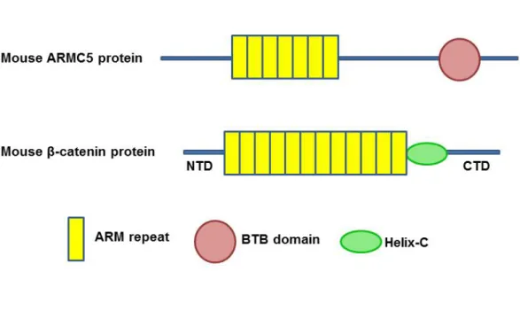
ARMC5 est une protéine intracellulaire contenant sept répétitions en tandem d’armadillo et un domaine BTB. Les fonctions du ARMC5 dans le système immunitaire ne sont pas encore connues. Nos résultats d'hybridation in situ ont montré une expression élevée de Armc5 dans le thymus, et une expression modérée dans les ganglions lymphatiques et la rate. Nous avons généré des souris KO Armc5. Fait interessant, les cellules T Armc5 KO présentaient de la prolifération diminuée et de la différenciation compromise vers Th1 et Th17 in vitro. Les souris KO étaient résistantes à l'induction expérimentale d’encéphalite auto-immune, et ont été compromises dans les réponses immunitaires anti-LCMV. En utilisant de la levure 2-hybride test, nous avons identifié 8 protéines ARMC5-associantes, qui sont connues pour les rôles dans l'activation de la cellule, le cycle cellulaire et l'apoptose. Une étude mécanique est en cours. Nos résultats montrent que Armc5 est essentiel dans la programme d'activation/de prolifération/de différenciation des lymphocytes T.
Nos études ont augmenté nos connaissances sur EFNB1, EFNB2, STRA6 et ARMC5 en biologie des lymphocytes T et leur pertinence à des troubles immunitaires dans des modèles animaux ainsi que chez l'être humaine.
Mots-clés : cellules T, EFNB1, EFNB2, STRA6, ARMC5, la polyarthrite rhumatoïde, la sclérose en plaques
Abstract
Receptor tyrosine kinases are a group of key signaling molecules, which have dual functions: sensing the environmental stimuli outside the cells and transmitting them into the cells. 20 years ago, our laboratory started to study the function of Ephb6 kinase, a receptor tyrosine kinase highly expressed in T lymphocytes. As both Efnb1 and Efnb2 are the ligands for Ephb6, we then proceeded to study their roles in T cell function in vitro and in vivo. T cell-specific Efnb1, Efnb2 single gene knockout (KO), as well as Efnb1/b2 double KO mice were generated, but only the double KO mice showed compromised thymocyte development, Th1 and Th17 function, IL-6 receptor signaling, and anti-virus responses.
Strong evidence indicates that T cells play a crucial role in the pathogenesis of rheumatoid arthritis (RA). Thus, we postulated that T cell-specific Efnb1/b2 double KO in mice may protect mice from collagen-induced arthritis (CIA), a mouse model for human RA. We found that Efnb1 and Efnb2 in T cells were essential for pathogenic antibody production and T cell migration to the inflamed paws in mice with CIA. Our clinical study suggests that the expression of EFNB1 in T cells might be a useful parameter for monitoring RA disease activity and treatment responses.
Naïve T cells have the ability to expansion and differentiation into effector cells once they encounter foreign antigens, during which a large number of molecules are modulated. Some of these molecules play essential regulatory roles, while others exert house keeping functions and/or act as supporters to cope with increased or changed metabolic demands. To fully elucidate events in the T cell activation program, we undertook unbiased exploration with DNA microarray analysis to identify molecules that were differentially expressed in WT versus Ephb6 KO T cells in the early T-cell activation stage. About 30 molecules were up- or down-regulated more than three folds in WT T cells compared with KO T cells. Stra6 (stimulated by retinoic acid gene 6) and Armc5 (Armadillo repeat-containing 5) were among those that had been validated for their altered expression. We generated mice with these two genes deleted to study their roles in T cell function in vitro and in vivo.
STRA6 is a high-affinity receptor for plasma retinol-binding protein (RBP) and mediates cellular vitamin A uptake. Stra6 KO mice manifest normal spleen and thymus in size,
cellularity and lymphocyte subpopulations. KO and WT T cells were similar regarding proliferation, differentiation and anti-viral immune responses to lymphocytic choriomeningitis virus (LCMV). Thus, the up-regulation of Stra6 is either a parallel event which is not essential for the T cell activation program or it is so critical that heavy redundancy exists.
ARMC5 is an intracellular protein containing seven tandem armadillo repeats and one BTB domain. Functions of ARMC5 in the immune system are not known previously. Our in situ hybridization results showed high expression of Armc5 in the thymus and moderate expression in the spleen and lymph nodes. A transient increase of Armc5 expression in T cells after TCR activation was found. To investigate its roles in T cell function, Armc5 KO mice were generated. The KO mice weighed 40% less than their WT counterparts. Lymphoid organs (the thymus, spleen and lymph nodes) of the KO mice appeared to be of normal size, weight, cellularity, and lymphocyte subpopulations. Intriguingly, Armc5 KO T cells presented decreased proliferation and compromised differentiation towards Th1 and Th17 in vitro. The KO mice were resistant to experimental autoimmune encephalitis induction and were compromised in anti-LCMV immune responses. Using yeast 2-hybrid assay, we have identified 8 ARMC5-assciating proteins, which have known functions in cell cycling and apoptosis. Further mechanistic study is underway. Our results reveal that Armc5 is vital in the T cell activation/proliferation /differentiation program.
Our studies have augmented our knowledge about EFNB1, EFNB2, STRA6 and ARMC5 in T cell biology and their relevance to immune disorders in animal models as well as in humans.
Keywords: T cells, STRA6, EFNB1, EFNB2, ARMC5, rheumatoid arthritis, multiple sclerosis
Table des matières
Résumé ... i
Abstract ... iii
Table des matières ... v
Liste des tableaux ... x
Liste des figures ... xi
Remerciements ... xiv
Statement of authorship ... xv
Liste des sigles ... 1
Liste des abréviations ... 5
Chapter 1 Introduction ... 6
1.1 Risk factors for autoimmune diseases ... 6
1.1.1 A variety of genetic factors related to distinct signaling pathways are involved in the initiation of autoimmune diseases ... 6
1.1.2 Infectious agents, sex hormones, smoking, and ultraviolet radiation play a role to initiate autoimmune diseases ... 7
1.2 Mechanism of autoimmune diseases ... 7
1.2.1 Rheumatoid arthritis ... 8
1.2.1.1 ‘‘RA is the most common inflammatory joint disease’’ ... 8
1.2.1.2 Clinical manifestation of RA ... 8
1.2.1.2.1 ‘‘The 2010 ACR/EULR classification criteria for RA’’ ... 8
1.2.1.2.2 Chronic pain and swelling leading to bone damage in RA joints ... 9
1.2.1.2.3 Systemic symptoms, elevated autoantibodies and acute phase proteins in RA patients ... 9
1.2.1.2.4 Subcutaneous nodule formation, pleuropulmonary disease, and cardiovascular disorders are also found in RA patients ... 10
1.2.1.3.1 CIA is ‘‘the gold standard animal model of human RA’’, while CAIA
proves a role of humoral immunity in RA ... 11
1.2.1.3.2 Spontaneous arthritis models, including human “TNF-α transgenic mice, K/BxN mice, and SKG mice”, have similarities with human RA ... 12
1.2.1.4 Pathogenesis of RA ... 12
1.2.1.4.1 Macrophages orchestrate in RA inflammatory responses ... 12
1.2.1.4.2 ‘‘B cells play a role in RA pathogenesis through autoantibody production’’, antigen presentation and costimulation, neogenesis of lymphoid microstructures and cytokine secretion ... 13
1.2.1.4.3 T cells are critical for disease onset and to sustain of RA ... 17
1.2.2 Multiple sclerosis ... 23
1.2.2.1 MS is the second cause of disability in young adults ... 23
1.2.2.2 Clinical manifestation of MS ... 24
1.2.2.2.1 Symptoms of MS ... 24
1.2.2.2.2 Relapsing-remitting with chronic, persistent progression is the most common clinical pattern of MS ... 24
1.2.2.2.3 McDonald criteria 2010 for the diagnosis of MS ... 25
1.2.2.3 Animal models of MS ... 25
1.2.2.3.1 Both EAE and TMEV-IDD are immune-models for human MS, with the former mainly induces CD4+ T cell responses ... 25
1.2.2.3.2 Administration of toxins enables the study of remyelination, and some spontaneous EAE models are explored ... 26
1.2.2.4 Innate immune system in MS ... 26
1.2.2.4.1 Viral infections can be the trigger of MS ... 26
1.2.2.4.1 γδ T cells undergo massive expansion, produce cytokines, chemokines, and cytokine receptors in the CNS ... 27
1.2.2.5 Adaptor immune system in MS ... 27
1.2.2.5.1 Role of B cells in MS ... 27
1.2.2.5.2 T cells are the major population in the pathogenesis of MS ... 28
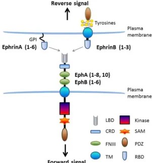
1.3 Eph receptors and ephrins ... 31
1.3.2 Signal transduction of EPHs and EFNs ... 32
1.3.2.1 Ligand-induced forward signaling transduction ... 33
1.3.2.2 Receptor-induced reverse signaling transduction ... 34
1.3.2.3 Both EPH receptors and EFNs have direct cross-talk with a variety of molecules ... 34
1.3.3 Functional roles of EPH/EFN signaling pathways ... 36
1.3.3.1 EPH/EFN signaling pathways control cell proliferation, adhesive function, and migration in neural development ... 36
1.3.3.3 Dysregulation of EPHs and ephrins are frequently found in cancer ... 37
1.3.3.4 EPH receptors and EFNBs can be found in different immune cells, their role in T cell biology are most extensively studied ... 38
1.4 STRA6 ... 41
1.4.1 Vitamin A homeostasis ... 41
1.4.2 STRA6 expression pattern and structure ... 42
1.4.2.1 STRA6 is a cellular transmembrane retinol binding protein for vitamin A transport ... 42
1.4.2.2 RBPR2 is a new retinol binding protein other than STRA6 ... 43
1.4.3 Role of vitamin A and STRA6 in T cell function ... 44
1.5 ARMC5 ... 45
1.5.1 Characteristic of Armadillo repeat ... 45
1.5.2 Structure and function of β-catenin ... 46
1.5.3 Role of β-catenin and other ARM proteins in T cell function ... 47
1.5.4 ARMC5 published papers mainly focus on the association of its mutations and PMAH ... 48
1.6 Hypotheses ... 49
1.7 Research Objectives ... 49
Chapter 2 Article-1 ... 50
2.1 The role of EFNB1 and EFNB2 in mouse collagen-induced arthritis and human rheumatoid arthritis ... 51
Introduction ... 53
Materials and Methods ... 55
Results ... 58 Discussion ... 63 References ... 66 Figure legends ... 70 Figures... 74 Chapter 3 Article-2 ... 80
3.1 To investigate the necessity of STRA6 upregulation in T cells during T cell immune responses ... 81
Abstract ... 82
Introduction ... 83
Materials and methods ... 86
Results ... 91 Discussion ... 95 Acknowledgements ... 99 References ... 100 Tables ... 105 Figure legends ... 106 Figures... 110 Chapter 4 Article-3 ... 118
4.1 Armc5 deletion causes developmental defects and compromises T cell immune responses ... 119
Abstract ... 120
Introduction ... 121
Materials and Methods ... 123
Results ... 135
Discussion ... 146
Acknowledgements ... 150
Tables ... 156
Figure legends ... 159
Figures... 165
Chapter 5 Discussion ... 176
Several issues arising from our studies are worth discussing. ... 177
The role of Efnb1/b2, Stra6 and Armc5 in CD4+ T cell activation ... 177
Redundancy ... 178
Spinal Bifida phenotype ... 179
The role of ARMC5 in cell proliferation versus apoptosis ... 181
Y2H data ... 183
Summary and Future Directions ... 184
Bibliographie ... 187
Appendix ... i
TNF-like ligand 1A (TL1A) gene knockout leads to ameliorated collagen-induced arthritis in mice: implication of TL1A in humoral immune responses ... ii
Abstract ... iii
Introduction ... iv
Materials and methods ... vi
Results ... xi
Discussion ... xvi
Acknowledgements ... xx
References ... xxi
Figure legends ... xxvii
Liste des tableaux
Chapter 3
Table 3.1 Antibodies for flow cytometry ... 105
Chapter 4
Liste des figures
Chapter 1
Figure 1.1 T cell activation ... 19
Figure 1.2 Signalling transduction of EPHs/EFNs ... 33
Figure 1.3 Diagram and structure of ARMC5 and β-catenin ... 46
Chapter 2
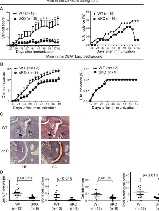
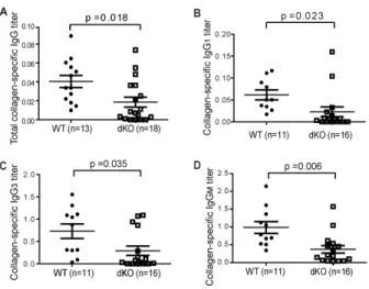
Figure 2.1 dKO mice were resistant to CIA induction ... 74Figure 2.2 Reduced serum collagen-specific Ab titres in dKO mice ... 75
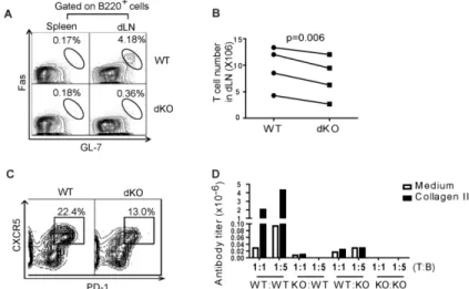
Figure 2.3 dKO T cells presented less help to B cells in mice of the C57BL/6 background ... 76
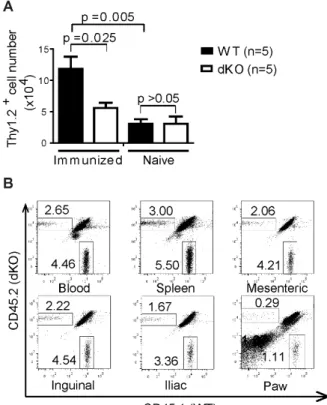
Figure 2.4 Compromised migration of dKO T cells to arthritic paws in mice of the C57BL/6 background ... 77
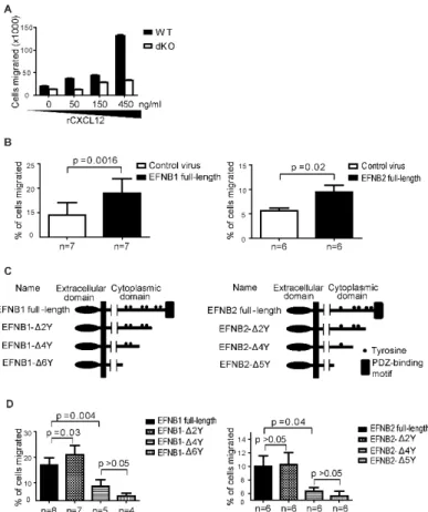
Figure 2.5 Efnb1 and Efnb2 expression affect T cell chemotaxis towards CXCL12 using mice in the C57BL/6 background ... 78
Figure 2.6 EFNB1 mRNA levels in T cells of RA patients ... 79
Chapter 3
Figure 3.1 Stra6 mRNA expression in organs and activated T cells ... 110Figure 3.2 Generation of Stra6 KO mice ... 111
Figure 3.3 Stra6 KO mice presented normal lymphoid organs and lymphocyte subpopulations ... 112
Figure 3.4 Normal activation and proliferation of KO T cells ... 113
Figure 3.5 Normal differentiation of Stra6 KO CD4 cells in vitro ... 114
Figure 3.6 Normal in vivo anti-LCMV immune responses of Stra6 KO mice ... 115
Figure 3.7 Glucose tolerance of KO and WT mice ... 116
Figure 3.8 Intracellular retinoid contents in lymphoid and other organs of Stra6 KO mice were comparable to those of WT mice ... 117
Chapter 4
Figure 4.1 Armc5 tissue-specific expression ... 165Figure 4.2 Generation of Armc5 KO mice ... 166
Figure 4.4 Armc5 KO mice presented normal thymus and spleen weight, cellularity and cell
subpopulations ... 168
Figure 4.5 KO T cells are compromised in proliferation and differentiation ... 169
Figure 4.6 KO T cells developed from the chimeric mice were compromised in proliferation and differentiation ... 170
Figure 4.7 KO mice are resistant to EAE induction ... 172
Figure 4.8 KO mice present compromised anti-LCMV immune response ... 174
Figure 4.9 Hypothetical model of ARMC5 action mechanisms ... 175
Chapter 5
Figure 5.1 Percentage of mice with kinky tails in 129/sv × CD-1 IGS F1 background ... 180Figure 5.3 Putative function of ARMC5... 185
Appendix
Figure 1 Generation of TL1A KO mice ... xxxiiiFigure 2 TL1A KO mice presented normal lymphocyte subpopulations ... xxxiv
Fugure 3 TL1A-KO mice manifested less severe CIA ... xxxv
Figure 4 Reduced collagen-specific Ab production in KO mice with CIA ... xxxvi
Figure 5 TL1A T cells presented normal help to B cells ... xxxvii
This thesis is dedicated to:
Remerciements
I would like to express my sincerest gratitude to my supervisors Dr. Jiangping Wu and Dr. Hongyu Luo, for their wisdom and patient in supervising me on my research in the past four years. I also appreciate them for their guidance for my career development and sharing their passion for science with me.
I would also like to thank all my colleagues in the lab; Dr. Terra Rafik, Dr. Bing Han, Dr. Jianning Mao, Dr. Xuehai Wang, Dr. Wei Jin, Dr. Zenghui Wu, Dr. Junzheng Peng, Dr. Shijie Qi, Charles Li, Michael Chen, Zeqin Zhang, Linjiang Lao and Wei Shi, for all the help, encouragement they offered. Special thanks to Yujia Wang for all the joy and sadness we’d shared.
Thanks to Dr. Johanne Martel-Pelletier, Dr. Mohit Kapoor, Dr. Guixiu Shi, Yongqiang Wu, Tania Charpentier, Dr. Alain Lamarre, Dr. Ming Zhong, Dr. Hui Sun for their wonderful collaborations. Special thanks to Dr. Xiangming Fang, Dr. Baoli Cheng for their collaboration and unconditional support.
Finally, I would like to express my deepest gratitude to all my jury members for their evaluation of this thesis.
Statement of authorship
Here is a statement of authorship regarding all the coauthors and myself to the papers included in this thesis:
Chapter 2
Hu Y, Wang X, Wu Y, Jin W, Cheng B, Fang X, Martel-Pelletier J, Kapoor M, Peng J, Qi S, Shi G, Wu J, Luo H. The role of EFNB1 and EFNB2 in mouse collagen-induced arthritis and human rheumatoid arthritis. Arthritis Rheumatol. 2015 Jul; 67(7):1778-88.
Conceived and designed the experiments: YH XW YW XF JM GS JW HL. Performed the experiments: YH XW YW WJ BC JP SQ. Analyzed the data: YH XW MK JP SQ GS JW HL. Wrote the paper: YH XW YW WJ BC XF JM MK JP SQ GS JW HL. For each particular figure
in this chapter, the authors who performed experiments are listed below: Figure 2.1: YH XW
SQ; Figure2.2: YH XW; Figure2.3: YH XW; Figure2.4: YH JP; Figure2.5: YH WJ; Figure2.6: YW BC.
Chapter 3:
Terra R, Wang Xh, Hu Y(Co-first), Charpentier T, Lamarre A, Zhong M, Sun H, Mao J, Qi S, Luo H, Wu J. To investigate the necessity of Stra6 upregulation in T cells during T cell
immune responses. PLoS One. 2013 Dec 31;8(12):e82808.
Conceived and designed the experiments: RT XW YH HL JW. Performed the experiments: RT XW YH TC MZ JM SQ. Analyzed the data: RT XW YH AL HS JW. Wrote the paper: RT XW YH AL HS JW. For each particular figure in this chapter, the authors who performed
experiments are listed below: Figure 3.1: RT XW; Figure3.2: RT XW SQ; Figure3.3: RT XW
JM; Figure3.4: RT XW YH; Figure3.5: YH XW; Figure3.6: TC; Figure3.7: YH; Figure3.8: YH MZ.
Chapter 4:
Hu Y, Mao J, Jin W, Luo H, Charpentier T, Lamarre A, Qi S, Peng J, Marcinkiewicz M, and Wu J. ARMC5 deletion causes developmental defects and compromises the immune system. Nature Communications (Accepted in principle).
Conceived and designed the experiments: YH JM JW. Performed the experiments: YH JM WJ HL TC SQ JP MM. Analyzed the data: YH JM WJ LH TC MM JW. Wrote the paper: YH AL JW. For each particular figure in this chapter, the authors who performed experiments are
listed below: Figure 4.1: MM YH WJ; Figure4.2: YH JP SQ; Figure4.3: YH JM HL; Figure4.4:
YH; Figure4.5: YH JP; Figure4.6: YH JP HL WJ; Figure4.7: YH JP HL; Figure4.8: TC;
Figure4.8: YH JW.
Appendix:
Wang X, Hu Y, Charpentier T, Lamarre A, Qi S, Wu J, Luo H. TNF-like ligand 1A (TL1A) gene knockout leads to ameliorated collagen-induced arthritis in mice: implication of TL1A in humoral immune responses. The Journal of Immunology. 2013 Dec 1;191(11):5420-9.
Conceived and designed the experiments: XW HL JW. Performed the experiments: XW YH TC SQ. Analyzed the data: XW YH AL HL JW. Wrote the paper: XW JW HL. For each particular
figure in this chapter, the authors who performed experiments are listed below: Figure1: XW; Figure2: XW TC; Figure3: XW YH SQ; Figure4: XW YH; Figure5: XW YH; Figure6: XW.
Liste des sigles
aa: Amino acidACPAs: Anti-citrullinated protein antibodies ACR: American College of Rheumatology ADH: Alcohol dehydrogenase
AF6: ALL1-fused gene from chromosome 6 AITD: Autoimmune thyroid disease
APCs: Antigen-presenting cells APC: Adenomatous polyposis coli ARMC5: Armadillo repeat containing 5 AS: Ankylosing spondylitis
BCR: B cell receptor Breg: Regulatory B cell
CAIA: Collagen-antibody-induced arthritis CBP: CREB-binding protein
CD: Crohn’s disease
CIA: Collagen-induced athritis CIS: Clinically isolated syndrome CK1α: Casein kinase 1α
CNV: Copy number variations
CRBP1: Cellular retinol binding protein-1 CRD: Cysteine-rich region
CRP: C-reactive protein CSF: Cerebrospinal fluid
CTD: Carboxyl-terminal domain
CTLA-4: Cytotoxic T lymphocyte associated antigen-4 DH-PH: Dbl-homology-pleckstrin-homology
DMARDs: Disease-modifying anti-rheumatic drugs EBV: Epstein-Barr virus
EFN: ephrin
EGF: Epidermal growth factor
Eph: Erythropoietin-producing hepatocyte kinase ESR: Erythrocytes sedimentation rate
EULAR: European league against rheumatism FAK: Focal adhesion kinase
FGF: Fibroblast growth factor FNIII: Fibronectin type III FoxP3: Forkhead box P3 GC: Germinal center
G-CSF: Granulocyte colony stimulating factor GD: Graves’ disease
GEF: Guaninenucleotide exchange factor
GITR: Glucocorticoidinduced tumor necrosis factor receptor GM-CSF: Granulocyte-macrophage colony-stimulating factor GRIP: Glutamate receptor interacting protein
GSK3β: Glycogen synthase kinase 3β GWAS: Genome-wide association scans
HGF/SF: Hepatocyte growth factor/scatter factor HHV-6: Human herpesvirus 6
HLA: Human leukocytes antigen HSC: Hepatic stellate cell
HSV-1: Human herpesvirus 1
ICOS: Inducible T cell co-stimulator
JAK/STAT: Janus kinase/Signal transducer and activator of transcription KO: Knockout
LAP: Latency-associated peptide LBD: Ligand binding globular domain LEF: Lymphoid enhancer-binding factor LRAT: Lechithin retinol acyl transferase MAPK: Mitogen-activated protein kinase
MHC: Major histocompatibility complex MMP: Matrix metalloproteinase
MRI: Magnetic resonance imaging MS: Multiple sclerosis
MTX: Methotrexate
MWS: Mathew Wood syndrome NTD: Amino-terminal domain OCB: Oligoclonal IgG band PD1: Programmed cell death 1 PI3K: Phosphatidylinositol 3’-kinase PICK: Protein interacting with C kinase
PMAH: Primary macronodular adrenal hyperplasia PPMS: Primary-progressive MS
PSA: Psoriatic arthritis
PTB: Phosphotyrosine binding
PTP-BL: Rrotein tyrosine phosphatase BAS-like RA: Rheumatoid arthritis
RALDH: Retinal dehydrogenase
RANK: Receptor activator of nuclear factor Κb RANKL: RANK ligand
RAR: Retinol acid receptor RBD: Receptor-binding domain RF: Rheumatoid factor
RRMS: Relapsing-remitting MS RTK: Receptor tyrosine kinases RXR: Retinoid X receptors
SCID: Severe combined immunodeficiency SF: Synovial fluid
SH2: Src homology 2
SLE: Systemic lupus erythematosus SM: Synovial membrane
STRA6: Stimulated by retinoic acid 6 T1D: Type 1 diabetes
T-bet: T-box transcription factor TCF: T cell-specific factor TEC: Thymic epithelial cell Tfh: T follicular helper TM: Transmembrane
TMEV-IDD: Theiler’s murine encephalomyelitis virus-induced demyelinating disease TPC: Tumor-propagating cell
Treg: Regulatory T cell
VEGF: Vascular endothelial growth factor WT: Wild type
Liste des abréviations
ARM: ArmadilloChapter 1 Introduction
Autoimmune diseases are conditions with an inappropriate activation of the immune responses, which can cause diseases such as rheumatoid arthritis (RA) and multiple sclerosis (MS) (Davidson & Diamond, 2001). There are about 80 types of autoimmune diseases. According to Cooper et al., more than 3% of the US population suffers from one or more of these diseases. The incidence of diseases are higher in women than in men (Cooper & Stroehla, 2003). It is a chronic disease which makes it a contributor to decreased life quality for individuals and also increase the costs of the whole health care system.
1.1 Risk factors for autoimmune diseases
Earlier twin studies and familial studies proved that genetic factors are essential for the development of autoimmune diseases. More recent studies show that non-genetic factors, such as hormones, infections, and environmental factors, interact with the existing genetic factors to trigger a particular kind of autoimmune disease in individuals bearing certain genetic variations.
1.1.1 A variety of genetic factors related to distinct signaling pathways are
involved in the initiation of autoimmune diseases
In the past decade, the extraordinary technical advances in this area have lead to the discovery of new factors that may contribute to human autoimmune diseases. For example, genome-wide association scans (GWAS) is a major approach to detect the relatively common (>1% or more) genetic variants, which is the dominant form of genetic variation for autoimmune diseases. These genetic changes also present in healthy populations. What’s more, the genetic associations found by GWAS turned out to be modest. The predisposition of autoimmune diseases represents the total effects of the genetic variations in different genes which influence the position and the strength of genetic signals and functions (Encinas & Kuchroo, 2000). Moreover, autoimmune diseases share a common set of susceptibility genes. For example, a variation within the human leukocytes antigen (HLA) is associated with almost every autoimmune disease. Copy number variations (CNV) and rare genetic variants are also present in the population. CNVs in genes like CCL3L1, FCGR3B, DEFB4, etc. have been linked to
many types of autoimmune diseases. A rare variant in TREX1, which is a DNA exonuclease, is found to be associated with sporadic lupus. Current published data suggest a variety of genetic heterogeneities related to distinct signaling pathways are involved in the initiation of autoimmuny
1.1.2 Infectious agents, sex hormones, smoking, and ultraviolet radiation
play a role to initiate autoimmune diseases
Environmental factors are also critical in the initiation of autoimmune diseases (Fagnani et al., 2015). Components of pathogens bear the potential to evoke autoreactivity through molecular mimicry (Cusick, Libbey, & Fujinami, 2012), which is due to the antigens from some microbes that share epitopes with endogenous antigens. For instance, peptides from Epstein-Barr virus can be presented to CD4+ T cells, which can also react with myelin basic protein (Wucherpfennig & Strominger, 1995). Moreover, infectious agents will lead to inflammation and lymphocytes activation, both of which lower the threshold of autoimmunity.
Women, especially women in their fertile period are most often affected, indicating an essential part of sex hormones in disease onset and perpetuation (Cutolo et al., 2006). Estrogen exerts dose- and time-dependent effects on cell growth and apoptosis for B cells and synovial fibroblasts in RA (Kawasaki, Ushiyama, Inoue, & Hukuda, 2000). It also has influences on Th1/Th2 cytokines in RA (Janele et al., 2006).
Smoking is related to SLE, RA, and myositis (Wahren-Herlenius & Dorner, 2013). It triggers inflammatory responses and sustains autoimmunity by the toll-like receptor pathways, autophagy, and apoptosis in the lung through reactive oxygen species.
Ultraviolet radiation can trigger systemic autoimmunity via immune pathways or by induction of apoptosis in diseases like SLE and dermatomyositis. Self-antigen-exposure during apoptosis prolongs the contact with autoantigen-specific B cells, which will lead to autoimmunity in individuals with genetic defects (Wahren-Herlenius & Dorner, 2013).
1.2 Mechanism of autoimmune diseases
Our immune system is designed to have a low level of autoreactivity. Exaggerated immune responses result in tissue damages and autoimmune diseases. For decades, studies from animal
models and humans have led to a hint that immune cells and the molecules they produce vary in different stages of disease pathogenesis.
1.2.1 Rheumatoid arthritis
RA is a chronic autoimmune disease that involves the activation of immune response both systemically and locally. Joints are the dominant organ that are targeted in the inflammatory responses.
1.2.1.1 ‘‘RA is the most common inflammatory joint disease’’
‘‘RA is the most common inflammatory joint disease’’ (Cooper & Stroehla, 2003). The annual incidence of RA has been estimated to be around 40 per 100, 000 (Cooper & Stroehla, 2003). The disease prevalence is about 0.9 percent of the Canadian population (Canada, 2011). However, the prevalence varies between 0.1 percent (in rural Africans) and 5 percent (some populations in India, Australia, New Zealand and Netherlands), indicating the initiation of RA can be influenced by ethnic and/or environmental elements (Helmick et al., 2008; Peschken & Esdaile, 1999). While affects all age groups, more than one-half of new RA cases occur between ages of 40 and 75 years (Cooper & Stroehla, 2003). Females have two to three times’ higher prevalence rate than that of males. A decreasing trend has been observed in the US (Doran, Pond, Crowson, O'Fallon, & Gabriel, 2002), as well as in high-risk populations in Indians (Jacobsson et al., 1994). RA causes economic burden due to disability in the workforce and the cost of treatment, especially the cost of biologic agents in recent years (Michaud, Messer, Choi, & Wolfe, 2003). RA increases the mortality rate in patients, and this death rate has not changed since 1990. Whether this is due to an inflammatory pathology, or due to the exposure to anti-rheumatic drugs, or due to both, is unclear (Wolfe et al., 1994). 1.2.1.2 Clinical manifestation of RA
1.2.1.2.1 ‘‘The 2010 ACR/EULR classification criteria for RA’’
RA is characterized by symmetrical synovitis involving multiple diarthrodial joints with extra-articular manifestations. In 2010, the criteria for the classification of ‘‘RA were revised by the American College of Rheumatology (ACR)/European League Against Rheumatism (EULAR) ’’ (Aletaha et al., 2010). The 2010 criteria help to identify patients earlier and also helps to
identify those who may respond to ‘‘disease-modifying anti-rheumatic drugs (DMARDs)’’ (Aletaha et al., 2010).
1.2.1.2.2 Chronic pain and swelling leading to bone damage in RA joints
The disease onset of RA is usually insidious, with the observation of tenderness and subtle swelling of one or more joints in hands and wrists. Some patients with RA have a slowly progressive course with exacerbations, which usually takes over weeks to months. However, some patients have a waxing and waning course known as “palindromic rheumatism”, which may take several years before the chronic, persistent features of RA become evident. Typically, RA involves the ‘‘metacarpophalangeal joints and the proximal interphalangeal joints of the fingers, the interphalangeal joints of the thumbs, the metatarsophalangeal joints of the toes, the wrists, elbows, shoulders, hips, knees, and ankles’’ (Aletaha et al., 2010). RA patients commonly have stiffness, which usually presents in the morning (morning stiffness) or after long periods of inactivity.
Seventy percent of patients with RA display radiographic evidence featured by ‘‘joint space narrowing and bone erosions’’ in small joints of hands and feet within few years after diagnosis. However, about 25% of patients with early RA may not develop erosive damage even after eight years of follow-up. The development of magnetic resonance imaging (MRI) is particularly useful for the measurement of the inflammatory process in the synovial membrane (McQueen, 2000). The use of ultrasonography enables the visualization of bone erosions, joints fluids, and synovial hypertrophy (Terslev et al., 2003).
1.2.1.2.3 Systemic symptoms, elevated autoantibodies and acute phase proteins in RA patients
Systemic symptoms, including fatigue, emaciation, low-grade fever, weakness, and depression often accompany the early signs of joint inflammation. Anemia of chronic inflammation, thrombocytosis, and sometimes mild leukocytosis are common hematologic abnormalities in active RA. The majority of RA patients have the positive test for rheumatoid factor (RF), anti-citrullinated protein antibodies (ACPA), or both. Some of them are antinuclear antibodies positive. The acute phase proteins, including the erythrocytes sedimentation rate (ESR) and C-reactive protein (CRP), are often elevated in patients with active RA. Significant correlations
are frequently observed between the degrees of elevation of these proteins and RA activity (Cylwik, Chrostek, Gindzienska-Sieskiewicz, Sierakowski, & Szmitkowski, 2010).
1.2.1.2.4 Subcutaneous nodule formation, pleuropulmonary disease, and cardiovascular disorders are also found in RA patients
Other systemic manifestations, including subcutaneous nodule formation, pleuropulmonary disease, and cardiovascular disorders are also found in RA patients. Rheumatoid nodules are the most frequently observed manifestation. Typically, subcutaneous nodules are hard and non-tender. These nodules vary in sizes, being associated with the present of serum rheumatoid factors, erosive joint destruction, necrotizing vasculitis and other ‘‘extra-articular manifestations of RA’’ (Vlak, 2003). The pleuropulmonary disease is a primary reason for the morbidity and mortality of RA. Interstitial lung disease, pleural thickening, and effusions are the most common pleuropulmonary manifestation of rheumatoid arthritis lung disease (Shaw, Collins, Ho, & Raghu, 2015). RA patients are more inclined to have myocardial infarction and stroke (Dhawan & Quyyumi, 2008). Rare manifestations, such as Felty’s syndrome which features with neutropenia and splenomegaly, were also reported. RA patients have a similar incidence of cancer than their healthy controls in general. However, RA patients have a higher incidence of lymphomas and lung cancer. Until now, how rheumatoid arthritis causes systemic illness remains obscure.
1.2.1.3 Animal models of RA
There are two major reasons for the extensive use of animal models in RA. Firstly, the mechanisms of RA, which can trigger articular disease onset and define chronicity of the local and systemic inflammation, remain obscure. Secondly, the rarely obtainable human tissues, such as synovial tissues, lymph node, bone marrow and spleen, hinds the research of finding out contributors to immune responses in human. Currently, numerous arthritis models including the induced- and spontaneous- models are developed (Asquith, Miller, McInnes, & Liew, 2009). Even though none of them can entirely mimic human RA, studies based on animal models have been proved to be helpful in understanding the potential mechanism and treatment of RA.
1.2.1.3.1 CIA is ‘‘the gold standard animal model of human RA’’, while CAIA proves a role of humoral immunity in RA
CIA is considered as ‘‘the gold standard animal model of human RA” (Brand, Latham, & Rosloniec, 2007). DBA/1 mice are the most popular strain for CIA model in research. These mice are susceptible to CIA because of their MHC class II haplotype (I-Aq). CIA bears similarity to human RA regarding the breach of tolerance, the role of lymphocytes in the initiation and persistence of the disease, and the histological findings in the arthritis joints. Studies reveal that mouse MHC Aq peptide binding part are conformationally similar to that of human HLA DR4 (DRB1*0401/DRA), which is the human MHC highly associated with RA (Holmdahl, Bockermann, Backlund, & Yamada, 2002). Also, the majority of collagen II-specific T cells in both human and mouse recognizes a peptide from aa256-270 of the collagen II peptide. Moreover, CIA mouse shares local features with human RA regarding the infiltration of immune cells in the rheumatoid synovium, the synovial lining cell hyperplasia, and the secretion of inflammation mediators, which resulting in local joint damage. The detailed mechanism of lymphocytes in the development of CIA will be discussed in detail in the “Pathogenesis of RA” part.
The biggest problem of the CIA model in DBA/1 background is its limited usefulness in genetically modified mice. This is due since most genetically modified mice are on the C57BL/6 background, which is not a CIA-susceptible strain. Studies showed that the use chicken collagen II and a boost with IFA (Incomplete Freund’s Adjuvant) overcome the resistant issue of C57BL/6 mice (Inglis, Simelyte, McCann, Criado, & Williams, 2008). After the refinement of the protocol, C57BL/6 mice share the basic pathological features with DBA/1 mice. However, they develop less severe CIA with later onset, lower incidence, and lower clinical scores. Also, intergroup inconsistency is observed in C57BL/6 mice, underscoring that precise, independent internal controls are necessary.
The injection of antibodies against type II collagen leads to collagen-antibody-induced arthritis (CAIA) (Khachigian, 2006). The antibodies trigger similar clinic development of arthritis as that of CIA. CAIA model itself proves the importance of auto-antibodies in the development of joint damage. Other antigens are also reported to induce arthritis (Asquith et al., 2009; Maffia et al., 2004). Arthritis can also be caused by other factors related to the T
cell-dependent signalling pathway as that of CIA; others may trigger cell signaling pathways in other immune cells. For example, zymosan, a component of Saccharomyces cerevisiae can trigger TLR2 signaling in macrophages (Frasnelli, Tarussio, Chobaz-Peclat, Busso, & So, 2005).
1.2.1.3.2 Spontaneous arthritis models, including human “TNF-α transgenic mice, K/BxN mice, and SKG mice”, have similarities with human RA
The spontaneous arthritis models are characterized by the development of chronic and progressive polyarthritis, which is similar to the human disease. These include “human TNF-α transgenic mice” (Li & Schwarz, 2003), “K/BxN mice” (Monach, Mathis, & Benoist, 2008) and “SKG mice” (Hata et al., 2004). In the human TNF-α transgenic arthritis model, treatment with anti-human TNF-α monoclonal antibody completely rescued mice from the disease. This model is particularly useful in exploring novel treatment targeting downstream signaling molecules of the TNF-α pathway. The K/BxN mice also develop spontaneous arthritis. The K/BxN mice express the TCR transgene “KRN, the MHC class II molecule Ag7”, as well as
autoantibodies against “glucose-6-phosphate isomerase” in sera (Monach, Mathis, & Benoist, 2008). This model provides a useful tool for investigating the innate immune systems in the initiation of arthritis. SKG model is caused by a point mutation of ZAP-70, which depends on environmental factors to induce inflammatory arthritis. Severe combined immunodeficiency (SCID) mice have also been used to exploit the underlying mechanism of RA by implanting them with human synovial tissues (Pierer et al., 2003). This model reveals the contribution of a local factor in cartilage destruction and bone erosion. It is also useful in identifying novel targets for the prevention of local damage in RA.
1.2.1.4 Pathogenesis of RA
1.2.1.4.1 Macrophages orchestrate in RA inflammatory responses
Macrophages are a subpopulation of white blood cells, which function in infectious microbe clearance, normal tissue remodeling, wound repair and angiogenesis. The classically activated M1 and the alternatively activated M2 are activated macrophages with distinct functions (Sica & Mantovani, 2012). In normal synovial membrane, macrophages (type A synovial cells) are
not the predominant cell type. However, in rheumatoid condition, the macrophage number significantly increases (Maruotti, Cantatore, Crivellato, Vacca, & Ribatti, 2007).
Macrophages play important roles in RA by phagocytosis. They differentiate into M1 macrophages in response to LPS and secrete proinflammatory cytokines like IFN-γ and TNF-α. These proinflammatory cytokines act as autocrine stimulator, as well as potent paracrine inducer of other inflammatory cytokines. For example, TNF-α can induce ‘‘IL-1, 6, 8 and granulocyte-macrophage-colony-stimulating factor (GM-CSF) in RA joints’’ (Butler, Maini, Feldmann, & Brennan, 1995; Haworth et al., 1991). It also induces matrix metalloproteinases (MMPs) in the synovium. MMPs are responsible for cartilage matrix destruction. Moreover, in the synovium, TNF-α induces NO production, which could promote synovial cells to produce TNF-α (Chae et al., 1997; McInnes et al., 1996).
Conversely, the activation of macrophages with IL-4 or IL-13 promotes the M2 phenotype, which suppressing destructive immunity, while promoting angiogenesis and matrix remodeling. Macrophages in rheumatoid arthritis synovium contribute to pathological angiogenesis by producing vascular endothelial growth factor (VEGF). Deletion of certain domain of VEGF receptor-1 in mice protects mice from a murine model of RA. This protection is due to the decrease of the phagocytosis and the secretion of IL-6 and VEGF-A (Murakami et al., 2006). Macrophages can also promote pathological angiogenesis through the release angiogenic chemokines and cytokines. The former includes CXCL5 (Koch et al., 1994), while the latter includes ‘‘IL-8, IL-15, IL-17, et al.’’ (Koch et al., 1991; Pickens et al., 2010). In contrast, antiangiogenic factors such as thrombospondin 2, are also produced (Park et al., 2004).
1.2.1.4.2 ‘‘B cells play a role in RA pathogenesis through autoantibody production’’, antigen presentation and costimulation, neogenesis of lymphoid microstructures and cytokine
secretion
The discovery of autoantibodies in the 1950s’ (Holman & Deicher, 1959; Rose, Ragan, & et al., 1948) had brought the humoral autoimmunity into the center stage, which was followed by the identification of numerous autoantibodies as the diagnostic markers of autoimmunity. However, as plasmapheresis showed disappointing results, treatment strategies no longer
focused on this area. It was the unexpected success of anti-CD20 antibody treatment to deplete B cells that brought them back to the central of the stage in autoimmunity.
Autoreactive B cells
Numerous evidence showed that subtle alterations in B cell receptor (BCR) signaling pathways triggered by autoantigens can be the predisposing causes for autoantibody production in both mice and human beings (LeBien & Tedder, 2008). Also, any aberrant in B-cell selection centrally in the bone marrow or peripherally in the lymphoid tissues during B cell development and/or disruption of the normal BCR somatic hypermutation during affinity maturation within the germinal center can lead to autoimmunity. An early break in B cell tolerance is proposed as different studies proved the presence of autoantibodies in the patients’ serum many years before the presence of any clinical symptoms. However, the exact role of autoantibodies in RA remains elusive.
Diverse functions of B cells in autoimmune diseases
1. Autoantibody production
Autoantibody production is a characteristic feature in most autoimmune diseases. Some autobodies have remarkable specificity and can be used as biomarkers for disease diagnosis and indicators of therapeutic interventions. Autoantibodies are usually high-affinity, somatically mutated IgG, which might lead to pathogenic consequences. Autoantibodies exert their function through direct binding or forming immune complexes. The former usually happens in organ-specific autoimmune diseases, like myasthenia gravis, where they directly damage the target organ. The latter often occurs in systemic autoimmune diseases, where tissue injury is caused by immune complex composed of autoantibodies conjugated with free molecules, cell surface antigens, and nucleoprotein antigens.
Autoantibodies may also cause damage through the engagement of complement and/or Fc receptor (FcR) effector pathways. In the K/BxN mouse model, the administration of autoantibodies activates the alternative complement pathway. This activation attracts neutrophils, leading to the accumulation of proteolytic enzymes and cytokines in the inflammatory joints (Monach, Benoist, & Mathis, 2004; Wipke, Wang, Nagengast, Reichert, & Allen, 2004).
It is still elusive to define the role of autoantibodies in the initiation of autoimmune diseases like RA. Autoantibodies alone are unable to initiate autoimmune diseases in multiple mice
models. Some of them are even proven to have protective functions (Shoenfeld & Toubi, 2005). What’s more, autoantibodies themselves are often not pathological. The co-existing secondary triggers of autoantibodies, such as infectious agents, will lead to a vicious chronic state of inflammation (McClain et al., 2005).
2. Antigen presentation and costimulation
Besides antibody production, B cells can also promote disease development by presenting antigens to T cells. B cells are required to start autoimmune diseases in mice model for arthritis, lupus, diabetes, etc. (Chan, Hannum, Haberman, Madaio, & Shlomchik, 1999; O'Neill et al., 2005; Wong et al., 2004). However, their antibody-secreting capabilities are not required that early. B cells capture antigen through their B-cell receptors, which is over 1,000-fold more efficient than other APCs. Therefore, B cells can present antigens at extraordinarily low concentrations and also focus the immune response to a specific antigen in certain autoimmune diseases (Rivera, Chen, Ron, Dougherty, & Ron, 2001). What’s more, B cell may have specific roles in providing costimulatory signals to antigen-specific T cells. In NZB/NZW mice, a spontaneous lupus model, elevated expression of costimulatory molecules are found in B cells from spleen (Wither, Roy, & Brennan, 2000).
3. Neogenesis of lymphoid microstructures
Neogenesis of lymphoid microstructures is found ectopically in multiple chronic inflammations. The size and organization of these neogenesis lymphoid structures vary depending on the context. These structures have been described in SLE, T1D, Sjögren’s syndrome, MS and RA (Aloisi & Pujol-Borrell, 2006; Drayton, Liao, Mounzer, & Ruddle, 2006). The exact function of these structures remains unknown. However, the locally generated plasma cells are found to secrete autoantibodies (Salomonsson et al., 2003), suggesting their potential role in the maintenance of autoimmunity.
4. Cytokine secretion
Activated B cells ‘‘produce multiple cytokines, including IL-6, IFN-γ, lymphotoxin-α, TGF-β, IL-4, and IL-10’’ (Duddy, Alter, & Bar-Or, 2004; Harris et al., 2000; Lund, Garvy, Randall, & Harris, 2005). B cell-derived cytokines play pathologic roles in regulating functions of multiple cells, such as macrophages, dendritic cells and T cells. In addition, signals from these cells can activate B cells in turn, creating a feedback loop They also play protective roles in immune responses to autoantigens, as in the case of IL-10, which will be described below.
5. Regulatory B cells
Regulatory B cells (Bregs) (Mizoguchi & Bhan, 2006) are B cells that can produce anti-inflammatory cytokines like IL-10, TGF-β. Bregs can be found in a variety of B-cell subsets and participate in the pathogenesis of autoimmune diseases, including SLE (Lenert, Brummel, Field, & Ashman, 2005), RA (Mauri, Gray, Mushtaq, & Londei, 2003), and MS (Matsushita, Yanaba, Bouaziz, Fujimoto, & Tedder, 2008). The regulatory properties of B cells in autoimmune diseases were originally reported in B10.PL mice. These mice are lack of B cells and are unable to recover from EAE as compared to their healthy counterparts (Wolf, Dittel, Hardardottir, & Janeway, 1996).
Like it is in Tregs, the IL-10 secreted by B cells also (Fillatreau, Sweenie, McGeachy, Gray, & Anderton, 2002) suppresses Th1 polarization and inhibits macrophage functions. B cells can regulate the T cell co-stimulation pathways by direct cell-cell contacts with CD40 and B7.
Autoantibodies in RA
The autoantibodies found in RA patients target quite diverse molecules, including ‘‘cartilage components, chaperons, enzymes, nuclear proteins and citrullinated proteins’’ (Song & Kang, 2010). In recent years, new technologies, like protein arrays, have been used to detect RA-associated autoantibodies with better specificity and sensitivity (Hansson et al., 2012; W. H. Robinson, Steinman, & Utz, 2003). However, only two of them are used in clinical practice currently.
1. Rheumatoid factor
RFs are autoantibodies that are against the Fc region of IgG. The IgM isotype is the primary type that exists in the sera of RA patients (Nell et al., 2005). RFs can be detected physically in ordinary old people, but also in other diseases like Sjögren’s syndrome, SLE. When the titer goes higher than 50IU/ml (RF50), it is quite specific for RA (Song & Kang, 2010). High titer IgM RF status is associated with poor disease prognosis. Notably, the co-presence of high titer IgM RF and IgA isotype usually indicate erosive RA (Jonsson et al., 1998; Scott, 2000). Furthermore, higher RF does not occur in experimental models of RA. Up to now, the role of RF in RA is still not entirely clear.
2. Anti-citrullinated protein antibodies (ACPAs)
The identification of ACPAs is one of the most meaningful event in RA diagnostic marker studies. Comparing with RF, it has a better diagnostic value regarding specificity and
sensitivity. ACPAs are referred to antibodies against ‘‘perinuclear factor, keratin, filaggrin and cyclic citrullinated peptide (CCP)’’ (Song & Kang, 2010). Citrulline is the common epitope of these antibodies, which can be generated post-translationally from arginine (Schellekens, de Jong, van den Hoogen, van de Putte, & van Venrooij, 2015). The ACPAs can be found in 70-90% of RA patients, with a specificity of 96% (Suzuki et al., 2003). They are highly specific for RA. As IgM RF, they are indicators of worse outcome in RA (Im et al., 2009; Kroot et al., 2000). Of all the ACPAs, anti-CCP is the most wildly used one in clinical diagnosis.
1.2.1.4.3 T cells are critical for disease onset and to sustain of RA Role of T cell activation and tolerance in RA
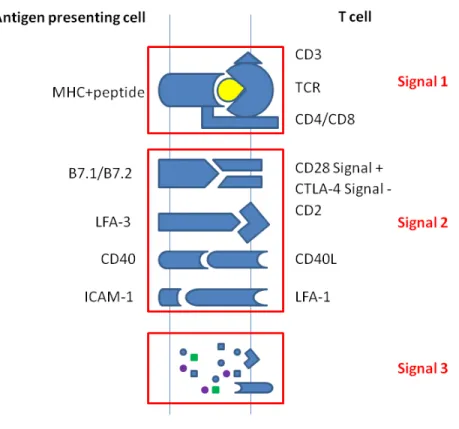
T cell activation is essential for the onset and maintenance of RA. To fully activate T cells, the signaling transduce through both TCR complex and the co-stimulators are needed. The former is identified as signal 1. It is stimulated when the major histocompatibility complex (MHC) with antigen peptide (pMHC) binds to TCR. The latter is called signal 2. It is stimulated when B7-1 and B7-2 on antigen presenting cells conjugate to CD28 on T cell surface (Figure 1.1). The engagement of signal 1 leads to TCR aggregation, which enables closer pMHC/TCR contact. TCR aggregation and activation of signal 2 will then initiate the phosphorylation and the activation of a variety of molecules, resulting in the signaling transduction through PI3K/AKT/mTOR pathway, MAPK/Erk pathway, PKCθ pathway, and Ca2+-mediated signaling pathways (Smith-Garvin, Koretzky, & Jordan, 2009). These signaling pathways are regulated by a variety of molecules at multiple levels in response to particular environment stimulation. Abnormal T cell activation involves both breakdowns of T cell tolerance and aberrant T cell response, leading to the autoimmune disease state.
CD4+ T helper cells control both the cell-mediated and humoral immune responses, which makes it the most important subpopulation of T cells in immune tolerance. T cell tolerance may occur during their maturation in the thymus (central tolerance) or occur in peripheral when they meet antigens (peripheral tolerance) (Xing & Hogquist, 2012). In central tolerance, the majority of immature T cells that recognize self-antigens with high avidity are deleted, while some of them differentiate into regulatory T cells (Tregs). In the past decades, the underlying mechanism of central tolerance has been well studied in animal models. However, it is still not known about how much it contributes to a human autoimmune disease. The
peripheral tolerance mechanism is another important aspect for T cell tolerance. Prolonged TCR activation may result in anergy. Defects and/or lack of the co-stimulation will contribute to peripheral tolerance. Moreover, the activation of death signal and the dysfunction of Tregs may also lead to the breakdown of peripheral tolerance.
Role of co-stimulation (signal 2) in T cell activation
As shown in Fig 1.1, signal 2 (co-stimulatory) is an essential part of T cell activation. The B7.1/B7.2 expressed on APCs conjugate both CD28 and (or) CTLA4 on T cells, depending on the cell type and context. The interaction of B7.1/B7.2 and CD28 promotes T cell activation, as well as T cell differentiation. In contrast, their interaction with CTLA-4 will lead to the suppression of T cell activation. The binding capacity of CTLA-4 with B7.1/B7.2 is much higher than that of CD28, making it efficient in regulating T cell activation signaling (Greene et al., 1996; Peach et al., 1994). CTLA-4-Ig (Abatacept) is approved by FDA for the treatment of RA (Kremer et al., 2005). It is a protein fuse with ‘‘the extracellular binding domain of CTLA-4 and the Fc region of IgG’’ (Kremer et al., 2005). Thus, abatacept is highly efficacious in treating RA, leading to well-improved physical function. It significantly reduces the progression of structural damage in patients with established RA (Provan et al., 2015; N. Takahashi et al., 2015). A combination of methotrexate (MTX) and abatacept can deliver significant clinical benefits and radiographic improvement (T. Hirota et al., 2015; Mochizuki et al., 2015). Abatacept is currently prescribed to patients with RA unresponsive to regular doses of existing DMARDs.
Later studies proved that many co-stimulation and co-inhibition molecules are involved in T cell regulation, such as ‘‘ICOS, PD1, OX40 (also known as CD134, TNFRSF4) et al.’’ (L. Chen & Flies, 2013).
Figure 1.1 T cell activation
Role of CD4+ T cell differentiation in RA
The etiology of autoimmune diseases has not been fully investigated up till now, however, accumulating evidence proves that CD4+ T cells is essential for the development and maintain of both systemic and local inflammation. Studies show that about one-third of cells infiltrated into the inflammatory joint are T cells. Naive CD4+ T cells will be driven into different subsets, named Th1, Th17, and Tregs, depending on the cytokine environments (signal 3) (Figure 1.1). Activated T cells can infiltrate into the inflammatory sites. These effector T cells will secrete molecules, such as chemokines and cytokines, which will further recruit other inflammatory cells and enhance local inflammation.
1. Th1 & Th17 cells are the principal players in the pathogenesis of RA
TCR activation and cytokine IL-12 will drive naive CD4+ T cells into IFN- γ-secreting Th1 subpopulation. T-box transcription factor (T-bet) is the master regulator that promotes Th1 differentiation. T-bet can also suppress the development of opposing cell lineages like Th2 and Th17. Th1 subpopulation mainly functions as the pro-inflammatory factor, while Th2
basically works as the anti-inflammatory factor. Studies showed that early administration of a monoclonal antibody against IFN-γ could protect mice from CIA (Boissier et al., 1995). The frequency of Th1 cells with citrulline-specific epitopes are increased in rheumatoid arthritis, and this is influenced by therapy and disease activity (James et al., 2014). In the past, the balance of Th1/Th2 was once thought to be critical in the pathogenesis of autoimmune disease including RA. In 2005, the recognition of Th17 subpopulation refreshed this notion (described in detail in the MS part) (Cua et al., 2003).
Th17 is a subpopulation of T helper cells, which secrete IL-17A, IL-17F, and IL-22. RORγt is the master transcription factor of Treg. Cytokines like TGF-β and IL-6 drive naive CD4+ T cells differentiation into Th17 subpopulation, while IL-23 promotes Th17 cells expansion. Fossiez and colleagues reported the first evidence of the involvement of Th17 in RA. They showed that IL-17 is capable of stimulating synovial fibroblast in vitro from RA patients. (Fossiez et al., 1996). Emerging reports coming from animal models show that less severe arthritis is found in genetically modified mice of Th17-related cytokines and transcription factors, including ‘‘IL-17 knockout mice, IL-23 p19 subunit knockout mice, and STAT4 knockout mice’’ (Langrish et al., 2005; Hildner et al., 2007).
Th17 cells are the primary T cell subpopulation in the pathogenesis of RA. The receptor of IL-17 is found to be expressed in cells from the immune system (monocytes and macrophages), as well as cells in the RA joint (fibroblasts and osteoblasts), which are essential for RA inflammation. First of all, Th17 cells exert their function through IL-17, which is able to generate other pro-inflammatory cytokines once the IL-17 receptors are activated. Secondly, these pro-inflammatory cytokines will further enhance the Th17 function in turn, which might trigger autoimmunity in genetic sensitive individuals. Thirdly, IL-17 can synergize with other proinflammation mediators, like TNF-α and IL-1β in RA. Particularly, IL-17 promotes germinal center (GC) formation in the K/BxN model (H. J. Wu et al., 2010). Recently, it was reported that Toll-like receptors expressed by synovial fibroblasts could induce IL-17 production (F. Hu et al., 2014).
Besides cytokines, Th17 also triggers chemokines production, which is critical for the recruitment of other inflammatory cells to the inflamed joint. This recruitment will lead to the local inflammatory cascade. Particularly, Th17 subset with the expression of CCR6+ will be recruited to the inflammatory site in response to the CCL-20 produced by synovial fibroblasts.
What makes it more interesting is the treatment against CCR6 in Th17 has proved to be effective. More over, IL-17 can also upregulate enzymes, small molecules, and cell surface receptors in cells such as synovial fibroblasts, causing bone and cartilage damage (Nakashima et al., 2011).
2. Tregs play a regulatory role in RA
Tregs, either natural or inducible, are subsets of T cells which play regulatory roles in immune inflammatory processes. Nature Tregs (nTregs) express CD25 (IL-2Rα) on the cell surface and FoxP3 in the cell nucleus. nTregs are developed in the thymus during the positive selection stage with a higher than the normal affinity for self-antigens. Inducible Tregs (iTregs) are generated out of thymus. IL-10 and TGF-β are two major cytokines which can drive naive CD4+ T cells to iTregs. Self-antigen-specific nTregs are the major subset of T cells that is critical for the maintenance of self-tolerance and the prevention of autoimmune diseases. iTregs, on the other hand, are a key subset of T cells in fighting against foreign pathogens.
CD25 and FoxP3 are cell surface markers for Tregs. However, recent studies showed that both CD25and FoxP3 could also be found in newly activated human effector CD4+ T cells. The IL-7 receptor α chain (CD12IL-7) is found to be inversely correlated with nTreg status (Liu et al., 2006). Thus, the combination marker CD4+CD25+FoxP3+CD127low is widely used to identify human nTregs. Fletcher and colleagues reported that CD39+FoxP3+ T cells also have the suppressive effect (Schuler, Harasymczuk, Schilling, Lang, & Whiteside, 2011). Further potential surface markers for the assessment of nTregs are ‘‘CTLA-4, GITR, CD73, and LAP’’ (T. Takahashi et al., 2000; McHugh et al., 2002; Deaglio et al., 2007; M. L. Chen, Yan, Bando, Kuchroo, & Weiner, 2008).
In autoimmune diseases, regulatory T cells not only offer a new perspective of self-tolerance establishment and maintenance, but also actually engaging in systemic and local inflammation, memory and resolution. Autoimmunity with immune dysregulation, polyendocrinopathy, enteropathy X-linked syndrome (IPEX) in children links to several mutations of Foxp3 gene (Bennett et al., 2001), while in mice mutation or depletion of the Foxp3 gene resulted in fatal autoimmune diseases (Brunkow et al., 2001). Data about the frequency of Tregs in RA are contradictory, especially in the peripheral blood (PB) (Kawashiri et al., 2011; van Amelsfort, Jacobs, Bijlsma, Lafeber, & Taams, 2004). Accumulating
evidence indicates the enrichment of Treg population in the synovial fluid (SF) and synovial membrane (SM) (Jiao et al., 2007; Moradi et al., 2014). The transfer of CD4+Foxp3+ Treg cells
reduces the severity of CIA when it is done prior to collagen immunization (Wright et al., 2009). The administration of monoclonal antibody specific for CD25 targeting CD4+CD25+ Tregs on collagen-induced arthritis (CIA) worsens the disease (Morgan et al., 2003). Some studies showed that Tregs from RA patients, especially Tregs from PB, have impaired suppressive function (Ehrenstein et al., 2004).
Tregs are capable of inhibiting other proinflammatory cell function in both contact-independent and contact-dependent manner. IL-10 impairs the ability of dendritic cells to promote Th1 differentiation and suppress the production of IL-12 by Th1 cells (Rubtsov et al., 2008). Blockade of TGF-β expression on Tregs with neutralizing antibodies resulted in the abolishment of Treg suppression (Nakamura, Kitani, & Strober, 2001). The contact-dependent inhibition involves the cell surface receptors on Tregs. Receptors like CTLA-4, GITR, and OX40 could trigger the suppressive effects of Tregs. CTLA-4 gene polymorphisms have been reported to be correlated with RA (Daha et al., 2009; Lei et al., 2005). Tregs from RA patients showed a less CTLA-4 expression, as well as a reduced function compared to healthy Tregs (Walker et al., 2009). Furthermore, signaling pathways triggered by CTLA-4 can regulate the balance between the anti- and pro-inflammatory cytokines, resulting in the inhibition effect (Kormendy et al., 2013). The third mechanism is that Tregs can deplete IL-2 through CD25, which is also known as the IL-2 receptor. With the high expression of CD25, Tregs will deprive IL-2, which is necessary for other effector CD4+ T cells. Effector CD4+ T cells will undergo apoptosis because of this deprivation (Pandiyan, Zheng, Ishihara, Reed, & Lenardo, 2007). Lastly, Tregs can execute target cells by making its own granzyme A and perforin (Cao et al., 2007) or releasing metabolites like adenosine to exert their immunosuppressive functions (Ohta & Sitkovsky, 2014).
3. Tfh cells are crucial for T-cell helped function in RA
T follicular helper (Tfh) cells are characterized by the expression of CXCR5 on CD4+ T cells. Bcl-6 is the signature transcription factor, and IL-21 is the signature cytokine. ‘‘PD-1, ICOS, CD40 ligand (CD40L)’’ (Ueno, Banchereau, & Vinuesa, 2015) et al. are their surface markers, which usually apply in combination with CXCR5. Tfh cells are essential for T-cell helped function, including B cell maturation and antibody production in germinal centers.
Tfh cells play important roles in the inflammatory responses of RA. Elevated circulating Tfh and IL-21 are found in patients with RA, and they are correlated with disease activity (Arroyo-Villa et al., 2014; Y. Zhang, Li, Lv, Yin, & Wang, 2015). Tfh cells could also be found in rheumatoid synovium tissues from RA patients, but are absent in OA synovium (Chu, Wang, Zhou, Chen, & Lu, 2014). In RA synovium and synovial fluid, enrichment of PD-1+ T cells is found, reflecting the existence of negative feedback at the inflammatory sites (Raptopoulou et al., 2010). Several strategies targeting Tfh-related molecules and cytokines showed impressive therapeutic effects, opening new venues for possible immune regulatory therapy in RA (Jang et al., 2009; Odegard et al., 2008).
4. CD8+ cells are predominantly proinflammatory T cells in RA
Earlier studies of CD8+ T cell functions in RA yield conflicting results, causing the research in this cell type became almost complete oblivion (Carvalheiro, da Silva, & Souto-Carneiro, 2013). However, recently, more and more evidence support that CD8+ T cells present in the inflammatory joint mainly contribute to the chronic inflammation locally (Fekete et al., 2007; Maldonado et al., 2003; Masuko-Hongo et al., 1997; Wagner et al., 1998). CD8+ T cells promote the local inflammation not only through their secretion of proinflammatory cytokines, but also kill the target cells by their lytic enzymes. It is also approved that CD8+ T cells can exert their suppressive role in an IL-10 dependent manner in RA (Carvalheiro et al., 2013).
1.2.2 Multiple sclerosis
1.2.2.1 MS is the second cause of disability in young adults
About 350,000 people suffer from MS in US, and this number is 2.5 million worldwide. MS affects adults of 20-40 years old. It is the second cause of disability in young people. The incidence of MS in women is about twice higher than that of men. Epidemic studies showed that MS has a familial distribution pattern (Weinshenker, 1996). It is also reported that monozygotic twins got higher chance to have MS than the dizygotic twins (Hansen et al., 2005). The prevalence rates of MS have been reported to be high in northern parts of Europe and North America (5-30 per 100,000), but low in Asia and South America (<5 per 100,000) (Pugliatti et al., 2006). Some reports show that the epidemic pattern of MS can be changed due to migration. In the majority of the case, MS does not influence longevity. However, due to