International Journal of Biological and Agricultural Research Saadi Leila et al. 1 (2) December2018 Pages 21- 27
Pages 1-9
21
Saadi Leila Département de Biologie et de Physiologie Cellulaire, Faculté SNV, Université Blida 1, (Algérie). E-mail: saadileila4@gmail.com
Abstract
The investigation of the toxicity of a neonicotinoid insecticide, thiamethoxam, in mammals has been the subject of several experimental studies. In the present work, we are particularly interested in studying the renal toxic effects of two doses (0.2 and 0.4 mg/kg/day) of thiamethoxam, an insecticide widely marketed in Algeria, administered orally in adult male mice. For this purpose, a renal histopathological study and plasma assays of the uremia and the creatinemia are carried out. Examination of the histological sections recorded dose-dependent lesions in the renal parenchyma. Renal impairment is characterized by the destruction of certain glomeruli and the presence of some hemorrhagic and inflammatory foci. These alterations are associated with leukocyte diapedesis presented in the blood vessels. The renal function is maintained in the normal state because the urea and creatinine contents remain unchanged. On the basis of these results, it turns out that thiamethoxam treatment induces a dose-dependent renal toxicity in male mice.
Key words: Renal Function; Histopathology; Adult male mice; Thiamethoxam; Toxicity; Dose-dependent.
Investigation
of
renal
toxicity
induced
by
neonicotinoid
(thiamethoxam) insecticide in mice
Saadi Leila
1,2*, Raki Ahmed Aziz
1, Bouazza Marwa
1(1)Département de Biologie et de Physiologie Cellulaire, Faculté SNV, Université Blida 1. (2)Laboratoire de recherche Ecobiologie Animale, ENS, Kouba, Alger.
Received 4 August 2019; Accepted 8 November; Available online 1 December 2019
I
nternational Journal of Biological and Agricultral Reasearch
( IJBAR)
Journal home page: www http://www.univ-eloued.dz/ijbar/
International Journal of Biological and Agricultural Research Saadi Leila et al. 1 (2) December2018 Pages 21- 27
Pages 1-9
22 Introduction
Pesticides are the most important group of chemicals in agriculture to prevent, destroy, repel or suppress any pests [1]. These pesticides contain biologically active substances that act on living things, hence the in-depth testing they normally do to determine their effects on humans and the environment.
Neonicotinoids, which are among the most widely used synthetic insecticides in the world, target its toxicity to the insect central nervous system [2] and constitute a class of insecticides in full expansion despite the questioning of their possible ecotoxicity, particularly on pollinating insects [3].
Thiamethoxam is a second-generation insecticide of the neonicotinoid family with a broad spectrum of action and is on the Algerian market under the trade name Actara 25WG. Like all pesticides, the intensive use of thiamethoxam can have negative effects on the environment, on non-target organisms and in particular on human health [4].
To further investigate the tissue and functional toxicity data for thiamethoxam, the present work aims to investigate the effects of two low doses of thiamethoxam, 0.2 and 0.4 mg/kg/day, administered orally on renal function in a laboratory animal model, male adult white mice.
Materials and methods Experimentation
This study is carried out in vivo in thirty male mice of the Naval Medical Research Institute strain (N.M.R.I.) which are divided into three groups. The first group represents controls who receive distilled water orally while the groups 2 and 3 receive orally thiamethoxam at 0.2 and 0.4 mg/kg/day respectively for 15 days. The evolution of body weight in all control and treated mice is measured every week. The oral median lethal dose (LD 50) being 871 mg/kg bw in mice.
Sacrifice of animals and sampling
The sacrifices are made in the morning on an empty stomach from 9h to 11h to avoid hormonal variations. Arteriovenous blood is collected in heparinized tubes numbered from each mouse. The renal chosen are rapidly removed, immersed in 10% formalin. The blood collected in heparinized tubes is centrifuged at 5000 rpm for 20 minutes. The plasma obtained is used in the determination of urea and creatinine.
Histopathological study
The fixed renal pieces were embedded in paraffin and cut with the microtome (Leica) to 5 μm thick. The sections are stained with Hemotoxylin-eosin and then observed under light microscopy at different magnifications.
Statistical analysis
The results of the weight and assays are presented as Mean ± SEM. Intra and intergroup comparisons are made by the t test (student test) after the application of the Ficher test. The difference is statistically
International Journal of Biological and Agricultural Research Saadi Leila et al. 1 (2) December2018 Pages 21- 27
Pages 1-9
23 significant (*) when 0.05> p> 0.02.
Results and discussion
Weight evolution
During the experiment, the control mice showed a slight weight gain (31,87g ± 1.8 vs 2.44 ± 32,5g). Whereas, the treated mice lost weight slightly (lot 1: 31 g ± 1.82 vs 1.76 ± 30.3 g) and (batch 2: 29.8 g ± 2.09 vs 2.82 ± 28.9 g) (fig.1). The changes are not statistically significant (p <0.05)
.
Figure 1. Evolution of the mean body weight (g) in control mice and treated with 0.2 and 0.4 mg/kg/day of thiamethoxam.
Same results are reported in male mice treated with 84.9; 168 and 329 mg/kg/day thiamethoxam for 90 days, these studies showed a significant reduction in body weight of rats [5] which is due to the low power consumption. This is implied as a sign of toxicity.
Plasma levels of urea and creatinine
Renal function exploration which was conducted by the determination of urea and creatinine levels in the blood.
In comparison with the plasma urea in the control mice (0.48 g/l ± 0.05), the results reveal a slight decrease of 0.41 g/l ± 0.12 and 0.37g/l ± 0.14in mice treated with 0.2 and 0.4 mg/kg/day of thiamethoxam, respectively (fig.2). The decrease appears to be dose-dependent. However, the differences noted remain statistically insignificant (p <0.05).
Bo
dy
Wi
ght (
g)
34
33
32
31
30
29
28
27
26
Lot 1 Lot 2 Lot 3
28.9 29.8 30.3 31 32.5 31.87
Mean
Before AfterInternational Journal of Biological and Agricultural Research Saadi Leila et al. 1 (2) December2018 Pages 21- 27
Pages 1-9
24
Figure 2. Thiamethoxam effect on urea rate (g/l) in treated mice with 0.2 and 0.4 mg/kg/day.
Treatment with thiamethoxamat rate of 0.2 and 0.4 mg/kg/day does not affect plasma creatinine. We note 3.62 mg/l ± 0.91 and 3 mg/l ± 1.41, in treated mice, respectively, compared to 3.57 mg/l ± 0.53 in control mice (fig.3). These differences remain statistically insignificant (p <0.05). Similar results are reported in male rats [6].
Figure 3. Thiamethoxam effect on creatininerate (mg/l) in treated mice with 0.2 and 0.4 mg/kg/day.
Lot 1 Lot 2 Lot 3
Mean 3,57 3,62 3
0
0,5
1
1,5
2
2,5
3
3,5
4
4,5
5
P
lasm
a
con
ce
n
tr
at
ion
of
c
re
at
in
in
e
(m
g/l
)
International Journal of Biological and Agricultural Research Saadi Leila et al. 1 (2) December2018 Pages 21- 27
Pages 1-9
25
Histology of renal parenchyma
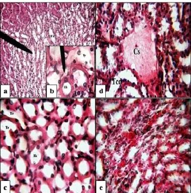
Cas: Blood capillary (wall thickening); Cb: Bowman's capsule; Cg: glomerular room; Cs: Blood congestion; Lc: Leukocytes during diapedesis; Tcd: Distal convoluted tube; Tcp: proximal convoluted tubule; Fleche (b): Podocyte nucleus; Flèche (c): Cell infiltration.
Figure 4. Renal cortical parenchyma in control (a and b), mice treated with 0.4 and 0.2 mg/kg/day of thiamethoxam (c, d, e, f and g, h respectively). H.E staining, Gr: x100 (a and c), x400 (b, d, e, f, g and h). Compared with those of controls(fig.4a and fig.4b), kidney structure of mice treated with 0.2 mg/kg/day of thiamethoxam preserves its normal general appearance. The cortex shows glomeruli without changes in shape or structure(fig.4d). However, frequent structural alterations are observed in the interstitial tissue surrounding the tubules. We found the presence of frequent bleeding sites, images of a blood congestion (fig.4c and fig.4f), and an important cellular infiltration testifying leukocyte diapedesis (fig.4c, fig.4fe and fig.4f). As well as the thickening of the capillary wall(fig.4e). In the medulla, the general appearance of tubules is normal (fig.5c) compared to that of the controls (fig.5a and fig.5b).
Histological examination of kidneys from mice treated with 0.4 mg/kg/day of thiamethoxam, revealed structural changes compared with controls and mice treated with 0.2 mg/kg/day. At the cortical level, we observed destruction of glomerulus in some areas (fig.4h). In the medulla, we noted several hemorrhagic area and blood congestion associated with the presence of leucocytes during diapedesis (fig5.d and fig.5 e).
International Journal of Biological and Agricultural Research Saadi Leila et al. 1 (2) December2018 Pages 21- 27
Pages 1-9
26
Cs: Blood congestion; Tcd: Distal convoluted tube; Tc: Collecting duct; Ah: Loop of Henle.
Figure 5. Renal medullain control (a and b), mice treated with 0.4 and 0.2 mg/kg/day of thiamethoxam (c, d and respectively). H.E staining, Gr: x100 (a), x400 (b, c, d and e).
The histological examination of renal parenchyma shows an altered structure which appear to be dose-dependent. In fact, the 0.2 mg/kg/day dose of thiamethoxam induced some cortical damage which manifested by the presence of some haemorrhagic foci and cellular infiltrates around the blood vessels. These lesions become clear with the dose 0.4 mg/kg/day and we observed some glomeruli in degeneration. At medulla level, the attack is limited to regions around the loop of Henle, presenting haemorrhagic appearance with the dose 0.4 mg/kg/day. This aspect is probably due to the destruction of the small capillaries that surround the loop. The alterations observed with treatment are considered as signs of nephrotoxicity. Similar results were found in male mice treated with thiamethoxam at rate of 26.39 and 78 mg/kg/day [6].
Conclusion
The present study shows that at low doses, thiamethoxam induces in the renal parenchyma male mice a low dose-dependent toxicity.
References
[1] Mencke N., Jeschke P., 2002 -Therapy and Prevention of Parasitic Insects in Veterinary Medicine Using Imidacloprid. Curr. Topics Med. Chem, 2 (7), 701–715.
[2] Elbert A., Haas M., Springer B., Thielert W., Nauen R., 2008 -Applied aspects of neonicotinoid uses in crop protection. Pest Management Science, 64:1099-1105.
[3] Jeschke P., Nauen R., Schindler M., Elbert A., 2011 -Overview of the status and global strategy for neonicotinoids. J Agric Food. Chem, 59:2897–2908.
International Journal of Biological and Agricultural Research Saadi Leila et al. 1 (2) December2018 Pages 21- 27
Pages 1-9
27
[4] Menandro N.A., 2008 -Insecticidal activity of thiamethoxam against the bamboo powder post beetle
Dinoderus minutus Fabr. (Coleoptera: Bostrichidae). Journal of Pesticides Sciences, 81: 109-113.
[5] FAO/OMS., 2010- Pesticide residues in food, part II: Toxicological evaluations, Rome, Italy, 21–30. [6] KhaldounOularbi H., Bouzid N., Boukreta S., Makhlouf C., Derriche F., Djennas N., 2017 -Thiamethoxam Actara® induced alterations in kidney liver cerebellum and hippocampus of male rats. Journal of xenobiotics, 7: 25-30.