THÈSE
En vue de l'obtention duDOCTORAT DE L’UNIVERSITÉ DE TOULOUSE
Délivré par Institut Polytechnique de Toulouse - INP Toulouse Discipline ou spécialité : Pathologie, Toxicologie, Génétique et Nutrition
JURY
Florence Mathieu, Professeur, INP-ENSAT (Président)
Siska Croubels, Professeur, Faculté de Médecine Vétérinaire de Ghent (Rapporteur) Yves François Pouchus, Professeur, Université de Nantes (Rapporteur)
Isabelle Oswald, Directeur de Recherche, INRA Sylviane Dragacci, Sous-Directrice de Recherche, ANSES
Olivier Puel, Ingénieur de Recherche, INRA
Ecole doctorale : Sciences Ecologiques, Vétérinaires, Agronomiques et Bioingénieries (SEVAB) Unité de recherche : Toxalim, Equipe Immuno-Mycotoxicologie
Directeur(s) de Thèse : Isabelle Oswald et Sylviane Dragacci Rapporteurs : Siska Croubels et Yves François Pouchus
Présentée et soutenue par Patricia Cano
Le Vendredi 6 Décembre 2013 Titre :
Caractérisation du Métabolome de F. graminearum: Détection et effets sur la barrière intestinale
Le travail ici présenté a été possible grâce à un partenariat entre l’Institut National de la Recherche Agronomique (INRA) et l’Agence Nationale de Sécurité Sanitaire des Aliments (ANSES).
Je tiens tout d’abord à remercier mes encadrants de thèse :
Dr. Isabelle Oswald, pour m’avoir accueillie au sein de son équipe et avoir assumé avec grande disponibilité et pertinence la direction scientifique de cette thèse. Merci aussi pour l’intérêt porté à la valorisation de ces travaux.
Dr. Sylviane Dragacci, pour avoir accepté de co-diriger cette thèse au pied levé.
Dr. Olivier Puel, pour avoir essayé de me transmettre sa passion ainsi que pour tout son enthousiasme quant à nos résultats. Je tiens aussi à te remercier d’avoir dirigé officieusement la plupart de mon travail. A quand l’HDR qui te sortira de l’ombre ?
Je tiens également à remercier les membres du jury :
Dr. Florence Mathieu, pour avoir accepté de présider le jury.
Dr. Siska Croubels et Dr. Yves François Pouchus pour avoir accepté d’être rapporteurs de cette thèse et d’avoir pris le temps de l’examiner.
J’adresse aussi tous mes remerciements aux membres de l’équipe d’immuno-mycotoxicologie pour leur gentillesse, leur aide et leurs conseils.
Plus particulièrement,
Aux étudiants avec qui j’ai partagé le bureau, la paillasse et les cafés. Merci à Selma, pour avoir tout partagé ces dernières années au labo, pour son soutient constant et pour son amitié.
A Philippe, Joëlle et Anne pour leur aide précieuse et leurs multi-compétences. Philippe, toujours là pour gérer les moments de panique à la paillasse mais aussi toujours là pour offrir encouragements et soutient. Joëlle, avec qui tous les sujets de conversation sont possibles et intéressants, pour ne jamais oublier que nous avons une vie en dehors du labo. Anne, qui rend possible l’impossible à l’animalerie, et m’a initiée patiemment dans l’expérimentation sur le porc.
Je voudrais aussi remercier les membres de Toxalim que j’ai côtoyé pendant ces trois années et qui ont participé au succès de ces travaux d’un point de vue technique mais aussi humain. Plus particulièrement,
Emilien et Laurent, pour leur accueil très chaleureux dans le monde mystérieux de la chimie analytique. Merci pour le temps que vous m’avez consacré et votre patience. Je tiens à souligner le plaisir que j’ai eu à travailler avec vous.
Mélanie et Alice, pour les moments partagés autour d’un café et qui ont rendu ces derniers mois plus faciles avec leurs sourires et leurs encouragements.
Hervé, Alex, Arnaud, Fred, Afi, Nicolas, Vincent et Edwin pour toute leur gentillesse, le réconfort que j’ai trouvé dans nos discussions, et surtout pour les moments partagés hors du labo.
Je remercie également toutes les personnes qui ont participé à l’élaboration de ces travaux de façon plus ponctuelle.
Marcel Delaforge, Christian Barreau, Vessela Atanasova Penichon pour avoir accepté de faire parti mon comité de thèse, et l’intérêt qu’ils ont porté à mes travaux.
Les équipes de Michel Péan, Thierry Langin et Christian Lannou pour leur aide pour la préparation des épis marqués et des épis infectés.
Finalement, j’adresse mes remerciements les plus sincères à ma famille et à mes amis pour leur soutient inconditionnel malgré la distance, mais aussi à celui qui m’a accompagnée et soutenue quotidiennement au cours de cette thèse et avec qui je souhaite maintenant partager la suite des aventures.
LISTE DES PUBLICATIONS ... 5 LISTE DES ABREVIATIONS ... 6 LISTE DES FIGURES ET DES TABLEAUX ... 9 I. INTRODUCTION ... 11 1. CONTEXTE ... 11 2. MOISISSURES, METABOLITES SECONDAIRES ET MYCOTOXINES ... 12 3. FUSARIUM GRAMINEARUM : PHYTOPATHOGENE ET PRODUCTEUR DE MYCOTOXINES ... 63 4. OBJECTIFS DE LA THESE... 73 II. TRAVAIL EXPERIMENTAL ... 75 1. CARACTERISATION DU METABOLOME DE F. GRAMINEARUM ... 75 1.1 Détection et identification de métabolites secondaires fongiques ... 76 1.2 Nouvelle méthode d’analyse non ciblée de métabolites fongiques (Article 1) ... 83 1.3 Etude du métabolome de F. graminearum (Article 2) ... 99
2. TOXICITE DU METABOLOME DE F. GRAMINEARUM SUR LA BARRIERE INTESTINALE ... 119
2.1 Exposition intestinale aux mycotoxines et réponse immunitaire ... 120 2.2 ‐ Effets pro‐inflammatoires du DON (Article 3) ... 126 2.3 ‐ Effets de la totalité du métabolome de F. graminearum ... 140 III. DISCUSSION ET CONCLUSIONS ... 153 1. CARACTERISATION DU METABOLOME DE F. GRAMINEARUM ... 155 1.1 Infections fongiques et contamination par les mycotoxines ... 155 1.2 Méthode d’analyse non ciblée pour caractériser le métabolome de F. graminearum ... 159 2. TOXICITE DU METABOLOME DE F. GRAMINEARUM ... 165 2.1 Discussion du protocole expérimental ... 165 2.2 Fusariotoxines et les systèmes de défense de l’organisme ... 169 3. CONCLUSIONS ... 177 REFERENCES BIBLIOGRAPHIQUES ... 179
5 Articles Deoxynivalenol as a new factor in the persistence of intestinal inflammatory diseases: an emerging hypothesis through possible modulation of Th17‐mediated response. Cano PM, Seeboth J, Meurens F, Cognie J, Abrami, R, Oswald IP, Guzylack‐Piriou L. PLoS ONE, 2013; 8(1): e53647.
New untargeted metabolic profiling combining mass spectrometry and isotopic labeling: application on A. fumigatus grown on wheat. Cano PM, Jamin EL, Tadrist S, Bourdaud’hui P, Péan M, Debrauwer L, Oswald IP, Delaforge M, Puel O. Analy Chem, 2013.
Isotopic labeling of the metabolome of F. graminearum in vitro and identification of new secondary metabolites by HPLC‐HRMS analysis. Cano PM, Jamin EL, Tadrist S, Bourdaud’hui P, Péan M, Debrauwer L, Oswald IP, Delaforge M, Puel O. Manuscrit en préparation.
Mycotoxins: Fungal Secondary Metabolites with Toxic Properties. Cano PM, Puel O, Oswald, IP. Progress in Mycological Research, publiée par IncEnfield (USA), CRC press. Chapitre en préparation.
Communications Orales
Induction de la réponse inflammatoire intestinale par le déoxynivalenol chez le porc: sensibilisation face aux pathogènes. Journées du Département de Santé Animale de l'INRA (Fréjus, Juin 2011).
Spéctrométrie de haute résolution et double marquage isotopique: une nouvelle méthode d'analyse non ciblée de métabolites fongiques. Journées Toxalim (Toulouse, Décembre 2012).
Une nouvelle méthode d'analyse non ciblée de métabolites fongiques : Spéctrométrie de haute résolution et double marquage isotopique. Journées des mycotoxines (Brest, Janvier 2013).
Fusarium graminearum in depth: a novel method to identify new metabolites by isotopic labeling and high resolution mass spectrometry. European Fusarium Seminar (Bordeaux, Mai 2013).
Combination of double isotopic labeling and high resolution mass spectrometry: a novel method for untargeted fungal metabolic profiling. Mycotoxins Workshop (Ghent, Mai 2013). Posters Implication du déoxynivalenol dans l’induction d’une réponse inflammatoire intestinale Th17 chez le porc. Immunologie des Animaux Domestiques (IAD, Lyon 2011).
Combination of double isotopic labeling and high resolution mass spectrometry: a novel method for untargeted fungal metabolic profiling. 61st ASMS Conference (Minneapolis, MN, Juin 2013).
6 Abréviations en anglais ANOVA Analysis Of Variance APC Antigen Presenting Cells APCI Atmospheric Pressure Chemical Ionization A.U. Arbitrary Units Aw Activity water b.w. body weight CAST Council for Agricultural and Sciences Technology CD Crohn’s Disease CCL20 Chemokine (C‐C motif) Ligand 20 CCR6 Chemokine (C‐C motif) Receptor 6 COX Cyclooxygenase CYP Cytochrome P450 DC Dendritic Cells DDGS Dried Distillers’ Grains and Solubles DMSO Dimethyl Sulfoxide EDTA Ethylene Diamine TetraAcetic acid EFSA European Food Safety Authority ESI Electrospray Ionization ELISA Enzyme Linked ImmunoSorbent Assay ERK Extracellular signal‐Regulated Kinases FAO Food and Agriculture Organization of the United Nations FBS Fetal Bovine Serum GALT Gut‐Associated Lymphoid Tissue GC Gas Chromatography HBSS Hank’s Balanced Salt Solution HIV Human Immunodeficiency Virus HPLC High Performance Liquid Chromatography HRMS High Resolution Mass Spectrometry HRP Horseradish Peroxydase IARC International Agency for Research on Cancer IEC Intestinal Epithelial Cell IBD Inflammatory Bowel Disease IPEC‐1 Intestinal Porcine Epithelial Cells‐1 MAPK Mitogen‐Activated Protein Kinase MLN Mesenteric Lymph Nodes mRNA messenger RiboNucléic Acid MS Mass Spectrometry MS/MS Tandem Mass Spectrometry NRPS Non‐Ribosomal Peptide Synthase NTC Non Template Control PBS Phosphate Buffered Saline
7 PGE2 Prostaglandin E2 PKS Polyketide Synthase PVC Polyvinyl Chloride qPCR quantitative Polymerase Chain Reaction QTL Quantitative Trait Locus RALDH1 Retinal Dehydrogenase RIN RNA Integrity Number ROR Retinoid‐related Orphan Receptor RPL32 Ribosomal Protein L32 RT Reverse Transcription SEM Standard Error Mean SFB Segmented Filamentous Bacteria STAT3 Signal Transducer and Activator of Transcription 3 TGF Transforming Growth Factor Th T helper cells TNF Tumor Necrosis Factor TLR Toll‐like Receptor TOF Time‐Of‐Flight UC Ulcerative Colitis USFDA United State Food and Drug Administration XCR1 Chemokine (C motif) Receptor 1 Abbréviations en français AB AntiBase ADN Acide Désoxyribonucléique AF Aflatoxine AFSSA Agence Française de Sécurité Sanitaire des Aliments ARN Acide Ribonucléique Bt Bacillus thuringiensis CMH Complexe Majeur d’Histocompatibilité CPA Cellule Présentatrice d’Antigène CX3CL Chemokine (C‐X3 motif) ligand DON Déoxynivalénol FB1 Fumonisine B1 IFN Interféron Ig Immunoglobuline IL Interleukine LD Limite de Détection LDA 22 Laboratoire de Développement et d’Analyses 22 M Molaire (mol/L) m/z Rapport masse sur charge MYCSA Mycologie et Sécurité des Aliments
8 ppb Partie par milliard (e.g. μg/kg) ppm Partie par million (e.g. mg/kg) pc Poids corporel RMN Résonance Magnétique Nucléaire Sa Sphinganine So Sphingosine TEER Résistance électrique transépithéliale TR Temps de Rétention Treg T régulateur UE/EU Union Européenne/European Union ZEA Zéaralénone
9 Figures FIG. I.1 : CYCLE DE REPRODUCTION CHEZ F. GRAMINEARUM (TÉLÉOMORPHE G. ZEAE) ... 64 FIG. I.2 : CYCLE BIOLOGIQUE DE FUSARIUM GRAMINEARUM SUR CÉRÉALES ... 65 FIG. I.3 : BIOSYNTHÈSE DE LA FUSARINE C ... 67 FIG. I.4 : BIOSYNTHÈSE DE L’AUROFUSARINE ... 68 FIG. II.1 : SCHÉMA DE L’ANALYSE D’UN MÉLANGE PAR CHROMATOGRAPHIE ... 76 FIG. II.2 : PRINCIPE DE FONCTIONNEMENT D’UN SPECTROMÈTRE DE MASSE ... 78 FIG. II.3 : SCHÉMA DE LA SOURCE ESI ET PRINCIPE DE IONISATION PAR ELECTROSPRAY ... 79 FIG. II.4 : SCHÉMA DE LA SOURCE APCI ET PRINCIPE DE L’IONISATION CHIMIQUE À PRESSION ATMOSPHÉRIQUE ... 79 FIG. II.5: SCHÉMA DU SPECTROMÈTRE DE MASSE HYBRIDE LTQ‐ORBITRAP® ... 80 FIG. II.6 : SCHÉMA DES DIFFÉRENTES TUNIQUES DE LA PAROI DIGESTIVE... 120 FIG. II. 7 : REPRÉSENTATION DE L’ÉPITHÉLIUM INTESTINAL ... 121 FIG. II.8 : REPRÉSENTATION DE DEUX ENTÉROCYTES ET DES PROTÉINES DE JONCTIONS ... 122 FIG. II.9 : MISE EN PLACE DE LA RÉPONSE IMMUNITAIRE INTESTINALE ... 125 FIG. II.10 : PLAN EXPÉRIMENTAL DE L’ÉTUDE IN VIVO DES EFFETS DU MÉTABOLOME DE F. GRAMINEARUM ... 144 FIG. II.11 : CONSOMMATION ET PRISE DE POIDS ... 148 FIG. II.12 : POIDS DES ORGANES ... 148 FIG. II.13 : MODULATION DE L’EXPRESSION DE CYTOKINES PAR L’EXPOSITION AU DON ET AU MÉLANGE DE FUSARIOTOXINES ... 149 FIG. III.1 : PROBLÉMATIQUE GÉNÉRALE DES MYCOTOXINES DANS LA CHAÎNE ALIMENTAIRE ... 156 FIG. III.2 : PERSPECTIVES DE TRAVAIL POUR LA CONTINUATION DE LA CARACTÉRISATION DU MÉTABOLOME DE F. GRAMINEARUM ... 162 FIG. III.3: FACTEURS AUGMENTANT LE RISQUE DE DÉVELOPPEMENT DES IBD ... 172 FIG. III.4: POSSIBLE RÔLE DU DÉOXYNIVALÉNOL DANS L’APPARITION DES MALADIES INFLAMMATOIRES CHRONIQUES INTESTINALES ... 174
10 Tableaux TABLEAU I.1 : PHYTOPATHOLOGIES ASSOCIÉES À LA CONTAMINATION PAR F. GRAMINEARUM ... 66 TABLEAU I.2 : COMPARAISON DES GÈNES PKS ET NRPS CHEZ F. GRAMINEARUM, F. VERTICILLOIDES, F. OXYSPORUM ET F. SOLANI ... 69 TABLEAU I.3 : FONCTIONS CONNUES DES PKS ET NRPS CHEZ F. GRAMINEARUM ... 69 TABLEAU I.4 : RECOMMANDATIONS DE L’AGENCE EUROPÉENNE POUR LA SÉCURITÉ ALIMENTAIRE (EUROPEAN FOOD SECURITY AGENCY, EFSA) CONCERNANT LES MYCOTOXINES DE F. GRAMINEARUM ... 70 TABLEAU I.5 : CONCENTRATIONS MAXIMALES AUTORISÉES POUR LES MYCOTOXINES DE F. GRAMINEARUM DANS LES PRODUITS ALIMENTAIRES DANS L’UNION EUROPÉENNE ... 70 TABLEAU II.1 : CONCENTRATIONS EN FUSARIOTOXINES DANS L’EXTRAIT DE CULTURE DE F. GRAMINEARUM ... 142 TABLEAU II.2 : RÉSULTATS DE L’ANALYSE DES MÉTABOLITES SECONDAIRES RETROUVÉS DANS L’EXTRAIT DE CULTURE DE F. GRAMINEARUM ... 146 TABLEAU II.3 : MODULATION DE L’EXPRESSION DE CYTOKINES PAR L’EXPOSITION AU DON ET AU MÉLANGE DE FUSARIOTOXINES ... 150 TABLEAU III.1: ANALYSE DES DENRÉES AGRICOLES DE JANVIER À DÉCEMBRE 2012 ET DÉTERMINATION DES NIVEAUX DE CONTAMINATION MONDIALES EN DON ... 165 TABLEAU III.2 : TENEURS MAXIMALES (EN MG DE DON /KG D’ALIMENT) RECOMMANDÉES PAR LA COMMISSION EUROPÉENNE POUR L’ALIMENTATION HUMAINE ET ANIMALE ... 165 TABLEAU III.3 : CALCULS DE CONTAMINATION EN DON DES RATIONS POUR LES PORCINS ... 166 TABLEAU III.4 : EFFETS DES MYCOTOXINES PRODUITES PAR F. GRAMINEARUM SUR L’INTESTIN ET LE SYSTÈME IMMUNITAIRE. ... 169
Introduction
11
I. Introduction
1. Contexte
Les moisissures sont des champignons filamenteux capables de coloniser de nombreux substrats dès que la présence d’éléments nutritifs et un taux d’humidité sont suffisants. Elles appartiennent au règne fongique, un des groupes majeurs du domaine des eucaryotes. Le nombre d’espèces de moisissures décrites jusqu’à présent s’élève à 99 000, et par extrapolation, les estimations les plus optimistes sur le nombre total d’espèces sur la planète considèrent qu’il y en aurait entre 3 et 5 millions. Parmi cette grande diversité d’espèces, certaines ont été utilisées depuis des siècles par l’Homme pour leurs propriétés médicinales ou pour l’élaboration d’aliments comme les fromages. Cependant, d’autres espèces sont responsables de pathologies humaines, animales et végétales, ainsi que de la dégradation des produits alimentaires. Les genres Aspergillus, Penicillium, Fusarium, Candida et Stachybotrys sont les plus pathogènes pour l’homme et peuvent déclencher des mycoses ou allergies pouvant devenir graves chez des patients immunodéprimés.
Les moisissures sont capables de produire une très grande variété de métabolites secondaires qui leur confèrent, en général, un avantage sélectif dans leur environnement. Ces métabolites sont dits secondaires car ils ne sont pas nécessaires au développement de la moisissure contrairement aux métabolites primaires tels que les acides nucléiques, les protéines ou les acides gras. De plus, contrairement aux métabolites primaires qui sont codés par seul un gène, la biosynthèse des métabolites secondaires est orchestrée par une cascade de réactions enzymatiques catalysées par des enzymes codées par des gènes regroupés en clusters. Certains de ces métabolites ont des propriétés bénéfiques pour l’homme telle que la pénicilline, mais d’autres peuvent avoir des conséquences néfastes pour la santé des vertébrés et sont connus sous le nom de mycotoxines. C’est le cas de l’aflatoxine qui est l’un des plus puissants agents cancérigènes naturels. Il existe une très grande diversité d’effets toxiques des mycotoxines qui découle directement de l’importante variété chimique de ces métabolites secondaires. Les genres Aspergillus, Penicillium et Fusarium sont les plus importants producteurs de ces toxines. Cependant, il a été observé que toutes les souches d’une même espèce ne produisent pas les mêmes mycotoxines et que certaines de ces souches ne sont pas toxinogènes. Les mycotoxines sont produites dans les champs ou pendant le stockage, lorsque les conditions sont favorables au développement fongique. Elles peuvent donc facilement contaminer les matières agricoles et perdurer bien plus que les moisissures au cours de la chaîne de production alimentaire du fait de leur grande stabilité. Les mycotoxines représentent donc un risque économique pour les agriculteurs puisqu’elles sont responsables d’importantes baisses de rendement, mais aussi des risques sanitaires pour les consommateurs. Ceci a fortement encouragé les pouvoirs publics à réglementer la présence et la concentration de certaines de ces toxines. Cependant, il y a encore un manque de connaissances important quant à la biosynthèse, la détection et la toxicité de ces mycotoxines qui empêche une bonne évaluation et prédiction des risques de contamination. C’est donc une thématique qui requière encore des efforts de recherche et c’est dans ce contexte que s’insèrent les travaux de cette thèse.
12
2. Moisissures, métabolites secondaires et mycotoxines
La partie qui est introduite par la suite présente la problématique des mycotoxines en portant une attention particulière aux mycotoxines majeures, de part leur toxicité, leur occurrence et leur réglementation. La description détaillée des voies de biosynthèse, des modes d’action, et des effets toxiques met en évidence la diversité et l’extrême complexité de cette thématique de recherche. Néanmoins, elle met aussi en exergue le manque de connaissances sur certains aspects qui poussent à poursuivre les études.
Cette partie sera publiée sous forme de chapitre dans une série de livres dédiée aux moisissures nommée : Progress in Mycological Research, publiée par IncEnfield (USA), CRC press. C’est pourquoi le texte est présenté en anglais, dans le format de publication.
Mycotoxins: Fungal Secondary Metabolites with Toxic Properties - 1
Mycotoxins: Fungal Secondary Metabolites with Toxic Properties
P.M. Cano INRA, UMR 1331, Toxalim, Research Center in Food Toxicology, FR 31027 Toulouse, France Université de Toulouse, INPT, 4-6 Allée de Monso, FR 31400 Toulouse, France Tel: 0033 (0)5 6128 5542 Fax: 33 (0) 5 61 28 51 45 Email: [email protected]
O. Puel INRA, UMR 1331, Toxalim, Research Center in Food Toxicology, FR 31027 Toulouse, France
Université de Toulouse, INPT, 4-6 Allée de Monso, FR 31400 Toulouse, France Tel: 0033 (0)5 6128 5154 Fax: 33 (0) 5 61 28 51 45 Email: [email protected]
I.P. Oswald INRA, UMR 1331, Toxalim, Research Center in Food Toxicology, FR 31027 Toulouse, France Université de Toulouse, INPT, 4-6 Allée de Monso, FR 31400 Toulouse, France Tel: 0033 (0)5 6128 5480 Fax: 33 (0) 5 61 28 51 45 Email: [email protected]
2- Mycotoxins: Fungal Secondary Metabolites with Toxic Properties Table of Contents
LIST OF FIGURES ... 3
LIST OF TABLES ... 3
I. INTRODUCTION ... 4
II. POLYKETIDES MYCOTOXINS ... 9
II.1.AFLATOXINS ... 9
II.2.FUMONISINS ... 12
II.3.OCHRATOXINS ... 15
II.4.PATULIN ... 18
II.5.ZEARALENONES ... 20
III. TERPENE MYCOTOXINS ... 24
III.1.GENERALITIES ON TRICHOTHECENES ... 24
Trichothecenes biosynthesis ... 25
Toxicity and mode of action ... 27
III.2.TYPE ATRICHOTHECENES ... 28
T-2 toxin ... 28
III.3.TYPE BTRICHOTHECENES ... 28
Nivalenol and Fusarenon X ... 28
Deoxynivalenol and its acetylated derivatives ... 29
III.4.TYPE DTRICHOTHECENES ... 30
Satratoxins ... 30
IV. NON-RIBOSOMAL PEPTIDE MYCOTOXINS ... 31
IV.1.ERGOT ALKALOIDS ... 32
IV.2.GLIOTOXIN ... 34
Mycotoxins: Fungal Secondary Metabolites with Toxic Properties - 3 List of figures
FIG. 1: WORLDWIDE OCCURRENCE OF THE MAJOR MYCOTOXINS ... 6
FIG. 2: BIOCHEMICAL MECHANISMS OF ACTION OF MYCOTOXINS ... 7
FIG.3: OVERVIEW OF THE DIFFERENT BIOSYNTHETIC PATHWAYS INVOLVED IN MYCOTOXIN PRODUCTION ... 8
FIG. 4: CHEMICAL STRUCTURES OF THE FOUR MOST OCCURRING AND NATURALLY PRESENT AFLATOXINS ... 10
FIG. 5: BIOTRANSFORMATION AND MODES OF ACTION OF AFB1 ... 12
FIG. 6: CHEMICAL STRUCTURES OF THE MAIN FUMONISINS ... 13
FIG. 7: MODES OF ACTION OF THE CARCINOGENIC FB1 ... 14
FIG. 8: DIFFERENT HYPOTHESIS FOR OCHRATOXIN A ... 17
FIG. 9: BIOSYNTHESIS OF PATULIN ... 19
FIG. 10: BIOSYNTHESIS OF ZEARALENONES ... 21
FIG. 11: MECHANISMS OF TOXICITY OF ZEARALENONE ... 23
FIG: 12: BIOSYNTHESIS OF TRICHOTHECENES ... 26
FIG. 13: EFFECTS OF DEOXYNIVALENOL ... 30
FIG. 14: ERGOT ALKALOIDS BIOSYNTHESIS: EXAMPLE OF ERGOTAMINE ... 33
FIG. 15: BIOSYNTHESIS OF GLIOTOXIN ... 35
List of tables Table 1: Most frequent mycotoxins produced worldwide (non-exhaustive). ... 4
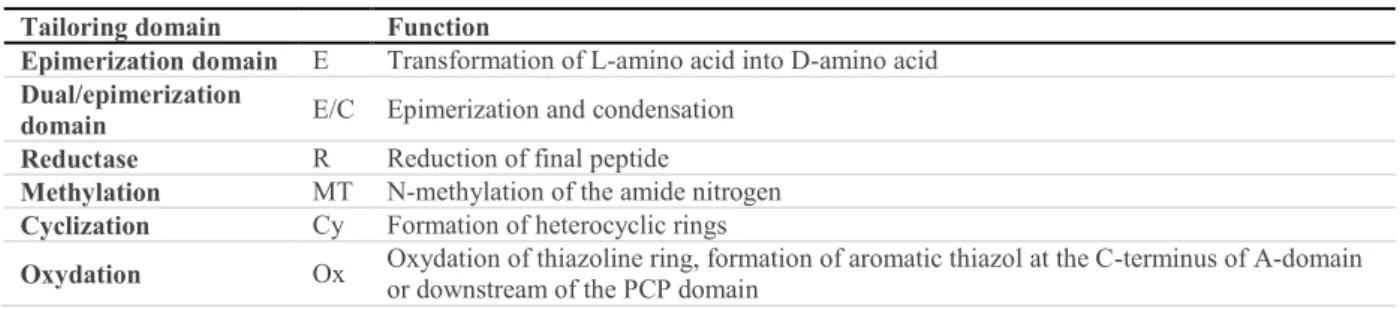
Table 2: Tailoring domains of non-ribosomal peptide synthases. ... 31
4- Mycotoxins: Fungal Secondary Metabolites with Toxic Properties I. INTRODUCTION
The fungal kingdom plays an important role in natural ecosystems, as fungi are essential decomposers of dead organic matter, thus restoring carbon, nitrogen, phosphorous and mineral levels in the biosphere. In addition to that, filamentous fungi constitute one of the most important sources of secondary metabolites. Many of these secondary metabolites present industrially interesting properties that have been beneficial for humans in the fields of medicine, food industry, cosmetics, energy and construction. However, other metabolites, known as mycotoxins, have been shown to have deleterious effects resulting in human, animal and phyto-pathologies. For instance, aflatoxin B1, one of the most potent carcinogenic and genotoxic natural substances is of fungal origin (Bennett and Klich 2003). The most important fungal genera that produce mycotoxins are Aspergillus, Penicillium and Fusarium. Aspergillus species are probably the most representative of spoilage fungi given their capacity to grow under a large array of environmental conditions. In fact, there are very few food commodities and raw materials in which Aspergillus cannot develop (Pitt and Hocking 2009). Penicillium species are also ubiquitous saprophytes that are encountered mainly as soil fungi and only accidentally in food/feed commodities, although some species are specifically used in the food production industry such as P. roqueforti (Pitt and Hocking 2009). Contrarily to Aspergillus and Penicillium species, Fusarium species are plant pathogens that are widely distributed in soils and particularly in cultivated soils. Since development of Fusarium species requires high humidity levels, these species are most frequently encountered on the fields pre-harvest or at early stages of storage (Leslie and Summerell 2006).
So far, over 1000 fungal secondary metabolites have been identified, among which, around 30 have proven toxicity and are considered as mycotoxins and only 6 are legally regulated (aflatoxins, zearalenone, deoxynivalenol, fumonisins, ochratoxin A and patulin). A list of the major mycotoxins, classified based on their biosynthetic origin is given in table 1. As it can be seen, there is a large variety of metabolites with very different toxicities. However, in most cases, there is not enough data to assess the exposure risks, which difficults the establishment of official regulations. For instance, toxicity of the so-called emerging mycotoxins, such as beauvericin, enniatins and moniliformin was acknowledged quite recently compared to other mycotoxins. Therefore, there is still not enough data for a proper assessment of the risks related to exposure to these mycotoxins, and no regulations have been established yet (Jestoi 2008).
Table 1: Most frequent mycotoxins produced worldwide (non-exhaustive).
Biosynthetic
pathway Mycotoxin Producing fungal genera Most Frequent Substrate Most notorious effects Regulation Polyketides Aflatoxins Aspergillus
Corn, Peanuts, Tree nuts
Carcinogenicity (group 1*)
Hepatotoxicity Yes
Citrinin Aspergillus Penicillium
Monascus
Grains,
Cheese Hepatotoxicity, Nephrotoxicity No
Fumonisins Fusarium Cereals (Maize)
Hepatotoxicity Disruption of lipid metabolism
Carcinogenicity (group 2B)
Yes
Moniliformin Fusarium Cereals Cardiotoxicity DNA damage No
Mycophenolic acid Penicillium Byssochlamys Blue cheeses Immunosuppressive No
Ochratoxins Aspergillus Penicillium Coffee, cocoa, spices, dried fruits, grapes Nephrotoxicity, Carcinogenicity (group 2B) Yes
Mycotoxins: Fungal Secondary Metabolites with Toxic Properties - 5
Biosynthetic
pathway Mycotoxin Producing fungal genera Most Frequent Substrate Most notorious effects Regulation
Penicillium Byssochlamys
DNA damage
Penicillic acid Aspergillus Penicillium Coffee, Cereals Synergism with Ochratoxin A No
Sterigmatocystin Aspergillus Cereals, Cheese Hepatotoxicity, Carcinogenicity (group 2B) No
Zearalenone Fusarium Cereals Hyperoestrogenism Yes
Terpenes Trichothecenes Fusarium Cereals Haematotoxicity, Immunosuppressive Yes
Non-ribosomal
peptides Beauvericin Fusarium Cereals, Fruits
Cytotoxic, ionophoric, Antibiotic and insecticidal
activity No
Enniatins Fusarium Cereals Fruits
Cytotoxic, ionophoric, Antibiotic and insecticidal
activity No
Ergot Alkaloids Claviceps Rye, Wheat Neurotoxicity Yes
Gliotoxin Aspergillus Animal feeds Immunosuppressive No
Roquefortine C Penicillium Blue cheeses (rare) Neurotoxicity No
Hybrids Cyclopiazonic acid Aspergillus Penicillium Oil seeds, nuts, peanuts, cereals, dried fruits Inhibition of Ca(necrosis) 2+-ATPase No
Fusarin C Fusarium Maize Carcinogenicity (group 2B) No
* Carcinogenicity groups according to the International Agency for Research on Cancer (IARC)
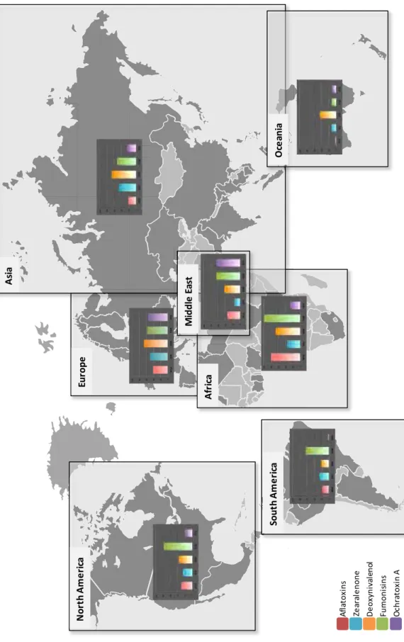
Mycotoxin-producing fungal species may develop all around the world in solid or liquid substrates as long as they are in presence of sufficient nutriments and humidity (aw > 0.6). Therefore, food and feed commodities are particularly affected. A worldwide survey performed in 2012 analyzed the presence of five of the main mycotoxins in 4,023 samples of food/feed commodities such as cereals, soybean and straw originating from North and South America, Europe, Africa, Middle East, Asia and Oceania (Fig. 1) (Biomin 2012). The results of this study show a worldwide occurrence of these mycotoxins but a heterogeneous distribution, mainly due to regional endemism of some species. Annual meteorological variations may also affect fungal development and mycotoxins distribution around the world.
Mycotoxins are produced during fungal development on the fields or during storage and therefore, in some cases, they can reach human and animal alimentary chains. Human exposure can also occur by ingestion of contaminated animal products. In addition to ingestion, which is the most common route of exposure, mycotoxins may also enter the organism through dermal, respiratory and parenteral routes (Omar 2013). Although cases of severe acute toxicity have already been reported for aflatoxin B1, and T-2 toxin, most of the concern related to mycotoxins exposure is due to chronic exposure. In any case, toxicity is dependent on the route, level and duration of exposure; on the age, health status, sex, genetics and dietary status of the exposed individuals and also on the possible interactions with other toxins (Omar 2013). At a molecular level, mycotoxins can affect the DNA template, the RNA polymerase thus hindering transcription, the ribosomes and polysomes thus hindering translation or different metabolic reactions. At a cellular level, these interactions may induce mutagenicity, carcinogenicity or teratogenicity (Fig. 2). All of this may disrupt the function of the different organs of the exposed individuals thus leading to hepatotoxicity, nephrotoxicity, neurotoxicity, immunotoxicity and so forth (Fig. 2) (Kiessling 1986, Omar 2013).
6- Mycotoxins: Fungal Secondary Metabolites with Toxic Properties
Fig. 1: Worldwide occurrence of the major mycotoxins (Biomin 2012). Occurrence of the 5 major mycotoxins (aflatoxins,
zearalenone, de oxynivalenol, f umonisins and oc hratoxin A ) were obt ained by a nalysis of 4, 023 s amples of food/feed commodities such as cereals, soybean and straw, coming from North and South America, Europe, Africa, Middle East, Asia and O ceania. The num ber of s amples a nalyzed for each mycotoxin i n e ach of t he r egions was different depending o n availabilities. Ho wever, for th e p urpose o f c larity, in th is figure, the re sults are e xpressed a s the p ercentage o f positive samples, independently of the total number of analyzed samples.
Eur ope A fr ica A sia M id d le E as t O ce an ia Sout h A m e ri ca No rt h A mer ic a A fl at o xi n s Ze ar al e n o n e D e o xyn iva le n ol Fum o ni si ns O chr at o xi n A
Mycotoxins: Fungal Secondary Metabolites with Toxic Properties - 7 Fig. 2: Biochemical mechanisms of action of mycotoxins (Kiessling 1986, Omar 2013). Given th e larg e d iversity o f
chemical structures of mycotoxins, they can induce a wide panel of toxic effects. At a molecular level, they can affect the DNA template, the RNA polymerase thus hindering transcription, the ribosomes and polysomes thus hindering translation or different metabolic reactions; at a cellular level these interactions may induce mutagenicity, carcinogenicity or teratogenicity. All o f th is may d isrupt the f unction o f th e d ifferent organs of th e e xposed in dividuals thus leading to h epatotoxicity, nephrotoxicity, neurotoxicity and immunotoxicity.
The variety of toxic effects induced by mycotoxins is directly related to the large diversity of these metabolites in terms of c hemical s tructures. H owever, i n sp ite of this diversity, bi osynthesis of mycotoxins derives from simple building blocks, all of which are initially metabolized from glucose: (1) ac etyl-CoA uni ts for p olyketides and terpenes and ( 2) ar omatic amino ac ids for non -ribosomal peptides (Fig. 3). Contrarily to bacterial toxins or fungal toxins from higher fungi, which are usually ribosomal pep tides, b iosynthesis of m ycotoxins require successive metabolic reactions t hat a re orchestrated by a cascade of enz ymes enc oded by di fferent g enes that ar e generally g athered in clusters (Keller and Hohn 1997) . The structural di versity of mycotoxins results from t he variety of sub-units and the large array of p rocessing r eactions ( cyclization, aromatization, alkylation, glycosylation, hydroxylation and epoxidation) involved in their biosynthesis (Boettger and Hertweck 2012). Additional diversity is further obtained by combination of biosynthetic pathways such as the fusion of polyketide and non-ribosomal peptide biosynthetic pathways. Although quite spread among microorganisms, this marriage remains enigmatic for the scientific community, especially concerning the biosynthetic programming and activity of the hybrid polyketide synthases (PKS) and the hybrid non-ribosomal pep tide synthases (NRPS) (Boettger and Hertweck 2012) . My cotoxins can t hus be
DNA
RNA
mRNA, tRNA, rRNA
Proteins Metabolic reactions Physiological effects Aflatoxin B1, G1 Sterigmatocystin Patulin Penicillic acid Trichothecenes TRANSCRIPTION: RNA polymerase Aflatoxin B1, G1 Sterigmatocystin Patulin Aflatoxin B1, G1 Trichothecenes Ochratoxin A TRANSLATION: Ribosomes/Polysomes Aflatoxin B1, G1 Patulin Trichothecenes Ochratoxin A Moniliformin Hepatotoxicity Nephrotoxicity Neurotoxicity Immunotoxicity Reprotoxicity
8- Mycotoxins: Fungal Secondary Metabolites with Toxic Properties
classified into four main categories: (1) polyketides, (2) terpenes, (3) non-ribosomal peptides and (4) hybrids (Fig. 3).
Fig.3: Overview of the different biosynthetic pathways involved in mycotoxin production. Mycotoxins can be classified
into four main categories (polyketides, terpenes, non-ribosomal peptides and hybrids) that derive from simple building blocks, all of which are initially metabolized from glucose: acetyl-CoA units for polyketides and terpenes and aromatic amino acids for non-ribosomal peptides. PK-NRP hybrids are obtained by the fusion of the polyketide and the non-ribosomal peptide biosynthetic pathways by the activity of hybrid polyketide synthases and hybrid non-ribosomal peptide synthases.
This chapter gives a detailed overview of some of the most representative mycotoxins of each of these categories. Special attention was paid to the mycotoxins that are regulated (aflatoxins, fumonisins, ochratoxins, patulin, zearalenone and trichothecenes). Gliotoxin and ergot alkaloids were chosen for their relevance in terms of occurrence and toxicity.
Glucose Triol Pyruvate Acetyl-CoA Polyketides (PK) Mevalonate Terpenes Pentose Shikimate
Aromatic amino acids
Phenylalanine Tryptophan
Non-ribosomal peptides (NRP)
Mycotoxins: Fungal Secondary Metabolites with Toxic Properties - 9 II. POLYKETIDES MYCOTOXINS
This group of secondary metabolites is characterized by the consecutive polymerization of ketide groups (CH2-CO)n, deriving from the metabolization of acetate. It is a large and very diverse group of metabolites that includes polyphenols, polyenes and macrolides. Given the central role of acetyl-coenzyme A (acetyl-CoA) in polyketides biosynthesis, a relationship can be drawn with the biosynthesis of fatty acids. As a matter of fact, these primary metabolites were initially also classified as polyketides (Bentley and Bennett 1999). However, two main differences exist between polyketides and fatty acids. Firstly, some of the polyketides synthases (PKS) involved in the biosynthesis of secondary metabolites require a different carboxylic acid than acetyl-CoA, which is not the case for the PKS involved in fatty acids biosynthesis, also known as fatty acid synthases (FAS). Secondly, unlike fatty acids, the oxidation state of polyketides is very variable. According to Nicholson et al., polyketides can be classified into three groups: non-reduced (like norsolorinic acid, an aflatoxin precursor), partially reduced (like 6-methylsalicylic acid, a patulin precursor) and highly reduced (like fumonisins) (Nicholson et al. 2001). This classification further correlates with the structure and biosynthetic origin of each metabolite. Fungal polyketides can have a wide variety of biological activities, which is not surprising given the large chemical and structural diversity of these metabolites. Some of these activities present industrial interests such as the well-known immunosuppressant properties of mycophenolic acid, which is produced by fungi belonging to the genera Penicillium. This metabolite has been widely used to prevent rejection in organ transplantation (Lipsky 1996) and also as a potent inhibitor of viral RNA synthesis in vitro (Gong et al. 1999). Another industrially interesting polyketide is lovastatin, which is mainly produced by Aspergillus terreus, and is used in the pharmaceutical industry as a cholesterol lowering drug (Seenivasan et al. 2008). However, many polyketides can also induce a large panel of toxic effects such as carcinogenic effects (aflatoxin, fumonisin B), hepatotoxicity (aflatoxin), nephrotoxicity (ochratoxin A), genotoxicity (patulin) and oestrogenic disruption (zearalenone). Interestingly, the impact of polyketides on public health is such that out of the 6 groups of mycotoxins that are regulated by the European Union, only the group of the trichothecenes does not belong to the polyketide family (European Commission 2006). More details will be given concerning the effects of these mycotoxins in this section.
II.1. Aflatoxins
This family of mycotoxins was one of the first to be discovered, over 40 years ago, as the cause of the Turkey X disease which triggered acute hepatotoxicity and death in poultry in Great Britain (Nesbitt et al. 1962). Since then, aflatoxins have been shown to have immunosuppressive, mutagenic, teratogenic and hepatocarcinogenic effects in both humans and experimental animals (Mathison 1997). They are now considered as the most carcinogenic and genotoxic substances of natural origin (Bennett and Klich 2003, CAST 2003). Contamination by aflatoxins generally comes before harvest in corn, peanuts, cotton seeds and tree nuts but also during improper storage of food commodities, generating important economical losses (CAST 2003). At a global level, contamination by aflatoxins affects over 5 billion people in the world through food and polluted air, most generally in warm and humid places, such as Asia, Africa and Central America, where it is associated with liver/lung cancer, HIV/AIDS, malaria, growth stunting, children malnutrition and increased risk of adverse birth (Gong et al. 2003, Jiang et al. 2008, Liu and Wu 2010, Shuaib et al. 2010, Khlangwiset et al. 2011, Rodrigues and Naehrer 2012).
10 - Mycotoxins: Fungal Secondary Metabolites with Toxic Properties
Aflatoxins are mainly produced by 13 Aspergillus species, among which A. flavus, A. ochraceoroseus and A. pse udotamarii produce t ype B af latoxins and A. paras iticus, A. bom bycis a nd A. nom ius produce both type B and G aflatoxins. A. flavus and A. parasiticus are the two main producing species (Varga et al . 2011 ). N ew Aspergillus species have been i dentified as producers of af latoxins: A. novoparasiticus, A. arac hidicola, A. m inisclerotigenes, A. ps eudocaelatus a nd A. p seudonomius (Pildain e t a l. 2008 , V arga et al. 2011, G oncalves et a l. 201 2, A djovi et al. 2014). I n addi tion to Aspergillus species, aflatoxins or aflatoxin-related metabolites such as sterigmatocystin have also been isolated from culture extracts of Fusarium kyusbuene (Schmidt-Heydt et al. 2009), Podospora anserin (Slot and Rokas 2011) and Dothistroma septosporum and D. pini (Bradshaw et al. 2006).
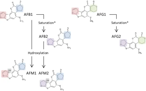
The aflatoxins family encompasses 16 structurally related furanocoumarins (Bhatnagar et al. 2003), AFB1, AFB2, AFG1 and AFG2 being the four most abundant and naturally occurring (Huffman et al. 2010). AFB1 and AFB2 are characterized by a cyclopentane E-ring whereas AFG1 and AFG2 contain a xanthone ring i nstead (Huffman et al . 2010 , Z eng et a l. 201 1). These differences result i n B lue fluorescence under long wave UV light for AFB1 and AFB2 and Green fluorescence for AFG1 and AFG2. AFB2 and AFG2 differ from AFB1 and AFG1 in that their bisfuranyl ring is saturated (Fig. 4). In addition, AFM1 and AFM2 are the major metabolites of AFB1 and AFB2, respectively and they are produced in the liver of exposed animals by hydroxylation of the parent compounds (Gallagher et al. 1994). AFM1 and AFM2 represent an important threat for public health because they are excreted in milk, linked to casein proteins, and they persist in dairy products and human breast milk (Galvano et al. 2008, Keskin et al. 2009, Motawee and McMahon 2009, Prandini et al. 2009). Recently, several studies hav e reported the presence of AFM1 in d airy products and, in so me cases, above t he maximum l imit ( 50 ng /kg) es tablished by t he European r egulation 1881 /2006 (Boudra et a l. 2007 , Delialioglu et al. 2010, Fallah 2010).
Fig. 4: Chemical structures of the four most occurring and naturally present aflatoxins: AFB1, AFB2, AFG1 and AFG2. AFB1 and AFB2 are characterized by a cyclopentane E-ring () and blue fluorescence under long wave UV light whereas AFG1 and AFG2 contain a xanthone ring () instead and display green fluorescence. AFB2 and AFG2 differ from AFB1 and AFG1 in that their bisfuranyl ring () is saturated (). AFM1 and AFM2 are the major metabolites of AFB1 and AFB2, respectively and they are produced in the liver of exposed animals by hydroxylation () of the parent compounds. * Saturation of the Bisfuranyl Ring
Biosynthesis of a flatoxins i s pr obably one of t he most st udied b iosynthetic pat hways of f ungal secondary metabolites, starting with the cloning of the gene nor-1 in A. parasiticus in the early 90’s
AFB1 AFB2 AFG1 AFG2 Saturation* Saturation* AFM1 AFM2 Hydroxylation
Mycotoxins: Fungal Secondary Metabolites with Toxic Properties - 11 (Chang et al. 1992). The complete aflatoxin biosynthetic pathway gene cluster is 70 kb long and contains 25 genes (Trail et al. 1995a, Trail et al. 1995b, Huffman et al. 2010). This biosynthetic pathway starts with the formation of hexanoate from acetate by the action of two fatty acid synthases (FASs). Hexanoate is then cyclisized by the action of a PKS into norsolorinic acid anthrone, which is the first stable intermediate. There are other stable intermediates such as averantin, versicolorin A and B and sterigmatocystin. The production of versicolorins A and B is a branching point, which determines whether AFB1 and AFG1 or AFB2 and AFG2 are synthesized. Versicolorin A is synthesized by desaturation of versicolorin B and therefore it is the precursor of the desaturated AFB1 and AFG1 whereas versicolorin B is the precursor of the saturated AFB2 and AFG2. Sterigmatocystin is the intermediate product in the versicolorin A pathway and it corresponds to dihydrosterigmatocystin in the versicolorin B pathway (Bhatnagar et al. 2003, Yu et al. 2004a, Yu et al. 2004b, Huffman et al. 2010, Roze et al. 2013). Notably, the last steps of the biosynthesis of aflatoxin take place in vesicules which play a key role in sterigmatocystin transformation and aflatoxin storage and export (Chanda et al. 2009). Interestingly, over 20 species, such as A. nidulans and A. versicolor, produce sterigmatocystin as a final product instead of aflatoxin. The enzymatic pathway involved in the biosynthesis of sterigmatocystin in A. nidulans is the same than in the biosynthesis of aflatoxin by A. parasiticus or A. flavus (Keller and Adams 1995, Brown et al. 1996, Keller and Hohn 1997). The main differences lie on different gene distribution in the genetic clusters and also, the presence of several additional genes in the aflatoxin cluster of A. parasiticus and A. flavus (Keller and Adams 1995, Brown et al. 1996, Yu et al. 2004a). That is why A. nidulans has been widely used as a model to study fungal genetics and more precisely the biosynthetic pathway of sterigmatocystin. It has also been used to study the regulation of secondary metabolism in fungi and that is how the velvet protein VeA, which plays a central role in the regulation of primary and secondary metabolism in ascomycetes and basidiomycetes was first described (Kafer 1965, Bayram and Braus 2012).
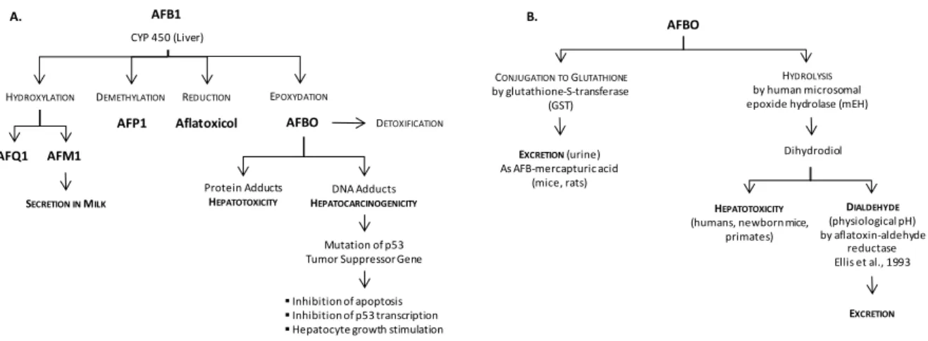
Toxicity of aflatoxins is mostly related to the genotoxic and hepatocarcigenic AFB1, which has been classified in the group 1 (i.e. sufficient evidence of carcinogenicity in humans) in the evaluation of carcinogenic risks to humans by the International Agency for Research on Cancer (IARC) of the World Health Organization (WHO) (IARC 2002). Toxicity of AFB1 follows its biotransformation mainly by cytochromes p450 in the liver (Fig. 5A) (Gallagher et al. 1994). The most reactive metabolite of AFB1 is the epoxydated exo-AFB-8,9-epoxide form, also known as AFBO (Gross-Steinmeyer and Eaton 2012, Hamid et al. 2013, Kim et al. 2013, Roze et al. 2013). This metabolite binds to proteins like albumin, which may result in hepatotoxicity (Sabbioni et al. 1987). It is also able to form DNA adducts which are responsible for hepatocarcinogenicity, and in humans, this is related to up to 50% of hepatocarcinomas (Bennett et al. 1981, Gross-Steinmeyer and Eaton 2012). Most generally, DNA adducts formed by AFBO lead to DNA base damage and oxidative damage, which may result in a mutation of p53 Tumor Suppressor Gene. Consequently, there is an inhibition of apoptosis and p53 transcription as well as a stimulation of hepatocyte growth, which all together may ultimately lead to cancer (Fig. 5A). There are two main pathways of AFBO detoxification that are more or less effective depending on the species (Fig. 5B). In mice, AFBO is conjugated to glutathione by a glutathione-S-transferase (GST), and the resulting AFB-mercapturic acid is easily excreted through urine (Nayak et al. 2009). However, this is not very effective in humans where AFBO is rather hydrolyzed by human microsomal epoxide hydrolases (mEH) (Slone et al. 1995). This results in the production of a dihydrodiol of AFB that can contribute indirectly to hepatotoxicity by forming protein adducts (Eaton et al. 2001). It is the subsequent reduction of the dihydrodiol into a dialdehyde by aflatoxine-aldehyde reductases that finally detoxifies AFBO (Ellis et al. 1993, Kelly et al. 2002). There are other metabolites of AFB1 that come from demethylation (AFP1), hydroxylation (AFQ1 and AFM1) and reduction (aflatoxicol) reactions, but contrarily to AFBO, these metabolites have a
12 - Mycotoxins: Fungal Secondary Metabolites with Toxic Properties
significantly lower toxicity compared to their parent compound (Slone et al. 1995, Eaton et al. 2001, Gross-Steinmeyer and Eaton 2012, Hamid et al. 2013, Kim et al. 2013) (Fig. 5A). However, AFM1 is also of great concern because of its presence in milk, and although it is less toxic than AFB1, it has been classified by IARC in the group 2B (i.e. possibly carcinogenic to humans) (Hsieh et al. 1984, IARC 2002). In addition to carcinogenic and genotoxic effects, AFB1 has also been reported to be immunotoxic. In vitro and in vivo studies have shown that it can reduce the number of circulating lymphocytes (Hinton et al. 2003), inhibit blastogenesis by lymphocytes (Meissonnier et al. 2008, Wada et al. 2008), alter the activity of natural killer cells and cytokine signalling (Methenitou et al. 2001), affect macrophage functions (Meissonnier et al. 2008, Bianco et al. 2012, Bruneau et al. 2012), and also impact on haematopoietic progenitors (Roda et al. 2010). Susceptibility to aflatoxins is very variable interspecies and interindividually mainly due to great differences in AFB1 biotransformation (Gross-Steinmeyer and Eaton 2012, Hamid et al. 2013, Kim et al. 2013, Roze et al. 2013). For instance, the LD50 (Lethal Dose 50) for ducklings is 0.3 mg/kg.bw whereas it is 9 mg/kg.bw for mice (Patterson and Allcroft 1970). The order of decreasing toxicity has been described as follows: AFB1> AFM1> AFG1> AFB2> AFG2.
Fig. 5: Biotransformation and modes of action of AFB1: a potent hepatotoxic and carcinogenic. Bioactivation of AFB1
takes place in the liver under the action of cytochromes P450 and results in the formation of the epoxydated exo-AFB-8,9-epoxide (AFBO), which is the most reactive metabolite of AFB1, but also other metabolites that come from demethylation (AFP1), hydroxylation (AFQ1 and AFM1) and reduction (aflatoxicol) of AFB1, but contrarily to AFBO, these metabolites have a significantly lower toxicity compared to their parent compounds (A). Detoxification AFBO can be performed by conjugation to glutathione or by hydrolysis (B). Dihydrodiol can contribute indirectly to hepatotoxicity by forming protein adducts unless it is reduced into a dialdehyde by aflatoxin-aldehyde reductases.
II.2. Fumonisins
They constitute a large family of mycotoxins mainly produced by Fusarium verticilloides, F. proliferatum and other Fusarium species (Gelderblom et al. 1988, Marasas 1996). These fungal species are important pathogens of corn responsible for “Fusarium kernel rot” or “pink ear rot” (Bullerman 1996, Marasas et al. 2004). Fumonisins have also been isolated from cultures of Alternaria alternata and Aspergillus niger (Chen et al. 1992, Frisvad et al. 2007, Mansson et al. 2010, Mogensen et al. 2010). Contamination can occur at different stages like crop growth, harvesting or storage, depending on temperature and humidity. However, development of Fusarium species mainly occurs prior harvest or in the early periods of storage when humidity levels are still high (aW > 0.9) (Dutton 2009). Human exposure to fumonisins is of greater importance in South American, Asian and African
AFB1 AFP1 AFQ1 DEMETHYLATION HYDROXYLATION DETOXIFICATION AFM1 AFBO EPOXYDATION Protein Adducts HEPATOTOXICITY DNA Adducts HEPATOCARCINOGENICITY Mutation of p53 Tumor Suppressor Gene
Inhibition of apoptosis Inhibition of p53 transcription Hepatocyte growth stimulation
Aflatoxicol REDUCTION CYP 450 (Liver) SECRETION INMILK A. CONJUGATION TOGLUTATHIONE by glutathione-S-transferase (GST) EXCRETION(urine) As AFB-mercapturic acid (mice, rats) HYDROLYSIS by human microsomal epoxide hydrolase (mEH)
Dihydrodiol DIALDEHYDE (physiological pH) by aflatoxin-aldehyde reductase Ellis et al., 1993 HEPATOTOXICITY
(humans, newborn mice, primates)
EXCRETION B.
Mycotoxins: Fungal Secondary Metabolites with Toxic Properties - 13 countries due to weather and storage conditions and also due to higher consumption levels of maize-based products (Dutton 2009).
The fumonisins family encompasses 28 structurally related metabolites that can be classified into 4 series: A, B, C and P (Rheeder et al. 2002). All of them share a long carbon chain backbone with an amine, but have different hydroxyl, methyl and tricarboxylic acid side chains (Fig. 6). A series have an acetylated terminal amine group in C2 position whereas B and C series have free amino groups and P series have a 3-hydroxypiridinium moiety at this position. C series differ from the rest in that their acyl chain is condensated with glycine instead of an alanine residue so they have a C19 backbone chain instead of C20 (Huffman et al. 2010). One particular feature of fumonisins structure is the presence of two tricarballylic esters on C14 and C15, which is rare in natural products. Type B fumonisins are the most abundant in naturally contaminated commodities. Esterification of the backbone structure gives FB4 and further oxidation gives FB2, FB3 and FB1 (Huffman et al. 2010). Among type B fumonisins, fumonisin B1 (FB1) represents 70% of total fumonisins content and FB2 generally accounts for up to 25%. Concerning the other series, only C fumonisins have been detected in corn samples but at much lower concentrations (Thiel et al. 1991).
Fig. 6: Chemical structures of the main fumonisins. The fumonisins family encompasses 28 structurally related
metabolites that can be classified into 4 series: A, B, C and P. All of them share a long carbon chain backbone with an amine, but have different hydroxyl, methyl and tricarboxylic acid side chains (R1 and R2). A series have an acetylated terminal amine group in C2 position whereas B and C series have free amino groups and P series have a 3-hydroxypiridinium moiety at this position. C series differ from the rest in that their acyl chain is condensated with glycine instead of an alanine residue so they have a C19 backbone chain instead of C20. Reproduced with modifications from (Lazzaro et al. 2012).
The biosynthesis of fumonisins has essentially been studied in the genome of Fusarium species (Proctor et al. 1999, Proctor et al. 2003, Proctor et al. 2008). The gene cluster of this biosynthetic pathway is concentrated in a 75 kb DNA region. Fumonisins biosynthetic pathway starts with the formation of a linear C18 backbone polyketide with two methyl groups and a terminal carbonyl, followed by condensation with alanine to form the C20 chain. Over 15 genes have been identified so far in the fumonisins biosynthetic cluster. However, the function of many of them remains poorly known (Brown et al. 2005). The first gene that was characterized was FUM1 which assembles the 18 carbon backbone chain from a molecule of acetyl-CoA and two molecules of S-adenosyl methionine (Huffman et al. 2010). FUM8, which encodes for an homologous of class II α-aminotransferases, is the next gene in the cluster and it is responsible for the condensation of the backbone chain (Huffman et al. 2010). In addition, FUM21, which encodes for a Zn(II)-2Cys6 DNA-binding transcription factor,
14 - Mycotoxins: Fungal Secondary Metabolites with Toxic Properties
is a specific pathway regulator (Brown et al. 2007). It positively regulates the expression of FUM genes and is thus necessary for fumonisins production.
Absorption of fumonisins and particularly of FB1 after oral ingestion is very low and remains under 4% (Martinez-Larranaga et al. 1999). However, it is rapidly distributed in all organs and tissues and more particularly in the liver and kidneys, which are the main target organs of this mycotoxin (Voss et al. 1993, Rotter et al. 1996b). Elimination occurs through feces, biliary and urinary secretions as the parent compound or after microbial deesterification, which results in partial or total hydrolization of FB1 into HFB1 (Shephard et al. 1994a, Shephard et al. 1994b, Shephard et al. 1994c, Martinez-Larranaga et al. 1999). There is no proof of FB1 biotransformation due to P450 cytochromes activities (Merrill et al. 1999). However, this mycotoxin is able to inhibit CYP2C11 and CYP1A2 activity by suppression of proteinase K (Spotti et al. 2000).
Toxicity of fumonisins was first reported for FB1 in farm animals in the late 80’s with leukoencephalomalacia in horses (Marasas et al. 1988, Kellerman et al. 1990) and pulmonary oedemas in pigs (Harrison et al. 1990), these two species being the most sensitive to FB1. Later, laboratory studies showed hepatotoxic, nephrotoxic, carcinogenic, immunotoxic, developmental and reprotoxic effects (Riley et al. 1994, Voss et al. 1995, Gelderblom et al. 2001, Marasas et al. 2004, Bracarense et al. 2012). In addition, embryotoxicity and embryolethality caused by fumonisins is strongly suspected in poultry (Javed et al. 1993, Rauber et al. 2012). In humans, there is evidence supporting a role of exposure to FB1 in neural tube defects (Marasas et al. 2004) and in liver and oesophagus cancer (Sun et al. 2007, Alizadeh et al. 2012). FB1 has thus been classified in the group 2B by IARC as “possible carcinogenic to humans” (IARC 2002).
Although FB1 increases micronucleus frequency and DNA strand breaks in vitro and in vivo, it is not considered as a genotoxic carcinogen since it lacks reactivity to DNA (Müller et al. 2012). Carcinogenicity of FB1 is rather explained by the induction of oxidative stress, which is related to the increase of reactive oxygen species (ROS) and the impairment of antioxidant defense mechanisms (Fig. 7A) (JECFA 2001, Müller et al. 2012). These result in lipid peroxidation and oxidative DNA damage. However the mechanisms behind the induction of oxidative stress by FB1 are still unknown.
Fig. 7: Modes of action of the carcinogenic FB1. The mode of action by which FB1 is carcinogenic is not very well
understood so far. Different mechanisms are possible: (A) induction of oxidative stress, related to the increase of reactive oxygen species (ROS) and the impairment of antioxidant defense mechanisms which results in lipid peroxidation and oxidative DNA damage and also (A) induction of apoptosis, which triggers the activation of defense mechanisms such as cell proliferation, possibly leading to tumorigenesis. Apoptosis can occur by (A) an increase of TNF-α, which initiates apoptosis through cell surface-mediated mechanisms, such as the caspase-3 pathway or (B) by the increase of pro-apoptotic shingoid bases (sphinganine, Sa and sphingosine, So) and the loss of complex sphingolipids (ceramide), which regulate cell adhesion. In order to counterbalance the apoptotic effects of Sa and So, cells induce the phosphorylation of these sphingoid bases by
FB1
OXYDATIVESTRESS
APOPTOSIS
↑ Reactive Oxygen Species
DNA BREAKS ↑ Caspase-3 Disruption of sphingolipids metabolism ↑ TNF-α CARCINOGENESIS A. ↓Ceramide ↑ ↑ Sphinganine* ↑ Sphingosine* FB1 Dihydro-ceramide synthase Ceramide synthase Sphinganine-1-phosphate ** Sphingosine-1-phosphate ** Sphk Sphk ↓Dihydroceramide ↑ 1-deoxy-sphinganine* B.
Mycotoxins: Fungal Secondary Metabolites with Toxic Properties - 15
Sphk1 into sphinganine 1-phosphate and sphingosine 1-phosphate, which are both antiapoptotic. In addition, FB1 also increases the production of 1-deoxy-sphinganine, which is also a potent pro-apoptotic.
* Pro-apoptotic metabolites ** Anti-apoptotic metabolites
Another possible mode of action for FB1 carcinogenicity is related to the induction of apoptosis, which has been shown in vitro and in vivo (Fig. 7A) (Müller et al. 2012). Induction of apoptosis triggers the activation of defense mechanisms such as cell proliferation, possibly leading to tumorigenesis. Again, the mechanisms by which FB1 induces apoptosis are not fully understood. Some studies provided evidence that apoptosis induction by FB1 is directly related to the increase of TNF-α, which initiates apoptosis through cell surface-mediated mechanisms, such as the caspase-3 pathway (Jones et al. 2001, Sharma et al. 2001, Bhandari and Sharma 2002).
Apoptosis could also be induced by the increase of pro-apoptotic sphingoid bases and the loss of complex sphingolipids, which regulate cell adhesion. FB1 is indeed a well-known sphingolipid disruptor since it inhibits ceramide synthases (Soriano et al. 2005) due to similarities with sphinganine (Sa) and sphingosine (So), two ceramide precursors (Fig. 7B) (Soriano et al. 2005). This results in a dose-dependent increase of Sa, and a lower increase of So, before toxicity symptoms appear (Riley et al. 1993). Thus the Sa/So ratio is commonly used as a biomarker of fumonisin exposure. In order to counterbalance the apoptotic effects of Sa and So, cells induce the phosphorylation of these sphingoid bases by Sphk1 into sphinganine 1-phosphate and sphingosine 1-phosphate, which are both antiapoptotic (Müller et al. 2012). In addition, it has been shown that FB1 also increases the production of 1-deoxy-sphinganine in vivo, which is also a potent pro-apoptotic that cannot be phosphorylated like Sa and So to reduce its toxicity due to the lack of the hydroxyl group (Zitomer et al. 2009). Other studies have shown that when FB1 or HFB1 bind to ceramide synthase, they are transformed into their N-acetyl derivatives, which are more toxic than the parent compounds (Harrer et al. 2013).
Concerning the toxicity of other fumonisins, FC1 and FC2, which are structurally equivalent to FB1 and FB2 are highly phytotoxic, and FC3 and FC4, which are structurally similar to FB3 and FB4 have moderate phytotoxicity (Abbas et al. 1998).
II.3. Ochratoxins
Ochratoxins (OTs) belong to a large family of mycotoxins that encompasses more than 20 different metabolites among which ochratoxin A (OTA) is the most naturally occurring and most toxic. Ochratoxins are produced by Aspergillus species and mainly by A. ochraceus and A. westerdijkiae, which are usually found on coffee, cocoa, spices and dried fruits in tropical regions of the globe. Other OT-producing Aspergillus species are A. alliaceus, which has been detected on figs and nuts, and A. carbonarius, which is responsible for the decay of grapes and the presence of OTs in wine. Many Penicillium species also produce OTs like P. verrucosum that develops on cereals during storage in temperate regions of the world and P. nordicum, which is found on cheese and fermented meat (Reddy and Bhoola 2010). As most mycotoxins, OTs are not degraded during most food-processing steps such as fermentation and cooking and they are thus frequently encountered in food and feed (Huffman et al. 2010). Cereals, wine, beer and pork meat are, in this order of importance, the main sources of human exposure to OTs (Huffman et al. 2010). In addition, another important feature of OTs is that they readily bind to proteins and therefore, they have also been detected in maternal milk, with the
16 - Mycotoxins: Fungal Secondary Metabolites with Toxic Properties
consequent exposure of infant babies (Pfohl-Leszkowicz and Manderville 2007). Ochratoxins are usually co-detected with other mycotoxins such as citrinin, aristolochic acid and fumonisins, with a resulting synergistic combination and an increased risk of genotoxicity. Nevertheless, very few studies have addressed the effects of co-exposure to these mycotoxins, and more data is needed (Reddy and Bhoola 2010).
Ochratoxins are hybrid molecules composed of a pentaketide dihydroisocoumarin moiety linked to the amino acid phenylalanine by an amide bond at C7. There is a chlorine atom on one of the hydroisocoumarin rings that strongly contributes to toxicity. That is why OTB which is the dechlorinated form of OTA is at least 10 times less toxic than OTA (Huffman et al. 2010). Other important metabolites are: OTα and OTβ, in which there is a cleavage of the amide bond of OTA or OTB, respectively; OTC, which is the ethyl ester form of OTA and the hydroxylated OTA. OTA and OTB also exist in open lactone form (O'Callaghan et al. 2013).
Compared to other mycotoxins, little is known about the biosynthesis of OTs. Most of the studies have used Penicillium species and thus, a putative genetic cluster for OTA production has been identified in P. nordicum (Karolewiez and Geisen 2005, Geisen et al. 2006). Given the chemical structure of OTA, three crucial steps for its biosynthesis have been defined: (1) the biosynthesis of the polyketide isocoumarin group, which is attributed to a PKS, (2) the ligation to phenylalanine, which is conducted by a NRPS and (3) the chlorination step, which is performed by a halogenase. Accordingly, sequences coding for a PKS (otapksPN), for a NRPS (otanpsPN) and for an enzyme homologous to a chlorinating enzyme (otachlPN) have been identified in the OTA genetic cluster in P. nordicum. In addition, other sequences have also been identified in the cluster coding for transporter proteins involved in OTA export (otatraPN) (Gallo et al. 2012). Homologous PKS genes have been identified in the genomes of A. carbonarius, A. niger, A. ochraceus, A. westerdijkae and P. nordicum and homologous NRPS genes have been identified in A. carbonarius (Gallo et al. 2013).
Little is known on the order of the different steps leading to OTA production. The first biosynthetic pathway was proposed by Huff and Hamilton in 1979 (Huffman et al. 2010) (Fig. 8A). They suggested that condensation of 1 acetyl-CoA and 4 malonyl-CoA by a PKS resulted in the formation of mellein which was oxidized into OTβ before chlorination into OTα. Then, ligation with phenylalanine would take places by the action of a NRPS, and would lead to the synthesis of OTC, which would finally be transformed into OTA by the action of an esterase. In 2001, Harris and Mantle suggested that chlorination took place in the final steps rather than before ligation to phenylalanine (Harris and Mantle 2001). Finally, in 2012, Gallo et al. observed, in A. carbonarius, that when the NRPS gene in OTA cluster (ACOTAnrps) was disrupted, the mutant did not produce OTA, as expected, but neither did it produce OTC, OTB nor OTα (Gallo et al. 2012). Only OTβ and phenylalanine were detected. They also observed that the mutant kept the ability to produce OTα after addition of exogenous OTA. Given these results, they proposed a new version of OTA biosynthesis in which OTβ is ligated to phenylalanine by the NRPS leading to OTB production (Fig. 8B). OTB is then chlorinated to give OTA which can be further metabolized to form OTα as well as other metabolites. This last step can be performed by the fungi itself or other microorganisms such as rumen microbiota (Gallo et al. 2012).