TITLE PAGE
Title: Are NREM sleep characteristics associated to subjective sleep complaints after mild traumatic brain injury?
Running title: Sleep after mild TBI
Authors’ name, degrees, and affiliations:
Caroline Arbour1-2, RN, PhD; Samar Khoury1, 3, PhD; Gilles J. Lavigne1, 4, DMD, PhD; Katia Gagnon1, 5, PhD (c); Gaétan Poirier1, PhD; Jacques Y. Montplaisir1, 6, MD, PhD; Julie Carrier1-2, PhD; Nadia
Gosselin1-2, PhD
1. Center for Advanced Research in Sleep Medicine (CARSM), Hôpital du Sacré-Coeur de Montréal, Montreal, Quebec, Canada
2. Université de Montréal, Department of Psychology, Montreal, Quebec, Canada 3. Université de Montréal, Department of Physiology, Montreal, Quebec, Canada 4. Faculty of Dental Medicine, Université de Montréal, Montreal, Quebec, Canada
5. Université du Québec à Montréal, Department of Psychology, Montreal, Quebec, Canada 6. Université de Montréal, Department of Psychiatry, Montreal, Quebec, Canada
Word count: 3738
Corresponding author: Dr Nadia Gosselin, Center for Advanced Research in Sleep Medicine, Hôpital du Sacré-Coeur de Montréal, 5400 boul. Gouin Ouest, local E-0330, Montreal, Qc, Canada H4J 1C5. Phone: (514) 338-2222 ext: 7717, Fax: (514) 338-3892, Email: nadia.gosselin@umontreal.ca
ABSTRACT
Introduction: Sleep complaints are common after mild traumatic brain injury (mTBI). While
recent findings suggest that sleep macro-architecture is preserved in mTBI, features of NREM sleep micro-architecture including EEG spectral power, slow waves (SW), and sleep spindles could be affected. This study aimed to compare NREM sleep in mTBI and healthy controls, and explore whether NREM sleep characteristics correlate with sleep complaints in these groups. Methods: Thirty-four mTBI participants (mean age: 34.2±11.9 yrs; post-injury delay: 10.5±10.4 weeks) and 29 age-matched controls (mean age: 32.4±8.2 yrs) were recruited for two consecutive nights of polysomnographic (PSG)
recording. Spectral power was computed, and SW and spindles were automatically detected in three derivations (F3, C3, O1) for the first three sleep cycles. Subjective sleep quality was assessed with the Pittsburgh Sleep Quality Index (PSQI). Results: mTBI participants reported significant poorer sleep quality than controls on the PSQI and showed significant increases in beta power during NREM sleep at the occipital derivation only. Conversely, no group differences were found in SW and spindle
characteristics. Interestingly, changes in NREM sleep characteristics were not associated with mTBI estimation of sleep quality.Conclusions: Compared to controls, mTBI were found to have enhanced
NREM beta power. However, these changes were not found to be associated with the subjective evaluation of sleep. While increases in beta bands during NREM sleep may be attributable to the occurrence of a brain injury, they also could be related to the presence of pain and anxiety as suggested in one prior study.
1. INTRODUCTION
Two-third of individuals who have experienced a traumatic brain injury (TBI) report altered sleep patterns and fatigue 3 years post-injury (Kempf, Werth, Kaiser, Bassetti, & Baumann, 2010). While sleep complaints are observed more frequently after mild TBI (mTBI), compared to moderate to severe TBI (Ouellet, Beaulieu-Bonneau, & Morin, 2006), our previous work found no change in the sleep macro-architecture of mTBI individuals (Gosselin et al., 2009; Khoury et al., 2013). Still, based on the hyperarousal model (Riemann et al., 2001), several components of sleep micro-architecture, such as the electroencephalographic (EEG) spectral power, could be affected. Accordingly, patients with primary insomnia often show elevated power values in the high beta and sigma frequency bands during non-rapid eye movement (NREM) sleep (Spiegelhalder et al., 2012). Reduced delta activity during NREM sleep has also been observed in several clinical conditions including depression and chronic fatigue (Armitage et al., 2007).
In addition to spectral power, slow waves (SW) and sleep spindles (two oscillatory features of NREM sleep) could be affected post-TBI. SW occur predominantly during stage 3 of NREM sleep. They are characterized as low frequency (4Hz) and high amplitude (75V) waves, with each oscillation presenting a negative phase (or hyperpolarized phase) and a positive phase (or depolarized phase), both lasting hundreds of msec (Csercsa et al., 2010). SW originate more frequently from the insula and the cingulate gyrus, and travel in an anteroposterior direction (Massimini, Huber, Ferrarelli, Hill , & Tononi, 2004; Murphy et al., 2009). As for sleep spindles, they consist of short (0.5–2.5 sec) synchronous bursts of 11-15 Hz fusiform waves generated in the thalamus and governed by the cortex through cortico-thalamic projections (Steriade, 2006). Spindle frequency varies with scalp topography, with slower mean spindle frequency in anterior derivations and higher mean spindle frequency in centroparietal derivations (Anderer et al., 2001). Moreover, they occur predominantly during stage 2 of NREM sleep (De Gennaro & Ferrera, 2003).
At the functional level, SW and sleep spindles are thought to play a crucial role in the plasticity of neural circuits, which is essential for learning and memory and to protect the sleeping brain from disruptive stimuli (Marshall, Helgadottir, Molle, & Born, 2006; Rioult-Pedotti, Friedman, & Donoghue, 2000; Steriade, 2006). Given that the rhythmicity of EEG activity is a marker of neuronal integrity (Siapas & Wilson, 1998), SW and spindles could unveil useful information about sleep after mTBI. This study aimed to examine the EEG spectral power and oscillatory features of NREM sleep, as well as their associations with subjective sleep complaints after mTBI. The specific objectives were to describe and contrast in mTBI and healthy controls: 1) the absolute and relative power frequencies during NREM sleep; and 2) the characteristics of SW and sleep spindles. We also wanted to explore the associations between NREM sleep and subjective sleep quality in both groups. Based on previous findings with mild to severe TBI (Ouellet, Beaulieu-Bonneau, & Morin, 2006; Rao, Bergey, Hill, Efron, & McCann, 2011), we hypothesized that increased NREM beta power would be found in mTBI compared to healthy controls. We also hypothesized that lower amplitude, frequency, slope, and density in SW, as well as lower amplitude, frequency, duration, and density in spindles would be noted in mTBI compared to controls. Finally, we expected that greater changes in NREM sleep would be associated with higher levels of sleep complaints in both groups.
2. METHODS
2.1. Design, sample ascertainment, and ethics
A cross-sectional design was used. The final sample was built upon data available from two previously published studies (Gosselin et al., 2009; Khoury et al., 2013) examining sleep quality in mTBI adults. The first study involved 10 athletes with a history of multiple concussions (at least one in the last year), and 11 age-matched non-concussed athletes (non-contact sports) as the control group (Gosselin et al., 2009). Study referral of concussed and non-concussed athletes in Gosselin et al. (2009) was made by the physicians irrespective of their sleep quality status after review of medical record and
clinical examination. Conversely, patients who came from the Khoury et al. (2013) study were screened from the admissions list of a Level 1 trauma hospital. They consisted of 24 adults with acute mTBI (less than 4 months post-injury), and 18 age-matched controls with no history of TBI. In contrast to Gosselin et al. (2009), mTBI patients from Khoury et al. (2013) were only recruited if they reported having poor sleep on the Pittsburgh Sleep Quality Index (PSQI: as per a score >5 on the scale). Still, they were excluded if they had consulted a sleep clinic or received any diagnosis of sleep disorder prior to study initiation. In both studies however (Gosselin et al., 2009; Khoury et al., 2013), the diagnosis of mTBI was made by a team of physicians specialized in sport medicine or neuro-traumatology based on the criteria from the World Health Organisation (WHO: Carroll et al., 2004). These criteria included a Glasgow Coma Scale score between 13-15 with a loss of consciousness or post-traumatic
amnesia/confusion for less than 30 min following the impact.
Overall, 34 mTBI adults (mean age: 34.21±11.94) and 29 age-matched controls (mean age: 32.41±8.28) were considered in this study, for a total of N=63 participants (Table 1). Injuries resulted either from a sport-related incident (n=14), a motor vehicle accident (n=10), a fall, (n=9), or an assault (n=1). Individuals with one or more of the following conditions were excluded: 1) gross cognitive or speech dysfunctions; 2) current nightshift work; 3) use of medication, drugs, or natural products known to influence sleep, affect and/or motor behaviors; 4) a documented history of neurological problem, psychiatric condition (including diagnosis of anxiety or depression disorders) or chronic substance abuse; 5) a documented history of sleep disorders including circadian cycle disruption; and 6) a history of chronic pain. Written consent was obtained from each participant and research protocols were individually approved by the ethics committee of the Hôpital du Sacré-Coeur de Montréal, Québec, Canada.
2.2. Procedure
2.2.1. Subjective sleep quality and depressive mood
All participants completed the Pittsburgh Sleep Quality Index (PSQI), a self-rated questionnaire that assesses 7 components of sleep quality over the previous month (Buysse, Reynolds, Monk, Berman, & Kupfer, 1989). A PSQI score 5 is indicative of sleep disturbance (Backhaus, Junghanns, Broocks, Riemann, & Hohagen, 2002). The second edition of the Beck Depression Inventory scale (BDI-II: Beck, Steer, & Brown, 1996) was used to assess participants’ levels of dysphoric mood. The BDI-II contains 21 items assessing symptoms of major depressive disorder, and a score between ≥10 indicates clinical depression (Beck, Steer, & Gardin, 1988).
2.2.2. Polysomnography
Participants underwent two consecutive nights of polysomnographic (PSG) recording at the sleep laboratory. Bedtime was set at 11:00 pm and sleep offset was set at 7:00 am. The first night was used for habituation and the second night for data collection and analysis. PSG recordings were performed using two different montages. In the project with concussed athletes (Gosselin et al., 2009), sleep architecture was recorded using 19 electroencephalogram (EEG) derivations (FP1, FP2, Fz, F3, F4, F7, F8, Cz, C3, C4, O1, O2, T7, T8, P7, P8), referred to linked earlobes. The montage used in the second project with mTBI (Khoury et al., 2013) comprised 11 EEG derivations (Fp1, Fp2, F3, F4, F7, F8, Fz, C3, C4, O1, O2), also referred to linked earlobes. In both studies, bilateral electrooculogram, chin, masseter, tibialis electromyograms (EMG), and three electrocardiogram (ECG) derivations were used. Thoracic and abdominal straps, a nasal airflow cannula, and a pulse oximeter were also used to monitor respiration during the first night allowing subjects who presented an apnea-hypopnea index 10 to be excluded. Signals were digitalized at 256Hz and 512Hz sampling rate for EEG and ECG respectively. Continuous audio and infrared video recordings were performed to detect abnormal movements and behaviors during sleep. Sleep stages were visually scored off-line on 20-sec epochs using Rechtschaffen and
Kales’ criteria (1968). Arousals, periodic leg movements, and respiratory disturbances were scored using standard criteria (AASM, 2005).
As for sleep macro-architecture, the following were computed: sleep latency, REM latency, sleep duration, and sleep efficiency (see Gosselin et al., 2009 for more details).The percentage of time (min) spent in each sleep stage was also computed for the entire sleep period. Sleep cycles were individually defined as a NREM sleep period lasting at least 15 min followed by a REM sleep period lasting at least 5 min and were automatically detected according to published criteria (Aeschbach & Borbély, 1993).
2.2.3. qEEG spectral analysis
Sleep frequency bands were analyzed off-line using Harmonie software version 6.21 (Stellate Systems) on artefact-free signals recorded on the left frontal (F3), central (C3), and occipital (O1) derivations, for the first three sleep cycles. For NREM sleep (stages 2, 3 and 4), Fast Fourier transform (FFT) was applied to the entire night and for each sleep cycle on NREM sleep EEG with a cosine filter on mini-epochs of 4 s with a spectral resolution of 0.25 Hz. Artifacts were detected automatically and through visual inspection. Sections with artifacts were not considered for further analysis. Frequency bands were defined as: delta (0.5–4 Hz), theta (4–8 Hz), alpha (8–12 Hz), sigma (12–16 Hz), and beta (16–32 Hz) according to standard nomenclature (IFSECN, 1974). Absolute and relative power for each frequency band and the ratio of slow to fast frequencies ((delta + theta)/(alpha + sigma + beta)) were calculated for each electrode (F3, C3, O1) and for each subject.
2.2.4. Automatic algorithm detection
SW and spindles were automatically detected on F3, C3, and O1 derivations and for the first three sleep cycles. For SW, data were initially bandpass filtered between 0.3 and 4.0 Hz using a linear phase Finite Impulse Response (FIR) filter (–3 dB), a method inspired from the work of Massimini et al. (2004). SW were detected on artifact-free stages 2, 3 and 4 of NREM sleep according to criteria
duration of negative deflection between 125-1500 ms; and 4) duration of positive deflection 1000 ms. Sleep spindles were detected during stages 2, 3 and 4 of NREM sleep using a validated automatic algorithm (Martin et al., 2013). Raw EEG data were filtered using a band-pass FIR filter (-3 dB) between 11 and 14.9Hz. The root mean square amplitude of the filtered signal was calculated on 0.25 s epochs and minimum spindle duration was set at 0.5 s. Artefactual epochs and those corresponding to REM sleep were not considered for analysis.
2.2.5. Morphologic characteristics and density of SW and sleep spindles
Once SW and sleep spindles had been identified, their morphologic characteristics were examined. Specifically, for each SW: 1) amplitude - considered as the difference in voltage between negative and positive peaks in V; 2) frequency - consisting in the number of SW cycles per min; 3) slope - referring to the velocity of the change between negative and positive peaks in V/s; 4) negative phase duration in s; and 5) positive phase duration in s, were computed. For each spindle: 1) amplitude – referred to the difference in voltage between highest negative peak and lowest positive peak in V; 2) frequency - consisting in the number of spindle cycles per min; and 3) duration – defined as the length of spindle in s, were computed. Adding to their morphologic characteristics, SW and spindles density (e.g. the number of SW or spindles per min of NREM sleep) was also calculated.
2.3. Statistical analyses
Statistical analyses were performed with SPSS version 22 (SPSS Inc, Chicago, IL) and statistical significance was set at p<0.05. Descriptive statistics e.g. frequencies with percentages, or means with standard deviations (SD) were performed for all study variables. Chi square and t-tests were used to compare mTBI and control participants in relation to their demographic, clinical, and sleep variables (including sleep macro-architecture variables). To examine the first research objective, three-way mixed measure ANOVA with two repeated within factors (e.g. three derivations and three sleep cycles) and one between factor (e.g. two groups) were performed to identify and assess any group differences
according to spectral power, SW, and spindle variables. P values for repeated measures with more than two levels were adjusted for sphericity with Greenhouse-Geisser corrections. Differences in interaction effects were assessed with post hoc Tukey HSD tests. Main effect analyses were performed when significant interactions were found. For the second research objective, Pearson correlations were performed between the different components of sleep macro-architecture (e.g. sleep latency, efficiency, stages duration), sleep micro-architecture (e.g. spectral power, SW, sleep spindles), and the severity of sleep complaints reported in both groups.
3. RESULTS
3.1. Sample characteristics, depressive mood, and sleep quality
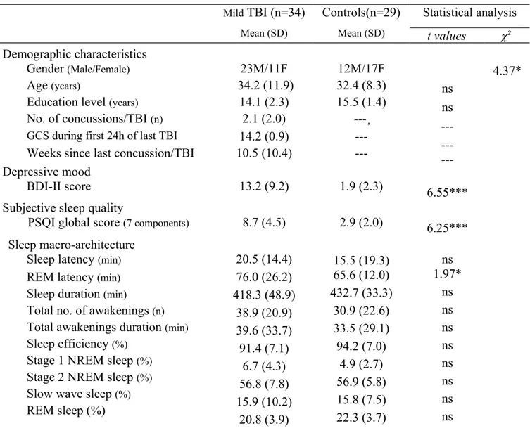
Although all participants were found to be similar regarding their demographic characteristics, those from the mTBI group had more severe depressive mood, as they scored significantly higher on the BDI-II scale than controls (Table 1). Participants with mTBI also reported lower sleep quality, with a global score on the PSQI about three times higher than that of controls. About sleep macro-architecture, mTBI participants were found to have significant longer REM latency than controls. No significant differences between both groups were found on the other components of sleep macro-architecture.
Table 1. Demographic, clinical, and PSG characteristics of mTBI and control participants Mild TBI (n=34) Mean (SD) Controls(n=29) Mean (SD) Statistical analysis t values 2 Demographic characteristics ns ns ---6.55*** 6.25*** 4.37* Gender (Male/Female) 23M/11F 12M/17F Age (years) 34.2 (11.9) 32.4 (8.3)
Education level (years) 14.1 (2.3) 15.5 (1.4) No. of concussions/TBI (n)
GCS during first 24h of last TBI
2.1 (2.0) 14.2 (0.9)
---¸ ---Weeks since last concussion/TBI 10.5 (10.4) ---Depressive mood
BDI-II score 13.2 (9.2) 1.9 (2.3)
Subjective sleep quality
PSQI global score (7 components) 8.7 (4.5) 2.9 (2.0)
Sleep macro-architecture Sleep latency (min)
REM latency (min)
Sleep duration (min)
Total no. of awakenings (n)
Total awakenings duration (min)
Sleep efficiency (%)
Stage 1 NREM sleep (%)
Stage 2 NREM sleep (%)
Slow wave sleep (%)
REM sleep (%) 20.5 (14.4) 76.0 (26.2) 418.3 (48.9) 38.9 (20.9) 39.6 (33.7) 91.4 (7.1) 6.7 (4.3) 56.8 (7.8) 15.9 (10.2) 20.8 (3.9) 15.5 (19.3) 65.6 (12.0) 432.7 (33.3) 30.9 (22.6) 33.5 (29.1) 94.2 (7.0) 4.9 (2.7) 56.9 (5.8) 15.8 (7.5) 22.3 (3.7) ns 1.97* ns ns ns ns ns ns ns ns *p0.05; ***p0.001; ns: non-significant; df=61 3.2. qEEG
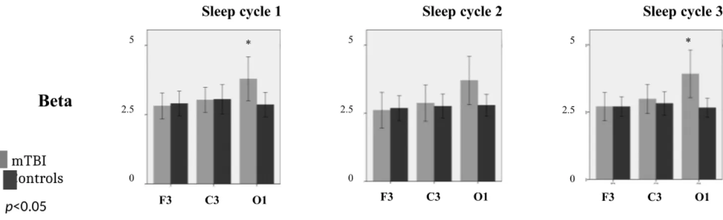
Spectral power analysis showed very few differences between mTBI and controls during NREM sleep periods. First, no significant differences in absolute delta, theta, alpha, or sigma power were found between both groups. Still, a significant Group by Derivation interaction was observed in beta bands. Based on Tukey post-hoc analysis, higher beta power was found in mTBI compared to controls at derivation O1 in all NREM sleep cycles Cycle 1 (F=4.454; p=0.039), Cycle 2 (F=3.761; p=0.047), Cycle 3 (F=7.455; p=0.008) (Figure 1).
Figure 1. Absolute beta spectral power (in V2/Hz) for NREM sleep across derivations (F3, C3, O1) and
sleep cycles (Cycles 1 to 3) in mTBI (grey) and controls (black)
Sleep cycle 1 Sleep cycle 2 Sleep cycle 3
Beta
mTBI Controls * p<0.05
A significant Group by Derivation by Cycle interaction was also found in relative theta, sigma, and beta power. However, after post-hoc analyses, only a significant change in beta band was observed between mTBI and controls. Similarly to what was found with absolute spectral power, the significant increases in relative beta power was observed at derivation O1 only, in all NREM sleep cycles Cycle 1 (F=4.306; p=0.049), Cycle 2 (F=3.343; p=0.048), Cycle 3 (F=7.193; p=0.010) (Figure 2). With both absolute and relative analyses, a statistical difference was reached for the NREM beta activity of the O1 derivation (see Figures 1 and 2 lower panels). Finally, no group differences were found in the ratio of slow to fast frequencies across electrodes (F3, C3, O1) and sleep cycles.
Figure 2. Relative beta spectral power (in %) for NREM sleep across derivations (F3, C3, O1) and sleep cycles (Cycles 1 to 3) in mTBI (grey) and controls (black)
Sleep cycle 1 Sleep cycle 2 Sleep cycle 3
Beta mTBI Controls * p<0.05 * 5 2.5 0 F3 C3 O1 5 2.5 0 F3 C3 O1 * F3 C3 O1 5 2.5 0 * F3 C3 O1 2 1 0 * 2 1 0 F3 C3 O1 * F3 C3 O1 2 1 0
3.3. Morphology and density of SW and sleep spindles
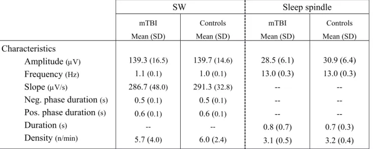
No significant between-group differences or interactions involving group were observed on any of the morphologic characteristics and density of SW and sleep spindles (Table 2). Even when
depressive mood (e.g. BDI-II scores) were considered as a covariate, no significant differences in SW and spindle characteristics were found between mTBI and control participants. Power analysis revealed that between 315 to 13,261 participants would have been required in each group to have a power of 0.80 for the detection of significant changes in the characteristics of SW and sleep spindles (Cohen, 2008). Table 2. Mean value of each SW and spindle characteristic observed in mTBI and controls ɸ
SW Sleep spindle mTBI Mean (SD) Controls Mean (SD) mTBI Mean (SD) Controls Mean (SD) Characteristics Amplitude (V) Frequency (Hz) Slope (V/s)
Neg. phase duration (s)
Pos. phase duration (s)
Duration (s) Density (n/min) 139.3 (16.5) 1.1 (0.1) 286.7 (48.0) 0.5 (0.1) 0.6 (0.1) --5.7 (4.0) 139.7 (14.6) 1.0 (0.1) 291.3 (32.8) 0.5 (0.1) 0.6 (0.1) --6.0 (2.4) 28.5 (6.1) 13.0 (0.3) --0.8 (0.7) 3.1 (0.5) 30.9 (6.4) 13.0 (0.3) --0.7 (0.3) 3.2 (0.4) ɸ As no main effect for derivation and sleep cycle were noted for SW and spindle characteristics, descriptive values were averaged across derivations (F3, C3, O1) and sleep cycles
2.4. Relationships between subjective sleep quality, sleep macro-architecture, and sleep micro-architecture
No correlations were found between PSQI scores and any of the PSG variables related to sleep macro-architecture (e.g. sleep latency, efficiency, stages duration). While significant differences in relative and absolute beta power values were observed during NREM sleep between mTBI and controls, they were not correlated with participants’ estimation of sleep quality (as per PSQI scores). Regarding
the other aspects of sleep micro-architecture, no correlations were found between PSQI scores, and SW and spindle morphology and density.
4. DISCUSSION
This study aimed to determine whether NREM sleep differs in mTBI patients compared to healthy controls, and if so, whether NREM sleep characteristics correlate with sleep complaints in this patient group. The main finding was that although mTBI reported poorer sleep quality than healthy controls, sleep macro-architecture was very similar in both groups. Additionally, no group differences in SW and sleep spindles were found. Still, mTBI showed significantly higher NREM sleep beta power in the occipital derivation, but these changes were not correlated with participants’ estimation of sleep quality.
It is well known that the different components of sleep micro-architecture such as spectral power bands, SW, and sleep spindles are a function of neuronal integrity (Siapas & Wilson, 1998). Considering a TBI can bring neuroanatomical changes similar to those observed in normal aging (Tsuno et al., 2002), we expected to observe some similarities between the sleep micro-architecture of mTBI and middle-aged adults. In light of our findings however, it appears that the disruptions of sleep EEG are different in individuals with mTBI than those observed in normal aging. Indeed, absolute power density in NREM sleep is often reduced in the entire delta, theta, alpha, and in the lower sigma bands in middle-aged adults (Landolt, Dijk, Archermann, & Borbéry, 1996), but one study also report increased beta activity (Carrier et al., 2011). In our sample of mTBI, significant higher beta power was observed in derivation O1, compared to controls. This result is similar to the one found in our previous study (Khoury et al. 2013) in which an increase in beta and gamma bands were found in derivation F8 during stage 2 NREM sleep. Heightened beta power in mTBI could represent a marker of cortical hyper-arousability during NREM sleep such as those observed in insomnia (Bader, Schäfer, Nissen, & Schenkel, 2013). In other words, mTBI individuals could have an increased tendency to be awakened during sleep.
Although significant changes in beta activity were found between mTBI and controls, they were not correlated with participants’ estimation of sleep quality. Even when depressive mood was considered as a covariate, no correlations were found between sleep characteristics and sleep quality. This
discrepancy between subjective and objective sleep has been observed previously in patients with mild to severe TBI, and is also well known in patients with insomnia (Ouellet, Beaulieu-Bonneau, & Morin, 2006). In a recent study involving women with severe premenstrual syndrome (Baker et al., 2012), higher anxiety levels were found to be correlated with poorer estimation of sleep quality in the absence of EEG sleep abnormalities. Also noteworthy are the results from our previous study (Khoury et al., 2013) in which PSQI scores were found to be best explained by mTBIs’ level of anxiety and pain rather than by their level of depression or their sleep architecture. Unfortunately, pain and anxiety were not assessed in all our participants, impeding our ability to further discuss our results on that aspect.
Finally, no changes in SW and sleep spindles were found between mTBI and healthy controls. This contrasts with findings from a previous study in which significant decreases in the amplitude and frequency of spindles was noted in moderate to severe TBI (Urakami, 2012). Based on these results, it appears that the composition of the brain structure could play a role in neuronal synchrony, and
consequently influence NREM sleep activities. As decreases in gray matter volume and reorganization of functional networks (i.e. white matter tracks) have been documented in moderate to severe TBI (Castellanos et al., 2011), and considering those elements are generally preserved after milder brain injuries, this may explain why no changes in SW and spindle were found in our sample of mTBI participants.
5. LIMITATIONS
This study was not without limitations. First, as the sample used was relatively small, the number of derivations that could be included in our analysis was limited. As such, significant results may have been found in other derivations than the ones we examined. Still, our sample is more than twice the size
of the sample used in previous studies on sleep architecture in mTBI civilians (Ouellet, Beaulieu-Bonneau, & Morin, 2006; Rao, Bergey, Hill, Efron, & McCann, 2011). In addition, clear group
differences were observed on the subjective sleep questionnaire and, inversely, very similar results were obtained in both groups in regard to almost all objective sleep measures. This suggests that our sample size was sufficient to reach our research objectives. A second limitation is the absence of matching for gender. Indeed, gender may influence NREM sleep oscillations as increased SW amplitude, frequency, as well as shorter SW duration and steeper slope were found in women compared to men (Carrier et al., 2011). In spite of this potential experimental biased, very few differences were found between mTBI (composed of male mostly) and healthy controls (composed of female mostly). A third concern is the fact that our numerous exclusion criteria may limit the generalizability of our findings. However, if participants with previous psychiatric conditions or substance abuse problems had been included, this would have limited our ability to assess the specific contribution of mTBI to the subjective and objective measures of sleep quality. Furthermore, considering that mTBI participants from Khoury et al. (2013) composed the majority of our final sample, and that they were recruited based on their self-report of poor sleep quality, our results may not be applicable to mTBI with no sleep complaints. Finally, although depression was not found to influence TBI participants’ sleep micro-architecture, it could undoubtedly have affected their perception of sleep quality as a positive correlation was found (r=0.502;
p=0.009) between BDI and PSQI scores.
6. CONCLUSIONS
No changes in SW and sleep spindles were found among mTBI participants compared to healthy controls. Still, mTBI participants were found to have increased beta power in the occipital derivation compared to controls in all sleep cycles. Higher beta power may be attributable to the occurrence of a brain injury. Most likely however, it could be related to the presence of other factors such as anxiety or pain (Khoury et al., 2013). At the physiological level, our findings about increased beta power suggest
that a converging elevation in wake-promoting patterns may contribute to the experience of non-restorative sleep in the context of mTBI. Future studies about sleep in mTBI should focus on the documentation of neuronal activities occurring during the transitions between sleep stages such as memory consolidation and information processing, as these elements may unveil important information about the restorative aspect of sleep after mTBI. Finally, the lack of abnormal findings about the sleep macro-architecture of our mTBI participants is consistent with previous findings (Gosselin et al., 2009; Khoury et al., 2013; Ouellet, Beaulieu-Bonneau, & Morin, 2006). While NREM sleep architecture seem to be preserved in mTBI with sleep complaints, PSG should still be performed in those patients to rule out the presence of other sleep disorders such as sleep apnea and pain.
7. ACKNOWLEGEMENTS
The authors would like to thank Dr Jean-François Giguère for his work in the confirmation of participants’ mTBI diagnosis. This research was partially supported by the Canadian Institutes of Health Research (CIHR) and by the Fonds de Recherche du Québec – Santé (FRQ-S) - both for Dr Gosselin. Part of this study was also funded by the FRQS’s Quebec Pain Research Network and the Research Centre of the study setting. Dr. Lavigne holds a Canada Research Chair on Pain, Sleep, and Traumatic Injuries. Dr Montplaisir holds a Canada Research Chair on Sleep Medicine.
8. CONFLICT OF INTERESTS
The authors declare that they have no commercial association, financial involvement or relationship with any organization or entity relevant to this manuscript that might be perceived as a conflict of interest.
9. REFERENCES
1. AASM. International Classification of Sleep Disorders. 2nd ed. Diagnostic and Coding Manual. American Academy of Sleep Medicine: Westchester, IL; 2005.
2. Aeschbach D, Borbély AA. All-night dynamics of the human sleep EEG. J Sleep Res 1993;2:70– 81.
3. Anderer P, Klosch G, Gruber G, et al. Low resolution brain electromagnetic topography revealed simultaneously active frontal and parietal sleep spindle sources in the human cortex. Neurosci 2001;103:581-92.
4. Armitage R, Landis C, Hoffmann R, et al. The impact of a 4-hour sleep delay on slow wave activity in twins discordant for chronic fatigue syndrome. Sleep 2007; 30:657–62.
5. Baker FC, Sassoon SA, Kahan T, et al. Perceived poor sleep quality in the absence of
polysomnographic sleep disturbance in women with severe premenstrual syndrome. J Sleep Res 2012;21:535-45.
6. Backhaus J, Junghanns K, Broocks A, et al. Test-retest reliability and validity of the Pittsburgh Sleep Quality Index in primary insomnia. J Psychosom Res 2002;53:737-40.
7. Bader K, Schäfer V, Nissen L, et al. Heightened beta EEG activity during nonrapid eye movement sleep in primary insomnia patients with reports of childhood maltreatment. J Clin Neurophysiol 2013;30:188-98.
8. Beck A, Steer R, Gardin M. Psychometric properties of the Beck Depression Inventory: Twenty-five years of evaluation. Clin Psych Reviews 1988;8:77-100.
9. Beck AT, Ward CH, Mendelson M, et al. An inventory for measuring depression. Arch Gen Psychiatry 1961;4:561-71.
10. Buysse DJ, Reynolds CF, Monk TH, et al. The Pittsburgh Sleep Quality Index: Anew instrument for psychiatric practice and research. Psychiatric Res 1989;28:193-213.
11. Carrier J, Viens I, Poirier G, et al. Sleep slow wave changes during the middle years of life. Eur J Neurosci 2011;33:758-66.
12. Carroll LJ, Cassidy JD, Holm L, et al. Methodological issues and research recommendations for mild traumatic brain injury: The WHO collaborating centre task force on mild traumatic brain injury. J. Rehabil. Med 2004;36:113–25.
13. Castellanos NP, Bajo R, Cuesta P, et al. Alteration and reorganization of functional networks: A new perspective in brain injury study. Front Hum Neurosci 2011;
doi:10.3389/fnhum.2011.00090.
14. Cohen BH. Explaining psychological statistics. 3rd ed. Hoboken, NJ: John Wiley & Sons; 2008. 15. Csercsa R, Dombovári B, Fabó D, et al. Laminar analysis of slow wave activity in humans. Brain
2010;133:2814-29.
16. De Gennaro L, Ferrara M. Sleep spindles: An overview. Sleep Med Rev 2003;7:423-40. 17. Gosselin N, Lassonde M, Petit D, et al. Sleep following sport-related concussions. Sleep Med
2009;10:35-46.
18. Kempf J, Werth E, Kaiser PR, et al. Sleep-wake disturbance 3 years after traumatic brain injury. J Neurol Neurosurg Psychiatry 2010;81:1402-05.
19. Khoury S, Chouchou F, Amzica F, et al. Rapid EEG activity during sleep dominates in mild traumatic brain injury patients with acute pain. J Neurotrauma 2013;30:633-41.
20. Landolt HP, Dijk DJ, Achermann P, et al. Effect of age on the sleep EEG: slow-wave activity and spindle frequency activity in young and middle-aged men. Brain Res 1996;738:205-12. 21. Marshall L., Helgadottir, H., Molle, M., & Born, J. (2006). Boosting slow oscillations during
22. Martin N, Lafortune M, Godbout J, et al. Topography of age-related changes in sleep spindles. Neurobiol Aging 2013;34:468-76.
23. Massimini M, Huber R, Ferrarelli F, et al. The sleep slow oscillation as a traveling wave. J Neurosci 2004;24:6862–70.
24. Murphy M, Riedner BA, Huber R, et al. Source modeling sleep slow waves. Proc Natl Sci USA 2009;106:1608-13.
25. Ouellet MC, Beaulieu-Bonneau S, Morin CM. Insomnia in patients with traumatic brain injury. J Head Trauma Rehabil 2006;21:199-212.
26. Rechtschaffen A, Kales AA. A Manual of Standardized Terminology, Techniques, and Scoring System for Sleep Stages of Human Subjects. Bethesda (MD): National Institute of Neurological Diseases and Blindness, 1968.
27. Riemann D, Spiegelhalder K, Feige B, et al. The hyperarousal model of insomnia: A review of the concept and its evidence. Sleep Med Rev 2010;14:19–31.
28. Siapas AG, Wilson MA. Coordinated interactions between hippocampal ripples and cortical spindles during slow-wave sleep. Neuron 1998; 21:1123-28.
29. Steriade M. Grouping of brain rhythms in corticothalamic systems. Neurosci 2006;137:1087– 1106.
30. Tononi G, Cirelli C. Sleep function and synaptic homeostasis. Sleep Med Rev 2006;10: 49-62. 31. Tsuno N, Shigeta M, Hyoki K, et al. Spatial organization of EEG activity from alertness to sleep
stage 2 in old and younger subjects. J Sleep Res 2002;11:43-51.
32. Urakami Y. Relationship between, sleep spindles and clinical recovery in patients with traumatic brain injury: A simultaneous EEG and MEG study. Clin EEG Neurosci 2012;43:39-47.