HAL Id: tel-01649221
https://pastel.archives-ouvertes.fr/tel-01649221
Submitted on 27 Nov 2017HAL is a multi-disciplinary open access archive for the deposit and dissemination of sci-entific research documents, whether they are pub-lished or not. The documents may come from teaching and research institutions in France or abroad, or from public or private research centers.
L’archive ouverte pluridisciplinaire HAL, est destinée au dépôt et à la diffusion de documents scientifiques de niveau recherche, publiés ou non, émanant des établissements d’enseignement et de recherche français ou étrangers, des laboratoires publics ou privés.
Al-4.5wt%Cu atomized droplet using an anisotropic
adaptive mesh
Carole Sarkis
To cite this version:
Carole Sarkis. Phase-field modeling of dendritic solidification for an Al-4.5wt%Cu atomized droplet using an anisotropic adaptive mesh. Material chemistry. Université Paris sciences et lettres, 2016. English. �NNT : 2016PSLEM048�. �tel-01649221�
THÈSE DE DOCTORAT
de l’Université de recherche Paris Sciences et Lettres
PSL Research University
Préparée à MINES ParisTech
Modélisation de la solidification dendritique d’un alliage Al-4.5%pdsCu atomisé avec
une méthode de champs de phase
anisotrope adaptative
~~~
Phase-field modeling of dendritic solidification for an Al-4.5wt%Cu atomized droplet
using an anisotropic adaptive mesh
École doctorale n° 364 :
Sciences Fondamentales et AppliquéesSpécialité
Mécanique numérique et matériaux
COMPOSITION DU JURY :
M. Patrice LAURE,
Directeur de recherche CNRS, Université de Nice Sophia-Antipolis, Président
M. Julien BRUCHON, Maître Assistant, Ecole des Mines de Saint-Etienne, Rapporteur
M. Steven LE CORRE, Professeur, Université de Nantes, Rapporteur
M. Gregory LEGRAIN, Maître de Conférences, Ecole Centrale de Nantes, Examinateur
M. Charles-André GANDIN, Directeur de recherche CNRS, MINES ParisTech, Examinateur
Mme Luisa SILVA, Chargée de recherche, Ecole Centrale de Nantes, Examinateur
Soutenue par :
Carole
SARKIS
Le 01 Décembre 2016
hDirigée par :
Charles-André
GANDIN
Luisa
SILVA
hAcknowledgments
First of all, I would like to convey my deepest gratitude to the director of my Ph.D., Charles-André Gandin, for recruiting me. I appreciate all his contributions and remarks and also for his time and fruitful ideas and for guiding me through the important phases of this thesis. I would like also to thank my supervisor Luisa Silva for her help, support and indications. I appreciate all her contributions, ideas, and remarks. I thank her also for giving me the opportunity to achieve this work
I’d like to thank Mathis Plapp for all the help and guidance he offered during my three years of research. A special thank to Gildas Guillemot and Patrice Laure for their contributions in this work.
I thank also our colleagues, from France and Canada, in the MIMOSA project.
I’d like to thank Mr. Julien Bruchon and Mr. Steven Le Corre to accept being the reviewers of this thesis and for their comments. I’d like to thank Mr. Patrice Laure and Mr. Gregory Legrain for their participation for the jury members.
I would like to express my gratitude for all my friends in CEMEF, Nadine, Lionel, Rebecca, Fadi, Ghina, Ali, Jeff, Jose, Thi Thuy My, Valentine, Shijia... I can’t forget my brothers and friends outside CEMEF who supported me and gave me good advices during these three years, Sophiadeo family.
A special "thanks" goes to the Ladies of CEMEF who make the working environment joyful, Marie-Françoise, Françoise, Geneviève, Florence, Carole, Murielle and the librarians Brigitte and Sylvie. I would like to thank also all the staff at CEMEF, Group EII and special mention to the director: Elisabeth Massoni.
I am very grateful to Patrick Coels for his advices, motivation, help, knowledge in life and managements and for the fruitful conversations we had.
Last but not least, I would like to thank my family and friends in Lebanon for their support. The achievement of this thesis requires: Trust in God, Patience and Work.
Thank you God for giving me power and strength to complete this work.
Contents
Chapter 1 ... 1
1 Introduction ... 1
1.1 Context of the thesis ... 1
1.2 Atomized droplets ... 2
1.3 Solidification ... 4
1.4 Dendrites ... 6
1.5 Models used for the simulation of dendritic growth ... 8
1.5.1 Front tracking through level-set approaches ... 10
1.5.2 Phase-field approach ... 11
1.5.3 Cellular automaton approach ... 12
1.5.4 Mesoscopic appraoch ... 12
1.5.5 Mean-field approach ... 14
1.6 Motivations and Objectives ... 14
Chapter 2 ... 17
2 Thermal model ... 17
2.1 Model equations ... 17
2.2 Numerical resolution ... 24
2.2.1 Finite element solver ... 24
2.2.2 Mesh adaptation ... 28
2.2.3 Parallel computing ... 33
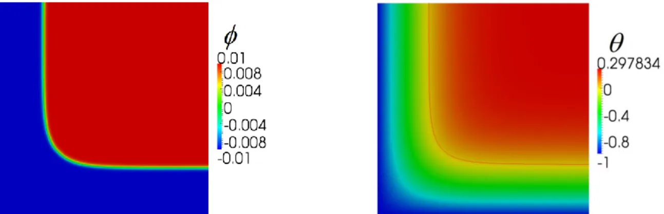
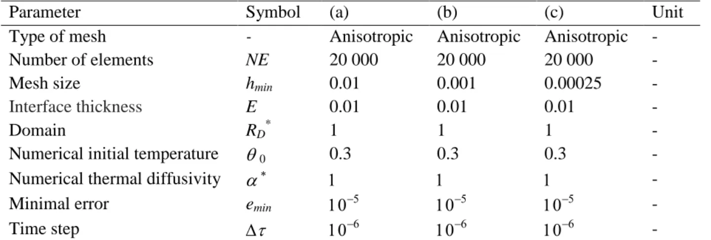
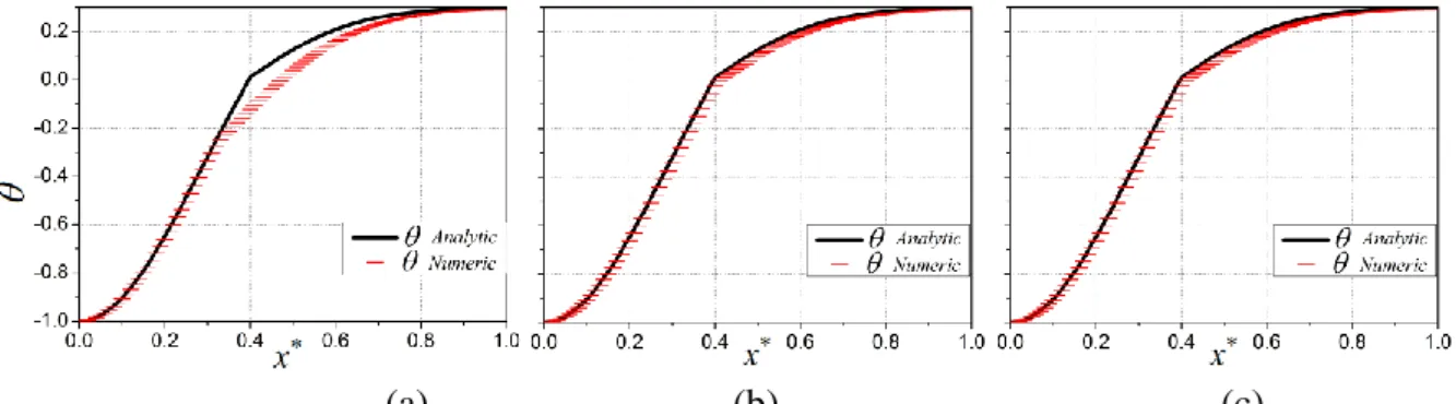
2.2.4 Validation on benchmark: temperature diffusion in a corner... 35
2.3 Thermal dendritic growth ... 43
2.3.1 Timestep and number of elements adaptation ... 46
2.3.2 Convergence of the tip velocity ... 48
2.3.3 Computational time ... 49
2.3.4 Study of the capillary anisotropy ... 52
2.3.5 3D thermal dendritic growth ... 54
2.4 Conclusion ... 59
Chapter 3 ... 61
3 Solutal model ... 61
3.1 Model equations ... 61
3.1.1 Mass conservation equation ... 63
3.2 Numerical resolution ... 65
3.2.1 Finite element resolution of the mass conservation equation... 65
3.2.2 Mesh adaptation ... 67
3.2.3 1D validation ... 68
3.3 2D dendritic growth ... 74
3.3.1 Comparison with a model of direct growth ... 74
3.3.3 Parameter sensitivity study ... 77 3.4 Conclusion ... 81 Chapter 4 ... 83 4 Thermo-solutal model ... 83 4.1 Model equations ... 83 4.2 1D thermo-solutal validation ... 84
4.3 Thermo-solutal dendritic growth ... 88
4.3.1 Mesh adaptation ... 88
4.3.2 2D thermo-solutal dendritic growth ... 89
4.3.3 3D thermo-solutal dendritic growth ... 94
4.4 Conclusion ... 96
Chapter 5 ... 99
5 Physical simulations ... 99
5.1 Modeling of equiaxed solidification in alloys ... 100
5.1.1 Phase-field model (PF) description ... 101
5.1.2 Mean-field model (MF) description ... 104
5.2 PF and MF simulations and methodologies for comparisons ... 110
5.2.1 Studied configurations ... 110
5.2.2 Phase-field simulation ... 113
5.2.3 Grain envelope extration ... 121
5.2.4 Computation of average and representative quantities... 123
5.2.5 Mean-field simulation ... 125
5.3 Parametric study ... 130
5.3.1 Microstructure parameter ... 131
5.3.2 Droplet radius ... 134
5.3.3 Initial droplet velocity ... 135
5.3.4 Nucleation temperature ... 136
5.3.5 External temperature ... 137
5.3.6 Atomisation gas ... 138
5.3.7 Timestep and initial radius ... 139
5.4 Conclusion ... 140
Chapter 6 ... 145
6 Conclusions and perspectives ... 145
6.1 Conclusions ... 145
6.2 Perspectives ... 146
6.2.1 3D PF simulations ... 146
6.2.2 Kinetic anisotropy ... 147
6.2.3 Growth with fluid flow ... 147
Appendix A Energy conservation equation ... 149
Appendix B Analytical solution for corner test ... 155
Appendix C 1D analytical solution ... 159
VII
List of symbols
Averaged thermal diffusivity m2s-1
l
/ s Thermal diffusivity in the liquid/solid m2s-1
Latent to sensible heat ratio = L
cs(TF TW) - Kinetic coefficient m-1s K Supercooling K c Layer thickness m Dirac function - Positive coefficient -
4 Constant intensity of anisotropy -
Signed function to determine the solid/liquid interface and vary between [-1;1] -
LS Signed function for the LevelSet model -
Gibbs-Thomson coefficient m K
Excess free energy of the solid/liquid interface J m-2
Signed distance -
Averaged thermal conductivity W m-1K-1
l
/ s Thermal conductivity in the liquid / solid W m-1K-1
r Interface curvature term m
-1
tip Tip curvature term m
-1
Dimensionless parameter related to the capillary length -
c Constant in one-diemsional solution- stationary interface position -
Secondary dendrite spacing m
E Chemical potential J mol
-1
f Kinematic viscosity of the atomization gas. Pa.s
k Kinetic coefficient m s
-1
K-1
p Fluid viscosity on the surface of the droplet Pa.s
Fluid viscosity far from the droplet Pa.s
Angle Degre°
0 Orientation angle Degre°
Non dimensional temperature -
0 Initial non dimensional temperature -
Anal Non dimensional analytic temperature -
ext Non dimensional external temperature -
i Interface temperature -
imp Imposed non dimensional temperature -
nucl Non dimensional nucleus temperature -
Density/Specific mass kg/m3
f Gas density kg/m
3
0 Reference density at temperature T0 and composition C0,i kg/m3
VIII
Stress tensor Pa
Non dimensional time -
0 Characteristic time of attachment of atoms at the interface s
Computational domain -
c Supersaturation wt%
l Liquid phase -
s Solid phase -
s/l Interface between the solid and liquid phases -
Vector used in the metric -
Aext The exchange surface of heat with the exterior m
2
A/ Surface of the interface / m2
Acc Acceleration m s-2
Arm Number of dendritic arms -
a1 Positive constant deduced from the asymptotic analysis -
a2 Positive constant deduced from the asymptotic analysis -
C Constant in super hyperbola -
C0 Initial composition wt%
cl Composition of the liquid phase wt%
cs Composition of the solid phase wt%
c l(intra) Composition of the liquid phase in the intradendritic zone wt%
c l(extra) Composition of the liquid phase in the extradendritic zone wt%
cs/l Composition of the solid phase at the s/l interface wt%
cl/s Composition of the liquid phase at the s/l interface wt%
c Composition wt%
ci Interfaec composition wt%
cp Specific heat at constant pressure J kg-1K-1
c Average concentration/Initial concentration wt%
D Solute diffusion coefficient m2s-1
Dl/ Ds Diffusion coefficient in the liquid/solid m2s-1
Di Diffusion coefficient in phase i m2s-1
Dj Diffusion coefficient in phase j m2s-1
d0 Capillary length m
E Interface thickness -
Ei Internal energy J
Ek Kinetic energy J
eij Edge error -
emin Minimum edge error -
Fv Volumetric force N m
-3
F Free energy J
FR Remesh frequency -
FH Ginzburg-Laudau free energy J
IX
f Volumetric fraction of the phase -
f Volumetric fraction of the phase -
fH Helmholtz free energy J
gl /gs Liquid/Solid volume fraction -
H Specific enthalpy of phase J kg-1
hext Heat extraction coefficient W m
-2
K-1
hmin Minimum mesh size -
i,j Notations used for solid and liquid phases -
j The diffusion flow in species W m-2
jt Heat flux W m
-2
jat Antitrapping term W m
-2
K Curvature m-1
Kc Stifness matrix from the convective term -
Kd Stifness matrix from the diffusive term -
K Distribution/Partition coefficient -
L Latent heat of fusion J kg-1
Le Number of Lewis -
l/ Diffusion lenght in at the interface / -
M Non dimensional liquidus slope -
M Positive mobility of the phase-field function m4J-1s-1
Mc Positive mobility related to the composition m
4
J-1s-1
Mi Metric -
Mm Mass matrix -
m Exponent in super hyperbola -
m Liquidus slope K wt%-1
NE Number of element -
NEM Number of edges -
Nu Nusselt number -
Nb_Proc Number of processors -
N1 Number of element to represent the interface -
N2 Number of element to represent outside the interface -
n Unit normal vector to the interface -
n/ Normal pointing to the exterior of the domain -
Pe Peclet number -
Pr Prandtl number -
Pcal Caloric power W
Pmech Mechanical power of the external forces W
p Pressure Pa p Stretching exponent - p Number of cores - q Space dimension - Re Reynolds number - Rg Gas constant JK -1 mol-1
X
R Radius at time t m
R0 Nucleus Radius m
S Solid region quantity -
S/ /interface density m-1
sij Edge stretching factor -
T Stress vector Pa
T Temperature K
T0 Initial temperature K
Text External temperature K
TF Fusion temperature K
Ti Interface temperature K
Timp Imposed temperature K
TL Liquidus temperature K
Tl/ Ts Temperature in the liquid/solid K
TM Melting temperature K
Tnucl Nucleus temperature K
TS Solidus temperature K
TW Wall temperature K
t Time s
tCPU CPU time S
U Non dimensional composition -
U0 Non dimensional initial composition -
UAnal Non dimensional analytic composition -
U L/ U S Non dimensional composition in the liquid/solid -
V Steady state growth velocity m s-1
V0 Initial droplet velocity m s
-1
Vinterface Volume of the interface -
VtipGF Velocity tip computed with Green function -
V Velocity m s-1
v0 Molar volume m
3
mol-1
v/ Local velocity of the interface -
vLS Velocity defined by the gibbs-Thomson equation m s
-1
vn Normal velocity -
vnst Stationary normal velocity -
vtip Velocity tip -
Vmax Maximum velocity m s
-1
WPF Interface thickness for the phase-field model m
W0 Initial interface thickness m
Xij Edge between node i and j -
X Coordinate m
Non dimensional thermal diffusivity -
XI
* Tip selection parameter/Constant marginal stability -
0* Non dimensional characteristic time of attachment of atoms at the interface -
D* Non dimensional solute diffusion coefficient -
d0* Non dimensional capillary length -
hext* Non dimensional heat extraction coefficient -
W0* Non dimensional interface thickness -
x* Non dimensional coordinate -
x0* Intersection of line x* = y* and interface curve x*, y* domain -
Non dimensional time step -
H Enthalpy J kg-1
Hf Latent heat of fusion J kg-1
t Time step s
V Volume of the subdomain defined by V m3
cp Heat capacity J m
-3
K-1
H Volume specific enthalpy J m-3
L Latent heat J m-3
∂ Surface of the domain/Domain boundary -
. Strain rate tensor s
-1
QT
.
Volumetric heat production term W m
-3
(i) Set of nodes connected to the node i -
q() Dimensionless function -
W(n) Capillary anisotropy -
(x – x/) Dirac function centered at the point x/ -
d(x*,) Real distance of x* from the interface at time -
erf x Error function -
Chapter 1
1 Introduction
__________________________________________________
1.1 Context of the thesis ... 1
1.2 Atomized droplets ... 2
1.3 Solidification ... 4
1.4 Dendrites ... 6
1.5 Models used for the simulation of dendritic growth ... 8
1.5.1 Front tracking through level-set approaches ... 10
1.5.2 Phase-field approach ... 11
1.5.3 Cellular automaton approach ... 12
1.5.4 Mean-field approach ... 14
1.6 Motivations and Objectives ... 14 ___________________________________________________________________________
1.1 Context of the thesis
The development of aluminum alloys with increased strength and ductility is an ongoing challenge for automotive and aerospace applications, as those illustrated in Figure 1.1. Improved properties are achievable through increased refinement of the microstructure, i.e. higher cooling rates (rapid solidification), as well as through alloying additions. The combination of using alloying additions and rapid solidification often results in a solidified primary phase that is supersaturated in alloying elements. This has been shown to occur in a number of aluminum alloys [HEN2010][ROY2005].
Figure 1.1 Examples of products with an aluminum alloy body for automotive and aerospace applications.
An undercooled melt corresponds to a non-equilibrium state of the liquid. Upon undercooling, driving forces are present in the melt. The number of possible solidification modes increases with undercooling, making accessible a broad range of metastable microstructures and structurally different phases. Crystal and dendrite growth velocities vary significantly with undercooling. Hence, models of the solidifying microstructure are tools to help in developing structure-property relationships for aluminum alloys under a range of high cooling rate
conditions. These models provide valuable insight into the relationship between process and material performance for the development of the next generation of aluminum alloys.
This study has been performed in the framework of MIMOSA (Microstructural Modelling of Rapidly Solidified Droplets and Spray Formed Strips of Aluminum-Copper-Scandium Alloys), a project funded by the French National Research Agency (ANR, France) in collaboration with the Natural Sciences and Engineering Research Council of Canada (NSERC, Canada).
The 3-year project is a collaborative effort between Canada and France involving three teams of researchers. Powder and spray formed samples were generated using Impulse Atomization, a rapid solidification technique of Al-Cu alloy was used.
The solidified samples were characterized using Scanning Electron Microscopy (SEM), X-Ray diffraction, differential scanning calorimetry and microhardness at AMPL (UofA, Edmonton, Canada). In addition, advanced characterizations were carried out, such as neutron diffraction and 3D-micro tomography together with automatic indexing of electron backscatter diffraction patterns (EBSD) at AMPL (UofA) and IM2NP (Univ d’Aix Mareille, Marseille, France). . The characterization data collected were also intended to be used for comparison with the models. Finally, Direct modelling of the dendritic microstructure for an Al-Cu binary alloy processed by atomization was developed at ARMINES CEMEF (MinesParisTech, Sophia Antipolis, France) using the phase-field method. This is the goal of the present work
1.2 Atomized droplets
Impulse Atomization (IA) is a single fluid atomization process. This technique has been extensively used for making metal powders, spray deposits, metal-matrix composites and spray refining of pig iron [DIN1997], [ELL2004], [HER2007], [PRA2006], [PRA2009]. It consists of a 0.5 m diameter and 4 m height cylindrical chamber, as schematized in Figure 1.2. This chamber is atmospherically sealed and can be filled with the gas of choice. In the case of atomization, an inert gas (such as He, N2 or Ar) is used. The top portion of the chamber consists
of an impulse unit where the material is melted at a controlled temperature and subsequently pushed through small orifices. The ensuing discontinuous melt streams break down into small droplets that fall through the gas in the chamber. The droplets attain a free-fall situation in the initial stages and therefore there is no gravity induced convection in these droplets. Droplets completely solidify as they fall through 3.5 m of the gas filled chamber and are collected in glass beakers filled with oil. It has been shown that IA produces rapidly solidified droplets and it is a useful technique for studying rapidly cooled systems, but also for varying alloy composition as it is more important to the fraction of phase distribution than cooling rate [PRA2009]. Successful pilot scale tests have been carried out with IA for the atomization of zinc through up to 400 orifices operating for 3 continuous hours, showing its potential as in the total process.
Figure 1.2 Schematic representation of an Impulse Atomization Unit and the atomized material before and after being melted [ELL2004].
Since the atomization temperature of an Al-Cu system is above 1 000 K, the liquid droplets may loose heat by radiation. However, since the droplets cool rapidly, the radiation heat loss component decreases, and therefore, has a small effect on the total heat loss [PRA2009]. The gas being stagnant, its primary function is to withdraw the heat from the liquid droplets, although the surface tension between the gas and liquid metal does play a part in breaking the liquid stream into droplets. Wiskel et al. [WIS2002] have shown that the cooling rate of Al-4.5wt%Cu atomized in He varies from 325 to 2 400 Ks-1 for droplets of diameter 950 to 275 m, respectively. On the other hand, the same range of droplets sizes atomized under N2 shows
cooling rates from 150 to 800Ks-1. Because of the better conductivity of He gas compared to N2,
the cooling rate is higher. Thus, a droplet of a given size in He shows a finer structure compared to the same droplet size atomized in N2. The data also shows that the microstructure length scale
decreases as the droplet size decreases, since smaller droplets cool at a faster rate. These cooling rate values show that IA can produce rapidly solidified powders.
To provide a better description of the microstructures, let us present shortly the experimental results obtained by Mimosa’s project partners [BED2015]. Firstly, IA leads to a size distribution of the droplets with diameter from less than 200 µm to more than 1 mm in the same batch. The droplets are thus sieved into several size classes by the technique described in [FED2012].
The microstructure morphologies were then investigated for different size ranges and for the two cooling gases, He and Ar. For this purpose, synchrotron X-ray micro-tomography was used post-mortem [NGU2012]. This technique provides a three-dimensional reconstruction of the droplets microstructure as the grey level depends on the X-ray transmission of the phase. The primary Al phase being less absorbing than the eutectic (mixture of the Al phase + the Al2Cu intermetallic),
the latter appears in lighter grey in the tomography reconstructions. The resolution used was of 0.56 µm/pixel (field of view of 1146.88 µm), which enables to study several small droplets at once. The statistical analysis of the droplet morphology has been carried out using the ImageJ
software [ABR2004]. The final droplet microstructure is the result of a complex three dimensional competition between dendrite arms, interdendritic intermetallic, as well as porosity. Therefore, only the cross-sections showing characteristic morphologies are shown in Figure 1.3, where four distinct morphologies were observed in the more than hundred studied droplets.
(a) highly branched (HB)
(b) highly branched with <111> primaries
(c) dendritic (d) finger bundle
Figure 1.3 Examples of the four morphologies identified in the Al-4.5wt.%Cu droplets, of diameter between 250 and 300 µm: (a) highly branched morphology, (b) highly branched morphology with primary arms oriented along <111> directions, (c) dendritic morphology and (d) finger bundle morphology. The nucleation position noted O is shown by a white dot and the primary arms noted OA and OB by white arrows [BED2015]. Grey level has been inverted, the
dendritic structure appearing darker than the eutectic region.
A major result of the synchrotron X-ray micro-tomography analysis was the variety of dendrite morphologies for Al-Cu droplets solidifying under the same process conditions, as shown in Figure 1.3. These morphologies were described in [BED2015]so only their main characteristics are here reminded. Some of the droplets grow in the usual <100> directions and present a highly branched microstructure, as illustrated in Figure 1.3(a). A structure growing first in <111> directions and then in <100> directions can also be observed as in Figure 1.3(b). The two other types of morphologies are fully growing along <111> directions, with a dendritic (Figure 1.3(c)) or a finger-bundle (Figure 1.3(d)) morphology. The growth orientations for this microstructure were validated by EBSD analyses [BED2015].
Interpretation given was that the first solid grow along the <111> direction if its growth velocity reaches values beyond a growth orientation transition. Eventually, its growth velocity decreases and the last part of the droplet would grow in the <100> direction, as observed in Figure 1.3(b). At lower velocity, the usual <100> is observed, as in standard foundary technologies.
1.3 Solidification
Solidification is the phase transformation studied here. It is involved in at least one of the manufacturing stages of almost every man-made object [KUR1998], [LUD2004]. Some important processes which involve solidification are
- casting: continuous, ingot, form, precision, die;
- welding: arc, resistance, plasma, electron beam, laser, friction; - soldering/brazing; O B B A O A O
- rapid solidification processing: melt spinning, planar flow casting, atomization, bulk undercooling, remelting surface and atomization;
- directional solidification: Bridgman, liquid metal cooling, Czochralski, electroslag remelting. In the forming of aluminum alloys, solidification is a transformation step during which the metal, initially liquid, gradually becomes solid upon cooling. The typical stages are: nucleation, primary growth dendritic structure, and secondary and further growth (peritectic or eutectic) [LUD2004]. Once a nucleus is formed, it is limited by capillarity and transport of heat and mass, leading to a morphological instability of the s/l interface and dendritic growth.
Figure 1.4 shows a temperature history measured of an Al-4wt%Cu melt. It reveals (I) heat transfer until the undercooling state appears and the nucleus is created, (II) recalescence due to dendritic growth, (III) another cooling, (IV) secondary nucleation of an eutectic structure and its associated reference and (V) cooling of the fully solid structure.
Figure 1.4 Temperature profile during solidification of an Al-4wt%Cu alloy [GAN2008]. The energy of the system is changed by heat extraction in several ways: firstly, there is a decrease in the enthalpy of the liquid and solid phases due to cooling; second, the transformation from liquid to solid releases the latent heat of fusion. But the transformation from the liquid to the solid also creates a curved and mobile solid/liquid interface, defined as an intermediate zone between the solid and the liquid, the thickness which is composed of a few atoms [MEC2010]. The curvature introduces capillary effects and microscopic heat and mass flows and the solid/liquid interface area is associated with an excess of interfacial energy. Therefore, systems that have a large interface have a higher energy.
In the case of an alloy, both heat and solute are rejected at the solid/liquid interface. Solute is released not only into the interdendritic liquid but also accumulates in a boundary layer outside the mushy zone or grain envelope. This is demonstrated by post mortum analysis of the average composition of Cu, as shown in Figure 1.5, revealing a non uniform distribution.
Figure 1.5 Cross-section through the center of a 250 m diameter Al–10 wt.%Cu droplet, produced by atomization in nitrogen showing, (a) dendritic microstructure as observed with scanning electron microscope and (b) corresponding average composition map (%Cu) deduced
from microprobe analysis [HER2006].
Figure 1.5 shows, on the left, a dendrite structure in grey surrounded with the interdendritic structure in white. On the right, the distribution map of Cu, with the presence of 10wt% of Cu in the alloy. Finally, diffusion flux is present at the very large scale of the dendritic arm.
1.4 Dendrites
The most frequently observed primary solidified microstructure is the dendrite. The descriptive term “dendrite” derives from the greck “”, a tree, with highly branched, arborescent appearance. It consists generally of a primary branch or trunk, secondary arms, eventually with tertiary branches growing from the secondaries and so on. This growth morphology is characterized by its paraboloid-like tip. There are different types of dendrites: the equiaxed dendrites that freely grow and are governed by solute and thermal diffusion, and the columnar alloy dendrites constrained by a temperature gradient and controlled by solute diffusion. In undercooled solidification processing, the highest nucleation temperature and the highest growth rate control the final appearance of microstructures and phases.
Figure 1.6 Equiaxed dendritic growth of a pure metal and an alloy showing the evolution of the temperature and composition in the liquid [KUR1998].
The growth of equiaxed dendrites of pure metals occurs under conditions where only heat flows from the interface to the surrounding liquid. The temperature gradient is negative at the interface and a thermal undercooling TT exists. In the case of equiaxed alloy growth, there exists a
negative temperature gradient and solute accumulation ahead of the dendrite tip leading to thermal and solutal undercooling, respectively Tc and Tt, as shown in Figure 1.6.
In the solidification of binary a alloy system, physical phenomena are usually described by stating the conservation for energy and the conservation for the solute species in each phase and using the Gibbs-Thomson relation to establish the normal velocity of propagation of the s/l interface [BOE2002], [TAN2006], [ZAR2009]. The mathematical description of these phenomena is given here after, while its approximation will be detailed later, in chapters 2-4.
∂c ∂t = D s sc s Species composition conservation in the solid ∂c l∂t = D l c l Species composition conservation in the liquid ∂Ts
∂t = s Ts Energy conservation in the solid ∂Tl
∂t = l T l
Energy conservation in the liquid
(
cl cs)n= (
D scsD l cl)·n Composition conservation at the s/l interface
L
n= (
s Tsl Tl )·nEnergy conservation at the s/l interface
T
i= TM + mclirn/
Gibbs-Thomson equation at the s/l interface( 1.1) ( 1.2) ( 1.3) ( 1.4) ( 1.5) ( 1.6) ( 1.7)
Here, T is the temperature and c the alloy compositions and l are the solid and liquid phases, t is
the time, Ds and Dl are the solute diffusion in the solid and in the liquid, s and l are the thermal diffusion in the solid and liquid, L is the latent heat, n is the normal velocity to the s/l interface
(n = v·n), s and l are the thermal conductivity in the solid and liquid, k is the kinetic
coefficient, n is the unit normal vector to the interface, is the Gibbs-Thomson coefficient related to the surface energy by the relation = TM
L , r is the interface curvature term, m is the
slope of the liquidus curve of the phase diagram for the alloy. Ti is the interface temperature, TM is the melting temperature of the pure solvent and cli is the concentration on the liquid side of
the interface. This system of equations provides the sharp interface formulation of our solidification problem. In a diffuse interface context, instead of solving the equations for each phase with the given interface conditions, we may obtain a set of equations valid in the whole domain [KAR1998].
A few measurements have been reported for alloy dendrites. A number of researchers have performed experiments to measure the dendrite tip velocity and radius for transparent alloy systems [AST2009], [BOU1989], [CHA1987], [CHO1988], [DOU1988], [GLI1988], [KAH1970]. Only the succinonitrile-acetone (SCN-acetone) experiments of Chopra et al. [CHO1988] resulted in data over a sufficiently large range of undercoolings and solute concentrations to allow a detailed comparison with theory.
Macroscopic conditions (such as undercooling upon equiaxed growth) affect the solidification but microcoscopic internal characteristics play also an important role. The most important factors of internal characteristics are the anisotropy of the properties at the s/l interface, key parameters affecting the evolution of crystal morphology [HOU2008]. Anisotropy at the s/l interface includes the energy, , and the kinetic coefficient, k [MUL1964].
1.5 Models used for the simulation of dendritic growth
Modeling of dendritic growth in solidification of pure metals and alloys remains a significant challenge in materials science and applied physics. Successful modelling of dendritic solidification requires both the solution of a complex free-boundary problem and an accurate account of the interface energy and kinetic anisotropy. The first task is difficult because of the difference in orders of length scale between the thickness of the diffusion boundary layer of heat/solute that surrounds the dendrite tip and grain envelope, and the microscopic capillary scale, while the second task is complicated by the need to compute the curvature of the interface [ZAB2006]. Figure 1.7 shows different scales for the s/l interface, experimental view and numerical modeling. The various scales are illustrative and not directly comparable, because all these quantities vary with the material, especially between metals and organic alloys. The smallest scale is for the atomic interface that can be simulated using molecular dynamics models, the dendritic scale is at the micrometer size, simulated often using phase-field methods, the grain structure which can be simulated using the CAFE (cellular automaton-finite element) model and the process scale of many meters simulated with finite volume or finite element methods.
Figure 1.7 Different scales for the s/l interface (top), and corresponding description of the microstructure with illustrations from experimental observations (middle) and numerical
simulations (bottom) [CAR2012].
In the last years, special attention has been givento the development of modeling techniques at various length scales for deeper understanding of microstructure formation. In the literature, at least four different approaches applied to dendritic growth can be identified. Firstly, sharp interface models [BAN1994], [NAK2006], [SAI1988], [UDA1999] are used to precisely reproduce the surface between solid and liquid by a dynamically refined mesh of the s/l interface. Secondly, phase-field (PF) models deal with the solid-liquid interface by introducing a continuous transitional layer of finite thickness using an additional quantity, thus eliminating the problem of explicitly tracking the interface and avoiding direct computations of the curvature. Several reviews on the methodology and capabilities of the PF models are available [AST2009], [BOE2002], [FRI2009], [HEC2004]. Thirdly, microscopic cellular automata (CA) were also employed for simulation of dendritic growth. When coupled with finite element modelling, this technique is referred to as CA-FE modeling [GAN1999], [RAP1993] and is then used to predict the development of the grain envelope, not directly simulating dendritic morphologiy. Fourthly, coarser-grained models have been developed at the mesoscale to predict the unsteady growth of dendritic grains and their internal solid fraction [STE1999], [STE2005], [ZAL2013], in 2D and 3D. These models track the evolution of the envelope of the dendritic grain, defined as an imaginary surface that passes through the tips of primary branches. However, they do not resolve exactly interactions between individual branches. Fifhtly, the mean-field approach, which cannot follow dendrite morphologies, but may give us an equivalent grain envelope, and repartition of phase fractions inside/outside the envelope as well as average solution fields, studied in 1D dimension by [TOU2009]. Other authors developed a virtual front tracking (VFT) method of the solid-liquid interface [BEL2003], [BEL2004]. Discretization methods most often used are the
finite element method (FEM), the finite volume method (FVM), the finite difference method (FDM) and the average volume method (AVM). One finds, in the literature, computations using structured isotropic meshes, structured anisotropic meshes (squares divided in smaller squares or triangles) as illustrated in Figure 1.8 (a), in 2D and 3D [BAN1994], [TON1998a], [XIE2013] and unstructured anisotropic meshes [NAR2007] as illustrated in Figure 1.8 (b).
(a) (b)
(c)
Figure 1.8 Examples of dendritic growth using: (a) the front tracking approach with an anisotropic structured mesh [BAN1994], (b) the phase-field approach with unstructured
anisotropic adaptive mesh [NAR2007], (c) a microscopic cellular automaton method [CHO2012].
1.5.1 Front tracking through level-set approaches
These models are mainly based on the use of the level-set functions, classically employed in many areas where it is necessary to follow an interface [BAN1994]. A level-set function, LS, is a
signed function that varies continuously from a positive value inside a phase, to a negative value outside. The interface is defined by the isovalue LS = 0. Most often, LS is the signed distance to
the interface. Interface is displaced by the resolution of the level-set equation: ∂LS
∂t + vLS |LS | = 0
where vLS is the velocity field, determined from the Gibbs-Thomson condition for solidification
problems. After the position of the front is calculated, the energy and solute conservation equations are solved. The main advantage of the level-set method is its ability to represent
complex topological changes. This method can be used to simulate growth with fluid flow [ZAB2006].
Among its claimed advantages, fairly easy implementation and fast computation time can be mentioned. However, the error associated with the estimation of curvature from the divergence of the normalized gradient of is large (10-30%). Since preferred growth directions and dendrite tip kinetics are governed by the small anisotropy of the interfacial energy (1-10% in metallic alloys), such methods can only give qualitative results, unless mesh sizes around the interface become very small in the perpendicular direction to it. The later condition may then become incompatible with the fast computation time advantage.
1.5.2 Phase-field approach
This method introduces a function which continuously varies from a constant value in a phase (eg. solid) to another constant in the other phase (eg. liquid). Most generally, the phase-field variable, , varies smoothly from -1 to 1 between the two phases over the diffused-interface region, which has a small but numerically resolvable thickness, WPF. The phase-field method
derives its attractiveness from the fact that explicit tracking of the interface and satisfaction of the interfacial boundary conditions are avoided. Furthermore, computation of interface normal and curvature is also avoided by solving an evolution equation for the phase-field variable which may be derived from the free energy or entropy formulation and coupled to the evolution of heat and solute. The fundamental difference with a classical level-set method is that a thickness WPF
is thus given to the interface itself, which becomes diffuse. The principle is to minimize the free energy of the system by stating:
∂
∂t = M
∂FH ()
∂t ( 1.8)
where M is the mobility of the phase-field, FH is the Ginzburg-Laudau free energy and is
defined by a Ginzburg-Laudau type of integration in the domain:
FH = fH () + W(n)2 2 | | 2 d ( 1.9)
fH() is the Helmholtz free energy and W(n) represents the anisotropy at the interface. The
gradient term is introduced because of the representation of the diffuse interface. The derivation of these equations allows to move the interface and to minimize the energy, FH. The results are
poor when using a large interface: it should be less than the capillary length to converge to a sharp interface solution [XIE2013], which may be problematic in problems with fluid flow, in particular without remeshing or grid adaptation [PRO1999].
Karma and Rappel [KAR1996] improved asymptotic coefficients for the thin-interface limit of the phase-field equations, which ameliorate the convergence of the method for a coarser grid density. It lowers the range of undercooling and allows the use of a larger width of the diffuse interface region (compared with the capillary length), and gives the possibility to choose the model parameters in a way to make interface kinetics vanishing. Other recent changes have been added to these methods such as the use of an adaptive FEM formulation that refines the zone near the diffuse interface [PRO1998] or the use of a stochastic Monte Carlo treatment of the large scale diffusion field [PLA2000b]. This method was implemented by adding fluid flow
effects [TON1998a], [TON1998b], as well as used to represent eutectic solidification [ELD1994], [KAR1994], [WHE1996] and the peritectic reaction [LO2001], i.e. including multiple solid phases. Yet, phase-field simulations are limited to represent growth at a grain scale, especially for concentrated alloys that need a large solidification range and have a low supersaturation.
For a classical problem of dendritic crystal growth, several multi-grid [BRA1997] or adaptive meshing algorithms [SCH1996] have been proposed in recent years. One particularly cited method proposed by Provatas et al. [PRO1998] uses the phase-field model on a regular grid to compute the dendrite, whereas the temperature diffusion field is integrated on an adaptive mesh using finite element techniques. While this method appears to be promising, it has yet to be implemented in three dimensions, where the difficulty of adaptive meshing becomes significantly enhanced.
1.5.3 Cellular automaton approach
The cellular automaton microscopic methods have been applied to the prediction of dendritic structures during solidification [CAR2012]. By the fact that global fields throughout the area (temperature, composition) indirectly influence the local state of the cells, these models were baptized Modified Cellular Automata, MCA. Several versions have been developed, but the most common models are based on the same principle as the phase-field. The difference is that the interface thickness is simply equal to a row of cells, that may have different sizes [KRA2009], [NAS1999], and they have been developed for their computational speed and their ability to be used on larger areas than the phase-field. Their main disadvantage is the anisotropy induced by a regular grid of cells (square or cubic), not obvious to erase. However, these methods were shown to achieve quantitative results [CHO2012], [YIN2011], [ZHU2007]. Finally, we note the existence of cellular automata models where the kinetics of the interface is calculated from analytical equations, such as the KGT method [ZHU2001].
1.5.4 Mesoscopic appraoch
The mesoscopic modeling technique consists on coupling numerical calculation of the temperature field at the macroscopic scale with an analytical model of dendrite tip growth. A Representative Elementary Volume (REV) is used and is large enough to include a representative sampling of the microscopic structures and, at the same time, small enough to enable a continuum description of the variables averaged over the REV on the macroscopic scale. This model can predict the evolution of the grain shapes, the growth interactions between multiple grains, and the nature of the thermal field in the melt between the dendrites. With this method, the computational power requirements are reduced compared to a direct microstructure simulation on a microscopic scale. A schematic illustration of the various length scales present, at the mesoscopic modeling scale, in equiaxed dendritic growth, is shown in Figure 1.9 (a).
Figure 1.9 Schematic illustration of equiaxed dendritic growth: (a) unit cell, where 1 is the
microscopic scale and 2 is the mesoscopic scale; (b) grain envelope and stagnant film
[STE1999].
The prediction of the tip growth speeds and radii requires the resolution of the thermal field at the scale 1, accomplished using a local analytical solution. The growth velocities of the grain
envelope can be obtained from dendrite tip speeds. The Ivantsov solution is used. The supercooling is applied at a confocal isothermal paraboloid located at a finite distance f away
from the dendrite tip and moving with the same speed as the tip. The tip speeds are calculated for every point on the envelope. Solid fraction can be deduced from the temperature gradient at the envelope.
Figure 1.10 Example of the evolution of the dendrite envelope for a single equiaxed dendrite [STE2005].
The model was later used for two-phase representation with a volume-averaged Euler-Euler method that consists of two parts: a macroscopic part with momentum, mass, heat, solute mass, and grain population conservation equations, and a microscopic part that describes the nucleation and growth of grains.
1.5.5 Mean-field approach
This model tracks the evolution of the envelope of the dendritic grain, defined as an imaginary surface that passes through the tips of primary branches. However, interactions between individual branches are not exactly solved. Moreover, conservation equations are solved by projecting the dendrite in 1D and are based on the average volume method. This model and its combination with finite element or finite difference methods may also be used to study microstructural selection mechanisms, such as the dendritic spacing selection and its history dependence, but do not approximate the microstructure selection within a grain under non-steady-state growth conditions. It requires dendrite arms spacings as an input, which have an influence on microsegregation [TOU2011a][TOU2011b] and macrosegregation [BEC2002]. This model is detailed in Chapter 5.
1.6 Motivations and Objectives
An adaptive phase-field model is developed and presented in this thesis. The aims of this present work are thus
- to describe the finite element implementation and programming of the proposed phase-field model with different homogenization schemes in a finite element code;
- to use parallel computing and automatic adaptive anisotropic unstructured meshes available with a C++ library developed at CEMEF, MINESParisTech, and their importance on the computational cost (time and memory);
- to obtain quantitative results using this model; - to simulate dendritic growth for Al-Cu droplets;
- to start comparisons with a mean-field approach, and justify the differences, i) of macroscopic integration (Thesis of Thi-Thuy-My NGUYEN [NGU2015]), ii) of computational time. This is based on the phase-field methodology, known to be quantitative.
- to show that we have a difficulty to do a computation for an Al-4.5wt%Cu system of big size. - to justify the advanced numerical methods (parallel computation, mesh adaptation, timestep adaptation, number of elements adaptation).
This manuscript is organized as follows, to model dendritic growth in a solidifying droplet as discussed in this chapter. Chapter 2 describes the phase-field model, for the solidification of a pure substance, by detailing the numerical resolution used for the diffusion, including mesh adaptation and parallel computation. Validation in 2D is presented, as well as capillary anisotropy implementation. Some symmetry assumptions are assumed for 3D simulations. Chapter 3 extends the phase-field model to represent the isothermal solidification of a binary alloy, explaining the difference from the previous one, in the numerical resolution and in the construction of the adapted mesh. Validations in 1D and 2D and sensitivity studies of the numerical parameters are presented. Chapter 4 couples thermal and solutal diffusion for a binary alloy. Finally, chapter 5 applies the model to the solidification of Al-Cu droplets and shows comparisons with a macroscopic mean-field model.
Résumé
Dans cette introduction, le cadre des travaux de thèse, ainsi que différentes définitions, sont introduits. Nous présentons le procédé d’atomisation de gouttes pendant lequel la solidification a lieu. La morphologie dendritique est alors décrite, aussi bien que les méthodes numériques utilisées dans la littérature pour sa simulation. Nous terminons par définir les objectives et les motivations de ces travaux: simulation de la croissance dendritique d’une goutte d’Al-Cu en utilisant le modèle de champs de phase et la méthode des éléments finis avec remaillage et calcul parallèle ainsi que d’autres optimisations pour diminuer le temps de calcul.
Chapter 2
2 Thermal model
__________________________________________________
2.1 Model equations ... 17 2.2 Numerical resolution ... 24 2.2.1 Finite element solver ... 24 2.2.2 Mesh adaptation ... 28 2.2.3 Parallel computation ... 33 2.2.4 Test: temperature diffusion on a corner ... 35 2.3 Thermal dendritic growth ... 43 2.3.1 Time adaptation of timestep and number of element ... 46 2.3.2 Convergence of the tip velocity ... 48 2.3.3 Computational time ... 49 2.3.4 Study of the capillary anisotropy ... 52 2.3.5 3D Thermal dendritic growth ... 54 2.4 Conclusion ... 59 __________________________________________________________________________ In this chapter, we present the mathematical model used for solidification of a pure substance under anisothermal conditions. The model is based mainly on a continuous formulation of the phase-field method, given in [KAR1998]. It is developed for solidification, assuming local thermal equilibrium. We use the equations in a dimensionless form, and show its numerical solution. Thermal dendritic growth is illustrated with a validation in 2D, including sensitivity studies for the computational time and capillary anisotropy magnitude, and also through 3D simulations.2.1 Model equations
Let us consider conservation equations written for the sharp interface problem and limited to a pure substance, with a solid, s, growing in the liquid, l. In the absence of phase motion, they lead to: T s t = s T s ( 2.1) T l t = l T l ( 2.2) Lvn = (s T slT l )∙n ( 2.3) Ti= TMr vn /k ( 2.4)
We have supposed that the pure substance has constant and equal density, [kg·m-3], thermal conductivity, [Wm-1K-1], and specific heat at constant pressure, cp [J·K-1kg-1], in both solid
Here, r = _.n [m-1] is the curvature term, k [m·s-1K-1] the kinetic coefficient, the
Gibbs-Thomson coefficient related to the surface energy [Jm-2]by the relation = TM
L [mK], TM
[K] the melting temperature of the pure solvent, Ti [K] the interface temperature, T [K] the
temperature, t [s] the time, L [J·m-3] the latent heat and n = v·n [m·s-1] the normal velocity
to the s/l interface.
It is convenient to define the dimensionless variable , measure of the undercooling, TM – T,
as:
= T TM
L/cp ( 2.5)
To use non-dimensional coordinates, in space and time, for the simulation, we define x* as
x* = x/W0 ( 2.6)
and as a dimensionless time:
= t
0 ( 2.7)
where 0 is the phase-field relaxation time, in [s].
The free-boundary problem described above becomes (Appendix A):
s = s*s l = l*l ( 2.8) vn = (sl ) ( 2.9) i= drvn ( 2.10)
where * is the thermal diffusivity:
*
= 0
W0 2 ( 2.11)
The thermal capillarity length d0 [m] is defined as:
d0 =
L/cp =
TMcp
L2 ( 2.12)
and [J·m-2] is the excess free energy of the solid/liquid interface. The kinetic coefficient [m-1s] is defined as:
= cp
k L ( 2.13)
In this approach, W0, the thickness of the interface, is assumed small compared to the scale of
the microstructure pattern, but not smaller than d0, the capillarity length.
The phase-field equation presented was derived for the anisothermal case, but has to be coupled to the energy equation. Let us consider equal diffusivities in the solid and in the
liquid, so <> = s = l = cte, as well as equal heat capacities <cp > = cps =cpl = cte. The
average equation of energy, developed in Appendix A, becomes:
T tT = 1 2 (L)s/l cp t ( 2.14)
Where (L)s/l is the latent heat associated with the transformation s→l. As defined before, the dimensionless temperature is used and the equation to solve becomes:
= 12
( 2.15)
Let us define as a function which describes the presence of the liquid and the solid phases in the computational domain Ω, made by the two subdomains, Ω s in the solid and Ω l in the liquid. An interface Ω s/l is defined between Ω s and Ω l. The function varies between and 1 as illustrated by Figure 2.1, andis defined as:
= tanh W0* 2 ( 2.16) In this expression, W0* is a non dimensional interface thickness (W0* = W/W0), where W0, as
previously introduced, measures a physical width and W represent the variable of this physical width. It is convenient to consider that it characterizes the diffuse solid/liquid interface, where
x is the physical coordinate system and W0 is a physical arbitrarly chosen length.
Figure 2.1 Variation of the phase function with the signed distance to a stationary flat liquid-solid interface, with W = W0.
The variable used in expression ( 2.16) is the signed distance to the solid/liquid interface. It is defined as = d(x*,) if x*s
0
ifxs/l
d(x*,) if x*l ( 2.17)where d(x*,) is the distance of x* to the interface Ω s/l at time .
Let us define the free energy functional F(,), which must decrease during any thermodynamic process, as [KAR1998]
F(, ) =
ΩF
int dV ( 2.18) with Fint = f () + W(n) 2 2 || 2. Fint is the sum of the volumetric energy given by the free energy density, f(, ), and the interfacial energy, respectively. W(n) is a measure of the anisotropy in the surface energy and n = -
|| , the normal vector, and may be defined as follows [KAR1998]: Wn = W0*(1 34) 1 + 44 1 34 (x*)4+ (y*)4 in 2D ( 2.19) or Wn = W0*(1 34) 1 + 44 1 34 (x*)4+ (y*)4 + (z*)4 in 3D ( 2.20) The instability of the solid/liquid interface that forms the dendrites is influenced by the anisotropy of the solid/liquid interfacial energy presented by the parameter 4
Allen-Cahn equation [CAH1979] may be used to guarantee that the total free energy decreases with time due to an excess in the interfacial energy:
F(,)
0 ∀ > 0 ( 2.21)
The Allen-Cahn equation is written by deriving the free energy to obtain, finally, the phase-field equation: = MF = MF int .F int ( 2.22)
where M is a positive mobility parameter. The free energy density, f(, ), for a pure material, can be given by:
f (, ) = g() + p() ( 2.23)
is a dimensionless parameter that controls the strength of the coupling between the phase and diffusion fields. It is typically of the order of unity. This term can correct the contribution of the heat added and ensure that the relation ([15/8(p(+1)) – 15/8(p(-1))]/2= 1) is verified,
whatever p() is chosen. In this expression of the free energy density of a pure element, p() is a function of that guarantees f / for and for and for all temperatures. Furthermore, g() must provide the energy hump between the solid and liquid phases, with a maximum value at the interface and a minima at = ±1. Hence, the following functions have been suggested by Karma and will be used in our model [KAR1998]:
g() = 2 + 4 ( 2.24) p() = 2 3 3 + 5 5 ( 2.25)
Figure 2.2 Illustration of the variations of f(,), g() and p() with = 2 and = - 0.5. From Eq.( 2.22), the evolution equation for variable , defining the position of the interface [EIK2010], is: = MF = MF int Fint = Mf x* Fint x* y* Fint y* z* Fint z* ( 2.26) Since F int = f () + W(n) 2 2 || 2 = f + 0 = f , then f = g( ) p( ) ( 2.27) This derivation is equal to
f
= + + 2 + ( 2.28)
and a factorization is used to obtain the simple form
f
= + 2 = () ( 2.29)
Replacing in equation ( 2.26), one obtains:
n Wn= [] x* | |2WnWn x*) y* | |2WnWn y*) z* | |2WnWn z*) ( 2.30) where n = 1 .
Our attention is focused on the growth in the limit of vanishing interface kinetics. This limit is obtained by setting (n) and equal to the values [KAR1998]:
n = (1 34) 1 + 44 1 34 (x*)4+ (y*)4+ (z*)4 2 ( 2.31) = Wa a ( 2.32)
This second expression is related with the fact that one may redefine the coefficient to include the variation of across the interface using the asymptotic analysis:
= a1 0 W a2W0 ( 2.33)
which vanishes when kinetic effects are eliminated. Langer [LAN1986] and then Caginalp [CAG1989] have derived:
d0 = a1
W0
( 2.34)
In these expressions, is the characteristic time of attachment of atoms at the interface (10-13 s for metallic systems), a1 is a positive constant of order unity that depends on the details of
the assumed form of free energy computed from the asymptotic analysis, a2 is a positive
constant of order unity that depends on the details of the functional forms chosen for f(), g() and p(). Karma and Rappel [KAR1998] deduced from the asymptotic analyses that a1 =
0.8839 and a2 = 0.6267.
To do the simulation of the dendritic growth, one needs to compute the value of
Wn x* | |2WnWn x*) , y* | |2WnWn y*) and z* | |2WnWn z*) , derived
from the anisotropy form of the interface, W(n). We can write W(n) in different forms as defined in eq.( 2.70) and eq( 2.71) for 2D, eq( 2.72) and eq( 2.73) for 3D where 1 and 2 define
the intensity of the anisotropy like 4. We compute Wn numerically and for the others we
have implemented their analytical derivation in our code to use it directly. The derivation forms are shown below in Table 2.1:
![Figure 1.10 Example of the evolution of the dendrite envelope for a single equiaxed dendrite [STE2005]](https://thumb-eu.123doks.com/thumbv2/123doknet/2891603.73858/26.892.134.764.642.957/figure-example-evolution-dendrite-envelope-single-equiaxed-dendrite.webp)


![Figure 2.27. Simulations were performed in a [ 200,200]x[ 200,200] domain and parameters are shown in Table 2.6](https://thumb-eu.123doks.com/thumbv2/123doknet/2891603.73858/57.892.111.780.637.1027/figure-simulations-performed-x-domain-parameters-shown-table.webp)