The Type II-A CRISPR-Cas system of Streptococcus
mutans: Characterisation of Bacteriophage-Insensitive
Mutan(t)s
Thèse
Cas Mosterd
Doctorat en microbiologie
Philosophiæ doctor (Ph. D.)
Québec, Canada
Résumé
Les bactéries sont continuellement exposées à un danger, la prédation par des bactériophages. Pour se défendre, elles ont développé une grande variété de mécanismes. Parmi ceux-ci, on retrouve CRISPR-Cas (« clustered regularly interspaced palindromic repeats »), un système adaptatif que possèdent environ 45% des bactéries. Une caractéristique unique du système CRISPR-Cas est qu’il constitue en quelque sorte la mémoire de l’hôte. Par exemple, le système peut emmagasiner des petits fragments d’un génome viral, appelés espaceurs, et les introduire dans son CRISPR. Cette mémoire lui permet de se défendre contre une réinfection par le même virus ou un virus hautement apparenté. Par contre, malgré que l’acquisition de nouveaux espaceurs semble fréquente dans la nature, ce phénomène n’est que très rarement observé en conditions de laboratoire. Néanmoins, quelques bactéries font exception à la règle et l’une d’entre elles est
Streptococcus mutans. Dans le cadre de cette étude, l’interaction entre la souche S. mutans
P42S et le bactériophage virulent M102AD a été analysée en détail. De plus, certaines applications potentielles du système CRISPR-Cas ont également été approfondies.
Le premier objectif de cette thèse était de caractériser le système CRISPR-Cas de
S. mutans P42S au niveau moléculaire et de déterminer son rôle dans les interactions
phage-bactérie. Le deuxième objectif était d’établir le potentiel de la protéine Cas9 de S.
mutans P42S (SmutCas9) comme nouvel outil d’édition génomique. S. mutans P42S
possède un système CRISPR-Cas de type II-A. Bien que ce type de système soit probablement le plus étudié, celui de S. mutans P42S présente plusieurs caractéristiques uniques lui permettant de se démarquer. En effet, ce dernier reconnaît un PAM différent de ce qui était auparavant connu pour cette espèce bactérienne, l’acquisition simultanée de multiples espaceurs semble fréquente, ce qui est probablement dû au phénomène de « priming ». Malgré le rôle de CRISPR-Cas dans la défense antivirale, S. mutans P42S dispose d’autres mécanismes de défense contre les phages. Des cellules mutantes sont résistantes aux phages en empêchant l’adsorption de particules virales à la cellule ont notamment été observées. D'autres mécanismes sont assurément impliqués dans la défense antivirale de S. mutans. Finalement, SmutCas9 s’est montrée efficace dans l’édition de génomes viraux et elle apparaît comme une candidate à explorer pour cette application.
Abstract
Bacteria are exposed to the constant threat of viral predation. To defend themselves, bacteria have developed a wide variety of different mechanisms. One of these mechanisms is CRISPR-Cas (clustered regularly interspaced palindromic repeats), an adaptive immune mechanism found in approximately 45% of bacteria. A unique feature of CRISPR-Cas systems compared to other antiviral defence mechanisms is that it has a memory. The system is capable of remembering previous viral encounters and protects the bacterial host from re-infection by the same or highly-related viruses. This memory is due to the acquisition of virus-derived genome fragments called spacers. Despite common acquisition of novel spacers in nature, and thereby the emergence of new immunity, acquisition of new spacers under laboratory conditions has been rarely observed. One of the few exceptions is
Streptococcus mutans. In this study, the interactions between S. mutans strain P42S and its
virulent bacteriophage M102AD are investigated in detail. In addition, possible applications of the CRISPR-Cas system are analysed.
The first objective of this thesis was to characterise the CRISPR-Cas system of S.
mutans P42S on the molecular level and to determine its role in antiviral defence. The
second objective was to determine the potential of the Cas9 protein of S. mutans P42S (SmutCas9) in genome editing. S. mutans P42S possesses a type II-A CRISPR-Cas system. Although this is arguably the best studied system, the one found in the strain S. mutans P42S has several features that makes it stand out. It recognises a PAM different from what was known for this species, multiple spacer acquisitions are frequent, and this appears to be partially due to priming. Although CRISPR-Cas plays a role in antiviral defence, there are additional antiviral defence mechanisms that protect S. mutans against phages. Adsorption resistance is one of them, although additional unidentified antiviral defence mechanisms are likely involved. Finally, SmutCas9 has been shown functional in editing of viral genomes and appears to be a candidate for human genome editing.
Table of contents
Résumé ... ii Abstract ... iii Table of contents ... iv List of figures... ix List of tables ... xiList of abbreviations ... xii
Acknowledgments ... xvi
Foreword ... xxii
Introduction... xxii
First research article (Chapter 1) ... xxii
Second research article (Chapter 2) ... xxxii
Third research article (Chapter 3) ... xxii
Fourth research article (Annex A) ... xxiii
Review (Annex B) ... xxiiiv
Introduction ... 1
Streptococcus mutans ... 1
Bacteriophages (phages) ... 4
Streptococcus mutans and its phages ... 7
Streptococcus mutans P42S and phage M102AD ... 7
Problematic, hypotheses and objectives of the study... 9
Chapter 1 – Article 1 (Characterisation of a type II-A CRISPR-Cas system in Streptococcus mutans) ... 11 Résumé ... 12 Abstract ... 12 Importance ... 13 Abbreviations ... 13 Introduction ... 15 Results ... 17
Analysis of the CRISPR-Cas systems of S. mutans P42S ... 17
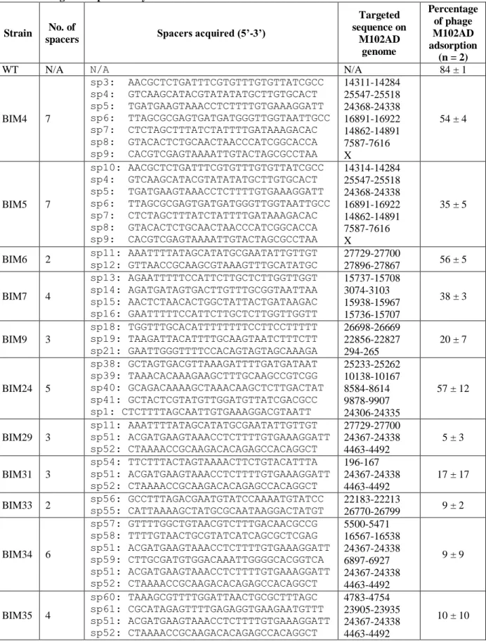
BIM assays ... 21
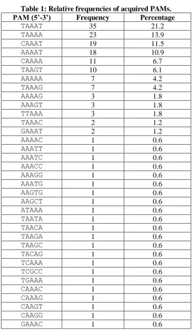
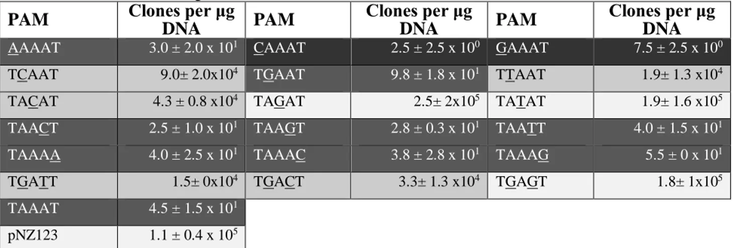
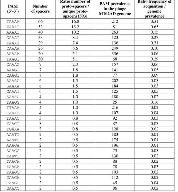
Identification of the proto-spacer adjacent motif (PAM) ... 22
Phage resistance assays ... 23
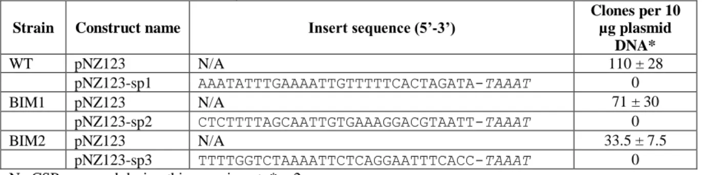
Plasmid interference assays ... 25
Plasmid-based determination of PAM sequence ... 26
Discussion ... 27
Materials and Methods ... 30
Strain, phage, and culture conditions ... 30
Identification and analysis of the CRISPR-Cas system in S. mutans P42S ... 31
BIM assay ... 31
Phage adsorption assay ... 32
Plasmid interference assay ... 32
Transformation of S. mutans ... 33
Determination of PAM sequence ... 33
Data availability ... 34
Acknowledgments ... 34
References... 345
Chapter 2 – Article 2 (CRISPR-Cas and adsorption resistance provide combined phage protection in Streptococcus mutans) ... 41
Résumé ... 42 Abstract ... 42 Abbreviations ... 43 Introduction ... 44 Results ... 45 BIM assays ... 45
Ectopic spacer acquisition ... 49
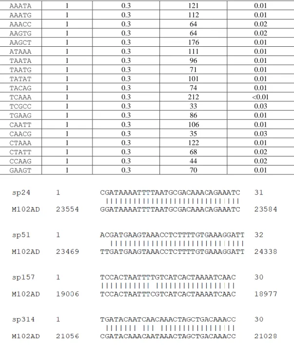
Acquisition of non-perfecly matching spacers ... 50
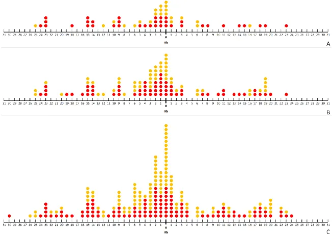
Priming ... 51
Impact of the multiplicity of phage infection on spacer acquisition... 55
Discussion ... 56
Materials and Methods ... 60
Strain, phage and culture conditions ... 60
BIM assays ... 60
Plasmid interference assays ... 61
Phage adsorption assay ... 62
Acknowledgments ... 62
References... 63
Chapter 3 – Article 3 (Application of Cas9 from Streptococcus mutans P42S in viral genome editing) ... 68 Résumé ... 69 Abstract ... 69 Abbreviations ... 720 Introduction ... 72 Results ... 74 RNA sequencing... 74 Construction of pTRKL-SmutCas9 ... 75
Genome editing of orf49 ... 76
Discussion ... 78
Materials and Methods ... 81
Bacterial strains, phages and growth conditions ... 81
RNA extraction and sequencing ... 81
Construction of pTRKL-SmutCas9 ... 82
Genome editing of phage p2... 84
Acknowledgments ... 85
References... 86
Conclusion and perspectives ... 90
References ... 96
Annex A: Article 4 (A mutation in the methionine aminopeptidase gene provides phage resistance in Streptococcus thermophilus) ... 106
Abstract ... 107
Abbreviations ... 108
Introduction ... 111
Materials and Methods ... 111
Bacterial growth and phage propagation ... 111
Bacteriophage-insensitive mutant isolation ... 113
DNA isolation, sequencing and bioinformatics analysis ... 113
Complementation assays ... 114
Proteomic analysis of the phage-infected S. thermophilus DGCC7796 cells ... 114
Directed and random mutagenesis ... 115
Directed metAP mutagenesis in Streptococcus mutans ... 116
Mutation stability test ... 117
Results and discussion ... 118
A mutation in the gene coding for the methionine aminopeptidase provides phage resistance ... 118
A mutation in the metAP gene confers phage resistance in S. thermophilus ... 119
Complementation with the wild-type allele restores phage sensitivity ... 120
The MetAP mutation has a broad range of action against cos-type phages ... 121
Other mutations in MetAP affect replication of phage DT1 ... 122
Phage adsorption and phage DNA replication are not affected ... 123
The MetAPH206Q mutation affects N-terminal methionine processing ... 124
The MetAPH206Q mutation affects growth of the bacterial strains ... 126
The MetAPH206Q mutation is stable ... 127
Conclusion ... 128
Acknowledgements ... 128
Author contributions ... 129
References... 128
Annex B: Review (A short overview of the CRISPR-Cas adaptation stage) ... 136
Résumé ... 137
Abstract ... 137
Introduction ... 139
General principle of CRISPR-Cas function ... 140
Classification ... 141
Adaptation... 143
Spacer acquisition under laboratory conditions ... 146
Type II-A CRISPR-Cas system of S. thermophilus as a model for spacer acquisition ... 148
Anti-CRISPR ... 152
Conclusion ... 153
Acknowledgements ... 154
References... 156
Annex C: Supplementary data ... 136
List of figures
Figure 1: Type II-A CRISPR-Cas system of S. mutans P42S.
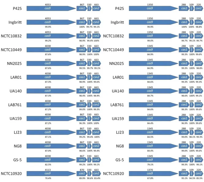
Figure 2: Comparison of cas genes and Cas proteins of nine S. mutans strains.
Figure 3: Percentage identity between Cas9 N-terminus and C-terminus found in several
S. mutans strains.
Figure 4: tracrRNA in S. mutans.
Figure 5: PAM downstream of protospacers. Figure 6: Distribution of spacer lengths.
Figure 7: PAM of all protospacers (A) versus PAM during single spacer acquisition events (B).
Figure 8: Non-perfectly matching spacers and their targets on M102AD.
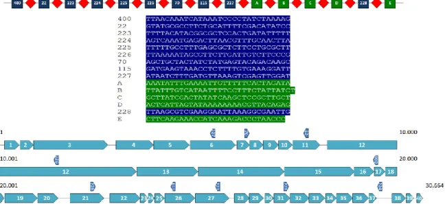
Figure 9: CRISPR locus of BIM 2.2-43CA and the protospacers on the genome of M102AD.
Figure 10: Interference activity of non-perfectly matching spacers. Figure 11: Priming in S. mutans P42S.
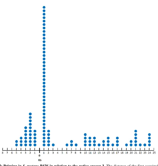
Figure 12: Priming in S. mutans P42S in relation to the native spacer 3. Figure 13: Editing of the orf49 of the lactococcal phage p2 using SmutCas9. Figure 14: Complementarity between crRNA and tracrRNA of S. mutans P42S and UA159.
Figure 15: Exchanging the spacer of pTRKL2-SmutCas9.
Figure 16: Transformability of plasmids containing protospacers flanked by different PAMs. Figure 17: Protein alignment of S. thermophilus SMQ-301 MetAP with four MetAP for which
the structure is available.
Figure 18: DNA replication of DT1 in the wild-type S. thermophilus SMQ-301 and S.
thermophilus SMQ-301:metAPH206Q.
Figure 19: Genetic alignment of phages D4090 and DT1.
Figure 20: Proteomic analysis of the N-terminal peptides from the proteome of the wild-type S. thermophilusstrain DGCC7796 and mutant DGCC7796:metAPH206Q.
Figure 21: Growth curves of the wild-type and mutant S. thermophilus strains. Figure 22: Classification of CRISPR-Cas systems.
Figure 23: Comparison of Cas9 from Streptococcus thermophilus and S. pyogenes. Figure 24: Spacer acquisition in CRISPR3 (type II-A) of S. thermophilus.
List of tables
Table 1: Relative frequencies of acquired PAMs. Table 2: Phage adsorption assays.
Table 3: Plasmid interference assays.
Table 4: Plasmid interference assays to determine the PAM. Table 5: Total and relative frequencies of each acquired PAM. Table 6: Spacer acquisition at different MOIs.
Table 7: Phage adsorption on BIMs obtained at various MOIs.
Table 8: Fragments cloned into pNZ123 for plasmid interference assays. Table 9: crRNA and tracrRNA sequences.
Table 10: Titres of phage p2 on various L. lactis strains and analysis of orf49 in the resulting phage plaques.
Table 11: Plasmids and bacterial strains used in this study. Table 12: Phages used in this study.
Table 13: Effect of MetAP mutations on phage efficiency of plaquing (EOP) and adsorption.
Table 14: Random mutagenesis of the metAP gene of S. thermophilus SMQ-301 and resistance to phage DT1.
Table 15: Generation time of S. thermophilus wild-type and mutant strains. Table S1: List of spacers acquired by different BIMs of S. mutans.
Table S2: Construct insert sequences.
Table S3: Phage adsorption percentages on all strains tested in this study. Table S4: List of primers in Article 3.
Table S5: List of primers in Article 4.
List of abbreviations
ABI abortive infection
aca anti-CRISPR associated
acr anti-CRISPR
BHI Brain Heart Infusion
BIM bacteriophage-insensitive mutant
bp base pair
BREX bacteriophage exclusion CaCl2 calcium chloride
cas CRISPR-associated
Cascade CRISPR-associated complex for antiviral defence
CBASS cyclic oligonucleotide-based anti-phage signalling system CDM chemically defined medium
cDNA complementary DNA
CEM CRISPR escaping mutant
Chi Crossover hotspot instigator
Cmr CRISPR RAMP module
CO2 carbon dioxide
cpf CRISPR-Cas substype as in Prevotella and Francisella CRISPR clustered regularly interspaced short palindromic repeats
crRNA CRISPR-RNA
csf CRISPR-Cas subtype as in Acidithiobacillus ferrooxidans CSP competence stimulating peptide
DISARM defence islands systems associated with restriction-modification DNA deoxyribonucleic acid
DSBs double-stranded DNA breaks
dsDNA double-stranded DNA
DTT DL-Dithiothreitol
EOP efficiency of plaquing GBP glucan-binding protein
iap isozyme alkaline phosphatase
kb kilo base
LB Luria Broth
LC-MS liquid chromatography–mass spectrometry LDH lactate dehydrogenase
LM17 M17 supplemented with lactose MOI multiplicity of infection
ng nanogram
nt nucleotide
OD600 optical density at 600 nanometres
ORF open reading frame
pAgo prokaryotic Argonaute PAM protospacer adjacent motif PCR polymerase chain reaction
PFU plaque forming unit
pH potential of hydrogen
RGP rhamnose-glucose polysaccharide R-M restriction-modification
RNA ribonucleic acid
rpm rotations per minute
rRNA ribosomal RNA
SIE superinfection exclusion
SmaCas9 Streptococcus macacae Cas9
SmutCas9 Streptococcus mutans Cas9
SpyCas9 Streptococcus pyogenes Cas9
ssDNA single-stranded DNA
ssRNA single-stranded RNA
TCTS two-component signal transduction system tracrRNA trans-activating crRNA
TSYE Tryptic Soy Broth supplemented with yeast extract and K2HPO4
Deze is voor jou, mama, dankjewel voor alles.
Acknowledgments
When you stay at the same place for a long time, you will end up thanking a lot of people. I would like to start with my supervisor Sylvain Moineau. If not for him, I would not have even started this amazing adventure of moving far away from home to not only learn so much about science, but also about life. With his enthusiasm and positive attitude he convinced me to cross the ocean and the positive attitude is something that helped me throughout the PhD. In addition, I would want to thank the research staff of the lab that keeps the lab running on a daily basis and has been a great all-round help. Therefore, many thanks to Denise Tremblay and Stéphanie Loignon, and especially to Geneviève Rousseau. She often thought along with me in the lab on moments that I needed it the most and also made a lot of effort to make me feel at home. Also the rest of the colleagues who helped me deserve a great thank you. In addition, thank you to my advisory committee consisting of Alex Culley and Jacques Corbeil as well, for taking the time to give advice throughout the years. And thank you to external examinator Peter Fineran for his great suggestions. NSERC of Canada is also acknowledged for funding the project.
Over the years, many people have been trained in the Moineau lab (“the bacteriophage bunker”) in the dentistry building of the Université Laval. I got the chance to meet some of the most wonderful and brilliant people that I’ve met in my life. Some stayed for a long time, some for a too short a period (particularly international visitors), but many people have helped me during my project and not only that. Many helped me to develop as a person and to know more about life. They showed me many new insights and different views that will stay with me for the rest of my life. Upon arrival, I got helped so much by Lynn el Haddad and Siham Ouennane, who were pretty much like two big sisters. They helped me with all the paper work and gave me tips and tricks of how everything works in Université Laval as a foreign student. Alex Hynes and Simon Labrie were also always available to help out whenever I had a question in the lab. It was a great opportunity to work with them. Giovanni Eraclio was a great companion during evenings and weekends in the lab. I will never forget the laughs and great conversations I’ve had with Alexia Lacelle-Côté. She was always there at moments when things were tough. Caroline Renaud and
Ariane Renaud always improved the atmosphere when they were there. Jessy Bélanger was such a great addition to the lab with his positive attitude. The ultimate good guy. Alessandra Gonçalves de Melo has always been there to give the advice a big sister would give to her little brother. Also, I’m sure neither of us will forget how we were both there during the great flood of the lab. Sana Hamdi was a great companion during weekends and holidays in the lab and we will always remember how we are survivors of that hell of a snowstorm during Christmas. Sébastien Lévesque definitely made the basement a better place with his sense of humour and always showed us that you could be more unlucky in the lab. Hanne Hendrix was a great colleague and housemate, although for a very short period. Françoise Leblanc-Bourque and Rachel Morin-Pelchat are among the funniest girls that I know and they always made me smile. Honghui Liu was such an inspiration to have around. She was truly a model colleague with good work ethics and with an amazing attitude towards other people and towards life. Nicolas ‘Timazing’ Lemire has been great to have as a colleague, but also as a drinking and sports buddy. It’s just difficult not to like Tim. Speaking of difficult not to like, of course there was Xiaojun ‘Xiaomazing’ Zhu. He is not only a very kind guy, but also the master of crystals and hotpots. Gabriel Byatt has among the most contagious laughs I’ve heard. It’s difficult not to be cheerful when Gab is around. Alice Perrault-Jolicoeur always has a fun anecdote to tell. Although short, during his stay, Torben Sølbeck Rasmussen was great to have around. It was nice to have a fellow northerner in the basement. Yuyu Shao is among the craziest, genius, creative and dynamic people I’ve ever met. With his unique sense of humour, his energy and brilliance he was a great addition to the team both inside and outside the lab. Jeffrey Cornuault always gave new insights when discussing a scientific problem, as well as when discussing football over a drink. The stay of Ville Hoikkala was too short, but he was good to have around and contributed to a positive vibe in the lab. Victoria Bureau-Lagarde was lots of fun in the lab and could switch easily between being the sweetest and the meanest person of the lab in a matter of seconds. Her sense of humour made the lab a good place to go to. The same can be said for Neil Chennoufi. Having Neil in the lab was like having a little brother. We shared so many great nights out and I will always keep those precious memories. Clément Fage was another great addition to the gang. His cheeky sense of humour was well appreciated in the basement or during nights out with the guys.
Outside the lab I’ve also had the chance to meet some wonderful people. First of all there were former housemates Edmond Haxaire, Félix Vallet and Toni Stoeber who welcomed me into the French community of Québec and with whom I will always share countless memories of our great nights in Québec. Through them I’ve also met my next former housemates, Manuel Pinard and Marc-André Rioux. I have had so many laughs and great nights with them and I feel lucky to have had them as my housemates. They also introduced me to other great people of the agronomie gang, such as Gabrielle Dumas, Renelle Giroux, Philippe Laliberté and Maxime Côté. And of course, there is my current housemate Alireza Yaseri. We went through the quarantine together and motivated each other while working from home. I’m happy to have spent this period in such good company. Apart from them, there is Martyne Audet, who accepted me in her family during Christmas and when I locked myself out. She was really someone I could count on. There was my aunt Tamam Ahmed Jama, who provided me with so much familial warmth far away from home and is one of the best and most impressive people I know. Besides them there were Deo Florence Onda and Adeel with whom I had many interesting discussions. The same can be said for my gym buddies Amar Laidani and Oliver Ernst. And of course, my tennis, badminton and drinking buddies Minja Velimirović and Billie-Tanisha Moar.
Back home in the Netherlands, there have been also people who have been regularly sending me long-distance support. In particular, I want to mention Dieke van Rees, Jeroen Koomen, Mirella Feenstra, Brenda van der Lee, Anne van Leeuwen, Daisy Hofman, Arja Grolleman, Lisette Erens, Tirsa Spruitenburg, Niels Floor and Wilbert Walraven. Your kind words and support have helped me through difficult moments.
I already mentioned a long list of colleagues who I owe a thank you. Some of them deserve some special attention. First of all, there were Maxime Bélanger and Bruno Martel, who warmly welcomed me into the lab. Both of them have always been available to think along with me during my project, but they have also been great friends and are just exceptionally thoughtful and caring people. They introduced me to the wonderful Canadian winter sports and have been great company during nights out, nights at home or late
evenings in the office. In the case of Maxime, I unfortunately only got to work with him for a couple of months. Bruno spent two periods in the lab so we spent a bit more time in the workspace. It was an absolute joy to work with them and get to know them. Also, there was Hany Geagea. He was there to welcome me into the lab and he has been there for me since the start. As my neighbour in the lab he was usually the first to know about my latest lab results and the first person I would talk to about it. But most of all, he has been an incredible friend who has supported me every single moment of my PhD. Together we got to know this new environment we lived in, shared our experiences and helped each other out with every struggle. His friendship has meant an awful lot and I could never have done this without Hany. Another great person I met in the lab was André da Silva Xavier. Very few people on this planet are as kindhearted as André. He has provided tremendous emotional support and friendship and his incredible work ethic was very motivational. There was also Witold Kot who unfortunately did not stay with us in the lab very long. But during the time he was there, he was an incredibly supportive friend, both inside and outside the lab. He is also among the people that had the biggest positive influence on the general atmosphere in the lab. Of course, I cannot forget Jéssica Fernandes Carvalhais. With her I had an amazing friend to keep me company in the lab during late evenings, weekends and holidays. But not only that, with her I had someone with whom I could talk about anything and with whom I have countless memories that I will cherish forever. I would like to mention Frank Oechslin, one of the most brilliant people I’ve met. His ideas and view on things have not only been a great help in the lab, but have also resulted in countless unforgettable nights out. I am happy to have such a friend that I can count on, both in and out of the lab. And finally there was the latest addition to our lab and twin sister Martina Scarrone, who has been an incredible help during the period I was writing my thesis and preparing for the defence, thinking along at so many important moments. In addition, her sense of humour, energy and friendship make life a more fun. And thanks to her being my training partner in confinement home workouts she helped me not to get fat during these last months of pizza-fueled thesis writing.
Then there have been some dear friends I’ve met outside the lab that also deserve a special mentioning. Deborah Tomaz has been a wonderful person to talk to who has
provided a lot of moral support. The same can be said about Erfan Dabaghi Zadeh. The many talks and good company in the gym, in the bar and wherever have meant a lot to me. Myriam Labbé, Krisztián Ratkovszki, Chenour Hasani and Samira Ebrahim have also been great friends throughout the PhD. The many nights out and good talks have been a great source of positive energy. I feel lucky to have met such wonderful friends. And last but definitely not least, Julien Fleuret. I have rarely met someone as warm, generous and loyal as Julien. We have spent a lot of hours together and I will always cherish all those good talks, the drinks, gym sessions that would take way too long and all the well meant advice. He has always been there for me and really made Québec city a better place.
There is a very special group that means the world to me. Donna dela Cruz is one of the people on this planet who understands me the most and with whom I got to share everything that was on my mind throughout this crazy journey called a PhD. No matter how far away we are from each other, I feel her support. Alice Lévesque has helped me in so many ways that I do not even know where to begin. She may actually sometimes know me better than I know myself. She also is the person who saw me at my worst, but still has my back. Alice and her mums really gave me the feeling of having a family of my own, far away from home. Words can never express my gratitude. Roberta Dagher has really improved my life ever since I know her. I consider myself very lucky to have her in my life. And whereas for many people 2020 will not be remembered as a highlight in their lives, for me it will always be the year when I met Miruna Anohim. Miruna has been amazing company in everyday life and gives me all the love and positive energy I need to make the most out of every day. I cannot thank her enough for her support.
But finally, the most important has been my mum. She has always done everything in her power and she has provided a solid base to make me succeed. If not for her, I would have never even started a PhD in the first place. She has always believed in me and had my back for the full 100%. All the motivational talks and all the love she has provided me with for all my life, including those years I have been far away, have made a huge difference. She has given me so much in this life and I will never be able to repay her, but I hope this
PhD will make her proud. I’m lucky to have her not only as my mum, but also as my best friend.
Foreword
This thesis consists of an introduction, which includes a review article; a section with the objectives of the study; and three chapters organised as research articles. These chapters are followed by a conclusion section containing, among others, perspectives.
Introduction
In the introduction section, I discuss bacteriophages, Streptococcus mutans and its phages.
First research article (Chapter 1)
“Characterisation of a Type II-A CRISPR-Cas System in Streptococcus mutans”. This article was published online in mSphere on the 24th of June 2020. The paper version of the article was published in the May/June edition of 2020 (Volume 5, Issue 3, e00235-20). I am the first author of this article which was prepared in collaboration with Sylvain Moineau. All authors are affiliated with the Département de biochimie, de microbiologie et de bio-informatique of Université Laval and with the Groupe de recherche en écologie buccale. Sylvain Moineau is also affiliated to the Félix d'Hérelle Reference Center for Bacterial Viruses.
Second research article (Chapter 2)
“CRISPR-Cas and adsorption resistance provide combined phage protection in
Streptococcus mutans”.
This article is in preparation for submission. I am the first author of this article which was prepared in collaboration with Sylvain Moineau. The affiliations of the authors are as in the previous chapters.
Third research article (Chapter 3)
This article is in preparation for submission. I am the first author of this article which was prepared in collaboration with Sylvain Moineau. Again, the affiliations of the authors are as previously mentioned.
Fourth research article (Annex A)
“A mutation in the methionine aminopeptidase gene provides phage resistance in
Streptococcus thermophilus”.
This article was published online in Nature Scientific Reports on the 25th of September 2019. The paper version of the article was published in Volume 9, Issue 13816. I am the second author of this paper, which was prepared in collaboration with Simon Labrie, Stéphanie Loignon, Marie-Ève Dupuis, Philippe Desjardins, Geneviève Rousseau, Denise Tremblay, Dennis Romero, Philippe Horvath, Christophe Fremaux and Sylvain Moineau. I performed the experiments related to Streptococcus mutans. Simon Labrie headed the project. Simon Labrie and Sylvain Moineau wrote the article and designed the experiments. Simon Labrie also performed the adsorption assays and growth curves. Marie-Ève Dupuis isolated the natural non-CRISPR BIMs. Geneviève Rousseau performed the mutation stability test and the growth curves. Denise Tremblay sequenced the genomes. Philippe Desjardins assisted Simon Labrie during Map experiments. Dennis Romero, Philippe Horvath and Christophe Fremaux helped with the design of the project. At the time of the project, Simon Labrie, Stéphanie Loignon, Marie-Ève Dupuis, Philippe Desjardins, Geneviève Rousseau, Denise Tremblay, Sylvain Moineau and myself were all affiliated with the Département de biochimie, de microbiologie et de bio-informatique of Université Laval and with the Groupe de recherche en écologie buccale. In addition, Simon Labrie is affiliated with SynthBio Lab Inc. and Sylvain Moineau with the Félix d'Hérelle Reference Center for Bacterial Viruses. Dennis Romero is affiliated with DuPont Nutrition and Biosciences (USA) and Philippe Horvath and Christophe Fremaux are affiliated with DuPont Nutrition and Biosciences (France).
Review (Annex B)
“A short overview of the CRISPR-Cas adaptation stage”
This review article was published online in the Canadian Journal of Microbiology on the 19th of June 2020. I am the first author of this review which was prepared in collaboration with Geneviève Rousseau and Sylvain Moineau. I performed the literature research and wrote most of the review. All authors are affiliated with the Département de biochimie, de microbiologie et de bio-informatique of Université Laval and with the Groupe de recherche en écologie buccale. In addition, Sylvain Moineau is affiliated with the Félix d'Hérelle Reference Center for Bacterial Viruses.
Introduction
Streptococcus mutans
Streptococcus mutans is a facultative anaerobic gram-positive bacterial species, primarily
associated with dental caries (Loesche, 1986). It was first isolated from a human tooth cavity sample in 1924. The isolated bacterium appeared to consist of ovaloid cocci rather than round ones, hence the name S. mutans (Clarke, 1924). There are a variety of virulence factors produced by S. mutans that lead to dental caries. These factors include, among others, adhesion factors and acid tolerance (Banas, 2004).
S. mutans encodes a wide variety of adhesion factors enabling the cell to adhere to tooth
surfaces. Glucose-independent adhesion occurs primarily by the means of the antigen I/II family. Proteins from this family enable the cell to adhere to various salivary components (Petersen et al., 2002). Furthermore, S. mutans makes use of glucose-dependent adhesion by relying on its glucosyl transferases (Munro et al., 1995; Ooshima et al., 2001). This group of enzymes splits sucrose into separate glucose and fructose moieties and catalyses a reaction to form glucans (glucose polymers) from glucose (Monchois et al., 1999). The glucans favour adherence of both the bacteria and the salivary pellicle, which is a protein-film covering the tooth surface (Kawabata and Hamada, 1999). S. mutans also encode several glucan-binding proteins (GBPs), of which several have been associated with virulence (Russell, 1979; Sato et al., 1997; Shah and Russell, 2004; Smith et al., 1994). In addition, the glucan-binding protein A (GbpA) is also involved in binding proteins and exopolysaccharides for biofilm formation (Banas et al., 2007). Other GBPs are equally essential for biofilm formation (Lynch et al., 2007). The biofilm structure allows S. mutans to withstand the constant fluctuation in nutrient availability and pH (Lemos and Burne, 2008; Yoshida and Kuramitsu, 2002).
Another factor of interest is the production of several bacteriocins. The bacteriocins encoded by S. mutans have been named mutacins (Hamada and Ooshima, 1975). These antimicrobial peptides can eliminate competitors for nutrients in the oral environment, perhaps functioning as virulence factors. Various stress-related genes, including several mutacin encoding genes are regulated by the competence ComDE system, a two-component signal
transduction system (TCSTS) (Li et al., 2002b, 2001; Perry et al., 2009; Van Der Ploeg, 2005). Mutacin-producing strains have been shown to encode bacteriocin-immunity proteins, also regulated by the ComCDE system (Matsumoto-Nakano, 2018). These observations have resulted in the hypothesis that S. mutans uses its mutacins to lyse competing bacteria and to take up free DNA (Kreth et al., 2005; Van Der Ploeg, 2005). This would lead to genetic diversity or for the nucleic acids to serve as nutrients (Spoering and Gilmore, 2006).
S. mutans is a highly acidogenic bacterium encoding a wide variety of fermentation
pathways with end products as lactate, formate and acetate (Ajdić et al., 2002). Depending on the conditions encountered, the ratios of these fermentation products may vary. Lactate is the primary end product of fermentation in high glucose conditions (Dashper and Reynolds, 1996). Although acidogenicity differs from strain to strain, acid production of S. mutans along with
Streptococcus sobrinus is significantly higher than that of other streptococci within the
environmental pH range of 5 to 7 (De Soet et al., 2000). The acid production of S. mutans alters the composition of the dental plaque flora, as growth of more acid-resistant bacteria is favoured. In addition, the low pH leads to demineralisation of tooth enamel which in the long term results in dental caries (Banas, 2004). A key factor for this outcome is lactate, as S. mutans strains deficient in lactate dehydrogenase (LDH) are significantly less cariogenic (Fitzgerald et al., 1989; Johnson et al., 1980). The importance of LDH is further underlined since its absence may even be lethal for S. mutans (Hillman et al., 1996).
Not only is S. mutans capable of producing a variety of acids, it is also highly resistant to them. In acidic conditions, S. mutans utilises its acid-tolerance response (Svensäter et al., 1997), which consists of altering the expression of genes involved in protecting the cell from the low pH and in particular an F1-F0-ATPase proton pump. Indeed, expression of the F1-F0-ATPase proton pump is increased as the pH decreases (Kuhnert et al., 2004). By pumping out protons the cell is capable of maintaining the internal pH at desired levels (Bender et al., 1986; Dashper and Reynolds, 1992; Kobayashi, 1985; Kobayashi et al., 1986). Furthermore, membrane fatty acid profiles are altered upon lower pH levels, thereby reducing the permeability to protons (Quivey et al., 2000). Acid-induced DNA repair enzymes have also been reported in S. mutans (Hahn et al., 1999). In addition, malolactic fermentation is employed by S. mutans to transform malate
into the less acidic lactic acid and CO2, thereby increasing the internal pH (Sheng and Marquis, 2007).
S. mutans is almost exclusively found in mixed-population biofilm conditions on the
tooth surface. Environmental factors determine the structure and composition of these biofilms. In order to respond to environmental changes within biofilms, TCSTSs are used to sense the variations and regulate gene expression accordingly. At least 14 TCTSs have been found in S.
mutans (Ajdić et al., 2002; Biswas et al., 2008). Mechanisms that are regulated by TCSTS
include virulence gene expression (Chen et al., 2008; Lévesque et al., 2007; Senadheera et al., 2005; Van Der Ploeg, 2005; Zeng et al., 2006), biofilm production (Li et al., 2002a; Qi et al., 2004), stress tolerance (Biswas et al., 2008; Chen et al., 2008; Deng et al., 2007; Li et al., 2001, 2002a; Qi et al., 2004) and competence (Li et al., 2002b; Qi et al., 2004; Senadheera et al., 2005; Van Der Ploeg, 2005).
Natural competence is a phenomenon found in multiple species of the Streptococcus genus (Håvarstein et al., 1997). It is driven by the Competence Stimulating Peptide (CSP), a signal peptide that is excreted by the cell to regulate competence-related genes in neighbouring cells. The unprocessed version of CSP is encoded by comC and together with comDE it forms the competence cassette comCDE. comD encodes a histidine kinase receptor protein for CSP, anchored in the membrane and comE encodes the intracellular response regulator which induces other competence related genes downstream in the process, ultimately sigma factor comX (Li et al., 2001), the master regulator of competence. Whereas in the orthologous system in
Streptococcus pneumoniae, comE directly activates comX, in S. mutans the intermediate
regulating system comSR is required downstream of the ComCDE system. ComE induces a number of pathways of which one undefined pathway results in expression of peptide precursor ComS, which is processed into a peptide regulating the expression of signal peptide ComR, ultimately activating comX (Mashburn-Warren et al., 2010).
Competence was correlated to proper biofilm formation as comCDE mutants generated biofilms with less integrity and biomass (Li et al., 2002b). Transformation efficiencies are highest under biofilm conditions since competence-related genes are upregulated in this setting (Li et al., 2001; Rathsam et al., 2005). In addition, at high concentrations of CSP, cell lysis is
induced (Qi et al., 2005), which suggests that the competence pathway functions in regulating cell density within biofilms (Lemos and Burne, 2008). The natural competence of S. mutans has been exploited in laboratory settings (Dufour et al., 2011). The exact sequence of CSP, the processed form of ComC, was identified (Hossain and Biswas, 2012) and its addition to an exponentially growing culture resulted in efficient transformation (Dufour et al., 2011).
Bacteriophages (phages)
Viruses are capable of infecting virtually all life forms, including bacteria. Viruses that infect only bacteria are called bacteriophages, or phages for short. With an estimated 1031 phage particles on the planet, they are considered the most numerous biological entities (Suttle, 2005) as well as the most diversified (Breitbart and Rowher, 2005). Since phages are responsible for a significant fraction of bacterial deaths in every ecosystem (Fuhrman, 1999), they have a role in the regulation of bacterial populations, particularly in marine environments (Suttle, 2005). Phages can also regulate the bacterial composition of our microbiota, such as in the gut (Kim and Bae, 2018) or the oral cavity (Bachrach et al., 2003).
The first description of filtrates capable of killing bacteria was in 1915 by Frederick Twort. Several theories were suggested. One of them was the responsibility of a bacterial virus (Twort, 1915). Independently, Félix d’Hérelle in 1917 stated the presence of bacterial viruses, invented the word “bacteriophage” and was able to isolate some of them (d’Herelle, 1917). Phage research has made remarkable advances over the past decades as they were the first models studied in virology. Nevertheless, phage research is currently still limited by the fact that the genomes of the majority of phages have not been sequenced. Many intact (or defective) phages are also found as prophages (dormant integrated phages) in bacterial genomes (Hendrix, 2003). In order to isolate or identify phages, first the bacterial host needs to be cultured, which is a significant bottleneck as the vast majority of the bacteria found in metagenomic data have yet to be cultured (Breitbart and Rohwer, 2005; Grimes et al., 1986; Torsvik et al., 1990; Torsvik and Øvreås, 2002). Development and improvement of culture-independent methods can help overcome this technical problem. For this, phage detection and identification methods have been developed involving techniques such as PCR (del Rio et al., 2007; Del Rio et al., 2008; Labrie and Moineau, 2000; Ly-Chatain et al., 2011; Moisan and Moineau, 2012). Whereas for bacteria,
the 16S rRNA harbours slow evolving sequences that allow a targeting site for bacteria-specific sequencing and identification (Woese et al., 1990), no such gene exists in phages (Breitbart and Rohwer, 2005). Therefore, PCR-based detection and identification methods have primarily been useful in cases where there is prior knowledge regarding the viral presence within the tested sample. A common viral signature gene used to identify viral sequences is g23, present in almost all viral genomes sequenced thus far (Adriaenssens and Cowan, 2014). However, this gene is identical in many related viruses and therefore does not allow the distinction between viruses as the 16S rRNA PCR does for bacteria (Nakayama et al., 2010).
The most common method to identify phages of which prior knowledge is lacking is metagenomics. Metagenomic approaches have high potential to reveal the true diversity of phages present on our planet (Dutilh, 2014; Koonin and Yutin, 2020). The taxonomy of phages is currently undergoing significant changes due to the avalanche of new and diverse viral genomes that have been identified through metagenomic studies and bioinformatic approaches (Walker et al, 2019; Koonin and Yutin, 2020).
Depending on the context, the capability of phages to infect and kill bacteria is considered disadvantageous or advantageous. Industrial food fermentations often make use of beneficial bacteria to change the organoleptic properties of a food product. The food fermentation process often occurs under non-sterile conditions in large vessels and phage infection is a significant undesired risk which can lead to fermentation failure or low-quality fermented products (Samson and Moineau, 2013). The most well-known advantageous application is phage therapy. Widespread antibiotic resistance among pathogenic bacteria is a major concern and due to the sharp decline of newly discovered antibiotics, alternatives such as phages may provide a positive avenue for control. Since phages are non-toxic to human cells, self-dosing and highly specific, they have been considered promising agents to combat bacterial infections. However, their high strain specificity is simultaneously considered a downside, since it requires identification of the exact bacterial pathogen (Domingo-Calap and Delgado-Martínez, 2018).
As phage infection is a constant threat to bacteria, they have evolved a variety of defences to protect themselves from predation. In turn, phages have developed mechanisms to circumvent
these defences which have led to the extension of the antiviral defence arsenal of bacteria. Many bacteria encode several antiviral defence mechanisms. In fact, they are estimated to make up 10% of bacterial genomes (Koonin et al., 2017). They are regularly gained due to horizontal gene transfer, but can as easily be lost if they create a fitness disadvantage in the absence of phage infection (van Houte et al., 2016). Because a single strain could never harbour all of these defence mechanisms, the pan-immune system has been proposed. This theory proposes that when strains with different defence mechanisms coexist within a population, this creates a reservoir of systems (the pan-genome), increasing the chance of survival of some of the members, and therefore the population, upon phage infection (Bernheim and Sorek, 2020).
The infection or replication cycle of phages consists of multiple steps, of which every single step can be a target of these antiviral defence mechanisms. The first step of phage infection involves adsorption to a receptor (or multiple receptors) on the surface of its host. The bacterium can defend itself by masking or modifying the receptor (Labrie et al., 2019). If the infection continues, the second step would involve the introduction of its genome, commonly by injection. A mechanism that interferes with the injection of DNA is the superinfection exclusion (SIE) system (Sun et al., 2006). Once the phage genome is introduced, the next line of defence interferes with the replication of this genome. For this purpose, bacteria can encode a wide variety of defence mechanisms, such as restriction-modification (R-M) systems (Oliveira et al., 2014), defence islands systems associated with R-M (DISARM) (Ofir et al., 2018), bacteriophage exclusion (BREX) (Barrangou and Oost, 2015; Goldfarb et al., 2015), prokaryotic Argonaute (pAgo) proteins (Swarts et al., 2014), and CRISPR-Cas (Barrangou et al., 2007). If the phage manages to bypass all these lines of defence, the phage DNA gets replicated, transcribed and translated and the phage proteins self-assemble to form new phages, ready to be released and infect their next prey. However, bacteria may encode a last resort in the form of abortive infection (ABI) systems (Samson et al., 2013), or cyclic oligonucleotide-based anti-phage signalling systems (CBASS) (Cohen et al., 2019). These systems kill the infected cell but protect the bacterial population from further infection. Many more antiviral systems exist or are under investigation (Doron et al., 2018). As the arm race between phages and bacteria continues, new antiviral mechanisms are still awaiting discovery.
Streptococcus mutans and its phages
Despite the significance of S. mutans in oral health, very few S. mutans phages have been isolated and described in the literature. The isolation of phages specific to S. mutans from saliva appears to be an inefficient process (Bachrach et al., 2003; Van Der Ploeg, 2007). Isolating S.
mutans specific phages from dental plaques has been proposed as a potentially more efficient
alternative (Mohamedhussein and Foley, 2020). There are currently only three S. mutans phage genomes available in public databases and described in the literature, namely phage M102 (Van Der Ploeg, 2007), M102AD (Delisle et al., 2012), and ɸAPCM01 (Dalmasso et al., 2015). These three phages are highly similar and have a narrow host range. This narrow host range has been proposed as another bottleneck in isolating novel S. mutans phages (Bachrach et al., 2003; Mohamedhussein and Foley, 2020). The genome of phage smHBZ8 has recently been made public, but no additional information is available. The first genome sequence of a S. mutans strain became available in 2001, when strain UA159 was sequenced (Ajdić et al., 2002). This strain is still often used as reference strain, including for CRISPR-Cas studies, which led to the identification of two CRISPR-Cas systems in this strain (CRISPR1 and CRISPR2). In a large-scale study of spacers acquired by different S. mutans strains, 172 of the 305 spacers acquired in CRISPR1 and 55 of the 155 spacers acquired in CRISPR2 showed significant sequence identity to the genome of phage M102. This indicates frequent encounters between S. mutans and M102-related phages (van der Ploeg, 2009) and even perhaps that S. mutans phages are not diverse. A screening of 171 S. mutans genomes revealed prophage-like elements in 24 of them. A total of 35 genomic elements that resembled (partial) prophages were identified, but only three appeared to be complete (Fu et al., 2017).
Streptococcus mutans P42S and phage M102AD
S. mutans P42S, also referred to as P42S-M (Delisle and Rostkowski, 1993), is a
spontaneous streptomycin-resistant mutant. It serves as the host strain for the virulent phage M102AD. Phage M102 was first isolated in 1988 in France and the sequencing of its dsDNA genome in Switzerland revealed a size of 31,147 bp (Van Der Ploeg, 2007). A stock of M102 was sent from France to the United States and re-sequenced. Initially thought to be M102, it appeared to be a different but closely related phage, and it was named M102AD. With 30,664 bp,
the M102AD genome is slightly shorter than the M102 genome. The genomes share 90.8% identity at the nucleotide level and out of the 40 M102AD orfs, 32 of them share at least 90% identity to their equivalent in M102 (Delisle et al., 2012). Electron microscopy revealed M102AD to be a member of the Siphoviridae family with a capsid size of approximately 67 nm and a non-contractile tail of approximately 283 nm in length and 8 nm in width, comparable to M102 (Delisle et al., 2012; Van Der Ploeg, 2007). Nine out of 25 tested S. mutans strains were sensitive to phage M102AD and all these strains belonged to serotype c. Among these, there was the strain S. mutans P42S (Delisle et al., 2012; Delisle and Rostkowski, 1993). The serotype-dependant host range was also observed for M102 (serotype c), e10 (serotype e) and f1 (serotype f) (Delisle and Rostkowski, 1993). Phage M102 was able to adsorb to the surface of all strains belonging to serotype c, even though it was not able to infect all these strains. The serotypes are differentiated from one another by the different linkage of the glucoside side chains of the rhamnose-glucose polysaccharides (RGPs) found in their cell walls. Transforming strains belonging to serotype e with the serotype c-specific locus resulted in drastically increased phage adsorption rates. This indicates that the receptor for M102 is found in the glucoside side chains of RGPs (Shibata et al., 2009). Considering the serotype-dependant host range of M102AD, the receptor may well be found in these glucoside side chains of RGPs. Phage M102 was not able to infect M102AD host strain P42S (unpublished), which suggests that these two phages do not use the same receptor.
Problematic, hypothesis and objectives of the study
Phage-host interactions are complex and despite much progress regarding this topic, still a lot is unknown for many bacterial species. It is even magnified considering that the large majority of host bacteria and phages have yet to be isolated in the laboratory. The evolution rate of bacteria and especially phages are the fastest on the planet. A better understanding of how bacteria and phages evolve, will allow a better understanding of evolution in general.
CRISPR-Cas is a fascinating natural mechanism by which bacteria have learned to defend themselves against viral predators. One aspect that makes it stand out among all other mechanisms is its capacity to acquire new spacers, ie new immunity. This microbial adaptability makes one think of the immune system as found in humans and animals. Yet, the adaptation phase of the CRISPR-Cas systems is the least understood. And with the current development of CRISPR-Cas9 genome editing technology and the set of derivative tools, which mostly rely on the interference step, attention has somewhat shifted away from the adaptation stage of this natural defence system.
Despite the diversity of CRISPR-Cas systems in microbes, very few systems are known to acquire new immunity under laboratory conditions. Streptococcus mutans is one of the few species known to harbour active CRISPR-Cas systems. To increase our knowledge regarding the adaptation phase of CRISPR-Cas, the strain S. mutans P42S was investigated. We hypothesise that S. mutans P42S harbours an active CRISPR-Cas system that enables it to protect itself from phage M102AD infection but using a different specifity compared to other S. mutans strains. This hypothesis is based on the fact that this strain carried a seemingly unique Cas9 protein (at least at the beginning of the project). The first objective of this study was therefore to characterise the CRISPR-Cas system of S. mutans P42S on the molecular level and to determine its role in antiviral defence (chapters 1 and 2).
As mentioned, CRISPR-Cas has received much attention in recent years due to the remarkable application in genome editing. One bottleneck is the lack of variety in PAMs targeted by Cas9 proteins. The discovery of new Cas9 proteins that recognise different
PAMs would allow a wider scope of sequences to be targeted by genome editing. We hypothesise that the Cas9 of S. mutans P42S can be applied in genome editing. The second objective of this study was to determine the potential of SmutCas9 in genome editing (chapter 3).
Chapter 1 – Article 1
Characterisation of a type II-A CRISPR-Cas system in Streptococcus mutans
Cas Mosterd1,2 and Sylvain Moineau1,2,3*
1 Département de biochimie, de microbiologie, et de bio-informatique, Faculté des sciences et de génie, Université Laval, Québec City, QC, G1V 0A6, Canada
2 Groupe de recherche en écologie buccale, Faculté de médecine dentaire, Université Laval, Québec City, QC, G1V 0A6, Canada
3 Félix d'Hérelle Reference Center for Bacterial Viruses, Faculté de médecine dentaire, Université Laval, Québec City, QC, G1V 0A6, Canada
* Corresponding author. Tel: +1 418 656 3712; Email: [email protected]
mSphere 2020. 5, e00235-20. doi: 10.1128/mSphere.00235-20
Keywords
CRISPR, CRISPR-Cas, Cas9, Streptococcus, mutans, bacteriophages, phage, resistance, plasmids, spacers
Résumé
Streptococcus mutans et les phages virulents pouvant l’infecter sont des membres
du microbiote oral humain. D’ailleurs, S. mutans est le principal agent responsable des caries dentaires. Pour survivre dans cette niche écologique, S. mutans code pour différents mécanismes de défense contre les phages, dont le système CRISPR-Cas. Dans ce chapitre, le système CRISPR-Cas de type II-A de S. mutans souche P42S est caractérisé en détail. Ce dernier démontre une activité naturelle d’adaptation et d’interférence en réponse à l’entrée d’ADN exogène, soit suite à une infection virale ou à la transformation d’un plasmide. Les espaceurs nouvellement acquis sont ajoutés à l’extrémité 5’ du locus CRISPR, mais aussi de manière ectopique dans quelques cas. La comparaison des gènes cas de la souche P42S à ceux d’autres souches de S. mutans a permis de constater que les gènes cas1, cas2 et csn2 sont fortement conservés chez cette espèce. Cependant, la diversité est plus élevée du côté de cas9. Bien que les domaines nucléases demeurent similaires, l'extrémité C-terminale de la protéine qui inclue le domaine de reconnaissance du PAM est moins conservée. En support à ces résultats, nos expériences ont démontré que les PAMs associés avec SmutCas9 de la souche P42S sont NAA et NGAA. Ces PAMs sont différents de ceux publiés précédemment pour le système CRISPR-Cas de la souche modèle S. mutans UA159. Cette étude illustre la diversité des systèmes CRISPR-Cas de type II-A que l’on retrouve à l’intérieur d’une même espèce bactérienne.
Abstract
Streptococcus mutans and its virulent phages are important members of the human
oral microbiota. S. mutans is also the primary causal agent of dental caries. To survive in this ecological niche, S. mutans must encode phage defence mechanisms, which include CRISPR-Cas systems. Here, we describe the CRISPR-Cas type II-A system of S. mutans strain P42S, which was found to display natural adaptation and interference activity in response to phage infection and plasmid transformation. Newly acquired spacers were integrated both at the 5’ end of the CRISPR locus and ectopically. In comparisons of the
cas genes of P42S to those of other S. mutans strains, cas1, cas2 and csn2 appear to be
While the nuclease domains of SmutCas9 are conserved, its C-terminus, including the PAM recognition domain, is less conserved. In support of these findings, we experimentally demonstrated that the PAMs associated with SmutCas9 of strain P42S are NAA and NGAA. These PAMs are different from those previously reported for the CRISPR-Cas system of the model strain S. mutans UA159. This study illustrates the diversity of CRISPR-Cas type II-A systems that can be found within the same bacterial species.
Importance
CRISPR-Cas is one of the mechanisms used by bacteria to defend against viral predation. Increasing our knowledge of the biology and diversity of CRISPR-Cas systems will also improve our understanding of viral-bacterial interactions. As CRISPR-Cas systems acquiring novel immunity under laboratory conditions are rare, S. mutans P42S provides an alternative model to study the adaptation step, which is still the least understood step in CRISPR-Cas biology. Furthermore, the availability of a natural Cas9 protein recognising an AT-rich PAM opens up new avenues for genome editing purposes.
Abbreviations
BHI Brain Heart Infusion
BIM bacteriophage-insensitive mutant
bp base pair
cas CRISPR-associated
CRISPR clustered regularly interspaced short palindromic repeats
crRNA CRISPR-RNA
CSP competence stimulating peptide OD600 optical density at 600 nanometres PAM protospacer adjacent motif
PFU plaque forming unit
tracrRNA trans-activating crRNA
WT wild-type
SmaCas9 Streptococcus macacae Cas9
SmutCas9 Streptococcus mutans Cas9
Introduction
More than 500 different bacterial species can be found in the human oral cavity, although very few of them can cause diseases (Paster et al., 2001). Streptococcus mutans is a Gram-positive bacterial species associated with dental caries, which is the most common oral disease. S. mutans metabolises carbohydrates transiently passing through the mouth into various acids, including lactic acid (Loesche, 1986). The resulting pH reduction demineralises the hard tissue of the teeth, and the net loss of minerals over time leads to the formation of dental caries (Fejerskov, 1997). S. mutans is also resistant to many environmental conditions (Lemos and Burne, 2008), and its capacity to favour dental caries is likely due to a combination of its adhesion abilities, production of acids and relative resistance to low pH (Banas, 2004).
Viruses are the most abundant biological entities in characterised Earth ecosystems and globally they can infect all hosts, including bacteria (Breitbart and Rohwer, 2005). Bacterial viruses (phages) play a role in the regulation of bacterial populations including in the oral microbiota (Bachrach et al., 2003). However, very few lytic S. mutans phages have been isolated and described in the literature (Delisle and Rostkowski, 1993; Van Der Ploeg, 2007). For example, only the genomic sequences of phage M102 (Van Der Ploeg, 2007), M102AD (Delisle et al., 2012) and ɸAPCM01 (Dalmasso et al., 2015) are currently available in public databases.
To survive in phage-containing environments, bacteria have developed an impressive arsenal of anti-phage mechanisms (Doron et al., 2018; Labrie et al., 2010). One of these numerous mechanisms is the CRISPR-Cas system. CRISPR (clustered regularly interspaced, short palindromic repeats) refers to a series of short palindromic nucleotide repeats interspaced with similarly sized ‘spacers’ and these arrays are found in less than half of bacteria (Ishino et al., 2018), including in S. mutans (van der Ploeg, 2009). Along with a set of associated genes (cas), this system acts as a microbial adaptive immune system (Barrangou et al., 2007). To date, six different types of CRISPR-Cas systems have been identified and divided into several subtypes (Makarova et al., 2019, 2018). Although
there are significant differences at the molecular level between the various types, they mostly function using a similar process.
First, short DNA proto-spacers from infecting phages (often from defective) (Hynes et al., 2014) or plasmid sequences (Garneau et al., 2010) are integrated into the CRISPR array as spacers in a process known as adaptation. An AT-rich sequence called the leader sequence, is often found directly upstream of the CRISPR array and usually contains a promoter that allows transcription of the array into pre-crRNA (Jansen et al., 2002; Pougach et al., 2010; Pul et al., 2010). The pre-crRNA is then matured into small RNA molecules (Brouns, 2008; Deltcheva et al., 2011). In type II systems, the small RNAs, also known as crRNA, are associated with Cas9 inside bacterial cells to recognise and cleave subsequent invading nucleic acids with sequences identical to the spacer (Garneau et al., 2010). The DNA cutting activity observed with type II systems also requires the presence of a short nucleotide motif called the proto-spacer adjacent motif (PAM), next to the target DNA (Deveau et al., 2008; Horvath et al., 2008). This ability to target and to specifically cleave DNA has led to many applications, including in genome editing (Gasiunas et al., 2012; Jinek et al., 2012). Another unique feature of type II systems is the requirement of tracrRNA (trans-activating RNA). These are small RNA molecules, which possess nucleotides of complementarity with the repeat regions of crRNAs. The complementarity will allow the formation of an RNA duplex which in turn facilitates crRNA maturation (Deltcheva et al., 2011).
In a previous study (van der Ploeg, 2009), it was noted that 19 out of 27 (70%) examined S. mutans strains possessed a type II-A CRISPR-Cas system, which consists of the four genes cas9, cas1, cas2 and csn2. Moreover, 9 of the same 27 strains (33%) possessed a type I-C CRISPR-Cas system, consisting of seven genes, which are cas3, cas5,
cas8c, cas7, cas4, cas1 and cas2, while 15% of them (4 out of 27) possessed both types.
Interestingly, 56% of the spacers (172 out of the 305 spacers) in the various CRISPR arrays of these S. mutans strains had homology to the genome of the virulent siphophage M102. Bioinformatic analyses also suggested that the type I-C system in the model S. mutans strain UA159 was inactive due to truncated cas1 and cas8c. On the other hand, spacer acquisition was experimentally demonstrated for the type II-A system. Indeed, a 5’-end
expansion of the CRISPR array was observed in bacteriophage-insensitive mutants (BIMs) isolated following exposure of the wild-type S. mutans strain UA159 to the phage M102 (van der Ploeg, 2009). Surprisingly, disruption of the type II CRISPR-Cas system did not restore phage sensitivity, suggesting the presence of additional antiviral systems (Serbanescu et al., 2015; van der Ploeg, 2009). Nonetheless, the PAM sequence recognised by the CRISPR-Cas system of strain UA159 was proposed to be 5’-NGG-3’ (van der Ploeg, 2009).
During the characterisation of the virulent siphophage M102AD (Delisle et al., 2012) it was demonstrated that it shares 90.8% identity at the nucleotide level with phage M102. Phage M102AD replicates on the host strain S. mutans P42S but not on S. mutans strain UA159. Here, we investigated the interactions between phage M102AD and its host
S. mutans P42S. We showed the presence of an active type II-A CRISPR-Cas system in S. mutans P42S following the characterisation of bacteriophage-insensitive mutants (BIMs)
obtained after a challenge with phage M102AD. However, bioinformatic analyses and functional studies indicated that this system recognises a different PAM.
Results
Analysis of the CRISPR-Cas systems of S. mutans P42S
Whole genome sequencing of S. mutans P42S revealed one CRISPR locus consisting of five spacers of 29 to 31 bp in length (Figure 1) separated by the five identical 36-bp repeat sequences (5’-GTTTTAGAGCTGTGTTGTTTCGAATGGTTCCAAAAC-3’) and a terminal repeat that possessed a mutation at the final bp (5’-GTTTTAGAGCTGTGTTGTTTCGAATGGTTCCAAAAT-3’). The same repeat sequence was observed in CRISPR arrays of other S. mutans genomes, including in UA159 (van der Ploeg, 2009). Of note, the third spacer in the CRISPR array of P42S had a stretch of 19 out of 20 bp identical to a segment of a gene of unknown function in the genome of phage M102AD. The other four spacers did not share any significant sequence identity with sequences in public databases, including spacers found in other S. mutans strains in the CRISPR database. Upstream of the CRISPR array, four cas genes associated to a type II-A