1
Uptake and subcellular distribution of aluminum in a marine diatom
Qingxia Liu1,3,4,†, Linbin Zhou1,2,3,4,†, Fengjie Liu2, Claude Fortin2, Yehui Tan1,3, Liangmin Huang1,3, 4, Peter G.C. Campbell2,*
1 CAS Key Laboratory of Tropical Marine Bio-resources and Ecology, South China Sea Institute of Oceanology, Chinese Academy of Sciences, Guangzhou, China 2 Institut national de la recherche scientifique, Centre Eau Terre Environnement,
Québec, Canada
3 Guangdong Provincial Key Laboratory of Applied Marine Biology, South China Sea Institute of Oceanology, Chinese Academy of Sciences, Guangzhou, China
4 University of Chinese Academy of Sciences, Beijing, China
† Theseauthors contributed equally
2 Abstract
Aluminum (Al) is widespread in the environment including the ocean. The effects of Al on marine organisms have attracted more and more attention in recent years. However, the mechanisms of uptake of Al by marine organisms and the subcellular distribution of Al once assimilated are unknown. Here we report the uptake and subcellular distribution of Al in a marine diatom Thalassiosira weissflogii. Short-term (< 120 min) uptake experiments showed that the Al uptake rate by the diatom was 0.033 ± 0.013 fmol/cell/min (internalization flux normalized to the exposure Al concentration of 2 µM = 0.034 ± 0.013 nmol·m-2·min-1·nM-1). Subcellular fractionation experiments showed that the internalized Al was partitioned to subcellular components in the following order: granules (69 ± 5%) > debris (17 ± 4%) > organelles (12 ± 2%) > heat-stable peptides (HSP) (~2%) > heat-denaturable proteins (HDP) (< 1%), indicating that the majority of intracellular Al was detoxified and stored in inorganic forms. The subcellular distribution of Al in the diatom is different from that of Al in freshwater green algae, in which most of the internalized Al is partitioned to organelles. We also evaluated an artificial seawater-based EDTA rinse solution to remove Al adsorbed on the diatom cell surface. Overall, our study provides new information to understand the mechanisms of uptake of Al by marine diatoms, and the mechanisms responsible for the biological effects (both toxic and beneficial) of Al on the growth of marine phytoplankton, especially diatoms.
Key words: aluminum; uptake; subcellular distribution; marine diatom; Thalassiosira
3
1 Introduction
Historically, research on the geochemical behaviour and toxicity of aluminum has focused primarily on the freshwater environment, notably in regions affected by acid precipitation (Driscoll and Schecher, 1988). However, Al is also widespread in the ocean and its toxicity towards marine organisms, including phytoplankton (Saçan et al., 2007; Wilson and Hyne, 1997), and its stimulatory effects on marine
phytoplankton growth (Stoffyn, 1979; Vrieling et al., 1999) have been sporadically reported in the oceanographic literature.
There has been an upsurge of interest in the effects of Al on marine organisms in recent years. With reference to marine phytoplankton, recent studies have reported both the toxicity (Gillmore et al., 2016; Golding et al., 2015) and the beneficial effects of Al on marine phytoplankton growth (Liu et al., 2018; Zhou et al., 2018a, 2018b, 2016). However, little is known about the uptake kinetics and the intracellular fate of Al in marine phytoplankton, which are important if we are to understand the
mechanisms responsible for the biological effects of Al.
Previous studies have reported the uptake of Al by freshwater algae, and its
subcellular distribution in the cells (Crémazy et al., 2013b; Pitre et al., 2014; Taylor et al., 2000). In contrast, the capacity of marine phytoplankton to take up Al, and the intracellular fate of Al once it has been assimilated, have not been examined. Some marine studies have shown that plankton, including phytoplankton, could accumulate (by adsorption and absorption) large amounts of Al both in the field and in the
laboratory (Kuss and Kremling, 1999; Saçan and Balcıoğlu, 2001), but in these earlier studies the authors did not differentiate between the amounts of Al adsorbed on the plankton cell surface and those internalized into the cells. Their failure to do so reflects how analytical difficulties have contributed to our lack of knowledge about Al uptake and subcellular distribution (Crémazy et al., 2013b). For example, the
4
tendency of Al to bind tightly to cell surfaces makes it difficult to operationally distinguish between Al merely adsorbed to biological surfaces and that actually internalized in the cell in uptake experiments. To the best of our knowledge, no specific method has yet been tested to remove Al adsorbed on cell surfaces in alkaline seawater.
The speciation of Al in seawater is very different from that in neutral or acidic freshwater (Driscoll and Schecher, 1990). The dominant species of Al in alkaline seawater are Al trihydroxide (Al(OH)30) and the aluminate anion (Al(OH)4-); the free trivalent Al ion exists only at a vanishingly low concentration in seawater (Pierrot and Millero, 2017). As the forms of Al present in the exposure medium are likely to influence its uptake by and toxicity towards living organisms, we speculated that the uptake and subcellular distribution of Al in marine phytoplankton could differ from the uptake and subcellular distribution of other metals in marine phytoplankton, and/or the uptake and subcellular distribution of Al in freshwater phytoplankton.
To examine this speculation, in the present study, an artificial seawater-based chelating rinse solution was designed and used to remove Al adsorbed on the cell surface, allowing us to determine the uptake rate of Al by the marine diatom
Thalassiosira weissflogii in short-term (< 2 h) uptake experiments in which the
diatom was exposed to 2 μM Al. We also examined the subcellular partitioning of Al in the diatom after long-term (4 day) exposures in an Aquil* medium with an initial Al concentration of 2 μM.
5
2 Material and Methods
2.1 Diatom culture
Cultures of T. weissflogii (CCMP1336) were obtained from the National Center for Marine Algae and Microbiota – Bigelow Laboratory for Ocean Sciences. Algal cultures were aseptically maintained in full Aquil* medium (Sunda et al., 2005) contained in polycarbonate bottles at 20 °C under constant illumination (100 μmol·m-2·s-1).
2.2 Preparation of reagents and solutions
Standard Aquil* medium (with 100 M nitrate, 10 M phosphate, 100 µM silicate, 1 M iron, 79.7 nM zinc, 121 nM manganese, 50.3 nM cobalt, 19.6 nM copper, 100 nM molybdenum, 10 nM selenium and vitamins), buffered with 100 M
ethylenediaminetetraacetic acid (EDTA), was prepared with trace-metal clean
artificial seawater (with an initial residual Al concentration of 24.7 ± 4.0 nM). Aquil* medium, without trace metals or vitamins (referred to as AquilW), was used as the washing solution for washing the harvested diatom cells before exposing them to Al and as the basis for the exposure solutions.
Dissolved Al in a stock solution (2000 µM AlCl3 in 0.01 M hydrochloric acid (Optima grade)) was added to the AquilW to prepare exposure media with a nominal Al
concentration of 2 μM for the short-term uptake experiments. The same Al stock solution was added to the full Aquil* medium to prepare the medium in which the diatom was incubated for 4 days, again with an initial Al concentration of 2 µM, and then harvested for examination of the subcellular partitioning of Al.
For the short-term uptake experiments, an EDTA rinse solution (AquilR) with 10 mM EDTA and a pH of 4.5 was designed and used to remove Al adsorbed on cell surfaces. Specifically, the EDTA rinse solution was prepared according to the following
6
procedure: 1) add all the chemicals for preparing 1 L artificial seawater according to the recipe described by Sunda et al. (2005), in 800 mL Milli-Q water (> 18.0 MΩ·cm) (first adding anhydrous salts, then hydrated salts); 2) dissolve 2.92 g EDTA and 1.6 g NaOH in 100 mL Milli-Q water, and add the EDTA-NaOH solution into the 800 mL salt solution; 3) add Milli-Q water to the salt solution to 1000 mL, and simultaneously adjust the pH of the solution to 4.5 by using diluted hydrochloric acid (HCl) solution (10% v/v, trace metal grade). The concentration of 10 mM EDTA was suggested as an upper limit below which EDTA should show no adverse effects on freshwater
phytoplankton (at least for the green alga Chlorella kesslerii grown in OECD medium at a pH of 6.0) (Hassler et al., 2004). The pH value of 4.5 was chosen to desorb the Al adsorbed on the diatom surface and to maximize the ability of the EDTA to complex the desorbed Al (see Supplementary Information, Fig. S1).
For the subcellular fractionation experiments, an oxalate reagent (0.1 M; see Supplementary Information) was prepared and used to desorb Al from the diatom surfaces, as it has been reported to be harmless to marine phytoplankton
(Tovar-Sanchez et al., 2003). The washing solution (AquilW), exposure medium (AquilW + Al), EDTA rinse solution (AquilR) and oxalate reagent were prepared and allowed to sit overnight to reach chemical equilibrium before being used.
2.3 Short-term uptake experiments
All the bottles and tubes used were trace-metal clean, and all the polycarbonate filters (Millipore Isopore) used for the uptake experiment were soaked in 10% HCl for 24 h, and then rinsed thoroughly with Milli-Q water before use in the experiments. Before the experiment, the exposure medium was first filtered through two layers of
polycarbonate filters (with a pore size of 0.2 µm). Both the upper and the lower filters were collected separately to check whether Al precipitation had occurred in the exposure medium or not. After the filtration, triplicate aliquots of the filtrate were
7
collected for determination of the initial dissolved Al concentration in the exposure medium, and 100 mL of the filtrate was dispensed into each polycarbonate bottle.
T. weissflogii cells in exponential growth phase were filtered and concentrated on a
polycarbonate filter (Isopore membrane, 2 µm pore size), and the algae were then washed three times with 10 mL AquilW. The collected algae were resuspended in a small volume (20 mL) of fresh AquilW. Aliquots of the algal suspension were added to each polycarbonate bottle with 100 mL exposure medium (AquilW + Al) to start the uptake experiment.
Two short-term uptake experiments with different exposure times were conducted. For the first experiment, diatoms at a density of 7.7 × 10³ cells·mL-1 were exposed to the exposure medium for 0, 30, 61, 91 and 121 min at 20 °C under constant
illumination (100 μmol·m-2·s-1). In this first experiment, we found that the cellular Al content varied significantly among replicates when the exposure time was longer than 1 h. We repeated the experiment by exposing the diatom cells at a higher density of 2.48 × 10⁴ cells·mL-1 to Al for 8.5, 23.5, 46.5, and 62 min under the same conditions. At each time point, the entire 100 mL exposure medium with algae was filtered through two superimposed polycarbonate filters (2 µm). The filtrate was collected for measuring the final dissolved Al concentration in the exposure medium. After the filtration, the cells on the filters were soaked with EDTA rinse solution (10 mL AquilR) for 10 min, rinsed twice with additional 10-mL portions of the rinse solution, and quickly rinsed with 2 mL Milli-Q water to wash off residual salts on the cells and filters. The upper filter with algae and the lower filter were then collected separately. The difference in Al amount between the upper filter with algae and the lower filter was treated as the total cellular Al. The time-zero (t0) samples were collected after the resuspended algae had been introduced into 100 mL of AquilW. Three replicates were obtained for each time point, including t0. Cellular Al was normalized on a per cell
8 basis (fmol/cell).
For comparing the relative efficiencies of removal of Al adsorbed on the cell surface using different solutions, T. weissflogii cells (7.7 × 10³ cells·mL-1)were suspendedin 100-mL portions of the exposure medium (2 µM Al) for 1 h and then collected on two superimposed acid-washed polycarbonate filters (2 μm). The cells were washed with the AquilR or the oxalate reagent, using the same protocol as that described above for the uptake experiments. Two replicates were used for each treatment. The cellular content of Al in the washed diatom cells was determined as described below.
2.4 Subcellular fractionation experiments
T. weissflogii cells in their exponential growth phase were added to 1 L of
experimental medium (Aquil* medium as the control, and Aquil* medium with 2 µM Al as the treatment) to reach up to an initial abundance of 130 cells·mL-1. Three replicates were used for the Al-enriched treatment. Before adding the inoculum, all the media were prepared and allowed to stand overnight to reach chemical
equilibrium.
After incubation for 4 days under continuous light of 100 µmol photon/m2/s at 20 oC, the T. weissflogii cells in the 1-L culture were collected on a polycarbonate filter (2 µm) by filtration under a low vacuum (< 1.7 ×10⁴ Pascal). After the filtration, the diatom cells were resuspended in 10 mL of the oxalate reagent for 10 min. Quick rinses of the cells with 10 mL of the oxalate reagent were conducted twice, followed by two washes of the cells with 10 mL artificial seawater. After the washing procedure, the collected T. weissflogii cells were resuspended in 3-mL artificial seawater, and were mixed thoroughly by vortexing. Small aliquots (0.2 mL) were transferred to 13 mL polypropylene (PP) vials for whole cell digestions. Additional small aliquots (30 µL) were diluted into 10 mL of Isoton II diluent for counting cell abundance and
9
determining cell surface areas in the cell suspension with an electronic particle counter (Multisizer 3 Coulter Counter with a 70-μm aperture; Beckman). Finally, 2-mL aliquots were added to 7-mL PP tubes for sonication and homogenization. The samples for sonication/homogenization were put on ice. All the filters, tubes and other containers were acid-washed prior to use and were trace-metal clean.
The sonication/homogenization was performed according to the procedure described by Crémazy et al. (2013b), but a lower sonication power of 10 W rather than 22 W was used (Branson 250 sonication probe, Danbury, CT, USA; 4 min, pulse frequency = 0.2 s/s, 4 oC). The homogenization efficiency (99% ± 5%) was estimated by comparing the particle size distribution of the samples before and after sonication, as determined with the particle counter. After the sonication treatment, a 1.5-mL aliquot of the disrupted cell suspension was used for subcellular fractionation. Five
subcellular fractions including organelles, granules, debris, heat-stable peptides (HSP) and heat-denaturable proteins (HDP) were obtained using a differential centrifugation approach developed for algal cells (Crémazy et al., 2013b). For each exposure, the metal content was analyzed in each subcellular fraction and normalized on a per cell basis (in fmol/cell).
2.5 Determination of dissolved Al in seawater and Al in the algae and subcellular components
Dissolved Al in the exposure medium was measured by using the Al-lumogallion complex method as described in Zhou et al. (2016). Fluorescence of the
Al-lumogallion complex was measured with a spectrofluorimeter (Cary Eclipse Fluorescence Spectrophotometer, Varian; 500-nm excitation/590-nm emission). For quality control, diluted samples of Al standard solution (1000 mg·L-1) were inserted randomly among seawater samples for measurement by inductively coupled plasma–
10
atomic emission spectroscopy (ICP-AES, Varian Vista Axial). The detect limit of the method was 0.3 µg Al·L-1 (or 11 nM).
To determine Al in the algae and filters collected in the uptake experiments, 0.5-mL concentrated nitric acid (Optima grade) was used to digest the samples at 90 oC for 2 h. After the digestion, 4.5 mL Milli-Q water was added to the sample, and Al
measurements were performed using an ICP-AES as described above. A standard reference material (citrus leaves, SRM 1572, National Institute of Standards and Technology (USA)), a multi-element tuning solution (PlasmaCAL PT-105 Te-08) and a single element standard (10,000 ppm Al, SCP Science) for ICP were used in tests of digestion efficiency (82% recovery for Al in the NIST citrus leaves), instrument optimization, and short-term stability.
For Al measurements in the pellet fractions (organelles, HDP and granules), samples were first digested with 400 µL concentrated HNO3 (trace metal grade) at room temperature for 48 h, followed by 160 µL 30% H2O2 (Optima grade) at room temperature for 24 h. After the first digestion, Milli-Q water was added to dilute the samples (final concentration of HNO3 of 10%), and then the samples were digested again with concentrated HNO3 (50%) at 90 oC for 2 h. The supernatant fractions (debris, HSP) were digested with concentrated HNO3 (50%) at 90 oC for 2 h. All the digested samples were diluted with Milli-Q water to reach a final HNO3 concentration of 10% prior to analysis by ICP-AES.
For measuring Al in the whole cells, the 0.2 mL samples of whole cells, which had been removed from the original cell suspension after the 4-day exposure to Al, were digested with 400 µL concentrated HNO3 at room temperature for 48 h, followed by 240 µL 30% H2O2 (Optima grade) at room temperature for 24 h. After the first
11
of 10%), then the samples were digested again with concentrated HNO3 (50%) at 90 oC for 2 h. Parallel digestions of the same samples without using the 30% H2O2 were also conducted to examine whether H2O2 digestion was necessary or not.
2.6 Data analysis
Regression analysis and t-tests were conducted by using the SPSS 17.0 software (SPSS Inc.).
3 Results
3.1 No Al precipitation occurred in the exposure medium with 2 μM Al
The exposure concentration of Al (2 M) in the present study was far lower than the solubility of Al in seawater (Angel et al., 2016). Calculations based on the chemical equilibrium model Visual MINTEQ (https://vminteq.lwr.kth.se/) indicate that Al(OH)4- (99%) was the dominant species in the exposure medium, followed by minor amounts of Al(OH)3 (1%) and Al(OH)2+ (<0.1%). The difference in Al
recovered from the upper and lower filters (with a pore size of 0.2 μm), collected after being used to filter the entire 2 L exposure medium without addition of algae, was very small (only 0.64 nmol). This difference was similar to the standard deviation (± 0.45 nmol) of Al amounts (1.64 ± 0.45 nmol filter-1 ) in all the lower filters (with a pore size of 2 μm) used for collecting T. weissflogii cells in the exposure medium after exposure. Aluminum adsorbed on the lower filters did not change significantly (with a coefficient of variation of 27%) during the exposure experiments (Fig. S2). These results indicate that no precipitation of Al occurred when the prepared exposure medium was allowed to sit overnight before use.
12
3.2 Similar performance for removing Al adsorbed on cell surface with the EDTA rinse solution or the oxalate reagent
After exposure to 2 µM Al for one hour, T. weissflogii cells washed with EDTA rinse solution (AquilR) had a slightly lower cellular content of Al (4.8 ± 0.5 fmol/cell) than those washed with the oxalate reagent (6.0 ± 1.3 fmol/cell), but the difference was not statistically significant (p > 0.05). The results showed that the two solutions have similar performance for removing Al adsorbed on the diatom cells.
3.3 Dissolved Al concentration in the exposure medium did not change significantly
For the first experiment with a low algae density of 7.7 × 10³ cells·mL-1, the dissolved Al concentration in the exposure medium did not change during the experiment (Fig. 1A). For the experiment with a higher algae density of 2.48 × 10⁴ cells·mL-1, the dissolved Al concentration in the exposure solution decreased (about 10%) quickly during the first 8.5 min after the algae were added to the exposure solution, but did not change significantly thereafter (from 8.5 min to the end of the experiment) (Fig. S3A). These results indicate that using a higher density of T. weissflogii cells in the exposure medium (about 3.2 times higher than that used in the first uptake experiment) may bring about rapid adsorption of Al to the diatom cells and a decrease of dissolved Al in the medium, with equilibrium being approached within 10 min.
Time (min) 0 20 40 60 80 100 120 140 Cell ular Al (fmol/ cell ) 0 3 6 9 12 15 B y = 0.038 x + 2.34 R2 = 0.91 Time (min) 0 20 40 60 80 100 120 140 Dis s olv e d A l c o nc e n tr at io n (nM ) 1600 1800 2000 2200 2400 A
13
Figure 1. Dissolved Al concentration in the exposure medium (A) and Al in Thalassiosira weissflogii cells after being exposed to 2 µM Al during the
uptake experiments (B). The solid circles and error bars on the left panel represent the mean and standard deviation (n = 3), respectively. The line on the right panel shows the increasing tendency of Al uptake by T.
weissflogii cells with exposure time (p < 0.05); the equation describing the
line is also illustrated, and R2 represents the determination coefficient. Each circle represents one replicate measurement at each time point. The empty circles show data at time points longer than 60 min, and one apparent outlier at the time point of 61 min, which were not included for the linear regression.
3.4 Uptake rate of Al by T. weissflogii
The Al content in T. weissflogii cells varied significantly among replicates when the exposure time was equal to or greater than 60 min (Fig. 1B). For the experiment with the lower algae density, if we remove data at time points longer than 61 min, and one apparent outlier (with a value more than three times higher than the mean of the other replicates) at the time point of 61 min, a significant increase of cellular Al with exposure time was observed (Fig. 1B), based on which we could estimate an Al uptake rate (0.038 ± 0.005 fmol·cell-1·min-1) (internalization flux normalized to Al concentration = 0.039 ± 0.005 nmol·m-2·min-1·nM-1). This rate is very close to the uptake rate (0.037 ± 0.012 fmol·cell-1·min-1) (internalization flux normalized to Al concentration = 0.043 ± 0.014 nmol·m-2·min-1·nM-1) estimated in the second uptake experiment with a higher algae density, if we exclude those data obtained at the first time point (at 8.5 min), and one apparent outlier at the last time point (62 min) (Fig. S3B). If we put all the data obtained in the two uptake experiments together, we could also see a significant increase of Al in T. weissflogii cells with exposure time (p <
14
0.05) (Fig. S4), and the estimated uptake rate was 0.033 ± 0.013 fmol·cell-1·min·-1 (internalization flux normalized to Al concentration = 0.034 ± 0.013
nmol·m-2·min-1·nM-1), which was similar to the rates estimated in each of the
individual uptake experiments. The reasons for the significant variation of the cellular Al amounts for replicate incubations when the exposure time was longer than 1 h remain obscure and are open for discussion.
3.5 Subcellular distribution of Al
After the 96-h incubation in Aquil* medium with 2 µM dissolved Al, approximately one-third of the Al initially present remained in solution (0.67 ± 0.08 µM) and the Al content of the exposed T. weissflogii cells (30.7 ± 1.7 fmol·cell-1) was more than one order of magnitude higher than that of the control cells (2.1 fmol·cell-1).
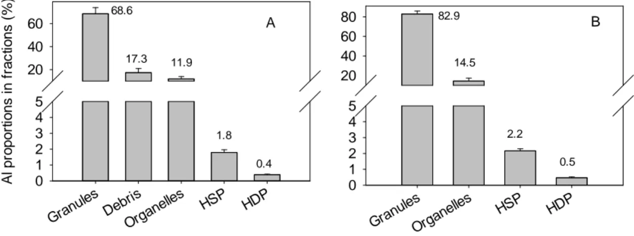
By using different digestion treatments for the whole cell samples, we found that there were no significant differences in the measured Al concentrations (Fig. S5), indicating that the H2O2 digestion step was not necessary, at least for our samples. Total metal recoveries from all the subcellular fractions accounted for 95.2 ± 5.3% of total cellular Al. The percentage of Al partitioning to subcellular components decreased in the order: granules (69 ± 5%) > debris (17 ± 4%) > organelles (12 ± 2%) > HSP (~ 2%) > HDP (< 1%) (Fig. 2A).
15 Gran ules Debri s Orga nelles HSP HDP A l pr op or ti on s i n f rac ti on s ( % ) 0 1 2 3 4 5 20 40 60 68.6 17.3 11.9 1.8 0.4 Gran ules Orga nelles HSP HDP 0 1 2 3 4 5 20 40 60 80 82.9 14.5 2.2 0.5 A B
Figure 2. Al distribution in subcellular fractions of Thalassiosira weissflogii following a 96-h incubation in Aquil* medium with an initial Al concentration of 2 µM: panel A with and panel B without the debris fraction. HSP = heat-stable peptides; HDP = heat-denaturable proteins; the error bars represent the standard deviation (n = 3).
As some of the Al in the debris fraction could be strongly bound to the biological surface (cell walls and membrane) (see Crémazy et al. (2013b)) and Al partitioning in the debris fraction was 17.3%, we can conservatively estimate that the proportion of intracellular Al was 82.7%. Without considering Al in the debris fraction, Al in the granules, organelles, HSP and HDP fractions accounted for 83 ± 3%, 14 ± 3%, ~2% and < 1% of the total intracellular Al, respectively (Fig. 2B). Since both the HSP and HDP fractions were originally present in the operationally defined cytosol, they thus represent an upper limit (2.7%) for the Al content of the ‘cytoplasm’.
16
4 Discussion
4.1 Methodological concerns
4.1.1 Composition of the extraction solution
The new seawater-based EDTA rinse solution was developed because no specific method had previously been tested to remove Al adsorbed on cell surfaces in alkaline seawater. The chemical composition of the oxalate reagent is very different from that of seawater in which marine phytoplankton grow, and its efficacy in desorbing Al from marine phytoplankton surfaces had not been examined.
To reduce the possible adverse effects of the rinse solution on algae, we adjusted the chemical composition of the rinse solution to be as similar as possible to that of the exposure medium without Al. However, a high EDTA concentration of 10 mM and a low pH of 4.5 were used for reasons explained in the Methods and Material section. As an even higher EDTA concentration was used in the oxalate reagent, which has been shown to be harmless to marine phytoplankton including T. weissflogii
(Tovar-Sanchez et al., 2003), we consider that the EDTA in the EDTA rinse solution (AquilR) is safe for the diatom.
The washing efficiency of the AquilR was slightly higher than that of the oxalate reagent, but the difference was not statistically significant, indicating that the washing efficiency of the AquilR is comparable to that of the oxalate reagent. The Y-intercept of the regression between cellular Al and exposure time (Fig. 1B) was 2.3 fmol·cell-1, a value that is similar to the background Al level (2.1 fmol·cell-1) in the diatom cells before exposure, indicating that the removal efficiency of the AquilR was quite good. The observation that the AquilR (pH 4.5) and the oxalate (pH 8.0) extractions gave similar values is an indication that brief contact with the acidic pH did not adversely affect the diatom cells (i.e., no loss of intracellular Al).
17
4.1.2 The influence of the background Al in the filter
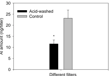
We determined that there was a substantial amount of Al in the filters used for collecting the diatom cells (23.2 ± 3.7 ng filter-1; Fig. 3); the Al amount in the filter was about 20% of the mean Al content in the diatom cells being exposed to 2 µM Al for 1 h or longer. After the acid-washing treatment, the Al content of the filter decreased markedly to 11.6 ± 1.8 ng filter-1 (Fig. 3), but the Al amount in the lower filter did not change significantly with exposure time (Fig. S2). The standard deviation of the Al amount in the lower filter was only 0.45 nmol or 3.2 ng (n = 15). The measured total amount of Al in algae that had been exposed to 2 µM Al (7.7 × 10⁵ cells) was 13–78 times higher than this standard deviation (Fig. S2). We conclude that the background Al content in the filters used for collecting the diatom cells did not contribute to the significant variation of the cellular Al amount for replicate incubations when the exposure time was longer than 1 h.
18 Different filters Al amo u n t (n g/fi lt er) 0 5 10 15 20 25 30 Acid-washed Control *
Figure 3. Al content in polycarbonate filters. Acid-washed polycarbonate filters were soaked in 10% (v/v) HCl for 24 h, and then washed with Milli-Q water thoroughly. Control = the unwashed control filter. The error bars represent the standard deviation (n = 3) and the asterisk indicates a significant difference between the two treatments (t-test, p < 0.05). The results indicated that the acid-wash procedure could significantly decrease Al content in the filter.
4.2 Aluminum uptake
To compare our data with values reported in other studies, we normalized the metal uptake rate to the exposure concentration of the metal, yielding the normalized internalization flux (NIF) of the metal into the algae. Generally, the algal uptake rates for metal increase with the concentration of exposed metals, following Michaelis- Menten saturation kinetics, a process that includes a quasi-linear part at low
concentrations and a saturation plateau at high concentrations. It follows that the NIF of a metal will tend to be higher for low ambient metal concentrations than for high exposures. Nevertheless, use of the NIF facilitates comparisons among published uptake rates.
19
nmol·m-2·min-1·nM-1; see Table S2) was comparable to those of divalent metals such as zinc, cadmium and manganese into a freshwater green alga, Chlamydomonas
reinhardtii (Sánchez-Marín et al., 2013). The NIF of Al into T. weissflogii was also
similar to that of another trivalent metal (scandium, Sc) into C. reinhardtii when the alga was exposed to Sc at µM levels (Crémazy et al., 2013a). However, our values were 3–10 times higher than the NIFs of other trivalent metals (lanthanum (La), neodymium (Nd), samarium (Sm), europium (Eu), thulium (Tm), and yttrium (Y)) into the same freshwater alga (C. reinhardtii) exposed to metal levels of 1 µM (Yang et al., 2014; Zhao and Wilkinson, 2015). Taylor et al. (2000) exposed giant intermodal cells of the macro alga Chara corallina to 50 µM Al and although most of the Al was sequestered in the cell wall, they were able to determine a much smaller flux across the plasmalemma (5.26–40×10-5 nmol·m-2·min-1·nM-1), i.e., values at least 100 times lower than those reported here for T. weissflogii.
Metal uptake rates by T. weissflogii have seldom been examined when the metal inorganic species in seawater were present at high concentrations (e.g., > 100 nM). The NIF of Al into T. weissflogii in our experiments was comparable to the NIF of Fe into the same diatom, when the dissolved Fe in seawater was present at a level of ≈100 nM and was not buffered by strong organic ligands (Anderson and Morel, 1982). Generally, substantial concentrations (e.g., 100 M) of EDTA (a strong organic ligand) were used to buffer metals (e.g., Fe) in seawater medium (e.g., Aquil* medium) and only a small proportion of the dissolved metal was present in inorganic form. As a result, the uptakes of metals such as Fe, Zn, Cu, Cd, and chromium (Cr(III)) by T.
weissflogii have normally been examined when the concentrations of their inorganic
species were present at sub-nM or pM concentrations (Table S2) and the measured uptake rates normalized to their inorganic concentrations were often one or two orders of magnitude higher than the NIF of Al into the diatom when exposed to 2 µM Al.
20
Physiological status could also influence the metal NIF into the diatom. For example, the NIF of inorganic Zn into Zn-limited T. weissflogii cells was about one order of magnitude higher than that for the Zn-replete cells, when both types of cells were exposed to a similar level (nM) of inorganic Zn (Table S2); the NIFs of Zn for the Zn-replete cells were comparable to the NIF of Al into the diatom exposed to 2 µM Al.
Overall, given the very different metal exposure concentrations in the experiments summarized in Table S2, and also the differences in inorganic metal speciation among metals, it is difficult to compare Al uptake fluxes with those reported for T. weissflogii and other metals.The reasons for choosing the 2 M concentration in the present study are that this concentration is less than the solubility limit for Al in seawater, and it does not show any adverse effect on the growth of the tested diatom in Aquil* medium (Zhou et al. 2016); furthermore, using a relatively high concentration of Al, we could more readily observe the uptake of Al in the diatom cells. Further work examining the uptake of Al at environmentally relevant levels (e.g., nM) is needed to determine the uptake parameters (KM and Vmax) and compare them to those for the other metals.
4.3 Mechanism of Al uptake
Little is known about how Al crosses algal cell membranes, even for freshwater algae. Exley and Mold (2015) have proposed five major routes by which Al could traverse cell membranes or epi-/endothelia, including paracellular transport, transcellular transport, active transport, ion channels and adsorptive or receptor-mediated
endocytosis. They also suggested that Al could be internalized in five forms: 1) free solvated trivalent cation, 2) low molecular weight, neutral, soluble complexes, 3) high molecular weight, neutral, soluble complexes, 4) low molecular weight, charged,
21
soluble complexes, and 5) nano- and micro-particulates. As far as the uptake of dissolved Al in an artificial seawater medium by a unicellular marine diatom is concerned, we consider that Al in the forms of free solvated trivalent cation and Al hydroxy-complexes (including the aluminate ion, Al(OH)4-), neutral Al(OH)30 and Al(OH)2+) are possible candidates as species assimilated by T. weissflogii, through possible routes including facilitated transport and ion channels. We have eliminated adsorptive or receptor-mediated endocytosis, given the frustule that surrounds the T.
weissflogii cell and the absence in the literature of any evidence of endocytotic
activity in this alga.
There is still disagreement in the literature as to whether or not the free trivalent Al cation (Al3+) could traverse plant cell membranes, but Xia et al. (2010) have suggested the possible involvement of cation transporters localized in plasma membranes. With respect to the transport of Al(OH)30, Al transporters belonging to the aquaporin family have been reported in higher plants (Negishi et al., 2012; Wang et al., 2017), and the involvement of an anion permease has also been proposed (Negishi et al., 2013). Nevertheless, criticisms on the methodologies and data
interpretation of some of the studies have been reported and, to quote Exley and Mold (2015), “there are no precedents across all biota for the active or passive
transmembrane transport of a trivalent metal cation”, including Al3+. Therefore, it is too early to suggest that Al could be transported across cell membranes through an Al-specific transporter for plants, let alone for marine diatoms.
To date, no established biological function of Al has been discovered and unlike iron, it cannot be reduced to a divalent state; therefore it might enter cells by “fooling” a transporter that is normally used to transport an essential metal or a ligand. For example, it has been suggested that Al might bind to specific ligands and masquerade as divalent, monovalent or even anionic species, making it possible to be transported
22
by the requisite membrane-bound transport system (Exley and Mold, 2015). For example, Mg2+, Ca2+ and the trivalent Fe3+ have been shown to be transported across a bacterial cell membrane in the form of a citrate complex (Blancato et al., 2006) and the aquaporin NIP1;2 has been reported to facilitate the transport of an Al-malate complex in a model plant species (Arabidopsis) (Wang et al., 2017). Whether organic molecules or ligands excreted by T. weissflogii could facilitate the uptake of Al is unknown.
As Al hydroxides are the dominant species of dissolved Al in seawater, they might be transported across the plasmalemma of T. weissflogii. Evidence for transmembrane transport of Al(OH)n(3-n)+ and AlFm(3-m)+ species, and Sc hydroxo-complexes has been reported for a freshwater alga (Crémazy et al., 2013a, 2014). In addition, Golding et al. (2015) suggested that the toxicity of Al to marine organisms, including diatoms, was the result of the dissolved Al forms of Al(OH)4- and Al(OH)3. The strong similarity among the conformations of As(OH)3, Sb(OH)3 and the glycerol molecule has been thought to be one of the reasons that As(OH)3 and Sb(OH)3 can be transported through aquaglyceroporins,channels that are specific for the transport of water, glycerol and other small, uncharged solutes (Bienert et al., 2008; Porquet and Filella, 2007). For the same reason, we speculate that Al(OH)30 might be transported via this route. The aluminate anion, Al(OH)4-, might also enter cells via an anion transporter, since anion transporters are known to be less selective than cation transporters (Barbier-Brygoo et al., 2011). A recent study has also indicated that aluminum bioavailability to freshwater algae under high pH conditions favoring anionic species differs from that at lower pH (Gensemer et al., 2018). For anionic species such as aluminate, the sparse literature suggests a wide range of bioavailability results for aluminate, ranging from not bioavailable to freshwater fish (Poleo and Hytterod, 2003) to likely bioavailable to at least the marine diatom T. weissflogii.
23
From a reading of the preceding discussion, it should be clear that the mechanisms by which Al enters T. weissflogii are speculative, that our understanding of Al transport across plasma membranes is very limited, and that further work is needed at the fundamental (biomembrane) level, especially for unicellular marine algae.
4.4 Subcellular distribution of Al in other phytoplankton
The subcellular distribution of Al in the diatom is very different from that reported for Al and another trivalent metal, Sc, in a green unicellular alga C. reinhardtii (Crémazy et al., 2013b). In the present study, we found that most of the Al internalized by T.
weissflogii was partitioning to the subcellular component granules (69%), followed by
debris (17%), organelles (12%), HSP (2%) and HDP (< 1%). A similar distribution was observed for two other trivalent metals (La3+, Eu3+) in the freshwater chlorophyte
C. reinhardtii (Fortin et al., 2017), but in the same alga the partitioning of Al and Sc
was very different, with the organelles dominating rather than the granules (i.e., organelles (47%), debris (38%) > granules (14%) > HSP (0.8%) > HDP (0.3%)).
Aluminum found in the granule fraction in the diatom might be bound to
polyphosphate bodies, given that Al is classified as a “hard” cation, a Class A metal, with a strong preference for ligands with oxygen donor atoms (e.g., phosphate) (Mujika et al., 2018). Polyphosphate-bound Fe has been identified as a potential vacuolar Fe storage pool in T. weissflogii (Nuester et al., 2012). Although the binding of Al to phosphate is not favored in high pH seawater (Zhou et al., 2016), the
internalized Al would bind to phosphate more readily at neutral pH or in a weakly-acid microenvironment within the cell. Clearly further work is needed to probe the forms of Al in the granule fraction in the diatom.
24
interactions, as it has confirmed that Al in diatoms can be truly internalized into the intracellular environment, rather than only being incorporated in the diatom siliceous frustules (Ren et al., 2013). The high percentage of partitioning of Al into the granule fraction may provide an explanation for why Al, even at concentrations as high as 20 M are not toxic to marine diatoms such as T. weissflogii and some other marine phytoplankton species (Gillmore et al., 2016; Golding et al., 2015; Zhou et al., 2018, 2016). Metal-rich granules are associated with metal tolerance in many organisms and are considered to play an important role in the detoxification of many trace metals (Crémazy et al., 2013b; Vijver et al., 2004). The presence of Al in the HSP and HDP subcellular components in the diatom is consistent with the findings that
oxygen-containing amino acids are the preferential coordination site of Al, and that Al can complex with amino acids (Mujika et al., 2018).
5 Conclusion
The uptake and subcellular distribution of Al in a marine diatom T. weissflogii were examined in the present study. The Al internalization flux observed in this work, normalized by the exposure concentration, is comparable to those observed for other trivalent elements with other algal species. Once assimilated, the majority of the internalized Al was partitioned into the detoxified granule fraction (69 ± 5%), with relatively little in the organelles (12 ± 2%), heat-stable peptides (HSP) (~2%) and heat-denaturable proteins (HDP) (< 1%). These results update our understanding the role of marine diatoms in the marine biogeochemistry of Al and they have also confirmed that Al can be truly internalized by diatoms into their intracellular
environment, rather than only being incorporated into the diatom siliceous frustules. These results also offer new information regarding the mechanisms responsible for the biological effects (both positive and negative) of Al on marine phytoplankton growth, especially diatoms. However, further work is needed to examine diatom uptake of Al at environmentally relevant concentrations, to explore how Al in
25
seawater is transported across the plasma membrane, and to understand the detailed location and speciation of Al within marine diatom cells.
Acknowledgement
We thank Kim Racine, Stéfane Prémont, Lise Rancourt, Sébastien Duval, Anissa Bensadoune, Julie Perreault and Jean-François Dutil at the Institut national de la Recherche scientifique for their technical assistance. Professor Neil Price and Dr. Liangliang Kong at McGill University provided the trace-metal clean artificial seawater. This work is supported by the National Key Basic Research Program of China (973 Program, 2015CB452904), the National Natural Science Foundation of China (41506150), the Natural Sciences and Engineering Research Council of Canada (NSERC), the Natural Science Foundation of Guangdong Province, China
(2015A030310169), the Science and Technology Planning Project of Guangdong Province, China (2017B030314052). C. Fortin and P.G.C. Campbell were supported by the Canada Research Chairs program.
26
References
Anderson, M.A., Morel, F.M.M., 1982. The influence of aqueous iron chemistry on the uptake of iron by the coastal diatom Thalassiosira weissflogii. Limnol. Oceanogr. 27, 789–813. Angel, B.M., Apte, S.C., Batley, G.E., Golding, L.A., 2016. Geochemical controls on aluminium concentrations in coastal waters. Environ. Chem. 13, 111–118. doi: 10.1071/EN15029. Barbier‐Brygoo, H., De Angeli, A., Filleur, S., Frachisse, J.M., Gambale, F., Thomine, S., Wege, S., 2011. Anion channels/transporters in plants: From molecular bases to regulatory networks. Annu. Rev. Plant. Biol. 62, 25–51. doi: 10.1146/annurev‐arplant‐042110‐103741. Bienert, G.P., Thorsen, M., Schussler, M.D., Nilsson, H.R., Wagner, A., Tamas, M.J., Jahn, T.P., 2008. A subgroup of plant aquaporins facilitate the bi‐directional diffusion of As(OH)3 and Sb(OH)3 across membranes. BMC Biol. 6, 26–26. Blancato, V.S., Magni, C., Lolkema, J.S., 2006. Functional characterization and Me2+ ion specificity of a Ca2+–citrate transporter from Enterococcus faecalis. FEBS J 273, 5121–5130. Crémazy, A., Campbell, P.G.C., Fortin, C., 2013a. The Biotic Ligand Model can successfully predict the uptake of a trivalent Ion by a unicellular alga below pH 6.50 but not above: possible role of hydroxo‐species. Environ. Sci. Technol. 47, 2408–2415. doi:10.1021/es3038388. Crémazy, A., Campbell, P.G.C., Fortin, C., 2014. In the presence of fluoride, free Sc3+ is not a good predictor of Sc bioaccumulation by two unicellular algae: Possible role of fluoro‐complexes. Environ. Sci. Technol. 48, 9754–9761. doi:10.1021/es5016247. Crémazy, A., Levy, J.L., Campbell, P.G.C., Fortin, C., 2013b. Uptake and subcellular partitioning of trivalent metals in a green alga: comparison between Al and Sc. BioMetals 26, (6):989–1001. doi:10.1007/s10534‐013‐9675‐6. Driscoll, C.T., Schecher, W.D., 1988. Aluminum in the environment. In: Sigel, H., Sigel, A. (eds) Aluminum and its Role in Biology. Metal Ions in Biological Systems, Vol.24. Marcel Dekker Inc., New York, pp 59–122. Driscoll, C.T., Schecher, W.D., 1990. The chemistry of aluminum in the environment. Environ. Geochem. Health 12, 28–49. doi:10.1007/BF01734046. Exley, C., Mold, M.J., 2015. The binding, transport and fate of aluminium in biological cells. J. Trace Elem. Med. Bio. 30, 90–95. doi:10.1016/j.jtemb.2014.11.002. Fortin, C., Racine, K., Singing, B.N., Beaubien, C., Campbell, P.G.C., 2017. Bioavailability, uptake and toxicity of emerging trace elements in green algae. Paper presented at the ICOBTE2017 ETH Zurich, Switzerland, 16–20 July, 2017Gensemer, R. W., Gondek, J.D., Rodriquez, P., Arbildua, J. J., Stubblefield, W., Cardwell, A. Santore, R., Ryan, A., Adams, W., Nordheim, E. 2018. Evaluating the effects of pH, hardness, and dissolved organic carbon on the toxicity of aluminum to freshwater aquatic organisms under circumneutral conditions. Environ. Toxicol. Chem. 37, 49‐60. doi: 10.1002/etc.3920. Gillmore, M.L., Golding, L.A., Angel, B.M., Adams, M.S., Jolley, D.F., 2016. Toxicity of dissolved and precipitated aluminium to marine diatoms. Aquat. Toxicol. 174, 82–91. doi: 10.1016/j.aquatox.2016.02.004. Golding, L.A., Angel, B.M., Batley, G.E., Apte, S.C., Krassoi, R., Doyle, C.J., 2015. Derivation of a water
27 quality guideline for aluminium in marine waters. Environ. Toxicol. Chem. 34, 141–151. doi: 10.1002/etc.2771. Hassler, C.S., Slaveykova, V.I., Wilkinson, K.J., 2004. Discriminating between intra‐ and extracellular metals using chemical extractions. Limnol. Oceanogr.‐Meth. 2, 237–247. Kuss, J., Kremling, K., 1999. Spatial variability of particle associated trace elements in near‐surface waters of the North Atlantic (30°N/60°W to 60°N/2°W), derived by large volume sampling. Mar. Chem. 68, 71–86. doi: 10.1016/S0304‐4203(99)00066‐3. Liu, J., Zhou, L., Li, G., Ke, Z., Shi, R., Tan, Y., 2018. Beneficial effects of aluminum enrichment on nitrogen‐fixing cyanobacteria in the South China Sea. Mar. Pollut. Bull. 129, 142–150. doi:10.1016/j.marpolbul.2018.02.011. Mujika, J.I., Dalla Torre, G., Formoso, E., Grande‐Aztatzi, R., Grabowski, S.J., Exley, C., Lopez, X., 2018. Aluminum's preferential binding site in proteins: sidechain of amino acids versus backbone interactions. J. Inorg. Biochem. 181, 111–116. doi: 10.1016/j.jinorgbio.2017.10.014. Negishi, T., Oshima, K., Hattori, M., Kanai, M., Mano, S., Nishimura, M., Yoshida, K., 2012. Tonoplast‐ and plasma membrane‐localized aquaporin‐family transporters in blue hydrangea sepals of aluminum hyperaccumulating plant. PLoS ONE 7, e43189. doi: 10.1371/journal.pone.0043189. Negishi, T., Oshima, K., Hattori, M., Yoshida, K., 2013. Plasma membrane‐localized Al‐transporter from blue hydrangea sepals is a member of the anion permease family. Genes Cells 18, 341–352. doi:10.1111/gtc.12041. Nuester, J., Vogt, S., Twining, B.S., 2012. Localization of iron within centric diatoms of the genus Thalassiosira. J. Phycol. 48, 626–634. doi:10.1111/j.1529‐8817.2012.01165.x. Pierrot, D., Millero, F.J., 2017. The speciation of metals in natural waters. Aquat. Geochem. 23, 1–20. doi:10.1007/s10498‐016‐9292‐4. Pitre, D., Boullemant, A., Fortin, C., 2014. Uptake and sorption of aluminium and fluoride by four green algal species. Chem. Central J. 8, 8. doi:10.1186/1752‐153x‐8‐8. Poléo, A. B. S., Hytterød, S., 2003. The effect of aluminium in Atlantic salmon (Salmo salar) with special emphasis on alkaline water. J. Inorg. Biochem. 97, 89‐96. doi: 10.1016/S0162‐0134(03)00261‐7.
Porquet, A., Filella, M., 2007. Structural evidence of the similarity of Sb(OH)3 and As(OH)3 with glycerol:
Implications for their uptake. Chem. Res. Toxicol. 20, 1269–1276. Ren, H.J., Brunelle, B.G., Sigman, D.M., Robinson, R.S., 2013. Diagenetic aluminum uptake into diatom frustules and the preservation of diatom‐bound organic nitrogen. Mar. Chem. 155, 92–101. doi:10.1016/j.marchem.2013.05.016. Saçan, M.T., Balcıoğlu, I.A., 2001. Bioaccumulation of aluminium in Dunaliella tertiolecta in natural seawater: Aluminium‐metal (Cu, Pb, Se) interactions and influence of pH. Bull. Environ. Contam. Toxicol. 66, 214–221. Saçan, M.T., Oztay, F., Bolkent, S., 2007. Exposure of Dunaliella tertiolecta to lead and aluminum: Toxicity and effects on ultrastructure. Biol. Trace Elem. Res. 120, 264–272. doi:10.1007/s12011‐007‐8016‐4. Sánchez‐Marín, P., Fortin, C., Campbell, P.G.C., 2013. Copper and lead internalisation by freshwater microalgae at different carbonate concentrations. Environ. Chem. 10, 80–90.
28 Stoffyn, M., 1979. Biological control of dissolved aluminum in seawater: Experimental evidence. Science 203, 651–653. doi:10.1126/science.203.4381.651. Sunda, W.G., Price, N.M., Morel, F.M.M., 2005. Trace metal ion buffers and their use in culture studies. In: Andersen, R.A. (ed) Algal Culturing Techniques. Academic Press, Amsterdam, pp. 35–63. Taylor, G.J., McDonald‐Stephens, J.L., Hunter, D.B., Bertsch, P.M., Elmore, D., Rengel, Z., Reid, R.J., 2000. Direct measurement of aluminum uptake and distribution in single cells of Chara corallina Plant. Physiol. 123, 987–996. Tovar‐Sanchez, A., Sañudo‐Wilhelmy, S.A., Garcia‐Vargas, M., Weaver, R.S., Popels, L.C., Hutchins, D.A., 2003. A trace metal clean reagent to remove surface‐bound iron from marine phytoplankton. Mar. Chem. 82, 91–99. doi:10.1016/S0304‐4203(03)00054‐9. Vijver, M.G., Van Gestel, C.A.M., Lanno, R.P., Van Straalen, N.M., Peijnenburg, W.J.G.M., 2004. Internal metal sequestration and its ecotoxicological relevance: a review. Environ. Sci. Technol. 38, 4705–4712. doi:10.1021/es040354g. Vrieling, E.G., Poort, L., Beelen, T.P.M., Gieskes, W.W.C., 1999. Growth and silica content of the diatoms Thalassiosira weissflogii and Navicula salinarum at different salinities and enrichments with aluminium. Eur. J. Phycol. 34, 307–316. Wang, Y., Li, R., Li, D., Jia, X., Zhou, D., Li, J., Lyi, S.M., Hou, S., Huang, Y., Kochian, L.V., 2017. NIP1; 2 is a plasma membrane‐localized transporter mediating aluminum uptake, translocation, and tolerance in Arabidopsis. Proc. NatL. Acad. Sci. USA 114, 5047–5052. doi: 10.1073/pnas.1618557114. Wilson, S.P., Hyne, R.V., 1997. Toxicity of acid‐sulfate soil leachate and aluminum to embryos of the Sydney rock oyster. Ecotoxicol. Environ. Saf. 37, 30–36. doi: 10.1006/eesa.1996.1514. Xia, J.X., Yamaji, N., Kasai, T., Ma, J.A.F., 2010. Plasma membrane‐localized transporter for aluminum in rice. Proc. Natl. Acad. Sci. USA 107, 18381–18385. doi:10.1073/pnas.1004949107. Yang, G., Tan, Q.‐G., Zhu, L., Wilkinson, K.J., 2014. The role of complexation and competition in the biouptake of europium by a unicellular alga. Environ. Toxicol. Chem. 33, 2609–2615. doi:10.1002/etc.2722. Zhao, C.‐M., Wilkinson, K.J., 2015. Biotic ligand model does not predict the bioavailability of Rare Earth elements in the presence of organic ligands. Environ. Sci. Technol. 49, 2207–2214. doi:10.1021/es505443s. Zhou, L., Liu, J., Xing, S., Tan, Y., Huang, L., 2018a. Phytoplankton responses to aluminum enrichment in the South China Sea. J. Inorg. Biochem. 181, 117–131. doi:10.1016/j.jinorgbio.2017.09.022. Zhou, L., Tan, Y., Huang, L., Wang, W.‐X., 2016. Enhanced utilization of organic phosphorus in a marine diatom Thalassiosira weissflogii: A possible mechanism for aluminum effect under P limitation. J. Exp. Mar. Biol. Ecol. 478, 77–85. doi: 10.1016/j.jembe.2016.02.009. Zhou, L., Tan, Y., Huang, L., Fortin, C., Campbell, P.G.C., 2018b. Aluminum effects on marine phytoplankton: implications for a revised Iron Hypothesis (Iron‐Aluminum Hypothesis) Biogeochemistry 139, 123‐137. doi:10.1007/s10533‐018‐0458‐6.