Université de Sherbrooke
Effect of Haptic Guidance and Error Amplification Robotic Training Interventions on the Immediate Improvement of Timing among Individuals that had a Stroke
By Amy E. Bouchard, B.Sc. Research in Health Sciences Program
This Thesis is presented to the Faculty of Medicine and Health Sciences to obtain the Master of Science degree (M.Sc.) for the Research in Health Sciences Program
Sherbrooke, Québec, Canada August 2016
Jury members for the Thesis evaluation
Prof. Marie-Hélène Milot, P.T., Ph.D., Research Director, Research in Health Sciences Program,
Prof. Hélène Corriveau, P.T., Ph.D., Co-research Director, Research in Health Sciences Program,
Prof. Johanne Desrosiers, O.T., Ph.D., Internal Member, Research in Health Sciences Program
Prof. Philippe Archambault, O.T., Ph.D., External Member, School of Occupational and Physical Therapy, McGill University
Summary
Effect of Haptic Guidance and Error Amplification Robotic Training Interventions on the Immediate Improvement of Timing among Individuals that had a Stroke
By: Amy E. Bouchard
Research in Health Sciences Program
Thesis presented to the Faculty of Medicine and Health Sciences in order to obtain the Master in Science Diploma (M.Sc.) in Research in Health Sciences, Faculty of Medicine
and Health Sciences, Université de Sherbrooke, Québec, Canada, J1H 5N4
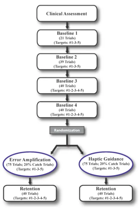
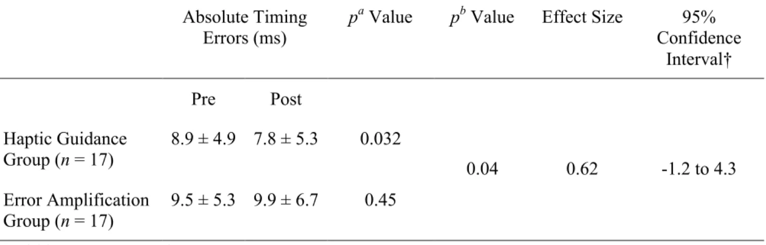
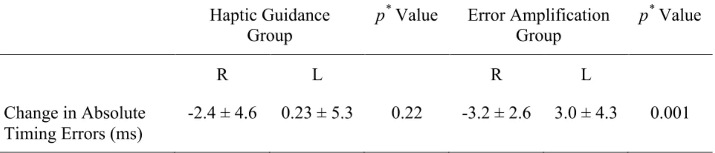
Many individuals that had a stroke have motor impairments such as timing deficits that hinder their ability to complete daily activities like getting dressed. Robotic rehabilitation is an increasingly popular therapeutic avenue in order to improve motor recovery among this population. Yet, most studies have focused on improving the spatial aspect of movement (e.g. reaching), and not the temporal one (e.g. timing). Hence, the main aim of this study was to compare two types of robotic rehabilitation on the immediate improvement of timing accuracy: haptic guidance (HG), which consists of guiding the person to make the correct movement, and thus decreasing his or her movement errors, and error amplification (EA), which consists of increasing the person’s movement errors. The secondary objective consisted of exploring whether the side of the stroke lesion had an effect on timing accuracy following HG and EA training. Thirty-four persons that had a stroke (average age 67 ± 7 years) participated in a single training session of a timing-based task (simulated pinball-like task), where they had to activate a robot at the correct moment to successfully hit targets that were presented a random on a computer screen. Participants were randomly divided into two groups, receiving either HG or EA. During the same session, a baseline phase and a retention phase were given before and after each training, and these phases were compared in order to evaluate and compare the immediate impact of HG and EA on movement timing accuracy. The results showed that HG helped improve the immediate timing accuracy (p=0.03), but not EA (p=0.45). After comparing both trainings, HG was revealed to be superior to EA at improving timing (p=0.04). Furthermore, a significant correlation was found between the side of stroke lesion and the change in timing accuracy following EA (rpb=0.7, p=0.001), but not HG (rpb=0.18, p=0.24). In other words, a deterioration in timing accuracy was found for participants with a lesion in the left hemisphere that had trained with EA. On the other hand, for the participants having a right-sided stroke lesion, an improvement in timing accuracy was noted following EA. In sum, it seems that HG helps improve the immediate timing accuracy for individuals that had a stroke. Still, the side of the stroke lesion seems to play a part in the participants’ response to training. This remains to be further explored, in addition to the impact of providing more training sessions in order to assess any long-term benefits of HG or EA.
Résumé
Effet de l’entrainement robotisé par réduction de l’erreur et augmentation de l’erreur sur le timing du mouvement chez la personne ayant eu un accident vasculaire cérébral
Par : Amy E. Bouchard
Programme Recherche en sciences de la santé
Mémoire présenté à la Faculté de médecine et des sciences de la santé en vue de l’obtention du diplôme de maitre ès sciences (M.Sc.) Recherche en sciences de la santé, Université de
Sherbrooke, Sherbrooke, Québec, Canada, J1H 5N4
À la suite d’un accident vasculaire cérébral (AVC), plusieurs atteintes, comme un déficit de timing, sont notées, et ce, même à la phase chronique d’un AVC, ce qui nuit à l’accomplissement de tâches quotidiennes comme se vêtir. L’entrainement robotisé est un entrainement qui est de plus en plus préconisé dans le but d’améliorer la récupération motrice à la suite d’un AVC. Par contre, la plupart des études ont étudié les effets de l’entrainement robotisé sur l’amélioration de l’aspect spatial du mouvement (ex : la direction du mouvement), et non l’aspect temporel (ex : timing). L’objectif principal de ce projet était donc d’évaluer et de comparer l’impact de deux entrainements robotisés sur l’amélioration immédiate du timing soit : la réduction de l’erreur (RE), qui consiste à guider la personne à faire le mouvement désiré, et l’augmentation de l’erreur (AE), qui nuit au mouvement de la personne. L’objectif secondaire consistait à explorer s’il y avait une relation entre le côté de la lésion cérébrale et le changement dans les erreurs de timing suivant l’entrainement par RE et AE. Trente-quatre personnes atteintes d’un AVC au stade chronique (âge moyen de 67 ± 7 années) ont participé à cette étude, où ils devaient jouer à un jeu simulé de machine à boules. Les participants devaient activer une main robotisée au bon moment pour atteindre des cibles présentées aléatoirement sur un écran d’ordinateur. Les participants recevaient soit RE ou AE. Une ligne de base et une phase de rétention étaient données avant et après chaque entrainement, et elles étaient utilisées pour évaluer et comparer l’effet immédiat de RE et AE sur le timing. Les résultats ont démontré que RE permet d’améliorer les erreurs de timing (p=0,03), mais pas AE (p=0,45). De plus, la comparaison entre les deux entrainements a démontré que RE était supérieur à AE pour améliorer le timing (p=0,04). Par ailleurs, une corrélation significative a été notée entre le côté de la lésion cérébrale et le changement des erreurs de timing suivant AE (rpb=0,70; p =0,001), mais pas RE (rpb=0,18; p=0,24). En d’autres mots, une détérioration de l’exécution de la tâche de timing a été notée pour les participants ayant leur lésion cérébrale à gauche. Par contre, ceux ayant leur lésion à droite ont bénéficié de l’entrainement par AE. Bref, l’entrainement par RE peut améliorer les erreurs de timing pour les survivants d’AVC au stade chronique. Toutefois, le côté de la lésion cérébrale semble jouer un rôle important dans la réponse à l’entrainement par AE. Ceci demeure à être exploré, ainsi que l’impact d’un entrainement par RE et AE de plus longue durée pour en déterminer leurs effets à long terme.
Mots clés : apprentissage moteur, réduction de l’erreur, augmentation de l’erreur, timing, accident vasculaire cérébral
Table of Contents
List of Tables ... VII List of Tables in the Scientific Article ... VII List of Figures ... VIII List of Figures in the Scientific Article ... VIII List of Abbreviations ... IX
Chapter I: Research Problem ... 1
Chapter II: Literature Review ... 2
II.1 Stroke ... 2
II.I.I Definition and Epidemiology ... 2
II.1.2 Post Stroke Impairments and Disabilities ... 3
II.2 Movement ... 5
II.2.1 Timing ... 5
II.2.2 Post Stroke Timing Deficit ... 6
II.3 Motor Learning ... 7
II.3.1 Definition ... 7
II.3.2 Principles ... 7
II.3.2.1 Characteristics of the Task: Practice ... 8
II.3.2.2 Characteristics of the Task: Feedback ... 9
II.3.2.3 Characteristics of the Task: Guidance versus Error ... 10
II.3.2.3.1 Guidance ... 10
II.3.2.3.2 Error ... 11
II.3.3 Measures of Motor Learning ... 12
II.3.4 Promoting Motor Learning among Individuals that had a Stroke ... 13
II.4 Stroke Rehabilitation Strategies ... 13
II.4.1 Best Stroke Practice Recommendations for Stroke Rehabilitation ... 13
II.4.2 Robotic Rehabilitation ... 14
II.4.2.1.1 Healthy Individuals ... 16
II.4.2.1.2 Individuals that had a Stroke ... 17
II.4.2.2 Error Amplification ... 18
II.4.2.2.1 Healthy Individuals ... 19
II.4.2.2.2 Individuals that had a Stroke ... 20
II.4.3 The use of Haptic Guidance and Error Amplification for Improving Temporal Components of Movement ... 21
II.5 Summary ... 21
Chapter III ... 23
III.1 Objectives ... 23
III.1.1 Primary Objective ... 23
III.1.2 Secondary Objective ... 23
III.2 Hypotheses ... 23
III.2.1 Primary Hypothesis ... 23
III.2.2 Secondary Hypothesis ... 23
Chapter IV: Materials and Methods ... 25
IV.1 Variables, Instrument and Procedure ... 25
IV.1.1 Descriptive Variables ... 25
IV.1.2 Independent Variable ... 28
IV.1.3 Dependent Variable ... 30
IV.1.4 Instrument ... 30
IV.2 Procedure ... 31
IV.2.1 Timing Task ... 31
IV.3 Sample Size Calculation ... 35
IV.4 Ethical Considerations ... 36
Chapter V: Scientific Article ... 37
Résumé de l’article scientifique ... 38
Chapter VI: Discussion ... 68
VI.1 Impact of Haptic Guidance on Motor Learning of Timing ... 68
VI.2 Impact of EA on Motor Learning of Timing ... 69
VI.4 Clinical Impact ... 71
VI.5 Limits and Strengths ... 72
VI.6 Conclusion ... 73
Acknowledgements ... 74
References……….75
Appendix I: Ethics Approvals………...99
Appendix II: English Version of the Consent Form………...101
Appendix III: French Version of the Consent Form………...109
Appendix IV: English Version of the Recruitment Ad ... 117
List of Tables
Table 1: Psychometric Qualities of the Clinical Tools used for Stroke Survivors………...27
List of Tables in the Scientific Article
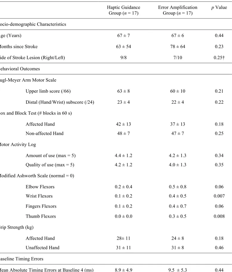
Table 1: Baseline characteristics for the haptic guidance and error amplification
groups………....53 Table 2: Changes in absolute timing errors following a single session of HG and EA robotic training interventions for the haptic guidance and error amplification groups……….55 Table 3: Impact of the side of the stroke lesion on the changes in performance at the timing task for the haptic guidance and error amplification groups……….56
List of Figures
Figure 1: Timing Exerciser Orthosis (TEO)………...28
Figure 2: Phases of the Timing Task………...32
Figure 3: Timing Task as Seen on the Computer Screen with the Lines………..33
Figure 4: Timing Task as Seen on the Computer Screen without the Lines………...34
List of Figures in the Scientific Article Figure 1: (A) Simulated Pinball-Like Task; (B) TEO; (C) A participant’s hand positioned in TEO………...47
List of Abbreviations
AVC Accident vasculaire cérébral CNS Central Nervous System EA Error Amplification
FMA Fugl-Meyer Stroke Assessment Scale
HG Haptic Guidance
MAS Modified Ashworth Scale TEO Timing Exerciser Orthosis
Chapter I: Research Problem
After having a stroke, survivors go through several recovery stages, starting with the hyper-acute, acute, sub-acute, and finally, the chronic stage, usually referred as more than six months post stroke (Demain et al., 2006; Reinkensmeyer et al., 2012; Richards, 2013). Individuals that had a stroke are often left with impairments, such as timing impairments (Bi and Wan, 2013), negatively impacting movement execution. Consequently, it is estimated that 80% of stroke survivors present some limitations in the accomplishment of activities of daily living (Richards, 2013). Fortunately, stroke survivors are capable of motor learning, even at the chronic stage (Teasell et al., 2012; Teasell et al., 2014), in order to improve movement execution. Both the spatial (e.g. direction) and temporal (ex: timing) components of movement are required in order to carry out daily tasks (Georgopoulos, 2002). As such, several studies have attempted to improve the spatial aspect of a task using two different robotic trainings for stroke survivors: error amplification (EA) and haptic guidance (HG) (Patton et al., 2006; Cesqui et al., 2008). HG consists of guiding the person to make the correct movement, and EA is used to increase movement errors. So far, as compared to HG, EA yields better results on the improvement of the spatial component of movement among individuals that had a stroke. However, it seems that no study has directly compared HG and EA to improve movement timing in this population, despite the negative impacts that a deficit in movement timing has on the performance of everyday tasks.
Chapter II: Literature Review
II.1 Stroke
II.1.1 Definition and Epidemiology
A stroke or a cerebrovascular accident is defined as “a sudden loss of brain function”, with symptoms lasting more than 24 hours (Heart and Stroke Foundation, 2016). There are two main types of stroke, depending on the cause: the first is an ischemic stroke, occurring 80% of the time, caused by a thrombus or an embolism (Lindsay et al., 2013). More specifically, thrombi are blood clots that block blood flow in the brain itself, and emboli are blood clots that occur elsewhere in the body that travel throughout the vascular system and proceed to block a cerebral artery (Mayfield Brain & Spine, 2016). The second type of stroke is called hemorrhagic, where a blood vessel ruptures, resulting in bleeding inside the brain tissue itself or around it (Heart and Stroke Foundation, 2016).
Every nine minutes, someone has a stroke in Canada, and there are more than 62,000 people that have a stroke each year HS report 2015. In addition, stroke is the third leading cause of death, and the number one cause of disability in the country. Due to the continuous aging of the population, more and more people are having strokes, and the risk of having a stroke doubles every 10 years, starting at the age of 55. The costs and needs for rehabilitation will only continue to increase because of this incessant increase in the aging of the population (Langhorne et al., 2011). At the moment, stroke costs the Canadian economy 3.6 billion dollars in health care and lost productivity (Canadian Vascular Network, 2013)
Fortunately, due to the continuous improvement in quality of care (e.g. technological innovations and health system service improvements), more than 83% of persons who have a stroke survive it (Heart and Stroke Foundation, 2015). Nevertheless, it is estimated that 405 000 Canadians are living with the consequences of stroke impairments and disabilities (Krueger et al., 2015).
II.1.2 Post Stroke Impairments and Disabilities
Only 10% of stroke survivors recover completely from their stroke, so the vast majority of survivors are left with impairments of the hemi body that is contralateral to the stroke lesion (Baak et al., 2015). Impairment is defined as a “change in function caused by a stroke” (p. 1.8) (Heart and Stroke Foundation, 2013). The occurrence and severity of post stroke impairments vary based on, among other things, the stroke survivor’s age, the severity and location of the lesion, and on how quickly they receive appropriate treatment once the stroke occurs (Heart and Stroke Foundation, 2013; Lindsay et al., 2013).
A variety of impairments can be observed after a stroke such as language problems (e.g. aphasia, dysarthria), cognitive problems, perception problems (e.g. apraxia, unilateral neglect) and sensory problems (Raghavan, 2015). However, motor impairments affect 80% of all stroke survivors (Langhorne et al., 2009). The central nervous system (CNS) is responsible for motor control, where it creates movements that are both purposeful and coordinated, while interacting with the body and the environment (Latash et al., 2010). Problems with motor control often include muscle weakness, spasticity, and incoordination (Raghavan, 2015).
Firstly, muscle weakness is common in 80% stroke survivors, and occurs mainly on the side of the body that is opposite to the brain lesion (National Stroke Association, 2016). Muscle weakness is often caused by damage to the cortico-spinal tract, which is one of the main motor pathways between the cortex and the spinal cord involved in voluntary movements (Jang, 2009). As such, a stroke-damaged cortico-spinal tract does not send the needed signal from the cortex to the spinal cord in order to create the desired movement (Canning et al., 2004; Wagner et al., 2007). In turn, this impaired signal can cause muscle contractions to have a slower initiation, termination, (Chae et al., 2002) and force (Canning et al., 1999). Consequently, muscles can undergo several changes following a stroke, such as decreases in muscle mass, muscle fibre length, among others, which may translate into muscle weakness of the affected limbs (Gray et al., 2012).
Secondly, spasticity, which is defined as an increase in muscle tone with increasing velocity, can affect up to 92% of stroke survivors (Malhotra et al., 2011; Li and Francisco, 2015; Raghavan, 2015). Spasticity can have a detrimental impact on stroke survivors’ motor ability, as it can reduce joint range of motion (Raghavan, 2015). For example, in the upper limb, spasticity is most often present in the flexor muscle groups such as in the wrist or elbow, making it difficult for stroke survivors to move their affected upper limb in order to pick up objects or get dressed, for instance (Thibaut et al., 2013; Li and Francisco, 2015).
Lastly, stroke survivors may suffer from uncoordinated movements, due to sensorimotor impairments that cause problems in the sequencing and timing of muscle contraction (Bourbonnais et al., 1992; Cheung et al., 2012). For example, stroke survivors can experience difficulty controlling the temporal aspect of antagonist and agonist muscles during reaching (Bourbonnais et al., 1992), where the contraction of antagonist and agonist muscles occurs at the same time. This phenomenon, called co-contraction, can thus hinder movement execution (Thibaut et al., 2013).
Altogether, post stroke impairments can cause individuals that had a stroke to develop learned non-use and/or learned bad use (Raghavan, 2015), leading them to make compensatory movements (e.g. flexing their trunk instead of their elbow when reaching), and hence cause disabilities (Cirstea and Levin, 2000). A disability can be defined as “the change in ability to meet daily demands or do things because of an impairment” (pg. 1.8; (Heart and Stroke Foundation, 2013). Indeed, stroke survivors have difficulty in performing everyday activities such as cooking, getting dressed, and bathing, which are all important for maintaining a good quality of life (Langhorne et al., 2011). More specifically, 80% of these individuals have difficulties carrying out activities of daily living (Richards, 2013). The numerous deficits and impairments following a stroke can hinder the social participation of these persons for recreational activities, like playing tennis or playing cards, and driving for instance (Carlsson et al., 2004; Edwards et al., 2006; Fougeyrollas, 2010; Rozon and Rochette, 2015). Luckily, it was demonstrated that rehabilitation could improve the social participation of stroke survivors, even if it takes time (Mayo et al., 2015; Obembe and Eng, 2016).
In view of these facts, there is no need to specify that impairments and disabilities can compromise post stroke movement execution. The components that are needed to properly carry out a movement will be discussed next.
II.2 Movement
Two aspects are required in order to effectively produce a movement: the first is spatial, such as the direction and fluidity of movement (Georgopoulos, 2002). The second is the temporal aspect of movement, like timing. Both of these components can be studied separately, depending on the nature of the task (Georgopoulos, 2002). Nevertheless, only the timing aspect of movement will be further discussed here since it is the variable of interest in this thesis.
II.2.1 Timing
Movement timing is defined as the ability to properly activate the muscles at the correct moment (Shumway-Cook and Woollacott, 2012). Also, the left side of the brain seems to be responsible for processing timing (Kwon et al., 2007; Freitas et al., 2011). For instance, in the study by Kwon et al. (Kwon et al., 2007), individuals that had a stroke who had a lesion in the left hemisphere displayed a much higher variability in timing during a tapping task as compared to those that had their lesion in the right hemisphere. Although timing can be divided into three components: execution time, the time required to stop a movement and reaction time, this work focused on reaction time, which is defined as “the duration from the onset of a stimulus to the initiation of a motor response” (p. 213) (Carnahan et al., 1997). However, the term timing will be used in this manuscript since it is the main term that is used in the literature.
Furthermore, movement timing is crucial in order to perform daily tasks such as driving either a car or a motorized wheelchair (Marchal Crespo and Reinkensmeyer, 2008; Marchal-Crespo et al., 2010), playing sports like golf or tennis (Marchal-Marchal-Crespo et al., 2013), or catching a ball (Ziherl et al., 2010). However, studies have found that post stroke timing deficits can hinder functional performance.
II.2.2 Post Stroke Timing Deficit
Stroke survivors can have timing deficits that are caused by impairments previously presented above (Hermsdörfer and Goldenberg, 2002; Gerritsen et al., 2003; Daly et al., 2006; Freitas et al., 2011; Bi and Wan, 2013).
A small number of studies have examined the relationship between reaction time and motor recovery post stroke (Hermsdörfer and Goldenberg, 2002; Werhahn et al., 2003; Miscio et al., 2006; Bi and Wan, 2013). For example, a study by Miscio et al. (Miscio et al., 2006) evaluated the reaction times of the flexors and extensors of the wrists of eight individuals that had a stroke (mean age=61 ± 13 years), using electromyography. They found that the reaction times were slower in both the affected and unaffected wrists (flexors and extensors). The authors explained that this might be due to a damaged cortico-spinal tract where there may be a slowing or interruption of its outflow. The authors also investigated whether there was a relationship between the reaction time and the Modified Ashworth Scale (MAS), a measure of spasticity (on a scale of 0 to 4, where 0 is normal), and the Medical Research Council scores, a measure of manual muscle strength (on a scale of 0 to 5, where 5 is normal). Although no relationship was found between reaction time and spasticity of the wrist, a significant relationship was obtained with muscle strength at the affected flexors (r=-0.55, p<0.0001) and extensors (r=-0.36; p=0.012), respectively. This meant that the stroke survivors with the most amount of motor strength had faster reaction times.
Moreover, individuals that had a stroke have been found to have reaction times for their affected limbs that are twice as long as that of their healthy side (Miscio et al., 2006; Bi and Wan, 2013) and when compared to healthy individuals (Bi and Wan, 2013), for tasks involving wrist extension and flexion. As such, this slowing of reaction time can compromise their performance of everyday functional activities (Freitas et al., 2011). It is to be noted that a smaller number of studies have investigated the relationship between cognitive impairment and timing, where higher cognitive impairments were associated with slower timings (Daly et al., 2006; Cumming et al., 2012; Cumming et al., 2014). However, this aspect was not studied in this
thesis, and it was ensured that all of the participants understood the timing task adequately (see Chapter IV).
Furthermore, an important part of rehabilitation for individuals that had a stroke is related to motor learning. Luckily, as mentioned earlier, they maintain their ability for motor learning in order to regain function of their affected limb (Teasell et al., 2012; Takeuchi and Izumi, 2013; Teasell et al., 2014). Motor learning, including its principles and measures, as well as the ways to promote it, will be further discussed.
II.3 Motor Learning
II.3.1 Definition
Motor learning, as defined by Schmidt and Lee (Schmidt and Lee, 2011), is “a set of internal processes associated with practice or experience leading to relatively permanent changes in the capability for skilled movement” (p. 327). Several aspects are required, such as temporal, spatial and hierarchical organizations of the CNS (Schmidt and Lee, 2011). Thus, motor learning can allow many things such as learning how to make a new movement, acquiring a new movement, or modifying a movement. Furthermore, motor learning occurs by the process of motor development, which is the “evolution of changes in motor behavior [that occur] as a result of growth, maturation, and experience” (p. 399) (Schmidt and Lee, 2011; O'Sullivan et al., 2014).
II.3.2 Principles
There are various aspects that can influence one’s motor learning (Schmidt and Lee, 2011; Lage et al., 2015), such as the person’s characteristics (e.g. attention and motivation), and the characteristics of the task to be learned (e.g. practice, feedback, guidance vs. error), which can all be modified in order to optimize motor learning. In this thesis, the characteristics of the task were manipulated in order to optimize movement timing and will thus be discussed in more detail.
II.3.2.1 Characteristics of the task: Practice
Practice includes two aspects: firstly, the more one practices, the more motor learning one will have. Secondly, with practice, substantial improvements can be noted towards the beginning of the task, and smaller improvements can be observed over time (Lage et al., 2015). Furthermore, there can be three main ways of providing practice.
The first is massed vs. distributed practice. In massed practice, the entire task is practiced at once with little rest periods. This kind of practice is often more efficient for people who have concentration problems (Schmidt and Lee, 2011). On the other hand, in distributed practice, the participant is given more rest periods and less practice of the actual task. The latter is better for individuals whom are more prone to fatigue (Schmidt and Lee, 2011). Both techniques have been shown to be efficient for stroke survivors (Dettmers et al., 2005; Geurts et al., 2005; Vearrier et al., 2005; Muratori et al., 2013). For example, massed practice was used to help improve balance among 10 individuals that had a stroke (Vearrier et al., 2005) where they practiced balance activities for 6 hours per day for 10 consecutive weekdays. Furthermore, a distributed form of constraint induced movement therapy was used in the study by Dettmers et al. (Dettmers et al., 2005), where 11 individuals that had a stroke received training of their affected arm 3 hours per day for 20 days where they had to repeatedly practice everyday tasks (e.g. opening and closing bottles). The participants served as their own control. The authors found significant gains for the affected trained upper limb such as in grip strength (4.2 ± 3.1 kg), and these gains were kept for as much as six months post therapy.
Secondly, practice can be given in blocks or randomly (Schmidt and Lee, 2011). More specifically, during random practice, different tasks are practiced in random order. During blocked practice, the same task is repeatedly practiced over and over. It has been shown that blocked practice provides better retention over the short term and that random practice is better over the long term (Immink and Wright, 2001). For example, both interventions, when combined with active neuromuscular stimulation, were shown to help individuals that had a stroke significantly improve their manual dexterity and reaction time, while they practiced the Box and
Block Test and a reaction time task involving the extension of their wrist and fingers, when compared to a control group (Cauraugh and Kim, 2003).
Thirdly, practice can be provided in part or in whole (Kurtz and Lee, 2003; Hansen et al., 2005; Schmidt and Lee, 2011; Klein et al., 2012). This applies to more complicated motor skills. In other words, one can learn the entire steps that are required to perform one task (whole) or one can learn a task in steps (in part). For example, (Klein et al., 2012) used robotic haptic guidance to compare part versus whole complex arm movement training in young healthy participants (n=40; age=28.6 ±5.5 years). The authors found that breaking down the task into components was more effective at improving learning than practicing the task in whole.
II.3.2.2 Characteristics of the task: Feedback
Feedback has been found to have a very important effect on motor learning (Krebs et al., 1998; Boian et al., 2002; Brewer et al., 2008; van Asseldonk et al., 2009). Feedback can be given extrinsically or intrinsically.
Extrinsic feedback occurs when feedback is given to the individual while he or she practices the task (concurrent feedback), or after the he or she practices the task (terminal feedback). In a healthy population, it has been shown that extrinsic feedback that is given during a task will improve performance but will decrease retention, whereas feedback that is given after a task will not improve performance while training but may increase retention (Carnahan et al., 1996). In a chronic stroke population, Secoli et al. (Secoli et al., 2011) provided concurrent sound feedback to individuals that had a stroke while they were practicing a tracking task with their affected upper limb positioned in a robotic device. The authors found that sound feedback increased the subjects’ effort during the task while at the same time decreasing their tracking errors as opposed to no sound feedback.
On the contrary, distorted feedback can be provided. For instance, visual feedback can be distorted by creating a difference between what the person perceives and what he/she experiences in reality (Brewer et al., 2006). Since this difference can be made imperceptible to the person, it can help promote improvements in performance (Brewer et al., 2008), especially
among neurologically impaired individuals, since it may help them overcome their resistance to move past their usual movements (Atkinson, 1964; Bandura and Cervone, 1986; Taub et al., 1994; Brewer et al., 2008). For instance, Brewer et al. (Brewer et al., 2008) used visual feedback distortion by manipulating the range of force that was mapped to a visual feedback bar during pinching and extension movements of the affected thumb and index finger among individuals that had a stroke. They noted that visual distortion feedback improved their performance by increasing their force production.
Furthermore, the human body naturally provides intrinsic feedback. For example, a mechanical perturbation to the arm triggers a sensory response that is sent to the nervous system. The nervous system then detects this sensory feedback and uses it to adjust for subsequent movements (Shadmehr and Mussa-Ivaldi, 1994; Wolpert et al., 1995; Conditt et al., 1997; Thoroughman and Shadmehr, 2000; Scheidt et al., 2001). It is thought that the nervous system then forms an internal model of the movement to allow motor learning. Thus, intrinsic feedback is crucial for motor planning and control (Flanagan et al., 1999; Desmurget and Grafton, 2000; Thoroughman and Shadmehr, 2000; Seidler et al., 2004; Emken and Reinkensmeyer, 2005; Halsband and Lange, 2006; Krakauer, 2006; Fine and Thoroughman, 2007; Tseng et al., 2007; Franklin et al., 2008; Izawa et al., 2008; Milot et al., 2010; Scott, 2012).
II.3.2.3 Characteristics of the task: Guidance versus Error
One can modify the characteristics of the task to be learned by guiding or disturbing movement (increasing error) in order to optimize motor learning. These two distinct concepts will be discussed in relation to what has been found in the literature. In the study for this thesis, guidance was given to one group of stroke participants and error was given to the other. Both principles were compared in order to determine which one optimized post-stroke motor learning.
II.3.2.3.1 Guidance
During guidance, the therapist or robot-mediated devices can physically guide the person to make the correct movement, decreasing overall errors such as timing errors, and improving the execution of movements (Liu et al., 2006; Crespo and Reinkensmeyer, 2009;
Marchal-Crespo et al., 2010; Milot et al., 2010; Lüttgen and Heuer, 2013; Marchal-Marchal-Crespo et al., 2014; Bouchard et al., 2015). It is the most common technique that is used in rehabilitation (Marchal-Crespo et al., 2014). It is hypothesized that guidance provides the CNS with additional somatosensory and proprioceptive cues, heightening neural reorganization and movement planning and thus motor learning (Kahn et al., 2006; Liu et al., 2006; Milot et al., 2010; Marchal-Crespo et al., 2014; Bouchard et al., 2015). Guidance also helps one learn how to perform new movements and relearn how to make movements. For example, Takahashi et al. (Takahashi et al., 2008) used HG to help improve arm motor function for individuals that had a stroke, as measured by the Fugl-Meyer Stroke Assessment Scale (FMA) and the Action Research Arm test.
However, when receiving guidance, some participants can become lazy, and let themselves be guided by the robotic movement or the therapist, which limits their retention and motor learning (Marchal Crespo and Reinkensmeyer, 2008; Ziherl et al., 2010; Reinkensmeyer et al., 2012). Also, because the errors are being reduced, it may limit the use of intrinsic feedback by the CNS to correct movements (Reinkensmeyer et al., 2012).
II.3.2.3.2 Error
The therapist or robot-mediated devices can induce error by disturbing the participant’s movement while he or she performs the task (Schmidt and Lee, 2011), which is referred to as extrinsic errors. For example, the therapist could push a participant’s arm to the left or right while he or she tries to grasp an object in a straight trajectory, increasing their trajectory error. It has also been reported that artificially augmenting errors creates a more rapid (Emken and Reinkensmeyer, 2005) and complete (Wei et al., 2005) motor learning. Likewise, errors can also be caused intrinsically. For example, after a stroke, it is thought that the increased errors related to motor impairments, such as spasticity or muscle weakness, become the norm and the CNS does not correct them anymore. It is thought that artificially increasing errors may awaken new inputs that were otherwise not stimulated (Wei et al., 2005) and promote motor learning post stroke.
In this thesis, the characteristics of the task were modified, where participants received either guidance or error amplification, provided by a robotic hand, while they performed a timing task.
II.3.3 Measures of Motor Learning
The first way of measuring motor learning is through one’s immediate change in performance (Carnahan et al., 1997; Wei et al., 2005; Marchal Crespo and Reinkensmeyer, 2008; Marchal-Crespo et al., 2010; Milot et al., 2010; Secoli et al., 2011; Reinkensmeyer et al., 2012; Milot et al., 2013; Marchal-Crespo et al., 2014; Bouchard et al., 2015). For example, Wei et al. (Wei et al., 2005) found that healthy participants were able to improve their performance following a robotic training for a reaching task by EA, where they were able to make faster and straighter movements compared to the control group. Another example is a study by Marchal-Crespo et al. (Marchal-Marchal-Crespo et al., 2014), which measured changes in performance through muscle activation with surface electromyography. More specifically, participants were asked to synchronize their non-dominant leg with their dominant leg, in terms of amplitude and frequency, to make stepping movements while receiving EA, HG or no robotic training. They found that motor learning was improved for those that had received the EA trainings.
A second approach to evaluating motor learning is by retention (Marchal-Crespo et al., 2010), which is defined as “the ability of the learner to demonstrate the skill over time and after a period of no practice” (p. 11) (O'Sullivan and Schmitz, 2016). For example, Marchal-Crespo et al. (Marchal-Crespo et al., 2010) found that robotic training allowed long-term retention (over one week) for a steering task among healthy seniors.
A third measure of motor learning is adaptability or generalization, which is “the extent to which learning generalizes (transfers) to the untrained eye or hand, to an untrained stimulus or movement, or to other contexts” (p. 201) (Censor, 2013). For example, several studies have investigated inter-manual transfer, in which subjects practiced learning with one hand and then were able to generalize their movements with the other hand, such as in drawing figures (Parlow and Kinsbourne, 1989; Thut et al., 1996; Howard and Howard, 1997; Japikse et al., 2003;
Obayashi, 2004; Perez et al., 2007; Perez et al., 2008) and cursive writing (Basteris et al., 2012). Studies also found that subjects were able to generalize to untrained targets from robotic trainings following HG (Milot et al., 2010) and EA (Wei et al., 2005).
All three measures of motor learning were retained in the study of this thesis. Based on the principles of motor learning that were described above, the strategies in which to promote motor learning will be discussed next in order to rehabilitate individuals that had a stroke.
II.3.4 Promoting Motor Learning among Individuals that had a Stroke
Firstly, as mentioned earlier, individuals that had a stroke are capable of motor learning (Winstein et al., 1999; Patton et al., 2006; Langhorne et al., 2011), even at the chronic stage (Pak and Patten, 2008).
Hence, the different strategies that are currently used in stroke rehabilitation to promote motor learning among individuals that had a stroke are outlined below.
II.4 Stroke Rehabilitation Strategies
According to the Canadian Best Practice Recommendations for Stroke Care, stroke rehabilitation is a “progressive, dynamic, goal orientated process aimed at enabling a person with impairment to reach their optimal physical, cognitive, emotional, communicative, and/or social functional levels” (Lindsay et al., 2013). Stroke rehabilitation is essential for stroke care, whether at the acute or chronic level, in order to optimize motor learning and recovery.
II.4.1 Best Stroke Practice Recommendations for Stroke Rehabilitation
In Canada, the Canadian Best Stroke Practice Recommendations for Stroke Care (Hebert et al., 2016) gives rehabilitation advice, starting at the stroke patient’s admittance to the hospital, and following his or her discharge. The guidelines recommend giving continuous rehabilitation that is suited to the person’s needs and for as long as he or she needs (e.g. months, years, etc.). Both the clinicians and the family members are a part of a team that helps the stroke survivor in
his or her rehabilitation process, and before he or she receives rehabilitation, the clinician must assess whether it is suitable for them to partake in it.
After leaving the acute hospital, it is recommended that stroke patients receive 45 to 180 minutes of rehabilitation per day, three to five times per week. Concerning the affected arm and hand, the Best Practice Recommendations (#5.5.1) recommends that the individual receives training that is “meaningful, engaging, progressively adapted, task-specific and goal-oriented […]” in order to optimize motor control and sensorimotor function (Lindsay et al., 2013). Individuals that had a stroke must be encouraged to use their affected limb during functional tasks, involving skills that are needed to perform everyday tasks (e.g. buttoning, lifting, pouring, etc.). More specifically, it is recommended that they receive rehabilitation treatments that focus on increasing the functional and active movement of the affected arm in a highly intense and repetitive manner, with performance feedback (Macko et al., 2005; Teasell et al., 2006; Ivey et al., 2008; Volpe et al., 2008; Hornby et al., 2011; Norouzi-Gheidari et al., 2012; Boyne et al., 2013; Boyne et al., 2015).
One way to achieve these crucial aspects is through robotic training, since it supports the guidelines for optimal rehabilitation, especially in terms of high intensity and repetition of training (Takeuchi and Izumi, 2013). The following sections describe the characteristics of robotic rehabilitation, which help emphasize the use of a robotic device in this thesis.
II.4.2 Robotic Rehabilitation
Robotic training is becoming increasingly popular to try to improve post stroke motor recovery (Norouzi-Gheidari et al., 2012). Robotic devices can use mathematical models of motor learning, which are thought to provide new somatosensory and proprioceptive stimulation, which may help brain reorganization and thus motor learning (Reinkensmeyer et al., 2012). In addition, knowing that intensity and repetition are key elements for optimal stroke rehabilitation, robotic training can prevent therapists’ fatigue by providing a more controlled, steady, intense, and frequent training, and more so to that of which a therapist could physically provide (Norouzi-Gheidari et al., 2012). Also, it has been shown that participants prefer robotic therapy over conventional therapy for several reasons such as it is more exciting (Reinkensmeyer et al., 2012).
Several studies have reported that robotic rehabilitation can improve post stroke motor recovery (Ferraro et al., 2003; Prange et al., 2006; Lo et al., 2010; Abdollahi et al., 2011; Reinkensmeyer et al., 2012; Milot et al., 2013). For instance, Lo et al. (Lo et al., 2010) found further improvements in motor recovery (differences in gains of approximately 3 points on the FMA), and time of completion of functional tasks (difference of approximately 8 seconds on the Wolf Motor Function Test). In addition, robotic training was found to significantly improve grip strength by 2.2 kgF in individuals that had a stroke, as compared to those who received conventional therapy (Reinkensmeyer et al., 2012). Finally, robotic training has also helped improve manual dexterity among individuals that had a stroke, illustrated by a significant increase of 6 blocks picked up and dropped on the Box and Block Test (Milot et al., 2013), regardless of whether the participants received single-joint or multi-joint robotic training. However, the high cost of robotic devices can prevent them from being a popular clinical tool (Norouzi-Gheidari et al., 2012), although experts in robotics have now established guidelines for robotic development (e.g. user-friendly interfaces, lower costs, etc.) to ensure that this technology can be better introduced into the clinical setting (Reinkensmeyer et al., 2012). Yet, when matched for the same parameters, robotic rehabilitation and usual care produce similar gains (Norouzi-Gheidari et al., 2012).
Several robotic modes are prevalent in the literature, such as assistive, assist-as-needed, and non-contacting coaching trainings (Marchal-Crespo and Reinkensmeyer, 2009; Reinkensmeyer et al., 2012). Within these modes of robotic training, two prevailing and opposing strategies, haptic guidance (HG) and error amplification (EA), are increasingly studied for stroke rehabilitation. These strategies form the basis of the current thesis and will be further explained in the following sections. However, the establishment of the appropriate type of robotic training that is needed to maximize motor learning is a current issue in the literature (Liu et al., 2006).
II.4.2.1 Haptic Guidance
Haptic guidance (HG) has been studied for the past 100 years to increase motor learning (Holding and Macrae, 1964). As mentioned previously, the principle of guidance consists of guiding the participant to make the appropriate movement, in order to promote motor learning. Thus, performance is improved, and movement errors are decreased (Liu et al., 2006; Marchal Crespo and Reinkensmeyer, 2008; Reinkensmeyer and Patton, 2009; Milot et al., 2010; Lüttgen and Heuer, 2012). This is thought to possibly maximize motor learning by increasing the amount of somatosensory (Carel et al., 2000; Marchal-Crespo and Reinkensmeyer, 2009), and proprioceptive cues (Patton and Mussa-Ivaldi, 2004) that are required for movement planning (Marchal-Crespo et al., 2014). The main results of using haptic guidance among healthy and stroke individuals will be presented below.
II.4.2.1.1 Healthy Individuals
Haptic guidance has been found to help people with several tasks such as learning calligraphy (Teo et al., 2002; Liu et al., 2006; Bluteau et al., 2008; Basteris et al., 2012), learning how to follow trajectories (Feygin et al., 2002), improving golf swings (Kümmel et al., 2014), making slalom movements on a ski simulator (Wulf and Toole, 1999), simulated steering of a motorized wheelchair (Marchal Crespo and Reinkensmeyer, 2008), learning difficult gymnastics routines (Domingo and Ferris, 2010) and training neurosurgeons (McBeth et al., 2004). Importantly, HG has been found to help people improve their timing (Wulf et al., 1998; Marchal Crespo and Reinkensmeyer, 2008; Marchal-Crespo et al., 2010; Milot et al., 2010; Lüttgen and Heuer, 2013; Heuer and Lüttgen, 2014; Bouchard et al., 2015). For most of these tasks, HG was provided by robotic means. For example, a study by Marchal-Crespo and Reinkensmeyer (Marchal Crespo and Reinkensmeyer, 2008) was conducted on young healthy participants (n=24). Participants were randomly divided into three groups. More precisely, one group received fixed HG, meaning that the guidance did not change throughout the training. Another group received guidance-as-needed HG, where the amount of guidance decreased as the participant improved his or her performance. Lastly, the other group received no guidance. The results showed that both groups that received HG had significant improvements in immediate motor learning as compared to the group that received no guidance, where they learned to initiate their turns earlier than the no guidance group. To explain these results, the authors hypothesize
that HG may provide sensory cues that the motor system uses in turn to improve motor learning of the trained task.
Yet, there are studies that have found that HG can worsen performance (Winstein et al., 1994; Domingo and Ferris, 2009; van Asseldonk et al., 2009; Heuer and Rapp, 2011; Heuer and Rapp, 2014). Interestingly, several studies have found that HG inhibits the learning of a reaching task while training with visuo-motor distortions when compared to training with no HG (van Asseldonk et al., 2009; Heuer and Rapp, 2011; Heuer and Rapp, 2014). Heuer and Rapp (Heuer and Rapp, 2011) suggest that this is due to the lack of necessary proprioceptive information and generation of errors, which are important for learning this kind of task. Furthermore, a study by Lüttgen and Heuer (Lüttgen and Heuer, 2012) found similar gains between two groups that received HG versus no guidance to draw circles. When comparing both groups, they found that the group that received robotic help had improved their performance during practice, but that both groups performed similarly in regards to timing and shape. To help explain the mixed results of HG, Feygin et al. (Feygin et al., 2002) have proposed that if a task is difficult to learn, such as a timing task, HG might be a good technique to favour motor learning, but if a task is simple or easy to learn, HG might not be the best technique. In addition, although it may increase performance, HG may inhibit long term retention because the person can become lazy (“slacking”) or even passive, relying too much on feedback or guidance (Winstein et al., 1994; Feygin et al., 2002; Kao et al., 2010). Also, it has been found in dynamic tasks that the motor system recruits less muscle when errors are reduced (Emken et al., 2007; Reinkensmeyer et al., 2009; Kao et al., 2010). Furthermore, some studies (Marchal-Crespo and Reinkensmeyer, 2008; Milot et al., 2010) did not find any generalization to other tasks following HG training, possibly due to the fact that errors are needed in order to form an internal model for motor learning and generalization, and since HG reduces errors, it limits generalization (Thoroughman and Shadmehr, 2000).
II.4.2.1.2 Individuals that had a Stroke
As was found for healthy individuals, the results of using HG among stroke survivors are also mixed.
Several studies have found many benefits to using HG among stroke survivors, such as making improvements in execution time (Squeri et al., 2009; Brokaw et al., 2011), fluidity of movement (Ziherl et al., 2010; Brokaw et al., 2011), drawing of a circle (Miyoshi et al., 2010), decreasing trajectory error (Squeri et al., 2009), improving gait and balance (Kim et al., 2015), and increasing motor recovery for the affected limb (Aisen et al., 1997; Lum et al., 2002; Fasoli et al., 2003; Colombo et al., 2005; Kahn et al., 2006).
On the other hand, some studies found a worsening or no benefit from receiving HG (Kahn et al., 2006; Hornby et al., 2008; Ziherl et al., 2010). A study by Kahn et al. (Kahn et al., 2006) compared one group of individuals that had a stroke that received an 8-week HG robotic training versus another that received no robotic assistance for a reaching task using the affected upper limb. Although both groups had gains in range of motion and speed, there was no significant difference between the two groups. In addition, a study by Hornby et al. (Hornby et al., 2008) found larger improvements in gait speed and symmetry for individuals that had a stroke that received a conventional physical therapy intervention versus those that received robotic guidance. Finally, one study by Ziherl et al. (Ziherl et al., 2010) encountered one of the limits of providing HG, where the participants became passive while receiving it, where they allowed the robot do the movement for them, which limited their motor learning when compared to a control group.
II.4.2.2 Error Amplification
In contrast to haptic guidance, error amplification (EA) is based on the principle that errors are a crucial stimulus in order to induce motor learning and the formation of an internal model of the movement (Emken and Reinkensmeyer, 2005; Patton et al., 2006; Reisman et al., 2007; Krakauer, 2009; Milot et al., 2010; Ziegler et al., 2010; Abdollahi et al., 2011; Kao et al., 2013; Marchal-Crespo et al., 2014). In that regard, as mentioned earlier, amplifying errors can lead to a more rapid (Emken and Reinkensmeyer, 2005) and complete (Wei et al., 2005) motor learning.
II.4.2.2.1 Healthy Individuals
EA has helped healthy individuals learn such tasks as stepping (Emken and Reinkensmeyer, 2005; Lam et al., 2006) and following ankle paths (Kao et al., 2013), reaching tasks (Wang et al., 2010; Sharp et al., 2011), as well as improving trajectory errors (Patton and Mussa-Ivaldi, 2004; Grafton et al., 2008; Izawa et al., 2008), adaptation time (Emken and Reinkensmeyer, 2005; Emken et al., 2007), and timing (Milot et al., 2010; Wang et al., 2010; Sharp et al., 2011; Shirzad and Van der Loos, 2012; Marchal-Crespo et al., 2014; Bouchard et al., 2015).
For example, Wei et al. (Wei et al., 2005) used a visuo-motor distortion task where participants had to hit targets while their hand was hidden. EA was provided by means of visual distortion by gains of 2, 3.1 or offset. Participants were randomized into four groups to receive only one treatment (one of the three types of EA or no robotic assistance). They found that those who received EA by a gain of 2 and the offset learned twice as fast as the other two groups. The offset group also doubled their amount of motor learning, more so than the other three groups. Interestingly, those who received the most EA training did not have the most motor learning. This result is in line with other studies that have found that giving too much EA can worsen motor learning (Takahashi and Reinkensmeyer, 2003; Domingo and Ferris, 2010). Reasons to explain this include the fact that if too much error is provided, it may increase frustration and decrease the participant’s motivation to learn the task (Domingo and Ferris, 2010). In addition, for someone that is not skilled at the task to begin with, it is possible that providing too much error may overwhelm the CNS with too much information to process (Milot et al., 2010).
In addition, as opposed to HG, some studies have found that participants could generalize their improvements in performance after receiving EA, as they were able to transfer their skills to other movement directions or tasks (Patton and Mussa-Ivaldi, 2004; Grafton et al., 2008). For instance, the study by Patton and Mussa-Ivaldi (Patton and Mussa-Ivaldi, 2004) found that participants were able to generalize to other targets with which they did not practice with, during a reaching task.
On the other hand, a study by van Asseldonk et al. 2009 found similar gains between receiving EA versus no robotic assistance for a reaching task with visuo-motor distortions (van Asseldonk et al., 2009). They suggest that during EA, the participants must have not only adapted to the task, but must also have reacted to the EA itself, which may have caused a reduction in the ability to adapt to the task.
II.4.2.2.2 Individuals that had a Stroke
Among individuals that had a stroke, EA has yielded positive results for gait performance (step length asymmetry) (Reisman et al., 2010; Reisman et al., 2013), functional recovery of the hand (Brewer et al., 2008), as well as the tracing of a circle (Huang and Patton, 2013). Many studies have also found that EA helped improve the performance of reaching tasks: more specifically, results showed that EA reduced the degree of error between the desired movements and those made by the participants (Patton et al., 2006), and improved the amplitude of movement (Abdollahi et al., 2011), coordination (Patton et al., 2006) and motor recovery (Abdollahi et al., 2011). For instance, Abdollahi et al. (Abdollahi et al., 2011) found a clinically significant gain of 7 points on the arm section of the FMA, after 19 individuals that had a stroke received both the control (mass practice) and robotic (massed practice with EA) conditions in a 6-week cross-over design where participants were asked to make several upper extremity movements with their affected arm (e.g. diagonal and side reaching across their body).
It seems that only two studies have directly compared the impact of EA and HG on improving the spatial component of movement (reaching) among individuals that had a stroke (Patton et al., 2006; Cesqui et al., 2008). The study by Patton et al. (Patton et al., 2006) compared HG and EA robotic trainings among 18 individuals that had a stroke, where participants were asked to make 834 reaching movements with their affected arm towards randomly placed targets using a robotic device. All of the participants received both trainings. The authors found that only the EA training helped the participants improve their performance.
In sum, practicing a task with both HG and EA can promote performance or motor learning of that specific natural task (that is without HG or EA), among individuals that had a stroke. However, all of the studies presented were conducted for spatial tasks (e.g. direction) and
not for temporal ones (e.g. timing), like in the study of this thesis, even though temporal deficits after a stroke can have a detrimental impact on the accomplishment of everyday tasks.
II.4.3 The use of Haptic Guidance and Error Amplification for Improving Temporal Components of Movement
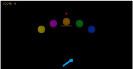
It seems that only two studies have used HG and EA among healthy individuals to try to improve timing (Milot et al., 2010; Bouchard et al., 2015). A study carried out by Milot et al. (Milot et al., 2010) was conducted in order to evaluate and compare the efficacy of HG and EA for young healthy individuals (average age of 24 ± 2.7 years) for a computerized pinball-like timing task, where the goal was to hit as many targets as possible with the proper timing while using a robotic hand device (TAPPER). They found that both EA and HG were effective at improving timing, but the effectiveness of each robotic training was dependent on the participant’s initial skill level, where: those that were better at the task to begin with benefited most from EA, and those that were less-skilled at the task gained from HG. A study by Bouchard et al. (Bouchard et al., 2015) used the same training paradigm with a modified version of TAPPER, TEO, to provide the pinball-like timing task to 32 healthy seniors (average age of 68 ± 4 years). Contrary to Milot et al.’s study (Milot et al., 2010), the authors found that only HG significantly improved the seniors’ timing (11.7 ± 4.4 ms vs. 9.7 ± 3.4 ms, p = 0.049), and EA had a tendency towards worsening their performance (9.8 ± 3.8 vs. 11.4 ± 5.9 ms, p = 0.13) They also found a correlation between age and the seniors’ timing performance following EA, where the older the participants were, the more EA worsened their performance (r = -0.59, p = 0.008). The project for this thesis is an extension of the study by Bouchard et al. (Bouchard et al., 2015), where the aim was to determine the effects of HG and EA on timing accuracy for a chronic stroke population.
II.5 Summary
In sum, both EA and HG have been used to improve movement execution in healthy and stroke individuals. Moreover, for both healthy and stroke populations, most studies have looked at improving the spatial aspect of a task and not the temporal (e.g. timing) component. Very few studies have directly compared EA and HG trainings (Patton et al., 2006; van Asseldonk et al.,
2009; Milot et al., 2010; Heuer and Rapp, 2011; Marchal-Crespo et al., 2014; Bouchard et al., 2015) and only one has directly compared them among a chronic stroke population (Patton et al., 2006).
Chapter III III.1 Objectives
III.1.1 Primary Objective
The main objective of this study was to evaluate and compare the effectiveness of a single training session by HG and EA on the immediate improvement of performance for a timing task among individuals that had a stroke.
III.1.2 Secondary Objective
The second objective consisted of exploring if there was a relationship between the side of the stroke lesion and the participants’ timing accuracy following HG or EA training.
III.2 Hypotheses
III.2.1 Primary Hypothesis
Since both stroke survivors and older individuals have been found to have slower timings (Carnahan et al., 1996; Wishart et al., 2000; Daly et al., 2006; McAuley et al., 2006; van Dijk et al., 2007; Marchal-Crespo et al., 2010; Seidler et al., 2010; Pietschmann et al., 2011; Cumming et al., 2012; Turgeon and Wing, 2012; Bi and Wan, 2013; Hoogendam et al., 2014) and based on the study by Bouchard et al. (Bouchard et al., 2015) that was conducted on healthy seniors, that found that HG was more beneficial to learning than EA, it was expected that the individuals that had a stroke would also benefit more from HG.
III.2.2 Secondary Hypothesis
Since it is believed that the left side of the brain is responsible for processing timing (Kwon et al., 2007; Freitas et al., 2011), it was hypothesized that the participants that had a
lesion on the left side of the brain would have an impaired learning of timing, as compared to those that had their stroke in the right hemisphere.
Chapter IV: Materials and Methods
The materials and methods that were used in this study were heavily based on a previous study (Bouchard et al., 2015). Based on the results of this Master’s thesis, an article was recently published (Bouchard et al., 2016) and can be found in this document (Chapter V). This thesis will nonetheless explain the most pertinent points, notably the functioning of the robotic hand and the variables of interest.
IV.1 Variables, Instrument and Procedure
IV.1.1 Descriptive Variables
To characterize the participants, socio-demographic variables were collected (e.g. age, stroke type, time since stroke) as well as descriptive variables, consisting of: manual dexterity (Box and Block Test) (Mathiowetz et al., 1985), motor recovery of the affected upper limb by the Fugl-Meyer Stroke Assessment Scale (FMA) (Fugl-Meyer et al., 1975), grip strength (Jamar dynamometer) (Mathiowetz, 2002), and the subjects’ self-perceived quantity and quality of the use of their affected upper limb in daily activities (Motor Activity Log) (Uswatte et al., 2006).
The Box and Block Test (Mathiowetz et al., 1985) consists of transporting the most blocks from one partition to another in one minute. The number of blocks transported by each hand is compared. The upper extremity section of the Fugl-Meyer Stroke Assessment Scale (Gladstone et al., 2002) consists of evaluating the motor recovery of the affected upper limb during various movements in and out of synergistic patterns. A total score of 66 signifies a normal motor recovery score. The JAMAR dynamometer is used to measure grip strength where the average score of three trials (in kg) is calculated for both hands (Mathiowetz, 2002). In the Motor Activity Log test, the participant is asked to answer how often he or she uses his or her affected upper limb in daily activities (i.e. turning on a light switch), in relation to the activities that were carried out in the previous week (Uswatte et al., 2006). A 0 to 5 Likert scale is used to quantify the amount of use of the affected upper limb (0 being “Did not use my weaker arm” and 5 “Used my weaker arm as often as before the stroke”) as well as the quality of use of the
affected upper limb (0 being “The weaker arm was not used at all for that activity” and 5 “The ability to use the weaker arm for that activity was as good as before the stroke”). The scores for both the quantity and quality of use of the affected limb are averaged. All of these assessment tools were chosen because they had good psychometric qualities for a stroke population as shown in Table 1 (p. 27).
Table 1: Psychometric Qualities of the Clinical Tools used for Stroke Survivors
Variable Measuring Tool
Reliability Validity
Manual dexterity Box and Block Test*
(Mathiowetz et al., 1985)
Excellent test-retest reliability for affected (r = 0.98) and non-affected hand (r = 0.93)
(Chen et al., 2009)
Excellent concurrent validity with the Action Research Arm Test (ARAT) (r = -0.80 and r = -0.71 for pre and post test respectively (Lin et al., 2010) as well as excellent convergent validity with the ARAT (r = 0.95)
(Desrosiers et al., 1994)
Motor recovery of the affected upper limb Fugl-Meyer Stroke Assessment (Gladstone et al., 2002) Excellent Interrater (CCI= 0.86-0.99) and intrarater (0.98-0.99) reliability (Platz et al., 2005) Excellent Test-retest Reliability (CCI = 0.97). (Platz et al., 2005)
Good construct validity with Barthel Index (r= 0.67) among chronic stroke survivors
(Dettmann et al., 1987)
Excellent criterion validity with Motor Assessment total scores (Malouin et al., 1994)
Grip strength Jamar
Dynamometer* (Peolsson et al., 2001; Mathiowetz, 2002) Excellent intrarater reliability (CCI = 0.86-0.95) (Boissy et al., 1999) Excellent test-retest Reliability (ICC = 0.80-0.89) (Bertrand et al., 2007)
Good construct validity with FMA (r= 0.84)
(Boissy et al., 1999)
Level and quality of use of the upper affected limb in activities of daily living Motor Activity Log (Uswatte et al., 2005; Uswatte et al., 2006) Excellent internal consistency (Cronbach’s alpha of 0.88) (Uswatte et al., 2006) Excellent test-retest reliability (r= 0.61-0.85)
(van der Lee et al., 2004)
Excellent concurrent validity with Stroke Impact Scale Hand Function scores (r = 0.72)
(Uswatte et al., 2005)
IV.1.2 Independent Variable
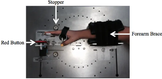
The independent variable was the type of robotic intervention that the participant received (EA or HG) during a computerized pinball game. To do so, Prof. Milot’s team at the Research Centre on Aging developed a robotic hand device, called Timing Exerciser Orthosis (TEO). TEO was used in a preliminary study by Bouchard et al. (Bouchard et al., 2015) among healthy seniors (see Figure 1).
Figure 1: Timing Exerciser Orthosis (TEO)
Furthermore, modified algorithms from Milot et al.’s study (Milot et al., 2010) were used in order to apply the HG and EA trainings in the current thesis. The same algorithms were used in Bouchard et al.’s study (Bouchard et al., 2015). More precisely, during HG training, the participant’s timing errors were decreased by delaying or speeding up TEO’s activation. For example, if the participant flexed their wrist too early, TEO would delay its activation, in order to decrease the person’s timing errors. On the contrary, timing errors were increased during EA. To illustrate, if the participant activated his or her wrist too late, TEO would delay its activation to increase the person’s timing errors even more.
More precisely, t = 0 represented when the red ball fell on the computer screen. The following formulas represent TEO’s activation (TBP):
1) TBP = TIP + DC
a. TIP was the time in which the motor sensors recognized that the participant moved his or her wrist;
b. DC was the programmed delay of when the participant made his or her wrist flexion, and that TEO moved.
The following formula includes the values that are needed in order to succeed at hitting the targets:
2) TBD = TID + DCD
a. TBD was the time needed for TEO to move and have the ball bounce up to hit the target;
b. TID was the time in which the participant should have flexed his or her wrist in order to be on time to hit the target;
c. DCD was a time constant (500 ms).
After, EP was equal to the participant’s timing error as to when he/she moved his or her wrist: 3) EP = TIP – TID
Next, EB was TEO’s timing error:
4) EB = TBP – TBD = EP + DC –DCD
Subsequently, it was expected that EB would be proportional to EP, where: 5) EB = kEP
a. k represented the EA gain.
More precisely, a k value that was greater than 1 resulted in an increase in timing errors, and a k value that was smaller than 1 provided a decrease in timing errors. The idea behind