Spectrum Inhibition of Virus Infectivity
Marie-Line Goulet1, David Olagnier2, Zhengyun Xu1,3, Suzanne Paz1, S. Mehdi Belgnaoui1, Erin I. Lafferty3, Vale´rie Janelle4, Meztli Arguello1, Marilene Paquet5, Khader Ghneim2, Stephanie Richards2, Andrew Smith2, Peter Wilkinson2, Mark Cameron2, Ulrich Kalinke6, Salman Qureshi3, Alain Lamarre4, Elias K. Haddad2, Rafick Pierre Sekaly2, Suraj Peri7, Siddharth Balachandran7, Rongtuan Lin1,3*, John Hiscott2*
1 Lady Davis Institute, Jewish General Hospital, McGill University, Montre´al, Canada, 2 Division of Infectious Diseases, Vaccine & Gene Therapy Institute of Florida, Port Saint Lucie, Florida, United States of America,3 Division of Experimental Medicine, McGill University, Montre´al, Canada, 4 Immunovirology Laboratory, INRS-Institut Armand-Frappier, Laval, Quebec, Canada,5 Comparative Medicine & Animal Resources Centre, McGill University, Montre´al, Canada, 6 Institute for Experimental Infection Research, TWINCORE, Hannover, Germany,7 Fox Chase Cancer Center, Philadelphia, Pennsylvania, United States of America
Abstract
The RIG-I like receptor pathway is stimulated during RNA virus infection by interaction between cytosolic RIG-I and viral RNA structures that contain short hairpin dsRNA and 59 triphosphate (59ppp) terminal structure. In the present study, an RNA agonist of RIG-I was synthesized in vitro and shown to stimulate RIG-I-dependent antiviral responses at concentrations in the picomolar range. In human lung epithelial A549 cells, 59pppRNA specifically stimulated multiple parameters of the innate antiviral response, including IRF3, IRF7 and STAT1 activation, and induction of inflammatory and interferon stimulated genes - hallmarks of a fully functional antiviral response. Evaluation of the magnitude and duration of gene expression by transcriptional profiling identified a robust, sustained and diversified antiviral and inflammatory response characterized by enhanced pathogen recognition and interferon (IFN) signaling. Bioinformatics analysis further identified a transcriptional signature uniquely induced by 59pppRNA, and not by IFNa-2b, that included a constellation of IRF7 and NF-kB target genes capable of mobilizing multiple arms of the innate and adaptive immune response. Treatment of primary PBMCs or lung epithelial A549 cells with 59pppRNA provided significant protection against a spectrum of RNA and DNA viruses. In C57Bl/6 mice, intravenous administration of 59pppRNA protected animals from a lethal challenge with H1N1 Influenza, reduced virus titers in mouse lungs and protected animals from virus-induced pneumonia. Strikingly, the RIG-I-specific transcriptional response afforded partial protection from influenza challenge, even in the absence of type I interferon signaling. This systems approach provides transcriptional, biochemical, and in vivo analysis of the antiviral efficacy of 59pppRNA and highlights the therapeutic potential associated with the use of RIG-I agonists as broad spectrum antiviral agents.
Citation: Goulet ML, Olagnier D, Xu Z, Paz S, Belgnaoui SM, et al. (2013) Systems Analysis of a RIG-I Agonist Inducing Broad Spectrum Inhibition of Virus Infectivity. PLoS Pathog 9(4): e1003298. doi:10.1371/journal.ppat.1003298
Editor: Ganes C. Sen, Cleveland Clinic, United States of America
Received September 12, 2012; Accepted February 24, 2013; Published April 25, 2013
Copyright: ß 2013 Goulet et al. This is an open-access article distributed under the terms of the Creative Commons Attribution License, which permits unrestricted use, distribution, and reproduction in any medium, provided the original author and source are credited.
Funding: This research was supported by grants from Canadian Institutes of Health Research (www.cihr-irsc.gc.ca/e/795.html; PAP 99022) and the Ministe`re du De´veloppement e´conomique, de l’Innovation et de l’Exportation (www.mdeie.gouv.qc.ca; PSR-SIIRI-297) to RL and JH. The funders had no role in study design, data collection and analysis, decision to publish, or preparation of the manuscript.
Competing Interests: The authors have declared that no competing interests exist. * E-mail: [email protected] (RL); [email protected] (JH)
Introduction
The innate immune system has evolved numerous molecular sensors and signaling pathways to detect, contain and clear viral infections [1–4]. Viruses are sensed by a subset of pattern recognition receptors (PRRs) that recognize evolutionarily con-served structures known as pathogen-associated molecular patterns (PAMPs). Classically, viral nucleic acids are the predominant PAMPs detected by these receptors during infection. These sensing steps contribute to the activation of signaling cascades that culminate in the early production of antiviral effector molecules, cytokines and chemokines responsible for the inhibition of viral replication and the induction of adaptive immune responses [2,5–7]. In addition to the nucleic acid sensing by a subset of endosome-associated Toll-like receptors (TLR), viral RNA structures within the cytoplasm are recognized by members
of the retinoic acid-inducible gene-I (RIG-I)-like receptors (RLRs) family, including the three DExD/H box RNA helicases RIG-I, Mda5 and LGP-2 [2,3,5,8–12].
RIG-I is a cytosolic multidomain protein that detects viral RNA through its helicase domain [13,14]. In addition to its RNA sensing domain, RIG-I also possesses an effector caspase activation and recruitment domain (CARD) that interacts with the mitochondrial adaptor MAVS, also known as VISA, IPS-1, Cardif [15,16]. Viral RNA binding alters RIG-I conformation from an auto-inhibitory state to an open conformation exposing the CARD domain, resulting in the generation of an activated state characterized by ATP hydrolysis and ATP-driven transloca-tion on RNA [17–19]. Activatransloca-tion of RIG-I also allows ubiquitina-tion and/or binding to polyubiquitin. In recent studies, poly-ubiquitin binding has been shown to induce formation of RIG-I tetramers that activate downstream signaling by inducing the
formation of prion-like fibrils composed of the MAVS adaptor [20]. MAVS then triggers the activation of IRF3 and NF-kB transcription proteins through the IKK-related serine kinases TBK1 and IKKe [10,21–23], leading to the primary activation of the antiviral program, involving production of type I interferons (IFNb and IFNa), as well as pro-inflammatory cytokines and antiviral factors [5,24,25]. A secondary response involving the induction of IFN stimulated genes (ISGs) is induced by the binding of IFN to its cognate receptor (IFNa/ bR), which triggers the JAK-STAT pathway to amplify the antiviral immune response [6,26–29].
The nature of the ligand recognized by RIG-I has been the subject of intense study given that these PAMPs are the initial triggers of the antiviral immune response. In vitro synthesized RNA carrying an exposed 59 terminal triphosphate (59ppp) moiety was first identified as RIG-I agonists [30–32]. The 59ppp moiety is present at the end of viral and self RNA molecules generated by RNA polymerization; however, in eukaryotic cells, RNA processing in the nucleus cleaves the 59ppp end and the RNA is capped prior to release into the cytoplasm. This mechanism distinguishes viral ‘non-self’ 59pppRNA from cellular ‘self’ RNA, and renders it recogniz-able to the innate RIG-I sensor [30,31,33]. Further character-ization of the RNA structure demonstrated that blunt base pairing at the 59 end of the RNA, with a minimum double strand (ds) length of 20 nucleotides was also important for RIG-I signaling [17,33,34]. Furthermore, short dsRNA (,300 bp) triggered RIG-I, whereas long dsRNA (.2000 bp) such as poly I:C and lacking 59ppp failed to trigger RIG-I, but was recognized by Mda5 [35].
Natural RNA extracted from virally infected cells, specifically the viral RNA genome or viral replicative intermediates, were also shown to activate RIG-I [30,31,36–38]. Interestingly, the highly conserved 59 and 39 untranslated regions (UTRs) of negative single strand RNA virus genomes display high base pair complementar-ity and the panhandle structure theoretically formed by the viral genome meets the requirements for RIG-I recognition [17]. The elucidation of the crystal structure of RIG-I highlighted the
molecular interactions between RIG-I and 59ppp dsRNA [18,39], providing a structural basis for the conformational changes involved in exposing the CARD domain for effective downstream signaling [18].
Given the level of molecular understanding of the RIG-I ligand and subsequent signaling leading to induction of antiviral immune response, we sought to investigate the range of the protective innate immune response triggered by RIG-I agonists against viral infections. A short in vitro-synthesized 59pppRNA derived from the 59 and 39 UTRs of the VSV genome activated the RIG-I signaling pathway and triggered a robust antiviral response that interfered with infection by several pathogenic viruses, including Dengue, HCV, H1N1 Influenza A/PR/8/34 and HIV-1. Furthermore, intravenous delivery of the RNA agonist stimulated an antiviral state in vivo that protected mice from lethal influenza virus challenge. This report highlights the therapeutic potential of naturally derived RIG-I agonists as potent stimulators of the innate antiviral response, with the capacity to mobilize genes essential for the generation of efficient immunity against multiple infections.
Results
59pppRNA stimulates an antiviral response in lung epithelial A549 cells
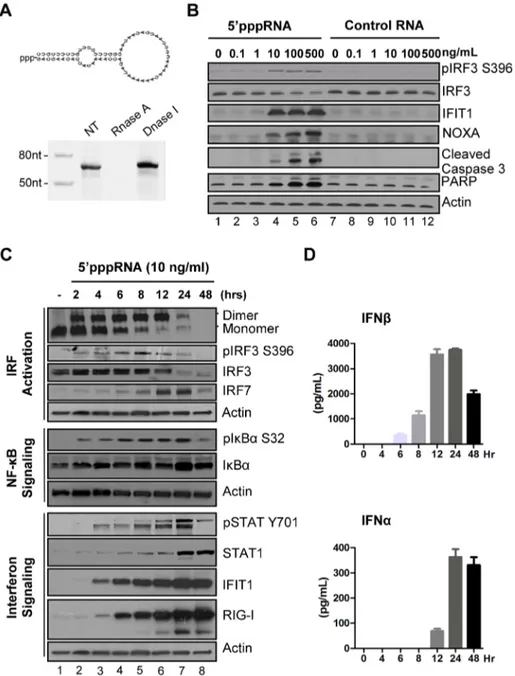
A 59 triphosphate containing RNA derived from the 59 and 39 UTRs of the negative-strand RNA virus Vesicular Stomatitis Virus (VSV) [17] was generated by in vitro transcription using T7 polymerase, an enzymatic reaction that synthesizes RNA mole-cules with a 59ppp terminus [17]. Predicted panhandle secondary structure of the 59pppRNA is depicted in Fig. 1A; gel analysis and nuclease sensitivity confirmed the generation of a single RNA product of the expected size (67 nucleotides). Transfection of increasing amounts of 59pppRNA resulted in Ser396 phosphor-ylation of IRF3 at 8 h – a hallmark of immediate early activation of the antiviral response (Fig. 1B, lane 2 to 6). Induction of apoptosis was detected following treatment with higher concen-trations of 59pppRNA; the pro-apoptotic BH3-only protein NOXA – a direct transcriptional target of IRF3 [40] – as well as cleavage products of caspase 3 and PARP were up-regulated in a dose dependent manner (Fig. 1B, lane 2–6). Optimal induction of antiviral signaling with limited cytotoxicity was achieved at a concentration of 10 ng/ml (,500 pM) (Fig. 1B; lane 4). Impor-tantly, the stimulation of immune signaling and apoptosis was dependent on the 59ppp moiety; a homologous RNA without a 59ppp terminus abrogated stimulation over a range of RNA concentrations (Fig. 1B, lane 8–12).
To characterize the antiviral response triggered by 59pppRNA, the kinetics of downstream RIG-I signaling were measured at different times (0–48 h) after stimulation of A549 cells (Fig. 1C). IRF3 homodimerization (1st panel) and IRF3 phosphorylation at Ser396 (2ndpanel) were detected as early as 2 h post treatment with 59pppRNA, and sustained until 24 h. Using a newly characterized anti-IRF7 antibody, induction of endogenous IRF7 was detected with kinetics that was delayed compared to IRF3 activation (4th vs. 3rd panel). IkBa phosphorylation was likewise detected as early as 2 h post-treatment and was sustained in A549 cells (6th panel). Altogether, IRF3, IRF7 and NF-kB are required for optimal induction of the IFNb promoter [27]. JAK-STAT signaling was detected at 4 h with Tyr701 phosphorylation of STAT1 (9th panel), as well as later accumulation at 24 h (10thpanel). IFIT1 and RIG-I itself, were up-regulated 4 h post-treatment (11thand 12thpanel) whereas a second group of ISGs (STAT1 and IRF7;
Author Summary
Development of safe and effective drugs that inhibit virus replication remains a challenge. Activation of natural host defense using interferon (IFN) therapy has proven an effective treatment of certain viral infections. As a distinct variation on this concept, we analyzed the capacity of small RNA molecules that mimic viral components to trigger the host antiviral response and to inhibit the replication of several pathogenic human viruses. Using gene expression profiling, we identified robust antiviral and inflammatory gene signatures after treatment with a 59-triphosphate containing RNA (59pppRNA), including an integrated set of genes that is not regulated by IFN treatment. Delivery of 59pppRNA into lung epithelial cells in vitro stimulated a strong antiviral immune response that inhibited the multiplication of several viruses. In a murine model of influenza infection, inoculation of the agonist protected animals from a lethal challenge of H1N1 influenza and inhibited virus replication in mouse lungs during the first 24–48 h after infection. This report highlights the therapeutic potential of naturally derived RIG-I agonists as potent stimulators of the innate antiviral response, with the capacity to block the replication of diverse human pathogenic viruses.
4thand 10thpanel) was induced between 6 h and 8 h after agonist treatment. IFNb was detectable in cell culture supernatant as early as 6 h after 59pppRNA treatment with a substantial release (4000 pg/ml) that peaked at 12–24 h (Fig. 1D, top panel). IFNa release was detected later at 12 h, and remained high thereafter (400 pg/ml) (Figure 1D). Thus 59pppRNA triggers a full antiviral response as demonstrated by the activation of transcription factors IRF3, IRF7 and NF-kB, release of interferons, JAK/STAT pathway activation and induction of ISGs.
59pppRNA induction of the antiviral response requires an intact RIG-I pathway
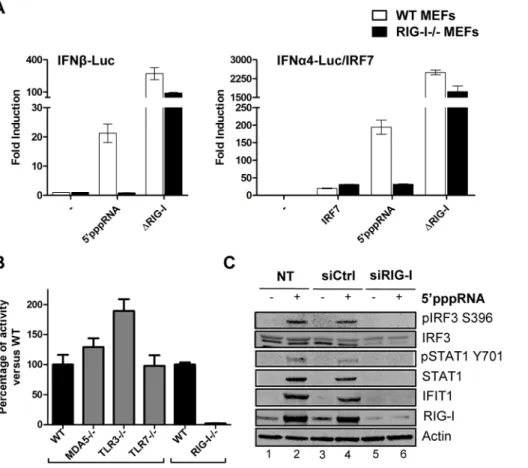
To address whether 59pppRNA exclusively activated the RIG-I sensor, wild type mouse embryonic fibroblasts (wt MEF) and RIG-I2/2 MEF were co-transfected with 59pppRNA and type 1 IFN reporter constructs to measure promoter activity. 59pppRNA activated the IFNb promoter 60-fold and the IFNa promoter 450-fold in wt MEF; promoter activity was dependent on RIG-I since these promoters were not stimulated in RIG-I2/2 MEF. As
Figure 1. 59pppRNA stimulates an antiviral and inflammatory response in lung epithelial A549 cells. (A) Schematic representation of VSV-derived 59pppRNA and gel analysis. The 59ppp-containing 67-mer RNA oligonucleotide is derived from the untranslated regions (UTRs) of VSV and the product of in vitro transcription runs as a single product degraded by RNase I. (B) 59pppRNA or a homologous control RNA lacking a 59-triphosphate end was mixed with Lipofectamine RNAiMax and transfected at different RNA concentrations (0.1–500 ng/ml) into A549 cells. At 8 h post treatment, whole cell extracts (WCEs) were prepared, resolved by SDS-page and analyzed by immunoblotting for IRF3 pSer396, IRF3, ISG56, NOXA, cleaved caspase 3, PARP and b-actin. Results are from a representative experiment; all immunoblots are from the same samples. (C) A549 cells were transfected with 10 ng/ml 59pppRNA and WCEs were prepared at different times after transfection (0–48 h), subjected to SDS-PAGE and probed with antibodies for IRF3 pSer-396, IRF3, IRF7, STAT1 pTyr-701, STAT1, ISG56, RIG-I, IkBa pSer-32, IkBa and b-actin; all immunoblots are from the same samples. To detect IRF3 dimerization, WCEs were resolved by native-PAGE and analyzed by immunoblotting for IRF3. (D) ELISA was performed on cell culture supernatants to quantify the release of IFNb and IFNa over time. Error bars represent SEM from two independent samples.
positive control, a constitutively active, CARD domain-containing RIG-I mutant [41] was used to bypass the requirement for RIG-I (Figure 2A). Furthermore, induction of the IFN response was exclusively dependent on intact RIG-I signaling, since IFNb promoter activity was not decreased in Mda52/2, TLR32/2 or TLR72/2 MEFs (Figure 2B). In A549 cells treated with 59pppRNA, knocking down RIG-I abolished IRF3 and STAT1 phosphorylation, as well as IFIT1 and RIG-I upregulation compared to control siRNA-treated cells (Fig. 2C; lane 6 vs. 4). Of note, generation of the knock down by transient transfection of short RNA did not activate immune signaling (Fig. 2C; lane 3 vs. 1). Hence, this 59pppRNA signals specifically via RIG-I.
Kinetics of the host response to 59pppRNA
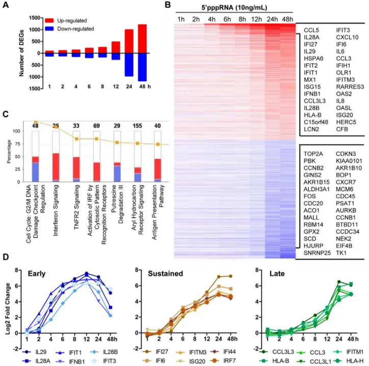
To evaluate the breadth of the host intrinsic response resulting from RNA agonist stimulation of RIG-I, modulation of the transcriptome of A549 cells stimulated with 59pppRNA from 1 to 48 h was analysed by gene array using the Illumina platform. Figure 3A shows a waterfall plot of differentially expressed genes (DEG; selected based on fold change $62, p-value #0.001) after
59pppRNA stimulation. The number of genes up-regulated by 59pppRNA administration steadily increased with time, while the majority of down-regulation occurred at 24–48 h (Fig. 3A). The heatmap presents DEG with emphasis on the most highly deregulated genes over time (Fig. 3B). Canonical pathway analysis using Ingenuity Pathway Analysis software identified IFN signal-ing, activation of IRFs by cytosolic PRRs, TNFR2 signaling and antigen presentation as the main up-regulated functional catego-ries, while functions related to cell metabolism and cell cycle were down-regulated by RIG-I agonist treatment (Fig. 3C). Subsequent kinetic analysis revealed that RIG-I agonist induced distinct temporal patterns of gene expression (Fig. 3D, S1 and S2A). For example, some genes were highly expressed early at 6–12 h, including IFNB1 and the IFNl family (IL29, IL-28A, IL28B), but the expression of these genes was not sustained throughout the time course and decreased at 24–48 h (Fig. 3D; lest panel). A second subset of genes associated with the antiviral response were induced early at 6–8 h, but expression was sustained and markedly augmented at 24–48 h, as exemplified by IFI family members, IRF7, and other ISGs (Fig. 3D; middle panel). A third subset of
Figure 2. Induction of the interferon response by 59pppRNA is dependent on functional RIG-I signaling. (A) WT and RIG-I2/2 MEFs were co-transfected with IFNa4 or IFNb promoter reporter plasmid (200 ng) along with 59pppRNA (500 ng/ml) or expression plasmids encoding a constitutively active form of RIG-I (DRIG-I) (100 ng). IRF-7 expression plasmid (100 ng) was added for transactivation of the IFNa4 promoter. Luciferase activity was analyzed 24 h post-transfection by the Dual-Luciferase Reporter assay. Relative luciferase activity was measured as fold induction relative to the basal level of reporter gene. Error bars represent SEM from nine replicates performed in three independent experiments. (B) Mda52/2, TLR32/2, TLR72/2 and RIG-I2/2 MEFs were co-transfected with IFNb promoter reporter plasmid (200 ng) along with 59pppRNA (500 ng/ml). Luciferase activity was analyzed 24 h post-transfection by the Dual-Luciferase Reporter assay. Relative luciferase activity was measured as fold induction relative to the basal level of reporter gene. Promoter activity in the knockout MEFs was then normalized against the activity in their respective wt MEFs to obtain the percentage of fold activation. Error bars represent SEM from nine replicates performed in three independent experiments. (C) A549 cells were either left untreated or transfected with a control siRNA or RIG-I siRNA. After 48 h, 59pppRNA (10 ng/mL) was transfected and at 8 h after treatment, WCEs were analyzed by SDS-PAGE and immunoblotted for pIRF3 Ser-396, IRF3, pSTAT1 Tyr 701, STAT1, IFIT1, RIG-I, and b-Actin. Results are from a representative experiment; all immunoblots are from the same samples.
doi:10.1371/journal.ppat.1003298.g002
genes was induced primarily at later time points, as part of the secondary response to 59pppRNA treatment, and included HLA and CCL3 families (Fig. 3D; right panel). Representative genes from different subsets were validated by quantitative real-time RT-PCR (Fig. S1). Overall, 59pppRNA induced a robust bi-phasic transcriptional response, characterized by strong activation of antiviral and inflammatory gene signatures; the kinetics of the transcriptional profile mirrored the biochemical activation events detected in Figures 1.
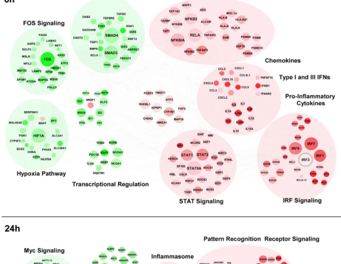
In order to gain systems-wide insight into the RIG-I transcriptome, a functional clustering of 59pppRNA-induced DEGs was performed. This functional clustering identified a variety of transcriptional sub-networks and biological processes regulated by RIG-I (Fig. 4). As expected, at 6 h (Fig. 4A), induction of antiviral and inflammatory response programs downstream of IRF, NF-kB, STAT signaling were identified (Fig. 4A and S2B); expression of several cytokine and chemokine genes were also up-regulated. Concomitantly, genes related to Fos
Figure 3. Transcriptome analysis of the host antiviral response to 59pppRNA. A549 cells were transfected with 10 ng/ml of 59pppRNA using Lipofectamine RNAiMax for designated periods of time. Samples were analyzed by Illumina gene expression array and DEG were identified based on fold change $62 and p-value #0.001 (A) Number of up-regulated and down-regulated DEG at each time point. (B) Heatmap of all DEG sorted by fold change; top 30 genes are listed. Red, Up-regulated; blue, down-regulated. (C) Functional characterization of DEGs following 59pppRNA treatment based on Ingenuity Pathway Analysis software. Bar height refers to the number of DEG in each pathway and the color refers to the contribution from up-regulated or down-regulated genes. (D) Genes among the top up-regulated genes were selected based on three different expression patterns: early, sustained, late.
Figure 4. Functional characterization of genes differentially expressed by RIG-I. The 6 h (A) and 24 h (B) time points from the kinetic analysis were selected to perform a functional classification of the up-regulated (red) and down-regulated (green) genes by 59pppRNA. Genes with a fold change $62 and p-value #0.001 were clustered using IPA. The intensity of the color is representative of the fold change. Larger circles indicate a transcription factor.
doi:10.1371/journal.ppat.1003298.g004
and TGF-b signaling, as well as hypoxic signaling via HIF-1a were down-regulated. At 24 h (Fig. 4B), genes associated with pathogen recognition receptor signaling, the ubiquitin pathway, inflamma-tion and apoptosis were also induced by RIG-I activainflamma-tion; interestingly, profiling of down-regulated genes identified func-tional clusters involved in cell cycle regulation, MYC signaling, and the heat shock response.
Comparative analysis of the gene expression profile in response to 59pppRNA and IFNa-2b
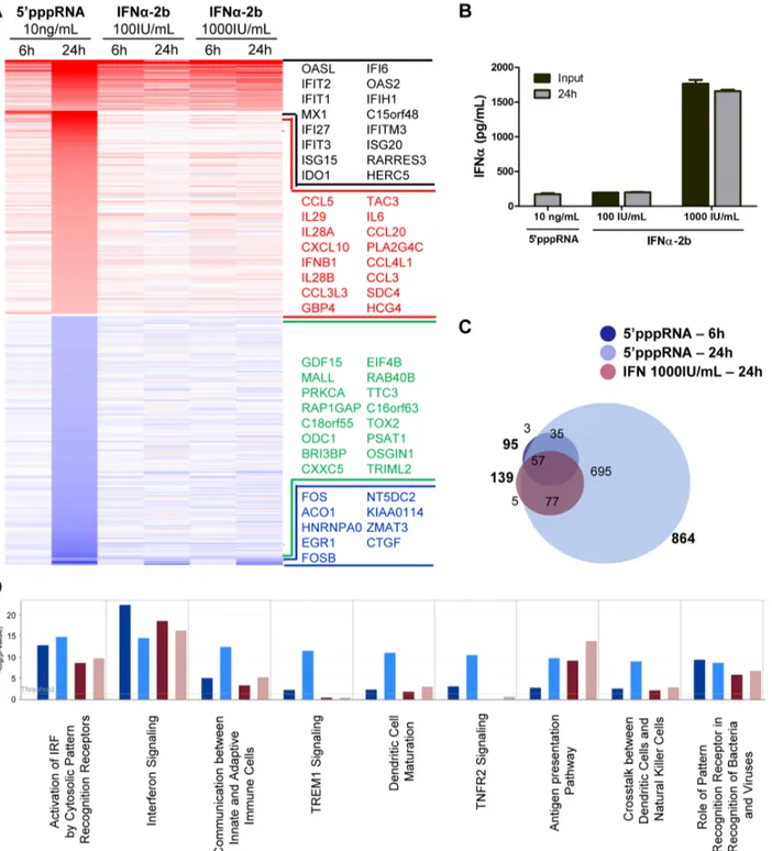
Although the contribution of type I IFN to the antiviral response stimulated by 59pppRNA is unquestionable, other factors may also augment the antiviral state established by 59pppRNA. To define gene uniquely induced by 59pppRNA, gene expression profiles of A549 cells stimulated with 59pppRNA or with IFNa-2b for 6 and 24 h were compared. The heatmap in Figure 5A displays the expression profile of genes differentially up- and down-regulated (fold change $2; p-value #0.001) by 59pppRNA and IFN (black and blue genes) or exclusively by 59pppRNA (red and green genes). Interestingly, 59pppRNA induced a significantly broader gene expression program compared to IFNa-2b, especially at 24 h. To determine whether differences in gene expression observed between 59pppRNA and IFNa-2b were due to sub-optimal stimulation by 2b, higher concentrations of IFNa-2b within the range reported for in vitro applications were tested. Treatment with 59pppRNA at 10 ng/ml was equivalent to treatment with IFNa-2b at 100 IU/ml in terms of IFNa levels released into cell culture supernatant. Increasing the concentration of IFNa-2b to 1000 IU/ml corresponded to levels of IFNa that were 8-times greater than physiological secretion following 10 ng/ ml 59pppRNA treatment (2500 pg/ml vs 480 pg/ml; Fig. 5B). Regardless of the amount of IFNa-2b used to activate cells, gene expression levels remained relatively unchanged (Fig. 5A; IFNa-2b 100 IU/ml vs 1000 IU/ml), indicating that IFNa-2b treatment was saturating.
Remarkably, the spectrum of genes differentially expressed by IFNa-2b treatment were virtually all contained within the transcriptome induced by 59pppRNA treatment - 57 at 6 h and 134 at 24 h, out of 139 genes (Fig. 5C) - demonstrating that 59pppRNA induced a complete IFN response by 24 h. Some of the genes characteristically activated as part of the IFN signature and maximally induced by 59pppRNA are MX1, IFIT1, ISG15 (Fig. 5A, black portion). This comparison also highlighted the fact that a surprisingly large number of genes are uniquely regulated by 59pppRNA - 38 genes at 6 h and 730 genes at 24 h (Fig. 5C). Most notably, IFNB1 and all three members of the IFNl family, as well as the cytokines CCL5, CXCL10, IL-6, and CCL3, were highly induced by 59pppRNA but not IFNa-2b treatment (Fig. 5A; red genes). While IFN signaling was highly induced by both treatments, IFNa-2b strongly activated antigen presentation machinery, and 59pppRNA preferentially stimulated dendritic cell maturation and crosstalk, linking innate and adaptive immunity as well as induction of a wider range of signaling pathways (Fig. 5D). Significantly, 59pppRNA preferentially stimulated a more exten-sive induction of IRF7 and NF-kB signaling nodes compared to IFNa-2b treatment (Fig. S2C). Thus, 59pppRNA treatment, besides inducing a complete IFN response, additionally stimulated the transcription of a large and unique set of inflammatory and antiviral genes.
59pppRNA acts as a broad-spectrum antiviral agent
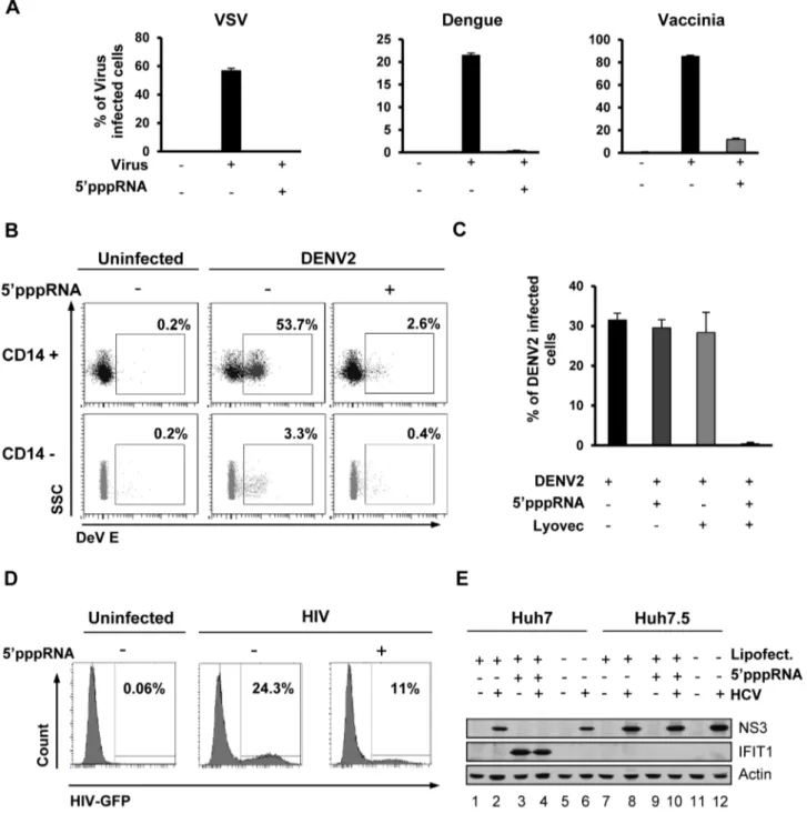
To determine if the RIG-I agonist was capable of inducing a functional antiviral response, A549 cells were treated with 59pppRNA, and 24 h later, challenged with VSV, Dengue
(DENV), or Vaccinia viruses. All viruses established infection in untreated cells as assessed by flow cytometry (60%, 20% and 80%, respectively) but in 59pppRNA-treated cells, VSV and DENV infectivity was reduced to ,0.5%, while infection with vaccinia was reduced to 10% (Fig. 6A). Release of infectious VSV and DENV virus was completely blocked by 59pppRNA treatment (1.76109and 4.36106PFU/mL in untreated cells, respectively vs. undetectable in treated cells; Fig. S3). Similarly, in primary human CD14+ monocytes, DENV infection decreased from 53.7% to 2.6% in the presence of 59pppRNA; in the CD142 fraction, DENV infectivity was lower (3%), but was likewise inhibited by RNA agonist treatment (0.4%; Fig. 6B). To demonstrate the requirement for intracellular delivery of 59pppRNA, primary CD14+ cells from three patients were treated with 59pppRNA alone, transfection reagent alone or the combination; DENV infectivity was reduced from ,30% to ,0.5% only upon transfection of the RNA agonist (Fig. 6C). To evaluate the antiviral effect of 59pppRNA against HIV infection, activated CD4+ T cells were pre-treated with supernatant isolated from 59pppRNA-treated monocytes and then infected with HIV-GFP. In the absence of treatment, 24% of the activated CD4+ T cells were actively infected by HIV as determined by GFP expression by flow cytometry, whereas infection of CD4+ T cells treated with 59pppRNA supernatants was reduced to 11% (Fig. 6D). 59pppRNA also had an antiviral effect against HCV in hepato-cellular carcinoma cell line Huh7; HCV NS3 expression was inhibited by 59pppRNA treatment (Fig. 6E; lane 4 vs. 2 and 6). The antiviral effect was fully dependent on RIG-I, as demonstrat-ed in Huh7.5 cells (that express a mutatdemonstrat-ed inactive RIG-I) by the absence of IFIT1 up-regulation following 59pppRNA treatment (Fig. 6E; lane 9) and NS3 expression comparable to untreated HCV-infected cells (Fig. 6E; lane 10 vs. 8 and 12). Thus, 59pppRNA is a broad-spectrum antiviral agent able to trigger an efficient innate immune response in different cell types and prevent infection by RNA and DNA viruses.
59pppRNA inhibits H1N1 Influenza infection in vitro
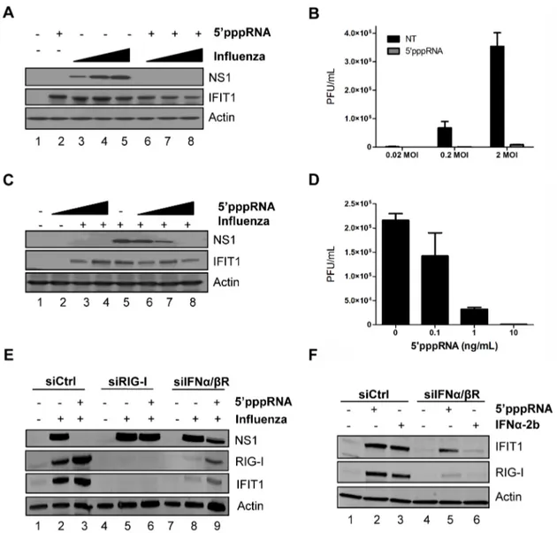
To further explore the inhibitory potential of 59pppRNA, A549 cells were pre-treated with 59pppRNA for 24 h and then infected with H1N1 A/PR/8/34 Influenza virus at increasing MOI (0.02, 0.2, 2); influenza replication was monitored by NS1 protein expression (Fig. 7A) and plaque assay (Fig. 7B). Viral replication was blocked by 59pppRNA pre-treatment, even at the highest MOI, as demonstrated by complete loss of NS1 expression and 40-fold decrease in viral titer at MOI 2. In A549 cells pre-treated with decreasing concentrations of 59pppRNA (10–0.1 ng/ml) prior to influenza virus challenge (0.2 MOI), 59pppRNA significantly blocked influenza replication at a concentration of 1 ng/ml, as demonstrated by a 3-fold reduction in NS1 protein expression (Fig. 7C; lane 7) and a 7-fold reduction in virus titer by plaque assay (Fig. 7D).
To demonstrate that the antiviral activity of 59pppRNA against influenza relies on RIG-I signaling, A549 cells were knocked down for RIG-I and infected with influenza; in the knockdown, ISGs were not induced (Fig. 7E, lanes 3 vs. 6) and 59pppRNA treatment failed to inhibit NS1 expression (Fig. 7E; lanes 5 vs. 6), indicating that the antiviral effect of 59pppRNA is exclusively dependent on RIG-I. Next, to determine whether the RIG-I ‘unique’ gene expression profile characterized in Figure 5 could compensate for the IFN response, A549 cells were knocked down for the IFNa/ bR; the knock down was efficient, as demonstrated by the absence of IFIT1 and RIG-I induction following IFNa-2b stimulation (Fig. 7F; lane 6). Interestingly, induction of ISGs was only partially reduced following 59pppRNA treatment (2.2-fold reduction of
Figure 5. Gene expression profiling of differentially expressed genes in response to 59pppRNA and IFNa-2b. A549 cells were transfected with 10 ng/ml of 59pppRNA using Lipofectamine RNAiMax or treated with IFNa-2b (100 IU/ml or 1000 IU/ml). Samples were collected at 6 h or 24 h post-treatment and were analyzed by Illumina gene expression array. Genes with a fold change $62.0 and p-value #0.001 were considered differentially expressed. (A) Heatmap showing top DEG affected by 59pppRNA and IFNa-2b treatments. Genes regulated by both 59pppRNA and IFNa-2b in at least one condition are indicated in black (up-regulated) or blue (down-regulated). Genes uniquely induced by 59pppRNA in at least one time point but not by IFNa-2b in any conditions are highlighted in red (up-regulated) or green (down-regulated). Top genes are listed in each instance. (B) Cell culture supernatant was collected at the time of treatment with IFNa-2b (0 h; input), or at 24 h following 59pppRNA or IFNa-2b treatment and assayed by ELISA for multiple subunits of IFNa. Error bars represent SEM from two independent samples. (C) Surface proportional Venn diagram illustrating the magnitude of the response by 59pppRNA and IFNa-2b at 6 h and 24 h. Number of DEG is indicated in each area. (D) Comparison of genes induced by each treatment - 59pppRNA 6 h (dark blue); 59pppRNA 24 h (light blue); IFNa-2b (1000 IU/ml) 6 h (red); IFNa-2b (1000 IU/ml) 24 h (pink) - based on functional classification by Ingenuity Pathway Analysis.
doi:10.1371/journal.ppat.1003298.g005
IFIT1 vs. siCtrl; Fig. 7F; lane 5 vs. 2); this IFN-independent activation of innate signaling was sufficient to reduce viral NS1 expression by 2.4-fold (Fig. 7E; lane 9 vs. 8). Thus, in lung
epithelial A549 cells, 59pppRNA treatment can efficiently inhibit influenza H1N1 replication in a RIG-I-dependent manner and stimulate an antiviral and inflammatory response
Figure 6. 59pppRNA acts as a broad-spectrum antiviral agent. (A) A549 cells were transfected with 10 ng/ml 59pppRNA 24 h prior to infection with VSVD51-GFP (MOI 0.1), Dengue virus (MOI 0.1), and Vaccinia-GFP virus (MOI 5), respectively. Percentage of infected cells was determined 24 h post-infection by flow cytometry analysis of GFP expression (VSV-GFP and Vaccinia-GFP) or intracellular staining of DENV E protein expression (Dengue virus). Data are from a representative experiment performed in triplicate 6 SD. (B) Human PBMCs were transfected with 100 ng/ ml 59pppRNA 24 h prior to infection with dengue virus at an MOI of 5. At 24 h post-infection, the percentage of Dengue infected CD14+ and CD142 cells was evaluated by intracellular staining of DENV E protein expression by flow cytometry. Data are from a representative experiment performed in triplicate 6 SD. (C) Human PBMCs from three different donors were transfected with 100 ng/ml 59pppRNA prior to infection with Dengue virus at an MOI of 5. The percentage of Dengue infected cells in the CD14+ population was evaluated by intracellular staining of DENV E protein expression using flow cytometry. Data are from an experiment performed in triplicate on three different patients 6 SD (D) CD4+ T cells isolated from human PBMCs and activated with anti-CD3 and anti-CD28 antibodies. Cells were incubated in the presence or absence of supernatant from 59pppRNA-treated monocytes for 4 h and infected with HIV-GFP (MOI 0.1) for 48 h. The percentage of HIV infected, activated CD4+ T cells (GFP positive) was assessed by flow cytometry. (E) Huh7 and Huh7.5 were transfected with 59pppRNA (10 ng/mL) and infected with HCV 24 h later. At 48 h post-infection, WCEs were collected and subjected to SDS PAGE and immunoblot to examine the expression of HCV viral protein NS3, IFIT1, and b-Actin. doi:10.1371/journal.ppat.1003298.g006
independently of IFN signaling to limit influenza infection in vitro.
59pppRNA activates innate immunity and protects mice from lethal influenza infection
To determine the potential of 59pppRNA in vivo, C57Bl/6 mice were inoculated intravenously with 59pppRNA (25mg) in complex with the in vivo-jetPEI transfection reagent. 59pppRNA stimulated a potent immune response in vivo characterized by IFNa and IFNb secretion in the serum and lungs (Fig. S4A) as well as antiviral gene up-regulation (Fig. S4B). The response was potent and rapid with serum IFNb levels increased ,20-fold compared to basal levels, as early as 6 h post administration (Fig. S4A; top left panel). The immune activation observed in vivo correlated with an early and
transient recruitment of neutrophils to the lungs along with a more sustained increase in macrophages and dendritic cells populations (Fig. S4C).
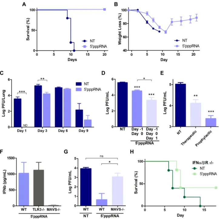
Next, to determine the antiviral potential of 59pppRNA in vivo, mice were treated with 59pppRNA 24 h before (day 21), and on the day of infection (day 0) with a lethal inoculum of H1N1 A/ PR/8/34 Influenza. Whereas all untreated, infected mice succumbed to infection by day 11, all 59pppRNA-treated mice fully recovered (100% survival) (Fig. 8A). Overall, weight loss was similar between the two groups (Fig. 8B), although a noticeable delay of 2–3 days in the onset of weight loss was observed in 59pppRNA-treated animals; treated mice then fully recovered within 12–14 days (Fig. 8B). Influenza replication in the lungs was monitored by plaque assay over the course of infection with virus
Figure 7. 59pppRNA inhibits H1N1 Influenza replicationin vitro. (A) A549 cells were treated with 59pppRNA (10 ng/ml); at 24 h post-treatment, cells were infected with an increasing MOI of A/PR8/34 H1N1 Influenza virus (0.02MOI, 0.2MOI, 2MOI) for 24 h. WCEs were subjected to SDS PAGE and immunoblot to examine the expression of influenza viral protein NS1, ISG56, and b-Actin. (B) Viral titers in cell culture supernatants from (A) were determined by plaque assay. Error bars represent SEM from two independent samples. (C) A549 cells were pre-treated with increasing concentrations of 59pppRNA (0.1 ng/ml to 10 ng/ml) for 24 h, prior to 0.2MOI influenza challenge. WCEs were examined for influenza NS1, ISG56, b-Actin by immunoblot. (D) Viral titers in cell culture supernatants from (C) were determined by plaque assay. Error bars represent SEM from two independent samples. (E) A549 cells were transfected with a control siRNA, RIG-I siRNA or IFNa/bR siRNA and then treated with 59pppRNA (10 ng/ml), followed by infection with Influenza (0.2MOI). WCEs were prepared 24 h later and subjected to SDS PAGE and immunoblot to examine the expression of influenza NS1, RIG-I, IFIT1, and b-Actin. (F) A549 cells were transfected with a control siRNA or IFNa/bR siRNA and then treated with 59pppRNA (10 ng/ml) or IFNa-2b (100 IU/mL) for 24 h. Expression of IFIT1, RIG-I and b-Actin was evaluated by western blotting.
doi:10.1371/journal.ppat.1003298.g007
titers in the lungs of untreated mice reaching a maximum at day 3 post-infection (Fig. 8C). A decrease in virus titer was noted by day 9 post-infection, possibly correlating with the onset of adaptive immunity, and all animals succumbed to influenza infection by day 11 (Fig. 8A). Interestingly, 59pppRNA treatment inhibited influenza virus replication in the lungs early after infection, within the first 24–48 h (Fig. 8C; Day 1); by day 3, virus titers in the lung had increased, although influenza titers were still ,10-fold lower compared to titers in untreated mice (Fig. 8C; Day 3). By day 9, the 59pppRNA-treated animals had controlled the infection, as demonstrated by the decrease in viral titers. Continuous admin-istration of 59pppRNA at 24 h intervals post-infection had an additive therapeutic effect that further delayed viral replication (Fig. 8D; 3 versus 2 doses of 59pppRNA), indicating that antiviral immunity may be sustained with repetitive administration of 59pppRNA. Furthermore, therapeutic administration of 59pppRNA also controlled influenza viral replication; although prophylactic treatment was most effective at blocking influenza dissemination in the lung, administration of the RNA agonist on day 1 and day 2 after influenza infection also reduced viral lung titers by ,10-fold (Fig. 8E).
The antiviral response triggered by 59pppRNA in vivo was dependent on an intact RIG-I signaling; serum IFNb release was abolished in MAVS2/2 mice, whereas the absence of TLR3 did not affect 59pppRNA-induced IFNb release (Fig. 8F). In agree-ment, MAVS2/2 mice treated with 59pppRNA did not control influenza lung titers (5-fold increase vs. wt mice) and the titer was comparable to untreated wt mice (Fig. 8G). To determine whether 59pppRNA treatment was sufficient to protect against influenza in the absence of IFN signaling, IFNa/bR2/2 mice were treated or not with 59pppRNA and challenged with influenza H1N1 virus. While untreated IFNa/bR2/2 animals succumbed to infection, 40% of the animals that received 59pppRNA treatment survived, suggesting that an IFN-independent effect of 59pppRNA func-tioned in the absence of the IFN response. Thus, intravenous administration of 59pppRNA stimulated a potent and rapid immune response in vivo that delayed influenza H1N1 virus replication in the lungs of infected animals and rescued mice from a lethal inoculation of influenza H1N1.
59pppRNA treatment limits influenza-mediated pneumonia
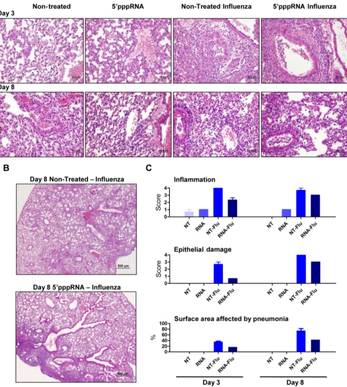
To further evaluate the effect of RNA agonist administration on influenza-mediated pathology, histological sections of lungs from untreated and treated mice were prepared and analysed. 59pppRNA treatment alone was characterized by a modest and rare leukocyte-to-endothelium attachment; mixed leukocyte pop-ulations (mononuclear/polymorphonuclear) infiltrated the peri-vascular space at 24 h after injection (data not shown) but the infiltration resolved and was limited to endothelial cell attachment at 3 and 8 days after intravenous administration (Fig. 9A). Influenza virus infection induced severe and extensive inflamma-tion and oedema in the perivascular space and the bronchial lumen at day 3 post-infection. In animals receiving the RNA agonist, influenza triggered a mild and infrequent inflammation that did not extend to the bronchial lumen at day 3 post-infection. Epithelial degeneration and loss of tissue integrity were more severe in the lungs of untreated, infected animals and correlated with epithelial hyperplasia observed at later times, compared to the lungs of animals treated with 59pppRNA. Inflammation and epithelial damage progressed in untreated mice by day 8 (Fig. 9E), and correlated with increased virus titer in the lungs (Fig. 8C); inflammation and epithelial damage was consistently less apparent in agonist-treated mice. Strikingly, the surface area of the lungs
affected by pneumonia was significantly reduced in 59pppRNA-treated mice compared to non-59pppRNA-treated mice – on day 3, 16% vs 35%; day 8, 41% vs 73% (Fig. 9C; bottom panel). Overall, influenza-mediated pneumonia was less severe in animals admin-istered 59pppRNA before influenza challenge, demonstrating that 59pppRNA possesses an antiviral effect in vivo that limits influenza replication in the lung, limits lung damage and prevents influenza-mediated pneumonia and mortality.
Discussion
RIG-I agonists are attractive potential antiviral agents, as triggering the innate cytosolic RIG-I pathway mimics the earliest steps of immune recognition and response to viral pathogens. In the present study, a short 59pppRNA agonist of RIG-I derived from the 59 and 39 UTRs of the VSV genome stimulated an antiviral response that protected human lung epithelial A549 cells or human PBMCs from challenge with several viruses, including DENV, Influenza, HIV, VSV, HCV and Vaccinia virus. Intravenous administration of the 59pppRNA agonist in mice stimulated an antiviral state in vivo that protected animals from lethal influenza virus challenge and controlled influenza virus-mediated pneumonia. Analysis of the dynamics of the host transcriptome following 59pppRNA stimulation was characterized by antiviral and inflammation related gene expression patterns with transcriptional nodes of genes regulated by IRF, NF-kB, and STAT families. Virtually all of the genes activated by IFNa-2b were encompassed within the 59pppRNA transcriptome; bioinfor-matics analysis also identified distinct gene patterns and functional processes that were uniquely induced or inhibited by 59pppRNA. Because of its potency both in vitro and in vivo, 59pppRNA represents a specific and powerful trigger of innate immunity and a novel approach to antiviral therapy.
For the first time, an RNA-based agonist of RIG-I was shown to block the replication of multiple viruses; this broad-spectrum antiviral activity of 59pppRNA is attributable in part to a potent stimulation of the inflammatory and antiviral response driven by the early induction of IRF, NF-kB, STAT, chemokines and pro-inflammatory cytokine genes. In parallel, we also observed an inhibition of genes involved in TGF-b signaling. Because of the immunosuppressive nature of the TGF-b pathway [42], inhibition of this transcriptional node could further potentiate immune activation in response to 59pppRNA agonist. The emergence of apoptosis and ubiquitin signaling nodes at later times (24 h) suggests a role for cell death and ubiquitin-based signal modification in the antiviral response.
59pppRNA stimulation of RIG-I triggered a complete IFN response. At 24 h post treatment, 59pppRNA induced the expression of 97% of the genes stimulated by IFNa-2b treatment and the magnitude of ISG induction by 59pppRNA was enhanced compared to IFNa-2b profile. Among the ISGs, the tripartite motif containing (TRIM) proteins, the IFITM proteins, MX1 and viperin exemplify the range of ISGs induced by RIG-I and all have been implicated as inhibitors of HIV-1, Influenza, VSV, West Nile, Dengue, and HCV [43–53]. In a recent study, high throughput screening of antiviral effectors identified a panel of broadly acting antiviral molecules, with the combined expression of multiple ISGs providing additive inhibitory effects against HCV replication [29]. Of the 28 validated antiviral ISGs identified, 19 were induced by 59pppRNA in A549, including IRF1, RIG-I, Mda5, IFITM3.
The transcriptome analysis also identified a distinct subset of 968 genes specifically induced by 59pppRNA and not IFNa2b -that additively or synergistically enhanced the antiviral response
stimulated by 59pppRNA treatment. Bioinformatics analysis identified a unique functional signature with up-regulated genes involved in inducing a wider range of signaling pathways and bridging innate and adaptive immune responses. The importance
of genes uniquely induced by 59pppRNA is highlighted by the antiviral response that limited influenza infection in vitro and in vivo, even in the absence of functional type I IFN signaling. We speculate that the extended range of genes induced by 59pppRNA,
Figure 8. 59pppRNA activates innate immunity and protects mice from lethal influenza infectionin vivo. C57Bl/6 mice were injected intravenously with 25 ug of 59pppRNA in complex with in vivo-jetPEI. (A–B–C) Mice were treated with 59pppRNA on the day prior to influenza infection (500 PFU; Day 21) and the day of infection (Day 0). Percent survival (A) and percent weight loss (B) were monitored. (C) Lung viral titers were measured by plaque assay at the indicated day post-infection. Error bars represent SEM from six different animals. ND: not detected. (D) Repetitive administration of 59pppRNA on the indicated days further decreases lung viral titers following infection with 500 PFU of influenza, as determined by plaque assay on Day 3 post-infection. Error bars represent SEM from five different animals. (E) Mice were infected with 50 PFU of influenza on Day 0. 59pppRNA was administered prophylactically (Day 21, Day 0) or therapeutically (Day 1, Day 2) and lung viral titers were determined on Day 3. Error bars represent SEM from five different animals. (F) WT, TLR32/2, MAVS2/2 mice were treated with 59pppRNA and serum IFNb was quantified by ELISA at 6 h. Error bars represent SEM from three different animals. (G) WT and MAVS2/2 mice were treated with 59pppRNA and infected with influenza (500 PFU). Lungs were collected and homogenized on Day 1 and lung viral titers were determined by plaque assay. Error bars represent SEM from four different animals. (H) IFNa/bR2/2 mice were either non-treated or treated with 59pppRNA and infected with influenza (100 PFU). Survival was monitored for 18 days. Statistical analysis was performed by Student’s t test (*, p#0.05; **, p#0.01; ***, p#0.001; ns, not statistically significant).
doi:10.1371/journal.ppat.1003298.g008
compared to IFNa-2b, reflect the activation of multiple signaling pathways downstream of RIG-I/MAVS [10], versus the more-limited transactivation potential of the IFN-regulated JAK-STAT axis [26].
Type III IFNs (IL29, IL28A, IL28B) were among the most highly stimulated genes uniquely up-regulated in response to 59pppRNA. Recent studies have demonstrated that both type I and type III IFNs activate similar components of the JAK-STAT pathways,
Figure 9. 59pppRNA treatment controls influenza-mediated pneumonia. Mice were treated with 59pppRNA in complex in vivo-jetPEI on the day prior to influenza infection (Day 21) and the day of infection (Day 0). Lungs were collected on Day 3 and Day 8 post-infection and stained with hematoxylin and eosin (H&E). For each group, a representative picture showing (A) inflammation and tissue damage and (B) the extent of pneumonia is presented. (C) Inflammation, tissue damage and surface area affected by pneumonia were scored by a veterinary pathologist. Grade 1 = minimal; Grade 2 = modest, rare; Grade 3 = moderate, frequent; Grade 4 = severe, extensive.
although type III IFNs were shown to prolong the activation of JAK-STAT signaling and induce a delayed and stronger induction of ISGs, compared to type I IFNs [54]. IFNls have been increasingly implicated in antiviral therapies: 1) IFNl administra-tion in mice stimulated expression of Mx1 and protected IFNaR2/2 mice from lethal influenza challenge [55]; 2) IL29 blocked HIV-1 replication by inhibiting virus integration and post-transcriptional events [56]; and 3) the combination of IL29 and IFNa or IL29 and IFNc enhanced the induction of antiviral genes and effectively inhibited HCV and VSV replication [57]. In addition, polymorphism in or near IFNl3 gene correlated with spontaneous or treatment-induced clearance of Hepatitis C infection and IFNl therapy is now actively investigated for the treatment of HCV [58]. Altogether these results indicate that specific induction of IFNl by 59pppRNA may contribute to immunity at the site of viral infection.
Of note, negative regulators of the innate immune response were also detected after 59pppRNA stimulation. In addition to the induction of SOCS1, USP18, and IFIT1 by both 59pppRNA and IFNa-2b treatment, unique negative regulators activated exclu-sively by 59pppRNA were also identified: SOCS3 contributes to the inhibition of the JAK/STAT signaling [59], and hence limits the amplification of the IFN response; A20 and IkBa inhibit the activation of the NFkB signaling complex [60,61], which would prevent excessive inflammation. This observation suggests that targeting an upstream viral sensor may provide activation as well as negative feed-back regulation to terminate the immune response and prevent uncontrolled inflammation; as such, this approach may offer an advantage over IFN therapy in terms of limiting potential toxic side-effects.
Activation of the RIG-I signaling pathway using 59pppRNA also induced an integrated set of genes and pathways that can efficiently bridge the innate and adaptive immune responses and utilize multiple arms of both systems. 59pppRNA mobilized genes that enhance trafficking of immune cells such as neutrophils, monocytes, naı¨ve and memory T cells and B cells, including CCL17, CCL20, CXCL10, CCL3, CCL5 and many others. We also observed the induction of genes important for the activation of the effector arm of the adaptive immune system such as IL-6, which has been shown to enhance CD8+ T cell survival and killing potential [62]. These cytokines and chemokines certainly play a role in initiating the innate and adaptive immune cell response, which is critical for the generation of efficient immunity against multiple viral infections in vivo.
Intravenous administration of the RIG-I agonist stimulated a potent immune response in vivo that reached the lungs and prevented mortality associated with virus challenge. Histopathol-ogy analysis revealed diminished influenza-mediated lung damage and recruitment of inflammatory cells in infected lungs following 59pppRNA treatment. The rapid control of virus replication, as demonstrated by significantly reduced virus titers in the lungs within 3 days of virus inoculation, may have prevented excessive immune cell recruitment early after infection. Influenza infection generates a complex pathogenesis mediated in part by viral- and immune-mediated damage [63]; therefore, the activation and recruitment of limited numbers of specific immune cell types, such as neutrophils, alveolar macrophages and dendritic cells may generate a beneficial antiviral microenvironment that additionally favor the initiation of adaptive immune response, which would eventually contribute to the in vivo efficacy of the agonist.
Other groups have recently reported that 59pppRNA induces a protective RIG-I mediated antiviral response that inhibits the replication of influenza virus [64–66]. The inhibition of influenza infection by 59pppRNA was dependent on the 59ppp moiety and
the secondary IFN response was crucial for mounting an effective antiviral response [64,65]. Recently, a short RNA molecule with dual functionality was developed - a siRNA against influenza NP gene and an agonist of the RIG-I pathway [66]. This 59pppRNA inhibited influenza infection in vitro and in vivo, but the contribution of RIG-I activation to the inhibition of influenza was not demonstrated. The defective interfering RNA produced during Sendai virus life cycle is the best characterized natural RIG-I ligand and is known to induce strong inflammatory response [67]. Interestingly, this molecule has adjuvant potential and could stimulate an antibody-dependent response directed to influenza antigens. Compared to these earlier studies, we adopted a systems approach to provide biochemical, transcriptional and biological mechanistic explanations for the antiviral efficacy observed in vitro and in vivo.
Activating natural host defense to prevent establishment and dissemination of viral infection is a valuable alternative strategy to antiviral drugs that specifically target viral processes. Interferon therapy has been used in the clinic for over two decades and has proven effective in the treatment of certain viral infections, mainly Hepatitis B and Hepatitis C [58], as well as malignancies and autoimmune diseases [68,69]. However, IFN therapy is also associated with significant side effects that limit its use [70]. PolyI:C, another dsRNA immune modulator, is also being tested in vitro and in vivo and has demonstrated efficacy against respiratory infections [71–73]. Along with antiviral drugs, vaccination is the primary approach to reduce morbidity and mortality associated with viral infection. Increasing the immunogenicity of vaccines with molecular adjuvants eliciting cytokines, co-stimulatory molecules, or immunomodulatory factors enhance the vaccine-elicited immune responses. 59pppRNA has the advantage of mimicking viral recognition to trigger an immune response analogous to natural viral infection. Furthermore, the response stimulated by 59pppRNA was reminiscent of the integrated and multipotent response elicited early following immunization by the most protective vaccine identified so far, the yellow fever YD17 vaccine [74]. Therefore, an immune modulator such as a RIG-I agonist may not only function as an antiviral therapeutic, but may also serve as a vaccine adjuvant to increase the magnitude of the antiviral immune response elicited by vaccine epitopes. Thus, the present study not only demonstrated the prophylactic and therapeutic antiviral potential of 59pppRNA, but also opens the door to further investigation of the potential of RIG-I agonists as vaccine adjuvants.
Materials and Methods In vitro synthesis of 59pppRNAs
The sequence of the 59pppRNA was derived from the 59 and 39 UTRs of the VSV genome as previously described [17]. In vitro transcribed RNA was prepared using the Ambion MEGAscript T7 High Yield Transcription Kit according to the manufacturer9s instruction (Invitrogen, NY, USA). The template consisted of two complementary viral sequences containing T7 promoter that were annealed at 95uC for 5 minutes and cooled down gradually over night (59-GAC GAA GAC AAA CAA ACC ATT ATT ATC ATT AAA ATT TTA TTT TTT ATC TGG TTT TGT GGT CTT CGT CTA TAG TGA GTC GTA TTA ATT TC-39). The in vitro transcription reactions proceeded for 16 hours. 59pppRNA was purified and isolated using the Qiagen miRNA Mini Kit (MD, USA). Homologous RNA without 59ppp moiety was purchased from IDT (Integrated DNA Technologies Inc, Iowa, USA); dephosphorylation of the 59pppRNA using CIAP (Invitrogen, NY, USA) generated identical results (data not
shown). Secondary structure was predicted using the RNAfold WebServer (University of Vienna, Vienna, Austria). RNA was analysed on a denaturing 17% polyacrylamide, 7 M urea gel following digestion with 50 ng/ul of RNase A (Ambion, CA, USA) or 100 mU/ul of DNase I (Ambion, CA, USA) for 30 min.
Cell culture, transfections, and luciferase assays
A549 were grown in F12K (Invitrogen, NY, USA) supplement-ed with 10% FBS and antibiotics. MEFs were grown in DMEM supplemented with 10% FBS, non-essential amino acids, and L-Glutamine (Wisent, Quebec, Canada). WT and RIG-I 2/2 MEFS were kind gifts from Dr. Shizuo Akira (Osaka University, Osaka, Japan) [75]. WT, Mda52/2, TLR32/2, TLR72/2 MEFS were kind gifts from Dr. Michael Diamond (Washington University, St Louis, USA) [76,77]. Lipofectamine RNAiMax (Invitrogen, NY, USA) was used for transfections in A549 according to manufacturer’s instructions. For luciferase assays, transfections were performed in wt and RIG-I2/2; wt and Mda52/2, TLR32/2, TLR72/2 MEFs using Lipofecta-mine 2000 (Invitrogen, New York, USA) or jetPRIME (PolyPlus, France), respectively. Plasmids encoding GFP-DRIG-I, IRF-7, pRLTK, IFNa4/pGL3 and IFNb/pGL3 were previously described [78]. MEFs were co-transfected with 200 ng pRLTK reporter (Renilla luciferase for internal control), 200 ng of reporter gene constructs, together with 59pppRNA (500 ng/ml) or 100 ng of a plasmid encoding a constitutively active form of RIG-I (DRIG-I) [41]. IRF7 plasmid (100 ng) was added for transactivation of IFNa4 promoter. At 24 h after transfection, reporter gene activity was measured by Dual-Luciferase Reporter Assay, according to manufacturer’s instruc-tions (Promega, Wisconsin, USA). Relative luciferase activity was measured as fold induction (relative to the basal level of reporter gene). For siRNA knock down, A549 cells were transfected with 50 nM (30 pmol) of human RIG-I (sc-61480), IFN-a/bR a (sc-35637) and b (sc-40091) chain, or control siRNA (sc-37007) (Santa Cruz Biotechnologies, Dallas, USA) using Lipofectamine RNAi Max (Invitrogen, NY, USA) according to the manufacturer’s guidelines. Treatment with 59pppRNA was performed 48 hrs later.
Immunoblot analyses
Whole cell extracts were separated in 8% acrylamide gel by SDS-PAGE and were transferred to a nitrocellulose membrane (BioRad, Mississauga, Canada) at 4uC for 1 h at 100 V in a buffer containing 30 mM Tris, 200 mM glycine and 20% methanol. Membranes were blocked for 1 h at room temperature in 5% dried milk (wt/vol) in PBS and 0.1% Tween-20 (vol/vol) and then were probed with primary antibodies: anti-pIRF3 at Ser396 (EMD Millipore, Massachusetts, USA), IRF3 (IBL, Japan), anti-RIG-I (EMD Millipore, Massachusetts, USA), anti-ISG56 (Thermo Fischer Scientific, Massachusetts, USA), anti-pSTAT1 at Tyr701 (Cell Signaling Technology, Inc, Massachusetts, USA), anti-STAT1 (Santa Cruz Biotechnology), anti-NS1(Santa Cruz Biotechnology), anti-pIkBa at Ser32 (Cell Signaling Technology, Inc, Massachusetts, USA), anti-IkBa (Cell Signaling Technology, Inc, Massachusetts, USA), anti-NOXA (EMD Millipore, Massa-chusetts, USA), anti-cleaved Caspase 3 (Cell Signaling Technol-ogy, Inc, Massachusetts, USA), anti-PARP (Cell Signaling Technology, Inc, Massachusetts, USA), anti-b-Actin (EMD Millipore, Massachusetts, USA). Antibody signals were detected by chemiluminescence using secondary antibodies conjugated to horseradish peroxidise and an ECL detection kit (Amersham Biosciences, Inc, NJ, USA)
IRF3 dimerization
Whole cell extracts were prepared in NP-40 lysis buffer (50 mM Tris, pH 7.4, 150 mM NaCl, 30 mM NaF, 5 mM EDTA, 10% glycerol, 1.0 mM Na3VO4, 40 mM b-glycerophosphate, 0.1 mM
phenylmethylsulfonyl fluoride, 5mg/ml of each leupeptin, pep-statin, and aproptinin, and 1% Nonidet P-40). WCE was then subjected to electrophoresis on 7.5% native acrylamide gel, which was pre-run for 30 min at 4uC. The electrophoresis buffers were composed of an upper chamber buffer (25 mM Tris, pH 8.4, 192 mM glycine, and 1% sodium deoxycholate) and a lower chamber buffer (25 mM Tris, pH 8.4, 192 mM glcine). Gels were soaked in SDS running buffer (25 mM Tris, pH 8.4, 192 mM glycine, 0.1% SDS) for 30 min at 25uC and were then transferred to nitrocellulose membrane (Amersham Biosciences). Membranes were blocked in PBS containing 5% milk (wt/vol) and 0.05% Tween-20 (vol/vol) for 1 h at 25uC and blotted with an antibody against IRF3 (IBL, Japan). Antibody signals were detected by chemiluminescence using secondary antibodies conjugated to horseradish peroxidise and an ECL detection kit (Amersham Biosciences, Inc, NJ, USA)
ELISA
The release of human IFNa (multiple subunits) and IFNb in culture supernatants of A549, and murine IFNa and IFNb in serum or lung homogenate (20% w/v) from mice in response to 59pppRNA were measured by ELISA according to manufacturer’s instructions (PBL Biomedical Laboratories, Piscataway, NJ).
Primary cell isolation
PBMCs were isolated from freshly collected blood using a Lymphocyte Separation Medium (Cellgro) as per manufacturer’s instructions. After isolation, total PBMCs were frozen in heat-inactivated FBS with 10% DMSO. On experimental days, PBMCs were thawed, washed and placed at 37uC for 1 hr in RPMI with 10% FBS supplemented with BenzonazeTMnuclease (Novagen) to prevent cell clumping. The optimal concentration of 59pppRNA to efficiently activate PBMC with minimal cytotoxicity was 100 ng/mL (data not shown). PBMCs were isolated from the blood of patients in a study both approved by IRB and by the VGTI Florida Institutional Biosafety Committee (2011-6-JH-1). Written informed consent approved by the Vaccine and Gene Therapy Institute Florida Inc. ethics review board (FWA#161) was provided and signed by study participants. Research was conformed to ethical guidelines established by the ethics commit-tee of the OHSU VGTI and Martin Health System.
Virus production and infection
VSV-GFP, which harbors the methionine 51 deletion in the matrix protein-coding sequence [79], was kindly provided by J. Bell (Ottawa Health Research Institute, CA). Virus stock was grown in Vero cells, concentrated from cell-free supernatants by centrifugation, and titrated by standard plaque assay as described previously [80]. The recombinant vaccinia-GFP virus (VVDE3L-REV), a revertant strain of the E3L deletion mutant, was kindly provided by Jingxin Cao (National Microbiology Laboratory, Public Health Agency of Canada, Winnipeg) [81,82]. Dengue virus serotype 2 (DENV-2) strain New Guinea C was grown in C6/36 insect cells for 7 days. Briefly, cells were infected at a MOI of 0.5, and 7 days after infection, cell supernatants were collected, clarified and stored at 280uC. Titers of DENV stocks were determined by serial dilution on Vero cells, with intracellular immunofluorescent staining of DENV E protein at 24 h post-infection and denoted as infectious units per ml. Titers of Dengue
virions were determined by standard plaque assay in Vero cells; plaques were fixed, stained and counted 5 days later. In infection experiments, both PBMCs and A549 cells were infected in a small volume of medium without FBS for 1 hour at 37uC and then incubated with complete medium for 24 h prior to analysis.
HIV-GFP virus is an NL4-3 based virus designed to co-express Nef and eGFP from a single bicistronic RNA. HIV-GFP particles were produced by transient transfection of pBR43IeG-nef+ plasmid into 293T cells as described previously [83,84]. Briefly, 293T cells were transfected with 22.5mg of pBR43IeG-nef+ plasmid by polyethylenimine precipitation. Media was replaced 14–16 h post-transfection, and viral supernatants were harvested 48 hrs later, cleared by low-speed centrifugation and filtered through a 0.45mm low binding protein filter. High-titer viral stocks were prepared by concentrating viral supernatants 100-fold through filtration columns (Amicon), then aliquoted and stored at 280uC. Viral titers were determined by p24 level (ELISA) and TCID50; briefly, 10-fold serial dilutions of concentrated viral supernatants were used to infect PBMCs from two donors pre-activated for 3 days with 10mg/ml of PHA. Half of the media was replaced on day 4, and 7 days after infection, supernatants were harvested and processed for p24 by ELISA. The Reed–Muench method was used to calculate the TCID50. For HIV infection, CD14+ monocytes were negatively selected using the EasySep Human Monocytes Enrichment Kit (Stem Cell) as per manufac-turer’s instructions. Isolated cells were transfected with 59pppRNA (100 ng/ml) using Lyovec (Invitrogen) according to the manufac-turer’s protocol. Supernatants were harvested 24 h after stimula-tion and briefly centrifuged to remove cell debris. CD4+ T cells were isolated using EasySepTMHuman CD4+ T cells Enrichment Kit (Stem Cell) according to the manufacturer’s guidelines. Purified CD14+ monocytes and CD4+ T cells were allowed to recover 1 h in RPMI containing 10% FBS at 37uC with 5% CO2
before experiments. For HIV infection, anti-CD3 Ab (0.5mg/ml) were immobilized for 2 hours in 24-well plate and CD4+ T cells were then added along with anti-CD28 Ab (1mg/ml) to allow activation of T cells for 2 days. After activation, cells were incubated for 4 hours with supernatant of 59pppRNA-stimulated monocytes and infected with HIV-GFP at an MOI of 0.1. Supernatant from the monocytes was left for another 4 h before adding complete medium.
HCV RNA was synthesized using the Ambion MEGAscript T7 High Yield Transcription Kit using linearized pJFH1 DNA (a generous gift Takaji Wakita; National Institute of Infectious Diseases, Shinjuku-ku) as template. Huh7 cells were electroporated with 10 mg of HCV RNA and at 5 days post-transfection, virus containing supernatant was collected, filtered (0.45mm) and stored at 280uC (HCVcc). Huh7 or Huh7.5 cells were pre-treated with 59pppRNA (10 ng/mL) for 24 h. Supernatants containing soluble factors induced following 59pppRNA treatment was removed and kept aside during infection. Cells were washed once with PBS and infected with 0.5 ml of undiluted HCVcc for 4 h at 37uC; then, supernatant was added back. At 48 h post-infection, WCEs were prepared; expression of HCV NS3 protein was detected by Western blot (Abcam, Toronto, Ca)
Influenza H1N1 strain A/Puerto Rico/8/34 was kindly provided by Veronika von Messling (Duke-NUS, Singapore). Viral stock was amplified in Madin-Darby canine kidney (MDCK) cells and virus titer was determined by standard plaque assay [85]. Cells were infected in 1 ml medium without FBS for 1 hour at 37uC. Inoculum was aspirated and cells were incubated with complete medium for 24 hours, prior to analysis. For viral infections, supernatants containing soluble factors induced follow-ing 59pppRNA treatment was removed and kept aside durfollow-ing
infection. Cells were washed once with PBS and infected in a small volume of medium without FBS for 1 h at 37uC; then supernatant was added back for the indicated period of time.
Flow cytometry analyses
The percentage of cells infected with VSV, Vaccinia and HIV was determined based on GFP expression. The percentage of cells infected with Dengue was determined by standard intra-cellular staining. Cells were stained with a mouse IgG2a mAb specific for DENV-E-protein (clone 4G2) followed by staining with a secondary anti-mouse antibody coupled to PE (Jackson Immuno Research). PBMCs infected with DENV were first stained with anti-human CD14 Alexa Fluor 700 Ab (BD Biosciences). Cells were analyzed on a LSRII flow cytometer (Becton Dickinson). Compensation calculations and cell population analysis were done using FACS Diva.
In vivo administration of 59pppRNA and influenza infection model
C57Bl/6 mice (8 weeks-old) were obtained from Charles River Laboratories. MAVS2/2 and WT (mixed 129/SvEv-C57Bl/6 background) were obtained from Z. Chen (The Howard Hughes Medical Institute, US). TLR32/2 mice were obtained from Taconic. IFNa/bR2/2 mice were bred on a C57Bl/6 background. For intra-cellular delivery, 25 ug of 59pppRNA was complexed with in vivo-jetPEI (PolyPlus, France) at an N/P ratio of 8 as per manufacturer’s instructions and administered intravenously via tail vein injection. Unless otherwise indicated, 59pppRNA was administered on the day prior to infection (Day 21) and on the day of infection (Day 0). Mice under 4% isoflurane anesthesia were infected intra-nasally with 500 PFU of Influenza A/PR/8/34 (Day 0). For viral titers, lungs were homogenized (20% wt/vol) in DMEM and titers were determined by standard plaque assay as previously described [85]. Briefly, confluent Madin-Darby Canine Kidney Cells (MDCK) were incubated with 250mL of serial Log10 dilutions for 30 minutes, the sample was aspirated, and cells overlaid with 3 ml of 1.6% agarose in DMEM. Plaques were fixed and counted 48 h later. All animal experi-mentations were performed according to the guidelines of the Canadian Council on Animal Care and approved by the McGill University Animal Care Committee. The IFNa/bR2/2 animal experimentations were approved by the INRS Institutional Animal Care and Use Committee.
Histology and histopathology
All five lobes of the lungs were collected and fixed in 10% neutral-buffered formalin for 24 h. The organs were paraffin-embedded and 4mm sections were cut and stained with hematoxyline and eosin staining (H&E). The slides were analysed by a board-certified independent veterinary pathologist.
Microarray analysis
The kinetics and the comparison to IFNa-2b were performed as two separate experiments. A549 cells were stimulated with either 59pppRNA (10 ng/ml) or IFNa-2b (100 IU/ml or 1000 IU/ml) for designated times. IFNa-2b (Intron A) was purchased from Schering Plough (Kenilworth, NJ). Cells were collected and lysed for RNA extraction (Qiagen, Valencia, CA, USA). Reverse transcription reactions were performed to obtain cDNAs which were hybridized to the Illumina Human HT-12 version 4 Expression BeadChip according to the manufacturer’s instruction, and quantified using an Illumina iScan System. The data were collected with Illumina GenomeStudio software. First, arrays
displaying unusually low median intensity, low variability, or low correlation relative to the bulk of the arrays were discarded from the rest of the analysis. Quantile normalization, followed by a log2 transformation using the Bioconductor package LIMMA was applied to process microarrays. To account for variability between batches, the data were adjusted using the ComBat procedure (http://dx.doi.org/10.1093/biostatistics/kxj037). Missing values were imputed with the R package (http://cran.r-project.org/web/ packages/impute/index.html). In order to identify differentially expressed genes between treated and controls (untreated) samples, the LIMMA package [86] from Bioconductor was used. For data mining and functional analyses, genes that satisfied a p-value (,0.001) with $2 fold change (up or down) were selected. Probes that do not map to annotated RefSeq genes and control probes were removed. The expected proportions of false positives (FDR) were estimated from the unadjusted p-value using the Benjamini and Hochberg method [87]. All network analysis was done with Ingenuity Pathway Analysis (IPA: Ingenuity systems, Redwood City, CA). The differentially expressed genes selected based on above criteria were mapped to the ingenuity pathway knowledge base with different colors (red: up-regulated; green: down-regulated). The significance of the association between the dataset and the canonical pathway was measured in two ways: (1) A ratio of the number of genes from the dataset that map to the pathway divided by the total number of genes that map to the canonical pathway was displayed; (2) by over-representation analysis Fisher’s exact test was used to calculate a p-value determining the probability that the association between the genes in the dataset and the canonical pathway is explained by chance alone. The pathways were ranked with 2log p-values. Microarray data have been deposited in the NCBI Gene Expression Omnibus.
Quantitative real-time PCR
Total RNA was isolated from cells using RNeasy Kit (Qiagen, Valencia, CA, USA). Spleen and lungs were homogenized in RLT buffer and RNA isolated as per manufacturer’s instruction. 1 ug of RNA was reverse transcribed using High-Capacity cDNA Reverse Transcription Kits from Applied Biosystems according to manu-facturer’s instructions. Parallel reactions without reverse transcrip-tase were included as negative controls. Relative amount of an intracellular RNA of interest was quantified by real-time PCR on a 7500 fast real-time PCR system and expressed as a fold change using SYBR Green (Roche) according to the manufacture’s protocol. All data presented are relative quantification with efficiency correction based on the relative expression of target genes versus GAPDH as reference gene. Primers sets used for these studies are presented in Table S1.
Supporting Information
Figure S1 Quantitative real-time RT-PCR validation of genes induced by 59pppRNA treatment. Quantitative real-time RT-PCR was performed to validate the kinetic observed in the microarray for selected genes. A549 cells were transfected with 10 ng/ml of 59pppRNA using Lipofectamine RNAiMax for designated periods of time. RNA was extracted from time-course samples and subjected to real-time RT-PCR analysis. Results are from a representative experiment; error bars represent SEM of three independent samples.
(TIF)
Figure S2 Transcriptional profiling following 59pppRNA stimulation of A549 cells. (A) Left panel: Temporal analysis of up-regulated genes at 6 h (FC$2 at 6 h). Right panel: Temporal analysis of genes that are exclusively
induced significantly at 24 h (FC#2 at 6 h and FC$2 at 24 h). Genes with highest fold change under these conditions are listed. (B) Node analyses of transcriptome activation 6 h (left panel) and 24 h (right panel) post 10 ng/ml 59pppRNA stimulation. Ingenu-ity Pathway Analysis software was used to generate transcriptome signalling networks centered around IRF1 and IRF7. Genes coloured in red were up-regulated, in green were down-regulated. (C) Node analyses of transcriptome activation 24 h post stimulation with either 10 ng/ml 59pppRNA (left panel) or 1000 IU/ml IFNa-2b (right panel). Ingenuity Pathway Analysis Software was used to generate signaling networks surrounding IRF7 and NF-kB. Genes in red were up-regulated (FC$2.0), in green were down-regulated (FC#2.0).
(TIF)
Figure S3 Quantification of VSV and Dengue virions. (A) Left panel: VSV virus titer from the supernatants from the experiment described in Fig. 6A was determined by standard plaque assay. Right panel: WCEs were analysed by western blotting for VSV protein expression. (B) Left panel: Dengue virus titer from the supernatants from the experiment described in Fig. 6A was determined by standard plaque assay. Right panel: RNA was extracted and analysed by real-time RT-PCR using primers specific for Dengue RNA (Forward 59 CAA GGC GAG ATG AAG CTG TA 39; Reverse 59 GGT CTT TCC CAG CGT CAA TA 39).
(TIF)
Figure S4 Stimulation of an immune response by 59pppRNAin vivo. C57Bl/6 mice were injected intravenously with 25 ug of 59pppRNA in complex with In vivo Jet-PEI or non-treated (NT, 0 h). (A) Serum (left panels) and lung homogenate (20% w/v in PBS; right panels) were collected at the indicated time point and ELISA for IFNb (top panels) and IFNa (bottom panels) were performed. Error bars represent SEM from three different animals. (B) Spleen (left panels) and lungs (right panels) were collected at the indicated time point. RNA was extracted subjected to real-time RT-PCR. Error bars represent SEM from three different animals. (C) Lungs were collected at 6 h and 24 h post 59pppRNA administration. Lungs were minced and digested with collagenase IV and DNase I for 30 minutes, with vigorous mixing after 15 minutes, and then filtered through a 70mm nylon filter. Cells were analysed by flow cytometry and the proportion of cells relative to CD45+ leukocyte is reported. Error bars represent SEM from four different animals.
(TIF)
Table S1 Primer sequences for real-time RT-PCR. List of forward and reverse primers used for real-time RT-PCR. (DOC)
Acknowledgments
The authors wish to thank J.X. Cao, J. Bell, S. Akira, Z. Chen, M. Diamond, Takaji Wakita and V. von Messling for reagents used in this study; Qiang Sun, Zhong He, Guylaine Gadoury, Tiejun Zhao, Michael J. Slifker and Naciba Benlimane technical assistance.
Author Contributions
Conceived and designed the experiments: M. Goulet, D. Olagnier, Z. Xu, S. Paz, M. Arguello, R. Lin, J. Hiscott. Performed the experiments: M. Goulet, D. Olagnier, Z. Xu, S. Paz, S. Belgnaoui, E. Lafferty, V. Janelle, S. Richards, A. Smith. Analyzed the data: M. Goulet, D. Olagnier, Z. Xu, S. Peri, M. Paquet, K. Ghneim, S. Belgnaoui. Contributed reagents/ materials/analysis tools: P. Wilkinson, M. Cameron, U. Kalinke, S. Qureshi, A. Lamarre, E. Haddad, R. Sekaly. Wrote the paper: M. Goulet, D. Olagnier, Z. Xu, R. Lin, J. Hiscott.