HAL Id: tel-01721983
https://tel.archives-ouvertes.fr/tel-01721983
Submitted on 2 Mar 2018HAL is a multi-disciplinary open access
archive for the deposit and dissemination of sci-entific research documents, whether they are pub-lished or not. The documents may come from teaching and research institutions in France or abroad, or from public or private research centers.
L’archive ouverte pluridisciplinaire HAL, est destinée au dépôt et à la diffusion de documents scientifiques de niveau recherche, publiés ou non, émanant des établissements d’enseignement et de recherche français ou étrangers, des laboratoires publics ou privés.
Mechanisms of regulation of P-glycoprotein and breast
cancer resistance protein at the blood-brain barrier :
focus on the role of morphine, and P-glycoprotein
activation
Catarina Alexandra da Silva Chaves
To cite this version:
Catarina Alexandra da Silva Chaves. Mechanisms of regulation of P-glycoprotein and breast cancer resistance protein at the blood-brain barrier : focus on the role of morphine, and P-glycoprotein activation. Human health and pathology. Université Sorbonne Paris Cité; Universidade do Porto, 2015. English. �NNT : 2015USPCB162�. �tel-01721983�
UNIVERSITÉ PARIS DESCARTES
École Doctorale Médicament, Toxicologie, Chimie, Imageries
UNIVERSIDADE DO PORTO
Faculdade de Farmácia, Departamento de Ciências Biológicas
Presented and defended by
Catarina Alexandra da Silva Chaves
Mechanisms of regulation of P-glycoprotein and Breast Cancer
Resistance Protein at the Blood-Brain Barrier: Focus on the role of
morphine, and P-glycoprotein Activation.
Thesis for Doctor of Philosophy Degree in Pharmaceutical Sciences
Toxicology Specialty
Elaborated under supervision of
Professor Xavier Declèves, PU-PH
Professor Doutor Fernando Remião
Date of Presentation: November 30th, 2015
Jury members:
Referees:Pr. Maria Alexandra BRITO Universidade de Lisboa (Portugal)
Pr. Robert FARINOTTI Université Paris-Sud (France)
Vogals:
Pr. Xavier DECLÈVES Université Paris Descartes (France)
Pr. Fernando REMIÃO Universidade do Porto (Portugal)
Dr. Nadia BENTURQUIA Université Paris Descartes (France)
Dr. Renata SILVA Universidade do Porto (Portugal)
iii É AUTORIZADA A REPRODUÇÃO INTEGRAL DESTA TESE APENAS PARA EFEITOS DE INVESTIGAÇÃO, MEDIANTE DECLARAÇÃO ESCRITA DO INTERESSADO, QUE A TAL SE COMPROMETE.
v If you can keep your head when all about you Are losing theirs and blaming it on you, If you can trust yourself when all men doubt you, But make allowance for their doubting too; If you can wait and not be tired by waiting, Or being lied about, don’t deal in lies, Or being hated, don’t give way to hating, And yet don’t look too good, nor talk too wise: If you can dream—and not make dreams your master; If you can think—and not make thoughts your aim; If you can meet with Triumph and Disaster And treat those two impostors just the same; If you can bear to hear the truth you’ve spoken Twisted by knaves to make a trap for fools, Or watch the things you gave your life to, broken, And stoop and build ’em up with worn-out tools: If you can make one heap of all your winnings And risk it on one turn of pitch-and-toss, And lose, and start again at your beginnings And never breathe a word about your loss; If you can force your heart and nerve and sinew To serve your turn long after they are gone, And so hold on when there is nothing in you Except the Will which says to them: ‘Hold on!’ If you can talk with crowds and keep your virtue, Or walk with Kings—nor lose the common touch, If neither foes nor loving friends can hurt you, If all men count with you, but none too much; If you can fill the unforgiving minute With sixty seconds’ worth of distance run, Yours is the Earth and everything that’s in it, And—which is more—you’ll be a Man, my son! Rudyard Kipling, from “Rewards and Fairies”
vii
To my Parents, Family, Friends,
and all those who contributed to
the construction of the person I am today
ix
ACKNOWLEDGMENTS
To the members of the jury:
Je tiens à exprimer ma profonde gratitude au Professeur Xavier Declèves, mon directeur de thèse. Je voudrais le remercier pour me donner l'occasion de découvrir tout ce domaine de recherche et pour partager ses connaissances et son expérience scientifique. Je suis profondément reconnaissante pour ses avis scientifiques, l'orientation donnée et les discussions fréquentes, les mots d’encouragement et l'attitude affectueuse qu’il m’a toujours fourni au long de ma période d'étude et de recherche, surtout pendant les moments les plus défavorables et décevants. Sans lui, et sans sa confiance, soutien et orientation, je n’aurais jamais pu, par moi-même, atteindre cet objectif.
Da mesma forma, é com todo o prazer que exprimo um enorme e sentido agradecimento ao Professor Fernando Remião, orientador da presente tese, pela orientação dos trabalhos conduzidos ao longo deste doutoramento, apoio científico e pelos preciosos conselhos e partilha do seu conhecimento. Acima de tudo, expresso aqui a minha enorme gratidão pelo constante incentivo, e por sempre ter acreditado no meu valor, e nas minhas capacidades para levar esta tese de doutoramento até ao fim. Quer em momentos de adversidade ou de sucesso, a confiança que constantemente depositou em mim, e as palavras de alento dadas no momento certo deram-me a energia e a motivação em momentos-chave, e que sem tal dificilmente teria conseguido este grande objectivo. O meu sincero agradecimento vai também para a Prof.ª Dr.ª Maria Alexandra Brito, Professora da Faculdade de Farmácia da Universidade de Lisboa, que me concedeu a honra de participar no presente júri e avaliar a qualidade do meu trabalho de tese. Tenho a certeza que os seus comentários serão preciosos para melhorar a qualidade desta dissertação.
Je tiens à remercier Monsieur le Professeur Robert FARINOTTI, Professeur à l’Université Paris Sud, qui m’a fait le grand honneur de participer à mon jury et d’être rapporteur de mon travail. Je le remercie également pour ses commentaires qui vont permettre d’enrichir ce document.
C’est aussi avec grand satisfaction que je tiens à remercier à Madame le Docteur Nadia
BENTURQUIA, Maître de Conférences à l’Université Paris Descartes, et aussi
chercheuse à l’Unité INSERM UMR-S1144 pour m’avoir fait le plaisir de participer à mon jury de thèse. Qu’elle trouve ici l’expression de ma gratitude.
x
Não posso ainda deixar de aqui manifestar o meu sincero e sentido apreço pela Drª
Renata Silva, que desde o primeiro dia me brindou com a sua ajuda, conselho e
amizade, e cujo contributo científico e incentivo foi precioso ao longo do último ano do meu doutoramento. Quando a sorte sistematicamente menos espreita, ela é capaz de uma perseverança e capacidade de acção admiráveis e inspiradoras. A sua boa disposição é contagiante, e mesmo nas alturas mais difíceis, ela como ninguém me fez arrancar um sorriso e enfrentar o dia-a-dia com mais garra e motivação. Muito obrigada Renata!
xi
I hereby also express my deepest gratitude to the members of the two research teams who have welcomed me and accompanied over the last four years.
Je tiens à remercier énormément à tous mes collègues et chercheurs de l’Unité INSERM UMR-S1144, que toujours m’on aidé pendant mon séjour en France. Je souhaite remercier en particulier :
Monsieur le Professeur Jean-Louis Laplanche, directeur de l’Unité INSERM UMR-S1144, bien comme au Monsieur le Professeur Jean-Michel Scherrmann, ancien directeur de l’Unité et actuel doyen de la Faculté de Pharmacie de l’Université Paris Descartes, pour m’avoir généreusement accueilli et m’avoir donné les moyens pour mener les recherches nécessaires au cours de ma thèse, et leur intérêt sur mon sujet de thèse. Je tiens à les remercier pour toute leur gentillesse, et je les assure toute ma reconnaissance.
Cynthia Marie-Claire pour sa gentillesse, sa disponibilité et ses très précieux conseils précieux en biologie moléculaire. La façon responsable, sérieuse et rigoureuse dans laquelle elle mène des recherches scientifiques est source d'inspiration et un vrai exemple pour tout futur chercheur. Je remercie aussi
Cindie Courtin, pour sa gentillesse, son aide au quotidien, et sa bonne humeur
communicative au sein du laboratoire.
Salvatore Cisternino, Bruno Saubaméa, Stéphanie Chasseignaux, Fanchon
Bourrasset et Lucie Chévillard, qui m’ont fait partager leurs expériences, m’ont
dispensé des très précieux conseils et avec qui j’ai eu l’opportunité d’avoir importantes discussions prolifiques. Leur contribution a conduit, sans aucun doute, à l’importante évolution de mes travaux.
Je remercie aussi très chaleureusement Véronique Cochois, Amina Karroubi,
Maria Smirnova, Sophie Nicolic, Ariane Tersac et notamment Dominique Creté, pour leur amitié, bonne humeur et disponibilité pour m’aider chaque fois
que j’ai eu besoin, toujours avec un doux sourire.
Je tiens à remercier également et vivement tous mes collègues de voyage au laboratoire, notre brésilien Jeverson Moreira et notre méxican David
Goméz-Zepeda, Hélène Chapy, Aude Jacob, Agnès Dodacki, Sylvain Auvity, Anne-Sophie Hanak, Olivia Campagne et Camille Cohier, qui ont partagés avec moi
les joies et les peines de la recherche scientifique, et qui ont toujours été de mon côté pendant trois ans de doctorat. Les partages de nos expériences, les conversations, à la fois plus grave ou plus léger, et les bons moments qu’on a
xii
passé ensemble ont fait de mon expérience à Paris une expérience vraiment agréable et inoubliable.
Aos membros do Laboratório de Toxicologia, do Departamento de Ciências Biológicas, da Faculdade de Farmácia da Universidade do Porto (FFUP), exprimo também aqui as minhas palavras de apreço, nomeadamente:
À Professora Maria de Lourdes Bastos, ao Professor Félix Carvalho e à
Professora Helena Carmo, que deram o seu importante contributo durante a
minha formação académica enquanto farmacêutica, e que nesta nova e crucial etapa da minha carreira profissional nunca deixaram de manifestar as suas palavras de apoio e coragem. A energia positiva e o entusiasmo com que conduzem e apoiam a investigação científica é, sem dúvida, fonte de grande admiração e inspiração.
À Drª. Emília Sousa, do Laboratório de Química Orgânica e Farmacêutica da FFUP, pelo entusiasmo e valioso contributo científico prestado na elaboração do presente trabalho.
À Dr.ª Vera Costa, agradeço a disponibilidade e pronta ajuda que sempre demonstrou para comigo. O rigor, dedicação e organização com que encara a ciência é, com certeza, inspirador.
À Diana Dias da Silva e Filipa Ponte, tenho a agradecer a ajuda e conselho que sempre me disponibilizaram, bem como a amizade que daí surgiu. À Diana, em particular, tenho-lhe a agradecer do fundo do meu coração as palavras de alento e confiança que me prestou num dos momentos do meu desespero, na recta final desta minha longa caminhada, e de que não esquecerei.
Agradeço, ainda, a todas as companheiras de jornada do laboratório, Maria João
Valente, Márcia Monteiro, Emanuele Alves e Juliana Garcia, bem como Ana Margarida Araújo, Débora Lima, Patrícia Moreira, Teresa Magalhães, e Maria Enea, que de maneira semelhante, enfrentam e ultrapassam as habituais
adversidades da investigação científica, e que estiveram a meu lado neste último ano de Doutoramento. A partilha de experiências, as conversas, por vezes sérias, outras vezes nem tanto, e os bons momentos passados juntos fizeram da minha experiência profissional algo muito mais enriquecedor e agradável.
Last but not the least, um infindável obrigado às admiráveis e incansáveis Cátia
Faria e Margarida Silva, sem as quais o Laboratório de Toxicologia não poderia
funcionar, e nunca tão eficazmente. Um especial e merecido agradecimento pela preciosa ajuda que sempre me ofereceram, a alegria e boa disposição que
xiii
emanam pelo laboratório, e que tornam mais agradável o dia de qualquer um. Obrigada pela vossa amizade, o vosso carinho, o incansável apoio, a palavra certa, e disponibilidade que sempre me prestaram!
As palavras de incentivo diárias são especial alvo do meu enorme reconhecimento e obrigado: a minha família, em particular os meus pais e o meu irmão, bem como dos meus mais queridos amigos, Joaquim Miguel Santos, Óscar Ramos, David Silva, Inês
Urbano, Melanie Salgado, Pedro Lourenço. Obrigada pelo incansável apoio, mesmo à
distância, e pelos momentos de descontração e boa disposição nas pausas da tese! Mais do que tudo, tenho a agradecer profundamente àqueles que constituíram a minha família e o meu lar durante os 3 anos passados em Paris, e que sempre me receberam de braços abertos, com um sorriso, e sempre uma palavra de apoio, tanto nos dias mais esperançosos como naqueles em que a frustração e o pessimismo pareciam vencer:
Mário Soares, Maria Clara Sanches, Bruno Vaz Moço, Carina Libório da Silva, Maria João Maia, João Pinto, Daniel Lima, Maria Ana Rafael, Vasco Laginha Rolo, Jonathan Rodrigues, João Nobre Cardoso, Mafalda Abreu, e Wilco Versteeg. Sem
eles, certamente este desafiante percurso não teria sido tão valioso. Palavras especiais vão para os companheiros de percurso, com quem partilhei esta montanha russa de emoções, e de batalhas pessoais e profissionais que um doutoramento representa, cujo mútuo apoio foi tão importante: Ricardo Soares, José Oliveira, e Renata Belo. Tenho um enorme orgulho em todos vocês e naquilo que construímos ao longo destes últimos 4 anos.
Catarina Chaves also acknowledges Fundação para a Ciência e Tecnologia (FCT) for her PhD grant (reference number SFRH/BD/79196/2011).
_____________________________________________________________________Publications
xvii Manuscripts in international peer-review journals:
Yousif S., Chaves C., Potin S., Margaill I., Scherrmann J.M., Declèves X. (2012)
Induction of P-glycoprotein and Bcrp at the rat blood-brain barrier following a subchronic morphine treatment is mediated through NMDA/COX-2 activation. Journal of Neurochemistry, 123 (4):491-503
Chaves C., Shawahna R., Jacob A., Scherrmann J.M., Declèves X. (2014) Human ABC
transporters at blood-CNS interfaces as determinants of CNS drug penetration. Current Pharmaceutical Design, 20 (10):1450-1462.
Chaves C., Gómez-Zepeda D., Auvity S., Menet M.C., Crété D., Labat L., Remião F.,
Cisternino S., Declèves X. (2015) Effect of subchronic intravenous morphine infusion and naloxone-precipitated morphine withdrawal on P-gp and Bcrp at the rat blood-brain barrier. Accepted for publication in Journal of Pharmaceutical Sciences
Unsubmitted Manuscripts:
Chaves C., Saubamea B., Chasseigneaux S., Remião F., Cisternino S., Declèves X.
(201-) Comparative analysis of the expression of neurotransmitter receptors in the rat brain cortex, and in rat isolated brain microvessels. Unsubmitted publication
Chaves C., Silva R., Palmeira A., Sousa E., Pinto M., Declèves X., Remião F. (201-)
Effect of newly synthetized thioxanthones on the activity of P-glycoprotein in RBE4 cells: a new approach to minimize the cytotoxicity of xenobiotics?. Unsubmitted publication
Oral Communications in scientific meetings:
Chaves C., Yousif S., Potin S., Scherrmann J.M., Declèves X. (2012) Le syndrome de
manque à la morphine induit la P-gp et la Bcrp au niveau de la BHE chez le rat par activation de la voie NMDA/COX-2. In Annual Meeting of the Société D’Études des Interfaces entre le Sang et le Cerveau (SEISC) (October 2012)
Chaves C., Crété D., Labat L., Remião F., Cisternino S., Declèves X. (2015)
Naloxone-precipitated morphine withdrawal does not induce P-gp and Bcrp in rat brain microvessels. In XLV Reunião Anual da Sociedade Portuguesa de Farmacologia (February 2015)
Publications_____________________________________________________________________
xviii
Poster Communications:
Chaves C., Yousif S., Potin S., Scherrmann J.M., Declèves X. (2013)
NMDAr/COX2-dependent induction of P-gp and Bcrp at the rat blood-brain barrier following a subchronic morphine treatment. In Multinational Meeting on Blood-Brain Interfaces, Arras, France (May 2013)
Chaves C., Yousif S., Potin S., Scherrmann J.M., Declèves X. (2013) Effect of the opioid subchronic exposure and withdrawal in the modulation of biomarkers of the blood-brain barrier. In 7th International Symposium on Microdialysis, Poitiers, France (May 2013)
Chaves, C., Auvity, S., Creté, D., Cisternino, S., Declèves, X. (2014) Naloxone-provoked
Opioid Withdrawal does not induce P-gp and Bcrp at the rat blood-brain barrier. In Barriers of the CNS: Expanding the Understanding of CNS Barriers in Health and Disease, New London, New Hampshire, USA (June 2014)
Chaves, C., Auvity, S., Creté, D. Cisternino, S., Declèves, X. (2014) Naloxone-provoked
Opioid Withdrawal does not induce P-gp and Bcrp at the rat blood-brain barrier. In 17th International Symposium on Signal Transduction at the Blood-Brain and Blood-Retina Barriers, Dublin, Ireland (September 2014)
Chaves, C., Silva, R., Palmeira, A., Sousa, E., Declèves, X., Remião, F. (2015) Effect of
innovative thioxantonic compounds on the activation of P-glycoprotein and on mitoxantrone’s cytotoxicity in RBE4 cells. In 11th International Conference on Cerebral Vascular Biology, Paris, France (July 2015)
Chaves, C., Silva, R., Palmeira, A., Sousa, E., Declèves, X., Remião, F. (2015) Innovative
thioxantonic compounds as P-glycoprotein activators and their role against mitoxantrone’s cytotoxicity in RBE4 cells. In 51st Congress of the European Societies of Toxicology (EUROTOX) (September 2015)
________________________________________________________________________Abstract
xxi
ABSTRACT
The blood-brain barrier (BBB) is the main interface of molecular exchange between the bloodstream and the central nervous system (CNS), where it plays an essential role on the control over the bi-directional passage of endogenous and exogenous compounds. At the BBB, P-glycoprotein (P-gp) and Breast Cancer Resistance Protein (BCRP) are the most important ABC drug efflux transporters preventing the entry into the brain of toxic compounds, drugs and xenobiotics circulating in the blood. There is increasing interest in understanding the molecular mechanisms underlying the modulation of P-gp and BCRP expression and function in order to control CNS accumulation of neurotoxicants and to overcome pharmacoresistance phenomena.
Recent studies showed that morphine, itself a substrate of P-gp, is implicated in the up-regulation of P-gp expression, which may contribute to its poor brain penetration and tolerance. However, it was unknown the mechanism underlying P-gp induction by morphine and its role on BCRP expression. Rats were used as an animal model for the study of the amplitude and the kinetics of the modulation of P-gp and Bcrp expressions at the BBB following a subchronic morphine treatment, in an escalating morphine dose regimen. Freshly isolated rat brain microvessels were used as BBB model to study P-gp and Bcrp contents following the in vivo treatment, while the hCMEC/D3 cell line was occasionally used for complementary studies. Our results demonstrated that a 5-day subchronic morphine regimen up-regulated both P-gp and Bcrp 12 to 24h after the last dose of morphine, which was not registered at earlier time-points of animal sacrifice, nor with a single dose of morphine. The animal treatment with a glutamatergic NMDA receptor antagonist, or a COX-2 inhibitor abolished the subchronic morphine-induced P-gp and Bcrp protein up-regulation, 24h after the last dose of morphine, suggesting that both are implicated in the morphine-dependent P-gp and Bcrp up-regulation.
Since the registered up-regulation only occurred from 12h after the last dose of morphine-onwards, we investigated whether it was a direct effect of continued exposure to morphine, or rather a consequence of the morphine withdrawal developed after discontinuation of treatment. Rats were treated either with a constant morphine infusion (5 days), or two chronic morphine regimens where withdrawal was precipitated by naloxone administration: an escalating dose (5 days) or a constant dose morphine regimen followed by a withdrawal period (2 days) and resume of the treatment for 3 additional days. Continuous i.v. morphine did not change P-gp and Bcrp levels in rat brain microvessels, it does not have a direct consequence on the cascade of regulation of these transporters at the BBB. Naloxone-precipitated withdrawal after escalating or chronic morphine dose regimen increased Mdr1a and Bcrp mRNA levels, but protein expression and activity
Abstract________________________________________________________________________
xxii
remained unchanged after naloxone administration. This latter result discrepancy may be due to posttranslational regulation or naloxone action at non-opioid receptors hampering P-gp and Bcrp up-regulation.
Subsequently, we did a large screening of the expression of several neurotransmitter receptors at the rat BBB, many of them implicated in the inflammatory cell-cell signaling, and which may have a role in the modulation of these ABC transporters. Also, we compared two different approaches of isolation of rat brain microvessels, mechanical dissection and enzymatic digestion, to assess which yield the purest microvessel fraction for the BBB study. The enzymatic digestion provided the highest enrichment of endothelial cells and pericytes, and the least contamination with astrocyte and neuron markers. Among the neurotransmitter receptors, rat brain microvessels have low expression of the adenosine receptor A1, the adrenoceptors α2A, β1, and cannabinoid receptors, while the ATP receptors P2Y1 and P2X7, and the adenosine receptor A2A assume a very significant expression, and so may play an important role in the cell signaling at the BBB.
Additionally, newly synthetized thioxanthonic derivatives, which previously demonstrated to directly increase P-gp activity without necessarily increase its expression in Caco-2 cells, were tested for P-gp activation in an in vitro BBB model, RBE4 cells, and it was evaluated whether they would afford protection against mitoxantrone-induced toxicity. Most of these compounds showed a prompt increase of Rho 123 efflux out of RBE4 cells, suggesting to be efficient P-gp activators in such model. However, when co-incubated with mitoxantrone, these compounds did not confer an increased protection against the mitoxantrone-induced cytotoxicity in RBE4 cells in culture for 24h. Still, thioxanthones remain as interesting drug candidates for an antidote strategy against the toxicity induced by harmful P-gp substrates, even though P-gp activation phenomenon should be interpreted carefully.
In summary, the present work developed under this PhD dissertation explored important pathways of regulation of both the expression and activity of the two major ABC transporters present at the BBB, and thus can be valuable tools to either overcome pharmacoresistance in the treatment of neurological diseases or to revert the neurotoxicity of substrates.
Keywords: Blood-brain barrier, P-glycoprotein, Breast Cancer Resistance Protein,
________________________________________________________________________Resumo
xxv
RESUMO
A barreira hemato-encefálica (BHE) representa a principal interface entre a corrente sanguínea e o sistema nervoso central (SNC), desempenhando um papel essencial no controlo da passagem sangue-cérebro de diversos compostos endógenos e exógenos. A glicoproteina P (P-gp) e a proteína de resistência ao cancro da mama (BCRP) são os principais transportadores de efluxo da família ABC presentes ao nível da BHE, limitando a passagem cerebral de compostos tóxicos, fármacos e xenobióticos circulantes na corrente sanguínea. Actualmente, regista-se um crescente interesse na comunidade científica para a melhor compreensão dos mecanismos moleculares subjacentes à modulação quer da expressão quer da função da P-gp e BCRP, no sentido de desenvolver medidas mais eficazes quer para prevenção da acumulação de compostos neurotóxicos no SNC, quer para superar fenómenos de farmacorresistência associados à terapêutica.
Estudos recentes evidenciam que a morfina, por si só um substrato da P-gp, está envolvida na indução da expressão da P-gp, o que poderá contribuir para a sua menor penetração cerebral, bem como para o desenvolvimento de tolerância. No entanto, não se conhece o mecanismo subjacente a tal indução da P-gp pela morfina, nem o seu eventual papel na expressão da BCRP. Com efeito, na condução da presente dissertação, realizamos um estudo da amplitude e a cinética da regulação da expressão da P-gp e BCRP ao nível da BHE na sequência de um tratamento subcrónico com morfina, em regime de doses crescentes, usando o rato como modelo animal. Para o efeito, foram isolados os capilares cerebrais dos animais sujeitos a tratamento, in vivo, enquanto que a linha celular hCMEC/D3 foi ocasionalmente utilizada para estudos complementares. Os nossos resultados demonstraram que um tratamento subcrónico com morfina (5 dias) foi capaz de induzir tanto a P-gp como a Bcrp 12 a 24 horas após a última dose de morfina administrada, mas não para tempos de sacrifício anteriores, bem como tal indução não foi registada quando a morfina foi administrada de forma aguda. O tratamento animal com um antagonista do receptor glutamatérgico NMDA, ou com um inibidor da COX-2 anulou este efeito de indução da P-gp e Bcrp pela administraçãosubcrónica de morfina, o que sugere o envolvimento destes dois componentes na indução da P-gp e Bcrp dependente da morfina.
Uma vez que este aumento da expressão só surgiu a partir de 12h após a última dose de morfina, decidimos investigar se tal seria um efeito direto da exposição continuada à morfina, ou por outro lado, uma consequência do síndrome de abstinência à morfina, desenvolvido após a descontinuação do tratamento. Desta forma, os animais foram tratados por um lado com uma infusão contínua de morfina (5 dias), ou sujeitos a dois
Resumo________________________________________________________________________
xxvi
diferentes regimes de exposição crónica à morfina, após os quais o síndrome de abstinência foi provocado pela administração de naloxona. A administração de morfina em contínuo, via i.v., não alterou os níveis de P-gp e BCRP nos capilares cerebrais de rato, o que indica a ausência de uma consequência directa da morfina na cascata de regulação destes transportadores ao nível da BHE. O síndrome de abstinência opióide provocado pela naloxona aumentou os níveis de mRNA Mdr1a e Bcrp, mas tanto a expressão e atividade proteicas mantiveram-se inalteradas após a administração de naloxona. Esta discrepância de resultados pode-se dever ou a um regulamento pós-translacional, ou a uma acção inespecífica da naloxona em receptores não opiáceos, impedindo a indução da P-gp e Bcrp.
Num outro estudo, foi feito um screening da expressão de vários receptores de neurotransmissores na BHE de rato, muitos deles envolvidos na sinalização célula-célula em processos inflamatórios, e que podem ter um papel na modulação destes transportadores ABC. Além disso, foram ainda comparadas duas técnicas de isolamento de capilares cerebrais de rato, o método de dissecção mecânica e o método de digestão enzimática, de modo de apurar qual a metodologia que permite a obtenção da fracção capilar mais enriquecida para o estudo da BHE. A digestão enzimática gerou um maior enriquecimento em células endoteliais e pericitos, com mínima contaminação em astrócitos e neurónios. Quanto aos receptores de neurotransmissores, os capilares cerebrais de rato exibiram uma baixa expressão de receptores de adenosina A1, adrenérgicos α2A, β1, e canabinóides, enquanto que os receptores de ATP P2Y1 e P2X7, bem como o receptor A2A de adenosina assumem uma expressão muito significativa, pelo que poderão desempenhar um papel importante na sinalização celular ao nível da BHE. Por fim, um conjunto de compostos tioxantónicos, que anteriormente haviam demonstrado aumentar directamente a actividade da P-gp sem um aumento da sua expressão na linha celular Caco-2, foram testados com vista a activação da P-gp num modelo de BHE in vitro, células RBE4, e protecção celular contra a toxicidade induzida pela mitoxantrona. A maioria destes compostos mostrou aumentar rapidamente o efluxo da Rho 123 das células RBE4, sugerindo que tais compostos são activadores de P-gp igualmente eficientes neste modelo. No entanto, quando co-incubados com mitoxantrona, estes compostos não conferiram um aumento da protecção contra a citotoxicidade induzida pela mitoxantrona nas RBE4 em cultura durante 24h desde o início da exposição. Ainda assim, as tioxantonas permanecem como interessantes candidatos no desenvolvimento de estratégias antidotais contra a toxicidade de substratos da P-gp, embora este fenómeno de ativação da P-gp deva ser interpretado com cuidado.
Em resumo, o presente trabalho desenvolvido no âmbito desta tese de doutoramento explorou importantes vias de regulação, quer ao nível da expressão como da actividade
________________________________________________________________________Resumo
xxvii
dos dois principais transportadores ABC presentes na BHE, e que, portanto, poderão ser ferramentas úteis quer na tentativa de contornar fenómemos de farmacorresistência no tratamento de doenças neurológicas, como na reversão da neurotoxicidade de substratos.
Palavras-chave: Barreira-hematoencefálica, Glicoproteína P, Proteína de Resistência ao
________________________________________________________________________Résumé
xxxi
RÉSUMÉ
La barrière hémato-encéphalique (BHE) représente la principale interface d'échange moléculaire entre la circulation sanguine et le système nerveux central (SNC), où elle joue un rôle essentiel sur le contrôle du passage bidirectionnel de composés endogènes et exogènes. À la BHE, la P-glycoprotéine (P-gp) et Breast Cancer Resistance Protein (BCRP) sont les transporteurs d’efflux ABC les plus importants, empêchant l'entrée de composés toxiques, des médicaments et des xénobiotiques circulant dans le sang dans le cerveau. Il y a un intérêt croissant pour la compréhension des mécanismes moléculaires sous-jacents à la modulation de l’expression et de la fonction de la P-gp et BCRP, afin de pouvoir contrôler l'accumulation de substances neurotoxiques dans le SNC et de surmonter les phénomènes de pharmaco-résistance.
Des études récentes ont montré que la morphine, elle-même un substrat de la P-gp, est impliquée dans l’augmentation de l'expression de la P-gp, qui peuvent contribuer à sa faible pénétration dans le cerveau et pour le développement de la tolérance. Cependant, le mécanisme sous-jacent à l’induction de la P-gp par la morphine, bien comme son rôle sur l'expression de BCRP était inconnu. Des rats ont été utilisés comme modèle animal pour l'étude de l'amplitude et la cinétique de la modulation de la P-gp et Bcrp à la BHE, après un traitement morphinique subchronique, en utilisant un protocole d’escalade de doses. Des microvaisseaux cérébraux isolés ont été utilisés comme modèle pour étudier la BHE, et les contenus en P-gp et Bcrp après le traitement in vivo, tandis que la lignée cellulaire hCMEC/D3 a parfois été utilisé pour des études complémentaires. Nos résultats ont montré qu’un régime subchronique de traitement à la morphine pendant 5 jours a induit la P-gp et Bcrp 12 à 24 heures après la dernière dose de morphine, un effet qui n'a pas été enregistrée lors des précédentes temps de sacrifices des animaux, ni avec une traitement aigue à la morphine. Le traitement des animaux avec un antagoniste de du récepteur glutamatergique NMDA, ou avec un inhibiteur de la COX-2 a aboli l’induction protéique de la P-gp et Bcrp par la morphine-subchronique, ce qui suggère que les deux facteurs sont impliqués dans l’up-régulation morphine-dépendante de la P-gp et BCRP. Sachant que l’induction a été enregistrée seulement à partir de 12h après la dernière dose de morphine, nous avons examiné si elle était un effet direct de l'exposition continue à la morphine, ou plutôt une conséquence du sevrage à la morphine développé après l'arrêt du traitement. Les rats ont été traités soit avec une perfusion constante de morphine (5 jours), soit avec deux schémas chroniques de morphine lorsque le sevrage a été précipité par l'administration de naloxone: un régime de doses croissantes (5 jours) ou un régime de doses constantes de morphine. La perfusion en continue de morphine n'a pas changé les niveaux de P-gp et Bcrp dans les microvaisseaux cérébraux de rat, et du
Résumé________________________________________________________________________
xxxii
coup n'a pas une conséquence directe sur la cascade de régulation de ces transporteurs à la BHE. Le sevrage provoqué par la naloxone a augmenté les niveaux d’ARNm pour le Mdr1a et Bcrp, mais l'expression et de l'activité protéiques sont restées inchangées après l'administration de naloxone. Cette disparité peut être dû soit à un effet de la régulation post-traductionnelle, soit à l’action de la naloxone dans des récepteurs non-opioïdes, qui peut entraver l’induction de la P-gp et Bcrp.
Par la suite, on a fait un large screening de l'expression de plusieurs récepteurs de neurotransmetteurs chez la BHE de rat, beaucoup d'entre eux impliqués dans la signalisation inflammatoire, et qui peut jouer un rôle dans la modulation de ces transporteurs ABC. Aussi, nous avons comparé deux approches différentes de l'isolement des microvaisseaux cérébraux de rat, la dissection mécanique et la digestion enzymatique, pour évaluer ce qui donne la fraction la plus pure des microvaisseaux pour l'étude de la BHE. La digestion enzymatique a donné le plus haut enrichissement en cellules endothéliales et pericytes, et la moindre contamination avec des marqueurs d'astrocytes et des neurones. Parmi les récepteurs de neurotransmetteurs, les microvaisseaux rat ont montré une faible expression du récepteur d’adénosine A1, adrénergiques α 2a, β1 et cannabinoides, tandis que les récepteurs d’ATP P2Y1 et P2X7 et les récepteurs d’adénosine A2A assument une expression très significative, et peut donc jouer un rôle important dans la signalisation cellulaire au niveau de la BHE.
Finalement, des nouvelles composés dérivés des thioxanthones, qui ont précédemment montré d’être capables d’augmenter directement l'activité P-gp, sans augmentation de l'expression dans les cellules Caco-2, ont été testés pour l'activation P-gp dans un modèle de BHE in vitro, les cellules RBE4, et ont été évalués dans le but de la protection cellulaire contre la toxicité induite par la mitoxantrone. La plupart de ces composés ont augmenté rapidement l’efflux de la Rho 123 des cellules RBE4, suggérant d'être activateurs de la P-gp efficaces dans ce modèle. Cependant, quand co-incubées avec la mitoxantrone, ces composés ne confèrent pas une protection contre la cytotoxicité induite par la mitoxantrone dans les cellules RBE4 en culture pendant 24h. Pourtant, les thioxanthones restent comme candidats intéressants dans une stratégie antidotal contre la toxicité induite par des substrats nocives de la P-gp, même si le phénomène d'activation de la P-gp doit être interprétée avec prudence.
En résumé, le présent travail au sein de cette thèse de doctorat a exploré des voies importantes de régulation de l'expression et de l'activité des deux principaux transporteurs ABC présents à la BHE, et peuvent donc être des outils précieux soit dans la tentative de contourner des phenomènes de résistance aux médicaments dans le traitement des maladies neurologiques, soit dans le renversement de la neurotoxicité liée à des substrats.
________________________________________________________________________Résumé
xxxiii Mots-clés: Barrière hémato-encéphalique, P-glycoprotéine, Breast Cancer Resistance
________________________________________________________________Table of Contents xxxvii
TABLE OF CONTENTS
ABSTRACT xix RESUMO xxiii RÉSUMÉ xxix TABLE OF CONTENTS xxxvINDEX OF FIGURES xli
INDEX OF TABLES xlv
ABBREVIATIONS LIST xlix
OUTLINE OF THE DISSERTATION lv
PART I 1
I. GENERAL INTRODUCTION 3
1. THE BLOOD-BRAIN BARRIER 3
1.1.THE CENTRAL NERVOUS SYSTEM: A HOMEOSTATIC NEED 3
1.2.CNS BARRIER LAYERS 3
1.3.OVERVIEW OF THE BBB AND ITS FUNCTION 4
1.4.THE NEUROVASCULAR UNIT: THE ARCHITECTURE OF THE BBB 6
1.4.1. Brain Endothelial Cells 6
1.4.1.1.Intercellular Junctions 8 1.4.2. Basement membrane 10 1.4.3. Astrocytes 10 1.4.4. Pericytes 12 1.4.5. Microglia 12 1.4.6. Neurons 13
1.5.TRANSPORT ACROSS THE BBB 13
1.5.1. Paracellular pathway 14
1.5.2. Carrier-mediated transport 14
1.5.2.1. Solute Carrier Transporters in the BBB 15
1.5.2.2. ATP-Binding Cassette Transporters in the BBB 16
Table of Contents_______________________________________________________________
xxxviii
1.5.4. Cell passage across the BBB 18
1.6. BBB STUDY APPROACHES 19
1.6.1. In vivo 20
1.6.2. Ex vivo 21
1.6.2.1. Isolation of brain capillaries 21
1.6.3. In silico 23
1.6.4. In vitro 23
1.6.4.1. hCMEC/D3 cell line 27
1.6.4.2. RBE4 cells 28
2. THE ATP-BINDING CASSETTE TRANSPORTERS IN THE BBB–FOCUS ON P-GLYCOPROTEIN
AND BREAST CANCER RESISTANCE PROTEIN 29
2.1. GENERAL CONSIDERATIONS ON THE ABCTRANSPORTER FAMILY 29
2.1.1. Nomenclature and Structure 31
2.1.2. Mechanism of Action 32
2.1.3.Physiological and pharmacological implications of the ABC
Transporters 33
2.1.4. Role of ABC Transporters in the brain microvessels 34
2.2. P-GLYCOPROTEIN 35
2.2.1. P-glycoprotein tissue distribution and main physiological roles 35
2.2.2. P-glycoprotein expression and function in the BBB 36
2.2.3. P-glycoprotein structure and mechanisms of drug efflux 38
2.2.3.1. Mechanisms of drug efflux by P-glycoprotein 40
2.2.4.P-glycoprotein substrates, substrate-binding sites and
pharmacoresistance: clinical implication 42
2.2.4.1. P-glycoprotein substrates 42
2.2.4.2. P-glycoprotein role in pharmacoresistance 43
2.2.4.3. P-glycoprotein substrate-binding sites 45
2.2.5. Modulation of P-glycoprotein at the BBB: inhibition, induction, and
activation 47
2.2.5.1. P-glycoprotein inhibition 47
2.2.5.2. P-glycoprotein inhibitors 48
2.2.5.3. P-glycoprotein induction 52
________________________________________________________________Table of Contents
xxxix 2.2.6. P-glycoprotein polymorphisms: implications in drug therapy and
disease 56
2.3. BREAST CANCER RESISTANCE PROTEIN 58
2.3.1. BCRP tissue distribution and main physiological roles 58
2.3.2. BCRP expression and function in the BBB 59
2.3.3. BCRP structure and mechanisms of mechanisms of drug efflux 61
2.3.4. BCRP substrates and substrate-binding sites 64
2.3.4.1. BCRP substrates 64
2.3.4.2. BCRP substrate-binding sites 66
2.3.5. Modulation of BCRP transport: BCRP inhibitors and inducers 67
2.3.5.1. BCRP inhibition 67
2.3.5.2. BCRP inhibitors 68
2.3.5.3. BCRP induction 69
2.3.6. BCRP polymorphisms: implications in drug therapy and disease 71
3. TRANSCRIPTIONAL REGULATION OF ABC TRANSPORTERS: FOCUS ON P-GLYCOPROTEIN
AND BCRP INDUCTION MECHANISMS 73
3.1.LIGAND-ACTIVATED RECEPTORS 74
3.1.1. Direct action of ligand-activated nuclear receptors 74
3.1.1.1. Regulation of BCRP activity through estrogen signaling 76
3.1.2. Increased expression by receptor-driven signaling 77
3.1.2.1. Wnt/β-catenin signaling 77
3.1.2.2. Inflammation 78
3.1.2.3. Oxidative Stress/Ischemia 79
3.1.2.4. Signaling mechanisms for decreased P-gp activity 80
3.2.EPILEPSY 81
4. MORPHINE AND THE BLOOD-BRAIN BARRIER:INTERPLAY WITH ABC TRANSPORTERS 85
4.1. HISTORICAL AND GENERAL OVERVIEW OF MORPHINE 85
4.2.PHARMACOKINETICS AND PHARMACOLOGICAL CONSIDERATIONS 87
4.2.1. Morphine Pharmacokinetics in the human body 87
4.2.2. Therapeutic applications and Pharmacological Effects 89
4.3.MECHANISM OF ACTION 91
Table of Contents_______________________________________________________________
xl
4.4.1.Development of Tolerance to Morphine 93
4.4.2. Opioid Addiction 93
4.4.3. Opioid Withdrawal 94
4.4.3.1. Cellular, molecular and neurotransmission adaptations during opioid addiction and withdrawal. The case of the glutamatergic
transmission. 96
4.5.MORPHINE AND THE BBB 99
4.5.1. Transport of morphine across the BBB – role of P-glycoprotein 99
4.5.2. Morphine influence on the BBB 101
4.5.2.1. Focus on the role of Morphine on the expression of ABC
transporters 102 PART II 105 II. OBJECTIVES 107 PART III 109 III. Manuscript 1 111 III. Manuscript 2 131 III. Manuscript 3 169 III. Manuscript 4 203
III. Co-written review manuscript 251
PART IV 267
IV. GENERAL DISCUSSION 269
GENERAL CONCLUSIONS 289
FUTURE PERSPECTIVES 291
PART V 293
xli
INDEX OF FIGURES
__________________________________________________________________Index of Figures
xliii
INDEX OF FIGURES
Figure 1. Representation of the BBB and the cells belonging to the NVU, an elaborate
interplay of central and peripheral cells. 7
Figure 2. Schematic representation of the intercellular junctions present at the endothelial
cells of the BBB. 9
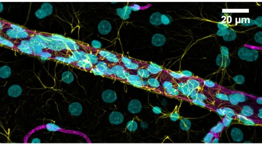
Figure 3. Confocal microscopy image of a rat brain microvessel surrounded by astrocytic
end-feet processes. 11
Figure 4. Major routes of molecular and cell trafficking across the endothelium of the
BBB. 14
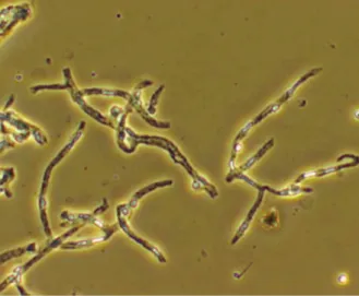
Figure 5. Contrast-phase microscopic visualization of brain microvessels isolated by
mechanical dissection of rat brain cortical grey matter. 22
Figure 6. Static VS Dynamic Models for the study of the BBB. 24
Figure 7. Schematic representation of a static co-culture model for the BBB study. 26 Figure 8. Phase-contrast microscopic view of the immortalized hCMEC/D3 and RBE4
cells. 28
Figure 9. Topological models for the structure of most ABC transporters. 32
Figure 10. Schematic representation of the mechanism of ABC transporter function. 33 Figure 11. Representation of the major ABC transporters expressed at the human
BBB. 34
Figure 12. Crystal structure of mouse P-gp. 38
Figure 13. Representation of a consensual model of P-gp substrate transport. 39
Figure 14. Proposed models of P-gp substrate efflux mechanisms. 40
Figure 15. Schematic representation of mechanisms of P-gp inhibition. 47
Figure 16. Proposed membrane topology of P-gp and BCRP (ABCG2) transporters. 62
Index of Figures__________________________________________________________________
xliv
Figure 18. Regulation of ABC transporters present at the BBB by direct action of
ligand-activated nuclear receptors. 75
Figure 19. Regulation of ABC transporters present at the BBB by inflammation and
oxidative stress. 79
Figure 20. Glutamatergic transmission as a trigger of transcriptional regulation of
P-glycoprotein. 83
Figure 21. Global potential opium production since 1998 until 2013. 86
__________________________________________________________________Index of Tables
xlvii
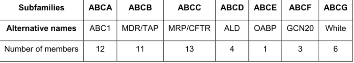
INDEX OF TABLES
Table 1. Non-exhaustive list of some SLC transporters expressed in the human BBB. 17 Table 2. Classification of the ABC transporter family transporters according to the Human
Genome Organization nomenclature. 31
Table 3. List of some endogenous compounds and xenobiotics that are substrates of
P-gp. 46
Table 4. List of some known P-gp inhibitors. 49
Table 5. List of some known P-gp inducers at the BBB. 53
Table 6. List of some endogenous compounds and xenobiotics that are substrates of
BCRP. 65
Table 7. List of some inhibitors of BCRP. 69
Table 8. List of some BCRP inducers. 70
________________________________________________________________Abbreviations List
li
ABBREVIATIONS LIST
ABC (transporters) – ATP-binding cassette (transporters) Aβ – β amyloid peptide
AhR – Aryl hydrocarbon receptor Akt – Protein kinase B
ALS – Amyotrophic lateral sclerosis AJ – Adherens junction
AMT – Adsorptive-mediated transcytosis AP – Alkaline phosphatase
AP-1 – Activator protein 1 AQP4 – Aquaporin-4
ATP – Adenosine triphosphate AUC – Area under the curve BBB – Blood-brain barrier
BCRP – Breast cancer resistance protein BCSFB – Brain-cerebrospinal fluid BEC – Brain endothelial cell
bFGF – Basic fibroblast growth factor BM – Basement membrane
cAMP – Cyclic AMP
CAM – Cell adhesion molecule
CAR – Constitutive androstane receptor CNS – Central nervous system
COX – Cyclooxygenase
CPA – Conditioned place aversion cPLA2 – Cytosolic phospholipase A2 CSF – Cerebrospinal fluid
CYP – Cytochrome P450
DHEA – Dehydroepiandrosterone ECE – Endothelin converting enzyme ECM – Extracellular matrix
EP1R – Prostaglandin E receptor 1 ER – Estrogen receptor
ERK – Extracellular signal-regulated kinase ET – Endothelin
ETR – Endothelin receptor FTC – Fumitremorgin C
GDNF – Glial-derived neurotrophic factor GFAP – Glial fibrillary acidic protein GLUT-1 – Glucose transporter-1
Abbreviations List________________________________________________________________
lii
GPCR – G-protein coupled receptor GR – Glucocorticoid receptor GSK – Glycogen synthase kinase HBEC – Human brain endothelial cells
hCMEC/D3 – Human cerebral microvessel endothelial cell line D3 clone HIV – Human immunodeficiency virus
iNOS – Inducible nitric oxide synthase I.C.V. – Intracerebroventricular I.V. – Intravenous
I.M. – Intramuscular I.P. – Intraperitoneal IL - Interleukin ISF – Interstitial fluid
JAM – Junctional adhesion molecule JNK – C-Jun N-terminal kinase LDL – Low-density lipoprotein LPS – Lipopolysaccharide LTD – Long-term depression LTP – Long-term potentiation M3G – Morphine-3-glucoronide M6G – Morphine-6-glucoronide
MAPK – Mitogen-activated protein kinase MDR – Multidrug resistance
miRNA – MicroRNA
NBD – Nucleotide-binding domain
Nfr2 – Nuclear factor (erythroid-derived 2)-like 2 NMDA – N-methyl-D-aspartate
NOP – Nociceptin or orphanin NR1 – NMDA-NR1 subunit NR2A – NMDA-NR2 subunit NVU – Neurovascular unit
ORL – Receptor-like orphan receptor
OST (transporters) – Organic solute carrier (transporters) PAG – Periaqueductal gray
PCN – 16α-carbonitrile
PECAM-1 – Platelet endothelial cell adhesion molecule-1 PET – Positron emission tomography
PGE2 – Prostaglandin-E2 P-gp – P-glycoprotein
________________________________________________________________Abbreviations List
liii PI3-K – Phosphatidylinositide-3-kinase
PKA – Protein kinase A PKC – Protein kinase C
PPAR – Peroxisome proliferator-activated receptor PQ – Paraquat
PTEN – Phosphatase and tensin homolog PXR – Pregnane X receptor
qRT-PCR – Quantitative real time-polymerase chain reaction RBE4 – Rat brain endothelial cell line clone 4
ROS – Reactive oxygen species Rho 123 – Rhodamine 123
RMT – Receptor-mediated transcytosis RVM – Rostral ventromedial medulla S1P – Sphingosine-1-phospate
SAPK – Stress-activated protein kinase S.C. – Subcutaneous
SLC (transporters) – Solute carrier (transporters) SNP – Single nucleotide polymorphism
TACE – TNF-α converting enzyme
TCDD – 2,3,7,8-tetrachlorodibenzo-p-dioxin TEER – Transendothelial electrical resistance TGF-β1 – Transforming growth factor-β1 TJ – Tight junction
TKI – Tyrosine kinase inhibitor TLR4 - Toll-like receptor 4 TM – Transmembrane
TMD – Transmembrane domain TMH – Transmembrane helices TNF-α – Tumor necrosis factor α TX – Thioxanthone
UGT – Uridine diphospho-glucuronosyltransferase UNODC – United Nations Office against Drugs and Crime VDR – Vitamin D receptor
VEGF – Vascular endothelial growth factor WB – Western blot
__________________________________________________________Outline of the dissertation
lvii OUTLINE OF THE DISSERTATION
Part I – General Introduction on the blood-brain barrier, ABC Transporters and
Regulation
In this section, a review on the existing literature on the blood-brain barrier, the main ABC transporters, and the known mechanisms of regulation of their expression at this barrier is presented, in order to provide a good basis for understanding the objectives and the obtained results of the experimental studies.
Part II – Objectives
The general objectives set for the preparation of the present PhD dissertation are presented in this dedicated section.
Part III – Experimental section
In part III, the manuscripts published or submitted for publication in the scope of this dissertation are presented.
Part IV – Discussion and Conclusions
In this section, an integrated discussion of the results obtained in the scope of this dissertation is presented. The discussion of their potential relevance and their connection with existing scientific reports is also addressed here. Moreover, part III includes the main conclusions taken from the work of the present dissertation and the future perspectives of research.
Part V – References
In this final part, all the literature references that were used in the introduction and discussion sections are listed.
______________________________________________________________General Introduction
3
I. GENERAL INTRODUCTION
1. The Blood-Brain Barrier
1.1. The Central Nervous System: a homeostatic need
The central nervous system (CNS), which comprises the brain and the spinal cord, is generally considered as one of the most important and complex systems in the human body. Through its coordination with the peripheral nervous system, the CNS is vital to the control and function of all the other systems present in the organism. The CNS contains two distinctive liquid compartments, the interstitial fluid (ISF) and the cerebrospinal fluid (CSF). The brain ISF bathes the neurons and the neuroglia (i.e microglia, astrocytes, oligodendrocytes, ependymocytes), whereas the CSF fills the ventricles and surrounds the meninges that protect the external surface of the brain. In order to ensure the proper functioning of the CNS and its neuronal signaling and cell:cell communication, it is essential to closely regulate the extracellular microenvironment and the composition of these extravascular fluids, and thus maintain local homeostasis. Simultaneously, the body needs to guarantee a proficient supply of oxygen and nutrients to the brain, and on the other hand prevent the accumulation of metabolic waste products and toxins through an effective clearance system.
In the human brain, the so-called CNS barriers represent barrier layers between the blood and the CNS and play an essential role in ensuring this constant supply of important nutritive elements, ions, amino acids and energy, and the removal of metabolic products, while at the same time, protect the brain against noxious substances (Daneman and Prat 2015; Zlokovic 2008).
1.2. CNS barrier layers
The first time there was evidence of the existence of a physical barrier between the CNS and the peripheral circulation was described by Paul Ehrlich in 1885, who noted that a dye injection into the blood circulation stained peripheral organs but not the brain and the spinal cord (Ehrlich 1885). Later in 1913, Ehrlich’s student Edwin Goldmann showed that an injection of trypan blue directly into the CSF stained cells within the CNS and not in the periphery (Goldmann 1913). Further support was later found by electron microscopy by Reese and Karnovsky, who demonstrated that when horseradish peroxidase was intravenously administrated it did not pass the capillary lumen, and thus evidencing the
General Introduction______________________________________________________________
4
existence of a solute exchange barrier between the blood and the brain (Reese and Karnovsky 1967).
Three CNS barrier layers are described to be present in the human brain and spinal cord. A first interface is formed by the highly specialized endothelium of parenchymal microvessels comprising the blood-brain barrier (BBB), partitioning the blood and brain ISF. The combined surface area of these capillaries (between 150 and 200 cm2.g-1 tissue) represents the largest CNS interface for blood-brain exchange, resulting in a total area for exchange in the average human adult brain of between 12 and 18 m2 (Abbott et al. 2010; Nag et al. 2005).
The second interface is provided by the epithelium of the choroid plexus (modified ependymal lining of the brain ventricles), which secretes the CSF into the brain ventricular system and forms the blood-CSF barrier (BCSFB) (Abbott et al. 2010; Brown et al. 2004). As a third interface present in the brain we have the arachnoid epithelium, which constitutes the middle layer of the meninges forming the outer covering of the CNS and separates the blood from the subarachnoid CSF. Due to its avascular nature and relatively small surface area, it does not represent a significant surface for exchange between the blood and the CNS (Abbott 2013; Abbott et al. 2010).
In this dissertation, we will give special focus to the BBB and its ability to restrict the brain entry of xenobiotics and other blood-circulating harmful compounds into the CNS.
1.3. Overview of the BBB and its function
It is well established that the human brain consumes over 20% of total body oxygen and energy, while it only corresponds to approximately 2% of total body mass (Shulman et al. 2003). Of the three mentioned CNS barriers, the BBB is distinguished as the most important interface for molecular exchanges between blood and the brain parenchyma, due to its dense network of microvessels throughout the brain and the proximity of their finest branches to individual neurons (Abbott et al. 2010). This network is composed of approximately 100 billion capillaries with diameters as small as 3 to 7 μm (Zlokovic and Apuzzo 1998), and the distance between capillaries is as short as 40 μm (Rodriguez-Baeza et al. 2003). These characteristics ensure that almost every neuron is perfused by its own blood capillary and therefore guarantee an efficient oxygen and nutrient supply to such high brain demand. No brain cell is over 25 μm from a brain capillary, meaning that once the solute diffuses through the BBB, it is considerably close to the neurons or glial cells. Despite the huge number of capillaries, they occupy only 0.1% of the brain volume
______________________________________________________________General Introduction
5
(Pardridge 1991; Pardridge 2003). The BBB is present in all brain regions, except in specialized and very size-limited sites of physiological cross-talks between the brain and the periphery, i.e. the circumventricular organs, where blood vessels allow diffusion of blood-borne molecules across the vessel wall (Ballabh et al. 2004; Cardoso et al. 2010; McKinley et al. 2003; Weiss et al. 2009).
BBB structure is formed by brain endothelial cells (BECs) composing the capillaries, which differ from endothelial cells present in the rest of the body due to the lack of fenestrations, and tight regulation of the movement of molecules, ions and cells across this barrier (Ballabh et al. 2004; Daneman 2012). The BBB exerts its barrier function at three different levels: (1) physical, through the extensive expression of tight junctions (TJs) along adjacent endothelial cells, reducing the paracellular pathway, (2) transporter-dependent passage, due to the existence of specific transport mechanisms mediating solute flux, and (3) metabolic, given the enzymes present in the BECs are capable of processing molecules in transit (Abbott et al. 2010). The resulting neurovascular coupling allows the maintenance of brain homeostasis by providing for the energy demands of neuronal activity, as well as it protects the CNS from toxins, pathogens, inflammation, injury, and disease, in association with various perivascular cells such as pericytes, microglia, astrocytes, and specialized cellular compartments such as the endothelial glycocalyx (Stanimirovic and Friedman 2012). In this way, the BBB plays a crucial role by exerting a bi-directional control over the passage of a large diversity of regulatory proteins, nutrients and electrolytes, as well as potential neurotoxins, and maintain a strict extracellular environment around synapses and axons. The BBB functions can be described as follows:
1. Brain nutrition: the BBB expresses specific transport systems in the luminal and abluminal membranes of BECs in order to ensure the appropriate supply of essential water-soluble nutrients (Abbott et al. 2010);
2. Regulation of ion homeostasis and preservation of neural signaling: the BBB provides a combination of specific ion channels and transporters to keep the optimal ionic composition for synaptic signaling function, as well as maintain the central and peripheral neurotransmitter pools separate, minimizing “cross-talk” (Abbott et al. 2010; Bernacki et al. 2008);
3. Control molecular trafficking: the BBB functions as a shield preventing neurotoxins (endogenous metabolites or ingested/environmental xenobiotics) and many macromolecules in the blood circulation from entering in the brain. The protein content of the CSF is much lower and markedly different to that of plasma, and the
General Introduction______________________________________________________________
6
leakage of several serum macromolecules into the brain can result in serious pathological consequences (Abbott et al. 2010). In this way, the BBB minimizes neuronal cell death, and preserves neural connectivity and immune quiescence. Thus, the BBB guarantees that the CNS environment is not easily affected by peripheral changes under physiological conditions. However, the BBB remains a dynamic interface that acknowledges the necessity to meet the demands of the whole organism, which are not static.
1.4. The Neurovascular Unit: the architecture of the BBB
The properties of the BBB are mostly defined by the BECs, but these are regulated and maintained by crucial interactions with the basement membrane and neighboring cells, such as microglia and astrocytes, as well as neurons and perivascular pericytes (Cardoso et al. 2010; Zlokovic 2008). The close proximity of these cells results in an effective unit of paracrine regulation critical for normal CNS functioning, often called the neurovascular unit (NVU) (see Figure 1).
1.4.1. Brain Endothelial Cells
The brain microvascular endothelium constitutes the most critical element of the BBB. BECs have been identified as morphologically and metabolically distinct of those present in the peripheral circulation. In comparison to those cells, BECs are characterized by (1) the presence of a narrow junctional complex at their adjacent margins, eliminating gaps or spaces between cells and preventing any free paracellular diffusion of blood-borne substances into the brain parenchymal space, (2) lack of fenestrations typically present in peripheral capillaries, (3) fewer pinocytic vesicles, and (4) higher number of cytosolic mitochondria, suggesting important metabolic activity (Abbott 2005; Bearer and Orci 1985; Correale and Villa 2009; Dorovini-Zis et al. 1991; Lane et al. 1992; Villegas and Broadwell 1993). In fact, BECs are said to be 50 to 100-fold tighter than those in peripheral microvessels (Abbott 2002) due to the expression of this elaborated junctional complex that includes mainly TJ and adherens junction (AJ) proteins (Hawkins and Davis 2005) [presented in more detail in section 1.4.1.1.]. Gap junctions have also been identified at the BBB, but their role in the barrier function is not clear (Zlokovic 2008). Additionally, the luminal surface of the BECs is covered by the so-called glycocalyx, which corresponds to a negatively charged mesh of proteoglycans, glycosaminoglycans, glycoproteins and glycolipids, making anionicity another determinant factor for BBB permeation (de Boer and