HAL Id: tel-01584465
https://tel.archives-ouvertes.fr/tel-01584465
Submitted on 8 Sep 2017HAL is a multi-disciplinary open access archive for the deposit and dissemination of sci-entific research documents, whether they are pub-lished or not. The documents may come from teaching and research institutions in France or
L’archive ouverte pluridisciplinaire HAL, est destinée au dépôt et à la diffusion de documents scientifiques de niveau recherche, publiés ou non, émanant des établissements d’enseignement et de recherche français ou étrangers, des laboratoires
Synaptic fluctuations in cerebellar interneurons
connected by a single synaptic contact
Maria Camila Pulido Puentes
To cite this version:
Maria Camila Pulido Puentes. Synaptic fluctuations in cerebellar interneurons connected by a single synaptic contact. Neuroscience. Université Sorbonne Paris Cité, 2016. English. �NNT : 2016US-PCB007�. �tel-01584465�
Université Paris Descartes
Faculté de Médecine
Ecole doctorale 474- Frontières du Vivant
Thèse de doctorat de Neurosciences
Synaptic fluctuations
in cerebellar interneurons
connected by a single synaptic contact
Par María Camila PULIDO PUENTES
Dirigée par Alain Marty
Présentée et soutenue publiquement le 11 Mars 2016 Devant un jury composé de :
CASADO Mariano, MC ENS, rapporteur
KAWAGUCHI Shinya, Associate Professor à Doshisha University, examinateur LEGAY Claire, Professor à Paris Descartes, examinateur
LEGENDRE Pascal, directeur de recherche, rapporteur MARTY Alain, DR CNRS, directeur de thèse
RYAN Timothy, directeur de recherche à Weill Cornell Medical College, examinateur
Faculté de Médecine
Ecole doctorale 474- Frontières du Vivant
Thèse de doctorat de Neurosciences
Synaptic fluctuations
in cerebellar interneurons
connected by a single synaptic contact
Par María Camila PULIDO PUENTES
Dirigée par Alain Marty
Présentée et soutenue publiquement le 11 Mars 2016 Devant un jury composé de :
CASADO Mariano, MC ENS, rapporteur
KAWAGUCHI Shinya, Associate Professor à Doshisha University, examinateur LEGAY Claire, Professor à Paris Descartes, examinateur
LEGENDRE Pascal, directeur de recherche, rapporteur MARTY Alain, DR CNRS, directeur de thèse
RYAN Timothy, Directeur de recherche à Weill Cornell Medical College, examinateur
L’élément constitutif des synapses centrales est le site synaptique individuel, comprenant une zone active du côté présynaptique et une densité postsynaptique associée. Du fait de limitations techniques nos connaissances sur le mode de fonctionnement d’un site synaptique restent insuffisantes. Pour faire progresser cette question nous projetons d’effectuer des enregistrements en paires entre interneurones de la couche moléculaire du cervelet. Ces neurones forment des synapses qui ont des signaux élémentaires quantiques de grande taille, et les synapses comprennent parfois un seul site synaptique, ce qui fait qu’ils offrent des avantages décisifs pour ce projet. Les réponses postsynaptiques à des trains de potentiels d’action seront étudiées dans différentes conditions expérimentales. Les résultats seront interprétés par un modèle supposant que les vésicules synaptiques doivent se lier à un petit groupe de sites d’arrimage avant l’exocytose.
Title:
Synaptic Fluctuations in Cerebellar Interneurons Connected by a single synaptic contact
Abstract:
The unitary element of central synaptic transmission is a single synaptic site, with one active zone as presynaptic component and the postsynaptic density as postsynaptic partner. Due to technical limitations there is much uncertainty on the mode of functioning of a single synaptic site. To address this issue it is planned to perform paired recordings between interneurons of the molecular layers of the cerebellum. These neurons form synapses with a large quantal size, and occasionally displaying a single release site, and are thus favorable for this study. Postsynaptic responses will be studied in response to trains of presynaptic action potentials under various conditions. The results will be compared to a model supposing the obligatory binding of vesicles to a small complement of docking sites prior to exocytosis.
Mots clés: Docking Sites, Single synaptic contacts, GABAergic interneurons. Keywords: Docking Sites, Single synaptic contacts, GABAergic interneurons.
I arrived to France directly from Colombia with the idea of starting my thesis project, already 4th years ago. However, since my arrival I have been enriched by different
experiences that make even better the process of making this PhD. I had the fortune of sharing with incredible people that, even some of them already left, make part of this adventure. There are tons of people with which I am thankful and happy to meet. First at all, I would like to thank Mariano, Shinya, Claire, Timothy and Pascal for accepting be part of my thesis jury.
I am grateful to Alain for guiding me during this process and allowing me to be part of this beautiful project. All his advices were always relevant in order to reach a happy ending. Also, I would like to thank Isabel not just for her enthusiasm helping with some experiments and for her useful comments, also for her support and concern during all these years in Paris. Without both of you this nice experience could not be the same.
As part of my formation, I will never forget to thank my mentors Enrico and Maria del Pilar. There are no words to express my gratitude and admiration for them. Those six months sharing with you here in Paris were the best!
I would like to thank all the current and previous members of 8118 which made my time here pleasant. I would like to thank Fede, Brandon, Taka, Javier, Jorge, Kris for their nice discussions during our lunch time at Crous, and during the extra-laboratory activities that were not few. Also, I would like to thank people from the laboratory 8119, which were always happy to make alternative activities together with us.
I am glad to meet Guadalupe, the best officemate and a really, really good friend. Since always, there were countless moments of joy... We never stop having fun and yet, time flew! I am grateful to her for advising and motivating me during these years of research, even from the distance.
Also, I am happy to share once again with Gerardo, who arrived with an excellent idea of making beer by ourselves, the best of all distractions!… well, there was also
the ping-pong table idea, which was better at our minds than in practice!
Likewise, I am glad to meet people that were part of this adventure. I am grateful to my friends Jimena, Eugenia, Camilo, Ana, Paula, Philippe, Jean, Joe, Johanna, Mad for their support during these years of studies. They have become a special part of my life.
I would like also to thank Paris Descartes University and the “Fondation pour la Recherche Médicale” for funding my studies.
Finally but not less important, I am grateful to my family that despite they were in Colombia, they were always thinking on me, giving me the best of their energies.
PREFACE...1
A historical introduction to the mechanisms of central synapses...3
Chapter 1: Synaptic transmission by vesicular release...3
1.1 Neuromuscular Junction Model...4
1.2 Miniature Spontaneous Events...5
1.3 Quantal Nature of end-plate Potentials...6
1.4 Vesicle hypothesis of Quantal secretion...7
1.5 Chemical transmission, speed and modulation...8
Chapter 2: Quantal transmission at central synapses: the release sites controversy...11
2.1 Are there quantum-like components in central synapses?...12
2.2 Looking for release sites at the CNS...15
2.2.1 One site-One vesicle hypothesis...16
2.2.2 Postsynaptic receptors occupancy and Multivesicular release hypothesis.. . .17
Chapter 3: Molecular nature of the building block of the synapses...23
3.1 Active zone Material and synaptic vesicle Docking...23
3.2 Molecular Machinery at the Active Zone...26
3.3 Distribution of calcium channels along the active zone...28
3.4 Active zone proteins and their macromolecules counterparts...29
Chapter 4: Physiology of docking sites...31
4.1 Defining the Readily releasable Pool...31
4.1.1 One site-one vesicle RRP models...31
4.1.2 Capacitance membrane changes in chromaffin cells...32
4.1.3 RRP at the Calyx of Held...32
4.1.4 Calcium uncaging at cerebellar MLIs...33
4.2 Fixed number of docking sites...35
4.3 Synaptic contact heterogeneity: a docking site concern...37
METHODS...41
Chapter 1: Cerebellar Slices preparation...43
1.1 Slices of young rat...43
1.2 Slices of adult rat...43
1.3 Cutting solutions...44
2.1 Recording set up...45
2.2 Interneuron selection...45
2.3 Electrophysiology...46
2.4 Paired MLI recordings...46
Chapter 3: Spontaneous events detection...47
Chapter 4: Monte Carlo simulations...47
RESULTS...49
Chapter 1: Vesicular Release Statistics and Unitary Postsynaptic Current at Single GABAergic Synapses...49
Chapter 2: Changes in IPSC amplitude and kinetics with extracellular calcium concentration...75
Chapter 3: Statistics of cumulative released vesicle numbers in elementary GABAergic synapses...79
3.1 Introduction...79
3.2 Extension of the 2-step model to single GABAergic synapses...80
Chapter 4: Modeling Changes in Synaptic Properties...86
4.1 Changes in success probability induced by augmentation of spontaneous synaptic activity... 86
4.2 Simulations of presynaptic parameters...90
Chapter 5: Developmental changes in the synaptic connectivity between cerebellar interneurons...96
5.1 Docking site number decreases with development...97
DISCUSSION...102
1.1 Importance of single synaptic contact recordings and docking sites calculation..102
1.2 An extension of the Variance-mean fluctuation analysis method...103
1.3 2-step Model and its implications in short-term synaptic depression and recovery. ... 105
1.4 Spontaneous release offers a way of modulating ρ, the occupancy of the recruitment site...106
CONCLUDING REMARKS...108
PERSPECTIVES...110
Figure 1. Quantal nature of the end-plate potential at the neuromuscular junction...5
Figure 2. Active Zone in Electron microscopy...9
Figure 3. Schematic diagram to explain quantal hypothesis...12
Figure 4. Quantal components in central synapses...13
Figure 5. Multivesicular release from a single synaptic contact recording...20
Figure 6. Docking sites are organized together with Active Zone Material at the neuromuscular junction...24
Figure 7. Molecular organization of the active zone...27
Figure 8. Vesicular readily releasable pool estimation at single synaptic contacts...34
Figure 9. Estimation of docking sites number and quantal parameters by a variance-mean analysis technique...36
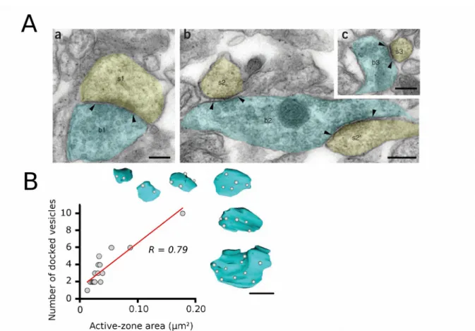
Figure 10. The number of docked vesicles correlates linearly with active zone size...38
Figure 11. Changes in PSC peak amplitude and kinetics with Cao...76
Figure 12. Statistics of cumulative released vesicle numbers in elementary GABAergic synapses...82
Figure 13. Increase in spontaneous release due to presynaptic depolarization affects evoked release...87
Figure 14. Simulation of synaptic depression during repetitive stimulation in two release models...90
Figure 15. The replacement site may not be fully occupied in control conditions...92
PREFACE
Neurons are the cells of the nervous system, characterized by having functional specialized regions, that allow the faithful transmission of information through specialized inputs and outputs. Neurons propagate codified information through a thin cable (the axon) until a junctional region (the synapse), where information is passed rapidly to another cell, usually another neuron, in a process known as synaptic transmission. Essentially, the electrical information carried by the first cell (presynaptic cell) causes a change in the electrical activity of the second cell (postsynaptic cell); where the codified information is retransmitted to a following neuron or specialized cell. Since neurons were discovered, near the end of nineteenth century, the problem arose as to how excitation was transmitted across the gap between pre- and postsynaptic cells, putting neurons and synapses in the limelight of neuroscience.
There is no doubt that the twentieth century has been the century of modern neuroscience, with an extraordinary explosion of knowledge. Since the discovery of action potentials in the giant fibers in the nerve cord of the earthworm by Lucas and Andrian in the first decades of that century, followed by the characterization of its electrical nature by Hodgkin and Huxley, an extraordinary variety of new phenomena have been described, leading to an ocean of mechanisms to interpret and understand. Many discoveries relate to the mechanisms of neuronal excitability and synaptic transmission, gradually reaching to the interpretation of whole circuits and global neuronal function.
Neuronal transmission is itself a whole universe of complexities, but for my purpose, we will focus on the precise moment when the action potential reaches the axon terminal and passes the electrical information to the following cell by the secretion of a neurotransmitter. While the nature of this step is now universally accepted, it is difficult to imagine the size of the collective effort that was involved in the elucidation of this fundamental process. And, while the main focus of modern neuroscience has
PREFACE
moved on to study more integrated phenomena, it is important to point out that this work is not actually over. Yes, we do know the most basic features of synaptic transmission, but we still need to agree on several aspects of this process. In spite of the incalculable amount of knowledge that has been produced during decades of studies, there are still fundamental questions that have been raised from the beginning of the study of synaptic physiology, and which remain unanswered. Worse, while decades of studies were not sufficient to fully explain synaptic function, they were more than sufficient to create contradictory theories and scientific disagreements. As may be guessed from what I have just said, my thesis is concerned about a problem that during years has created a lot of confusion and even some personal enmities in its path; but still, it is wonderful to see how with the patient integration of all the available evidence -obtained with fantastic and ingenious methods and experiments- there is light at the end of the tunnel, there is light at the end of synaptic transmission understanding.
Along the following introduction, I am taking you through a journey in history, by relating stories and experiments, showing how from one observation made 60 years ago, a long sequence of discoveries were made, raising new questions at each step, eventually raising the hope to discern the building blocks of the synaptic processes. This description is highly simplified and does not try to be exhaustive. Rather, I have tried to focus on conceptual advances, highlighting what in retrospect appears to pave the way to subsequent progress, and trying to extract a plausible, coherent picture from occasionally discrepant views.
A HISTORICAL INTRODUCTION TO
THE MECHANISMS OF CENTRAL
SYNAPSES
Chapter 1: Synaptic Transmission By
Vesicular Release.
During the first half of the last century, a controversy arose about how electrical information passes through that small gap that is formed between neurons, the
synaptic cleft. At that time, Hodgkin and Huxley had yet to elucidate how information
signals are generated in the neuronal axon membrane. However, it was already assumed that these signals are electrical in nature. Therefore, the most logical and natural suggestion was that an electrical process should be involved in passing that information to the next cell. Such a process was eventually demonstrated at so-called
electrical synapses. But in parallel to electrical synapses, a totally different type of
synaptic transmission was revealed at chemical synapses, as already anticipated by previous discoveries suggesting the involvement of chemical transmitter substances. The efforts of Dale (1914) and Loewi (1921) contributed to demonstrate that acetylcholine, a chemical originally isolated from the ergot, causes contraction of intestinal muscle, inhibits heartbeat and lowers blood pressure. Specifically, Loewi showed that stimulating the vagus, which innervates the heart muscle, inhibits heart beating by releasing a chemical substance which was eventually identified as acetylcholine. Later, Dale demonstrated that acetylcholine is naturally produced in the body and that it is released from the motor nerve endings in skeletal muscle . Even though these studies pointed out chemical transmission as a fundamental part of synaptic transmission, many neurophysiologists doubted whether chemical release
Synaptic transmission by vesicular release.
could be fast enough to account for the speed of the transmission of information. The assumed chemical substance must be released in the synaptic cleft, must act postsynaptically and must be removed all within a few milliseconds; in their eyes that was very difficult to accept. This difficulty was combined with the practical challenge of recording such rapid events in a living tissue. However the latter difficulty started to be lifted in the mid century, when it became possible to make electrical recordings from neurons by the insertion of a tiny intracellular electrode, allowing the recording of both sites involved in the synaptic transmission: the presynaptic and postsynaptic cells.
1.1 Neuromuscular Junction Model.
Depending on the scientific question that is raised, the model of study and the techniques have to be adapted. For instance, the first recordings of intracellular action potential obtained independently by Hodgkin & Huxley and by Cole and Curtis, took advantage of the special features of the giant axons of squids. At that time, the electrical recordings of cellular potential were made using sharp intracellular pipettes that were literally inserted inside the cells; it was fundamental to look for animal models that were adapted to those conditions. The size of squid axons, around 0.5mm, allows the insertion of recording electrodes, a process which in other cellular models would be physically impossible as it would damage all the tissue.
In the 50s, the neuromuscular junction emerged as a model to study synaptic transmission, because the size of the postsynaptic element, the muscle fiber, is sufficiently large to allow the stable insertion of a glass pipette. The neuromuscular junction is a synapse, but it is not a typical one, and it differs markedly from central interneuronal synapses, as I will discuss later. The goal of this synapse is to evoke contraction of the postsynaptic muscle cell. The motor axon is demyelinated near the end, where it branches and where its terminal makes synaptic contacts with the part of the muscle fiber called the motor end-plate. Typically when the muscle is stimulated by the arrival of an action potential at the terminal, it produces a change in its membrane potential, the end-plate potential, with an amplitude around 50mV, evoking its contraction (Katz, 1969). This particular synapse provides a fine introduction to the physiology of chemical synapses and it has become an excellent
model to understand how a chemical process can account for the speed of synaptic transmission, as I will be discussing in this chapter.
1.2 Miniature Spontaneous Events.
In 1952, Fatt and Katz used a high gain amplifier with a micro-electrode inserted into a frog sartorius muscle fiber at the end plate region. They examined closely recordings from the resting muscle where they observed small fluctuations in the membrane potential. These electric discharges were similar to normal end-plate
potentials (EPPs) except for their size. They were just 0.5mV in height, and therefore
received the name of miniature end-plate potentials (mEPPs; Figure 1A; Fatt & Katz, 1952).
Statistical analysis of the time intervals between successive mEPPs showed that their occurrence was randomly distributed in time, having a spontaneous behavior. mEPPs were just found at the end-plate region and they were not present in denervated muscle, indicating a close association with the nerve terminal. Moreover, mEPP amplitudes were reduced by curare and increased by prostigmine (an
Figure 1. Quantal nature of the end-plate potential at the neuromuscular junction. (A) series of records of spontaneous changes in the membrane potential, known as miniature end-plate potentials (mEPPs, adapted from Fatt & Katz, 1952). (B) Step-wise fluctuations of EPP responses during calcium deficiency. (C) The amplitude distribution mEPPs (upper panel) and evoked EPPs responses (lower panel) after magnesium block. The expected number of failures of transmission is indicated by an arrow, and the continuous curve was calculated from a Poisson distribution, modified to take account of the variability in size of the quantal units. Peaks of the EPP amplitude histogram occur at 1, 2 and 3 times the mean amplitude of the spontaneous potentials. (B)–(C) from (del Castillo & Katz, 1954b).
Synaptic transmission by vesicular release.
anticholinesterase), demonstrating that mEPPS were produced by the action of acetylcholine on nicotinic receptors. These findings indicated that spontaneous activity was directly related with synaptic transmission (Fatt & Katz, 1952).
The next question was the amount of acetylcholine involved in a miniature response. An initial guess could have been to attribute a mEPP to a random collision of a single molecule. But Fatt and Katz doubted that a single molecule would be enough to cause a depolarization of 0.5mV, because that necessarily implied that just a thousand molecules are needed to produce a normal EPP of 50mV. Previously, it was estimated that the quantity of acetylcholine released by an impulse is about 10⁶ molecules (Acheson, 1948), indicating that each single mEPP must be produced by several thousands of molecules. Moreover, Fatt and Katz highlighted that the response to externally applied acetylcholine is a smooth depolarization, not a series of unitary events of about 0.5mV height. Taking together these findings, it is reasonable to ask, how are those thousands of molecules released into the synaptic cleft? What is the release mechanism involved to produce the miniature discharge?
1.3 Quantal Nature of end-plate Potentials.
The neuromuscular transmission is blocked by an excess of magnesium ions. This effect is due to a reduction of the quantity of acetylcholine released per impulse (Del Castillo & Engbaek, 1954). Taking advantage of this well known effect, Fatt and Katz made remarkable findings. When the EPPs were recorded in a high Mg² and low⁺ Ca² solution its mean amplitude was smaller and ⁺ surprisingly, successive EPPs responses exhibited random fluctuations in a stepwise manner. It was then found that the size of these steps approaches that of mEPPs (F igure 1B; Fatt & Katz, 1952). Integrating these results, Katz and collaborators tentatively suggested that the EPP is built by the summation of units which are identical in size with mEPPs. They further suggested that acetylcholine is released in discrete packets or quanta (Del Castillo & Katz, 1954). This is now known as the quantal hypothesis. Experimental support for this theory was initially based on impressive quantitative analysis made in collaboration with Del Castillo in 1954. This analysis was designed to estimate the mean number of quanta responding to one impulse, m.
Under normal conditions, the EPP is large and statistical fluctuations are small. The size of responses is maintained by a large population of packets. Each packet is assumed to be independent of the others, and to have a probability, p, to be released for each stimulus. Consequently, one might expect that quantal release would be distributed binomially under normal conditions. However, when the probability of any individual event is low (where mostly failures are observed together with occasional responses), as in quantal release recorded in high magnesium and low calcium solution, the binomial distribution degenerates and reduces to a Poisson distribution (Katz, 1969).
Using this clever idea and using the proportion of failures, Del Castillo and Katz proposed one way of calculating the quantum content (m) from Poisson's law, where
m is given by the natural logarithm of the reciprocal of the proportion of failures.
Alternatively, if the minis are the least unit and an EPP is made up of units of the same size, as was postulated, another way to calculate m should be the relation between the mean amplitude of the EPP responses (recorded in high magnesium) and the mean amplitude of spontaneous potentials. Furthermore, if the hypothesis is correct these two ways of calculating m should be in agreement. In fact, they showed a perfect agreement between both methods and furthermore they showed that the proportions of EPPs of different sizes are described by a Poisson distribution (Figure1C; Del Castillo & Katz, 1954).
1.4 Vesicle hypothesis of Quantal secretion.
At the same time when Katz and colleagues were making that fantastic work on quantal transmission, important observations were made by De Robertis and Bennett (1955) and Palade and Palay (1954). They showed for the first time that the nerve terminal contains large number of tiny, membrane-bound structure called synaptic
vesicles. Taking advantage of these findings, Del Castillo and Katz (1956) suggested
that vesicles represent the packets of acetylcholine that are released in response to a presynaptic action potential. They further suggested that a quantum of approximately 10,000 molecules of neurotransmitter represents the number of acetylcholine molecules contained in a single vesicle.
Synaptic transmission by vesicular release.
While the hypothesis that a quantum is represented by a single vesicle appeared very attractive, still there was not any proof in favor of that hypothesis, leaving the scene open for other suggestions about the mechanism of neurotransmitter release. Alternative options suggested that a quantum of information could be represented by the synchronized release of a group of around 7 to 10 vesicles in to the synaptic cleft (Kriebel & Gross, 1974). Moreover, there was an even more drastic suggestion that the release mechanism involves the diffusion of neurotransmitter through a pore (channel) that opens during a short time interval, between the synaptic vesicle and the synaptic cleft (Falk-Vairant et al., 1996).
The controversy went on until Hurlbut and colleagues (1990) made impressive measurements on the terminal. They compared the numbers of quanta of acetylcholine released and of the synaptic vesicles lost after stimulating the nerve. Briefly, Hurlbut and colleagues measured on one side, the quantal release in low calcium/high magnesium conditions obtained with -latrotoxin, a potent toxin extracted from the black widow spider, which causes a massive quantal release from the nerve terminals. On the other side, some muscles were fixed at various times during the secretion period and the number of synaptic vesicles remaining in the cross-sections of the terminals were counted on the electron micrographs. The relation between the quanta released and the vesicles remaining, had almost a perfect correlation, with a slope of regression line around -1.1 to -1.2 (Hurlbut et al., 1990), which means that a quantum of transmitter is derived from a single vesicle.
1.5 Chemical transmission, speed and modulation.
Now that we know that a quantum is represented by a single vesicle, it is pertinent to ask: how are these vesicles released into the extracellular medium?. Since vesicles are enclosed by a membrane, the release of neurotransmitter into the synaptic cleft requires the fusion of the vesicle membrane with the plasma membrane of the presynaptic terminal, a process called exocytosis.
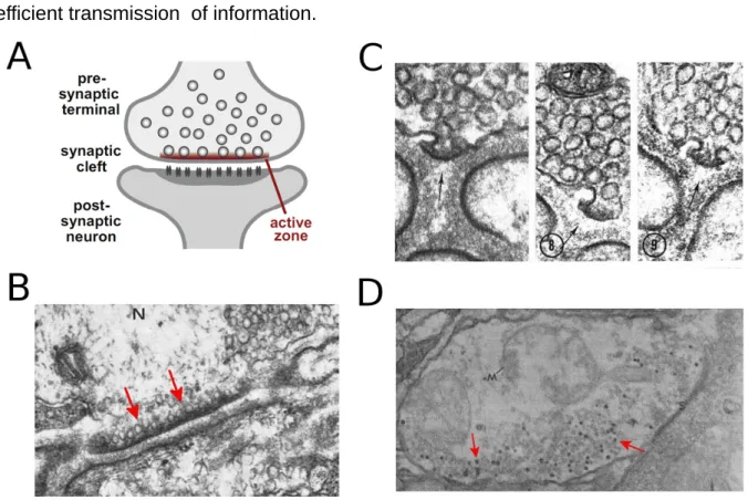
The French neuroscientists, Couteaux and Pécot-Dechavassine (1970) first suggested that exocytotic sites are not randomly distributed, indicating that vesicles do not fuse indiscriminately with the plasma membrane. They are localized at the
active zone, (F igure 2A-B). Each active zone is represented as a dense material close to the presynaptic membrane, together with a heap of vesicles. At the end plate, active zone is localized opposite the folds of the postsynaptic membrane (Südhof, 2012). Which makes sense, since the receptors of acetylcholine are localized just in that postsynaptic membrane, making this mode of release a very efficient transmission of information.
Figure 2. Active Zone in Electron microscopy. (A) Schematic drawing of a synapse (from Südhof, 2012). (B) Electron micrograph of the active zone (arrows) of the frog neuromuscular junction. Synaptic vesicles are clustered close to the active zone membrane. (C) Exocytotic opening of the synaptic vesicles into the synaptic cleft at the level of the active zone. (D) Vesicular peroxidase uptake at lobster neuromuscular junction reveals synaptic vesicle endocytosis. A few vesicles are indicated by arrows (from Holzman et al., 1971). (B) and (C) from (Couteaux & Pécot-Dechavassine, 1970).
The first direct evidence for vesicular membrane fusion was achieved by Heuser and colleagues in 1973. They produced a prolongation of action potential duration using a blocker of potassium channels, to enhance transmitter release. Then, a few milliseconds after the nerve was stimulated, the muscle was quickly cooled by slamming it onto a block of copper cooled to -269°C by liquid helium. The resulting freeze-fracture electron-micrographs showed vesicles held in the precise moment of exocytosis (F igure 2C; Heuser & Reese, 1973).
Synaptic transmission by vesicular release.
The existence of an exocytotic process, necessarily implies the existence of a counterpart, otherwise the presynaptic membrane will grow indefinitely. Evidently neurons do not behave like that. Let me describe a simple but ingenious experiment on the neuromuscular junction. Suppose that we have a certain enzyme, which oxidizes a variety of compounds and their reaction products could produce an electron-dense material. We have created a biological marker, which can be detected by electron microscopy. If we use this enzyme as a marker in the external solution in our neuromuscular junction model, and if we find that after stimulating the muscle the marker appears inside the cell, this indicates uptake of plasma membrane and some extracellular solution, proving the existence of endocytosis (Figure 2D; Holzmann et al., 1971). This uptake process was first established by Holzmann in the early 70s, and it occurs just after the exocytosis. Since that, it has been shown one major form of endocytosis begins with the formation of invaginations and subsequent clathrin coating, starting once again the formation of synaptic vesicles.
Chapter 2: Quantal Transmission At Central
Synapses: The Release Sites
Controversy.
In the previous chapter, we described synaptic transmission in the neuromuscular junction and we discussed arguments indicating that this transmission is mediated by neurotransmitter packed into vesicles. Each vesicle corresponding to a single quantum of information, fuses in the active zone at the plasma membrane and releases its content into the synaptic cleft. The transmitter reacts with the postsynaptic receptors, propagating the signal from the presynaptic neuron to the postsynaptic or cell. In this chapter, we will discuss whether the quantal statistical analysis developed by Katz and colleagues to explain the packing of information at the neuromuscular junction, also applies to synapses of the central nervous system. According to Katz, vesicular release can occur only at specific locations, the so-called
reactive sites or release sites, and he assumed a number N for these sites. As was
explained before, when an impulse arrives, release of neurotransmitter occurs with a certain probability at each site, assuming that they function independently of each other. In other words, release follows binomial statistics; the average number of released quanta is the product of the total number of release sites (N) with the probability of release (p; Figure 3; Katz, 1969).
Unfortunately, attempts to adapt Del Castillo and Katz's hypothesis to the central nervous system have not been entirely successful. Trying to make sense of this literature, it is difficult for readers to disentangle the terminology created around synaptic transmission, particularly regarding release sites. Katz worked in a model where many synapses are activated by a single nerve impulse. In the context of a central synapses however, what is a release site? Is a release site a single fusion ready vesicle? A single active zone? A single synapse with perhaps, multiple active zones? There are a lot of questions and I will try to address them one by one.
Quantal transmission at central synapses: the release sites controversy.
2.1 Are there quantum-like components in central synapses?
Each segment of the spinal cord includes thousands of motoneurons. The
motoneurons, which originate from the central nervous system, are innervated by
several afferent Ia neurons, the stretch-activated sensory neurons. The motoneurons are efferent nerves that propagate signals from the spinal cord to the muscle, and they emerged as a suitable model to study central synapses.
Motoneurons display excitatory postsynaptic potentials (EPSPs) in response to presynaptic stimuli. These EPSPs are composed of discrete units, suggesting the presence of quantal components (Kuno, 1964). However, in each motoneuron EPSPs are arising from the action of several afferent fibers; in order to distinguish responses from single terminals or see quantal release coming from a single vesicle, it is necessary to reduce the input to a single presynaptic axon, a single fiber, as was done by Redman and colleagues in the 1980ies.
Figure 3. Schematic diagram to explain quantal hypothesis. Reactive sites on the vesicles and the active zone membrane are shown as dots. Transmitter release only occurs when two sites meet. The probability of occurrence varies directly with the number of reactive sites in the axon membrane (from Katz, 1969).
In a first approach, they were able to record postsynaptic responses evoked from the stimulus of a single axon, called unitary EPSPs (Figure 4A). Interestingly, they showed that unitary EPSPs display amplitude variability, which cannot be explained from the background noise distribution, and they attributed this phenomenon directly to synaptic transmission (Figure 4B). Further, using a statistical computing technique known as deconvolution, where it is assumed that variations are discrete rather than continuous, they were able to show that EPSP amplitudes fluctuated between four discrete voltages, each with an increasing step of about 100µV (Figure 4C). They associated this step with a quantal unit in the synaptic transmission, showing for the first time quantal transmission at the CNS (Jack, Redman, & Wong, 1981).
Figure 4. Quantal components in central synapses. (A) Four individual EPSPs evoked from the stimulus of a single axon. (B) The histogram of the noise, which was fitted with a Gaussian, and distribution of the peak voltage of the evoked EPSP. (C) The deconvolved release probability calculated using the Gaussian curve for the noise and the histogram in B. This result indicates that EPSP fluctuates between four discrete peak voltages, with the probabilities of each as indicated. (D) Synaptic contacts in motoneurons. Upper Panel: The dendrite of the motoneuron, together with the pre-terminal branches of the group Ia axon (black line) and synaptic contacts (arrows). Lower Panel Right: scheme representing the calculated space constant for dendritic branch. The diameter of each cylinder has been drawn to scale to represent the average diameter of the branch it represent. The 2µm bar provides the scaling for dendrite diameter. (A)-(C) adapted from (Jack et al., 1981). (D) adapted from (Redman et al., 1983).
Quantal transmission at central synapses: the release sites controversy.
This raises the following question: does this novel quantal unit at the CNS represent a single vesicle, as was shown in the neuromuscular junction? Unfortunately at this stage the information is not sufficient to know whether this quantum represents the release of one vesicle in a single active zone, or represents the activity of a single active zone in a multi-active zone synapse. In order to find what is the source of synaptic potential fluctuation, it becomes important to consider the morphology of the recorded neuron.
Redman and colleagues brilliantly thought that the best way to find the relation between the discrete fluctuations and their sources, was to investigate the origins of the synaptic potentials. Motoneurons and single axon fiber were filled with HRP, an enzyme that was typically used as marker for electron microscopy. Using this technique, it was possible to detect the points of contact between dendrites and axons, the Boutons (F igure 4D-top,Redman & Walmsley, 1983a). To corroborate that these sites were physiologically involved in the synaptic transmission, they calculated the electrical distances from the soma to each synaptic bouton, using the shape index of each recorded EPSP and the cable model (Figure 4D-bottom). Both measurements, the electrical distance and the distance obtained from the reconstruction of the synaptic contacts, were in complete agreement, concluding that fluctuations originated from different contact sources between the motoneuron and its innervating axon (Redman & Walmsley, 1983). They concluded that an evoked EPSP obtained by stimulation of a single axon, originates from a wide range of electrotonic locations on the dendritic tree, which are correlated with the location of each bouton. Redman called each location a release site, thinking that each contact represented the minimal unit in synaptic transmission1. However up to here, the
above findings are not enough to reach that conclusion, and it is important to clarify that a CNS release site as defined by Redman cannot be confused with the release site, with total number N, proposed by Katz; rather, it will be more associated with the active zone description.
In summary, this research showed the existence of quanta in synapses of the central nervous system. It also showed that these quanta are directly associated with the
number of synaptic boutons between axon and dendrites. Nevertheless, we still do not know whether each quantum shown by Redman represents a single vesicle or whether these discrete components are associated to the net release in each bouton. In the second alternative, this component could involve the release of multiple vesicles per active zone, which due to insufficient resolution would appear as a single component.
2.2 Looking for release sites at the CNS.
This chapter has attempted to gain insight into the mechanism by which neurotransmitters are released at the CNS. So far, it has been shown that neurons can be connected by more than one bouton. Moreover, amplitude histograms of evoked synaptic potentials can be fitted with binomial distributions, and the number of peaks are believed to correspond to the number of release sites. This correspondence requires that each synaptic contact contributes at most one quantum, either because it releases at most one vesicle (the so-called “one site-one-vesicle hypothesis”, as proposed by Triller & Korn, 1982); or because the postsynaptic receptors are close to saturation so that multivesicular release is indistinguishable from singular events (Redman, 1990).
The next logical step is to resolve the number of vesicles that are released in one active zone from the arrival of an action potential. Is there one vesicle release or multivesicular release per action potential? Reaching this apparently simple question, it will help to finally address another question raised 45 years ago: what is the “release site”, with total number N, proposed by Katz?.
In the case that the one site-one vesicle hypothesis holds true, that would imply that the release site proposed by Katz would be represented by a single active zone. Then, the probability of release p, would be the probability that one of the multiple available vesicles will be released. In the other case, if multivesicular release can be elicited by a single action potential, the release site would be each individual vesicle ready to release and p would be the fusion probability for each vesicle (Stevens, 2003). These two vesicular release hypotheses represent different interpretations of Katz's model, and distinguishing which of the two is correct becomes vital for our
Quantal transmission at central synapses: the release sites controversy.
comprehension of central synaptic transmission.
2.2.1 One site-One vesicle hypothesis.
Korn and Faber used the inhibitory synapses on Mauthner (M) cells as a preparation to support their one site-one vesicle hypothesis. As in motoneurons, it was found that the number of active zones established on this neuron, equals the number of binomial components obtained by statistical analysis of their inhibitory postsynaptic
potential amplitudes (IPSPs). The estimated quantal amplitude was relatively small in
size, resulting from the opening of around 1000 postsynaptic Cl channels. For them,⁻ the most parsimonious explanation of their findingswas that one quantum corresponds to the amount of neurotransmitter released by one vesicle in a given active zone (Korn & Faber, 1991).
Despite the lack of direct evidence to support their interpretations, Korn and Faber proposed that the active zones were the building block of synaptic connections, or in other words that active zones and release sites were the same. They justified this assumption with the fact that in the NMJ there are 300-1000 active zones in one junction, while the mean quantal content is only about 200 (Korn & Faber, 1991). However, discerning the number of vesicles involved per active zone is difficult, particularly when a whole population of synapses are activated simultaneously. To get more accurate numbers, it is necessary to look for synaptic models where it is possible to record synaptic release from a single bouton or active zone.
As usual technical progress came to the rescue. A major improvement came with the whole-cell variant of the patch-clamp technique. This made possible an accurate control of the postsynaptic membrane voltage and a faithful recording of synaptic currents.
When a neuron is under whole-cell recording, a second micro-pipette can be approached close to the neuronal dendrites. From this second electrode, an external voltage stimulation can be applied. With some luck it is possible to excite an individual presynaptic axon and to record responses from our 'clamped' neuron. The
minimal stimulation technique is based on these foundations. If such a connection is
current response is obtained together with a high probability of failures (non responses). When these two conditions are met, likely the neuron is stimulated in a single bouton.
Stevens and collaborators used whole-cell recordings from CA1 hippocampal pyramidal neurons and minimal stimulation of axons to study what seems to be single synapses. However, the analysis of minimal stimulation outcome is difficult because of the uncertainty at different levels of the single synapse components: Is the number of boutons really 1? Is the number of active zones per bouton also 1? Is there multivesicular release in one active zone? Is there postsynaptic receptor saturation?. Stevens knew about these difficulties but he was forced to simplify the analysis, assuming that a single synapse had only a single active zone that can release zero or one quanta (Stevens & Wang, 1995).
In agreement with the one-site one-vesicle hypothesis, it was concluded that in one site, only one vesicle is released per action potential and then Katz's N, the number of release sites, would correspond to the number of active zones. But still, the question that remained open is whether the interpretation made by some researchers assuming inhibition of multivesicular release is correct, or whether a big fraction of postsynaptic receptors can be saturated by transmitter released by one vesicle, so that simultaneous release by several vesicles could remain undetected.
2.2.2 Postsynaptic receptors occupancy and Multivesicular release hypothesis.
How can we assay multivesicular release? What about thinking in other terms: if only one vesicle is released by a spike, the transmitter concentration in the cleft will merely depend on the amount of molecules contained in one vesicle, and it will not vary with any change in the synaptic release probability. If however, transmitter from multiple vesicles can act all at once in the same population of receptors, the peak of transmitter concentration will increase according to the number of vesicles, and thus will vary together with the release probability (Tong & Jahr, 1994).
Quantal transmission at central synapses: the release sites controversy.
Jahr and colleagues proposed this direct assay of multivesicular release. They estimated the concentration of free neurotransmitter (glutamate) in the synaptic cleft of hippocampal interneurons, using a rapidly competitive antagonist of the postsynaptic receptors. They measured the degree of inhibition on the excitatory postsynaptic current (EPSC), by taking into account the number of receptors free of antagonist at the instant of release and, the number of receptors that turns free of antagonism by competition at the peak of transmitter release. In theory, if there is multivesicular release and an increase in the release probability, the transmitter transient in the cleft will increase and the degree of inhibition of the EPSC will decrease. And in practice, that was exactly what they found. When they raised the probability of transmitter release by increasing calcium levels, the amount of receptor inhibition was decreased, showing for the first time that multivesicular release occurs in a single active zone (Tong & Jahr, 1994).
Although these observations are in favor of multivesicular release, some groups had still some doubt pointing out that these findings can be equally explained by
cross-talk, where transmitter release from one synapse leads to significant activation of
receptors of the neighboring synapses (Barbour & Häusser, 1997; Scanziani et al., 1997). The implications of neurotransmitter spill-over in the synaptic transmission had been estimated by the implementation of theoretical models of transmitter diffusion. These models were able to estimate, during highly synaptic activity, the levels of neurotransmitter involved in cross-talk processes, which can end activating high-affinity receptors and also desensitizing certain receptors from neighboring neurons. In that sense, two vesicles coming from independent active zones can activate the same postsynaptic receptors cluster, making difficult to distinguish between cross-talk and multivesicular release (Barbour & Häusser, 1997). Once again comes the necessity of have a single bouton, single active zone model to finally avoid any doubt.
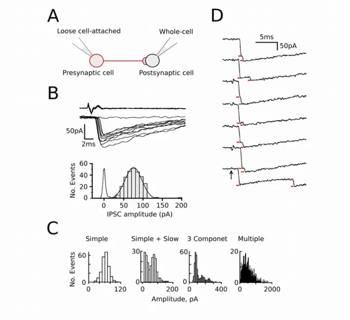
This leads us to yet another experimental situation: Two synaptically connected inhibitory interneurons, the presynaptic cell recorded under loose cell-attach clamp and the postsynaptic cell under whole-cell clamp (Figure 5A). When the presynaptic interneuron is electrically stimulated, the postsynaptic counterpart responds with an
inhibitory postsynaptic current (IPSC, top panel in Figure 5B). Sometimes failures can occur and in most of the cases one or a few peaks can be distinguished in the amplitude histogram. These results indicated that this type of synapse includes few active zones and, several categories of connections can be distinguished by the shape of the amplitude histogram (Figure 5C). It is reasonable to propose that a connected pair of interneurons that presents low variance in amplitude and a single component in its histogram is connected by a single active zone (Figure 5B; Kondo & Marty, 1998).
Now that we have a single active zone model, let us come back to the multivesicular release issue, and try to solve another relevant problem in all this controversy: the saturation level of the postsynaptic receptors. By analogy with the neuromuscular junction, low receptor occupancy was implicitly assumed in the early formulation of the one site-one vesicle hypothesis. However, in central synapses, if one release event is sufficient to highly saturate the postsynaptic receptors, a second simultaneous vesicle signal will be reduced in amplitude or perhaps undetectable (Auger et al., 1998). Examining closely timed pairs of synaptic currents (doublets), and studying the relation between amplitude of the second event and the time interval, it was possible to conclude that the degree of receptor occupancy after release of a vesicle is high at cerebellar interneurons (Auger & Marty, 1997). In such a case, multivesicular release would be revealed in the IPSCs as closely successive events that do not sum linearly, and display small amplitude current increments as shown in the Figure 5D (red lines). In fact, the lack of linear summation of IPSCs doublets confirms that they activate the same set of receptors and come from the same active zone. Which also explains the low variance in the amplitude histogram that has just one component, interpreted as one active zone connection (Auger et al., 1998). These connections were called simple synapses.
Cerebellar interneuron connections have become an important model to understand synaptic transmission because they provide high resolution recordings from a single synaptic contact. Even though interneurons present high postsynaptic receptor occupancy, they were one of the first synapses showing multivesicular release, an important fact to consider along the pursuit for the building block of synapses.
Quantal transmission at central synapses: the release sites controversy.
Considerable evidence has been obtained in recent years in favor of multivesicular release, including electro-micrographs that show the exact moment when simultaneous vesicular membrane fusion occurs (Abenavoli et al., 2002). In fact
Figure 5. Multivesicular release from a single synaptic contact recording. (A) Schematic drawing of the pair-recording configuration. (B) Recording of evoked IPSCs in cerebellar interneuron. Upper Panel: pre- and postsynaptic traces at a faster time scale. Only one failure is shown, but the failure rate was 0.63. Lower Panel: amplitude distribution for successful IPSCs. The eIPSC distribution can be fitted to a single Gaussian. The histogram of the recording noise is also shown. (C) Classification of paired recording according to the shape of he amplitude histograms. The amplitude variability increases with the number of synaptic connections. The distributions fitted by one Gaussian are called Simple synapses. (D) Displaying IPSCs traces on a fast time scale, sometimes it reveals two events in close succession (red bars), showing multivesicular release at single connections. (A) - (C) adapted from (Kondo & Marty, 1998). (D) adapted from (Auger & Marty, 2000).
multivesicular release occurs at many inhibitory and excitatory synapses throughout the brain, including hippocampus, cerebral cortex, cerebellum, hypothalamus and sensory synapses (Rudolph et al., 2015).
However, it appears in certain synapses that transmitter from one vesicle is far from fully occupying postsynaptic receptors and that multivesicular release can still happen (Rudolph et al., 2015). A particular case occurs at hippocampal GABAergic synapses, where due to low postsynaptic receptor occupancy, the IPSC amplitude increases by several folds in a high release probability condition. The analyses of the variance and mean of those IPSC amplitudes (method adapted by Clements and Silver in 2000 and explained in chapter 4), together with anatomical reconstructions, have shown that actually there is an increase in the signal provided by a given active zone when increasing external calcium, which indeed reflects multivesicular release (Biró et al., 2006).
Receptor occupancy is an important consideration in assessing the consequences of simultaneous release. Multivesicular release allows synapses with low receptor occupancy to enhance their dynamic range. While high occupancy synapses forfeit dynamic range due to insensitivity to changes in release probability, they operate with greater fidelity (Rudolph et al., 2015). Since multivesicular release occurs at single CNS active zones, the definition of release site should be reconsidered and may move down by one level in the synaptic hierarchy, being associated with the vesicles that are attached to the active zone, as will be discussed in the next chapter.
Chapter 3: Molecular Nature Of The Building
Block Of The Synapses.
When an impulse arrives at the synaptic terminal, calcium inflow triggers vesicle fusion and neurotransmitter release. By this mechanism multiple vesicles can fuse simultaneously at the presynaptic membrane, secreting neurotransmitter into the synaptic cleft. Thus the release site contemplated by Katz, would be the physical site where each vesicle docks in close association with the active zone at the presynaptic membrane, called docking sites (Figure 3; Katz, 1969). Those sites where the vesicles are docked would be the building block of the synapses.
In recent years, morphological, biochemical and functional evidence has been accumulated supporting the existence of several docking sites per bouton. Docking sites are specific macromolecular structures, where vesicles must bind before undergoing exocytosis. In this chapter we will describe dense aggregates of macromolecules called active zone material (AZM) and discuss their role in the docking of synaptic vesicles on the presynaptic membrane. I will start describing how these macromolecules are physically distributed on the AZ and later on, I will give names to these molecules, trying to describe proteins and complexes involved in the docking sites structural formation. Finally, I will introduce some models that explain the physiology of the docking sites at the active zone.
3.1 Active zone Material and synaptic vesicle Docking.
Vesicular release is an extremely fast process triggered by the influx of calcium ions. Llinas and colleagues measured the time course of calcium ions concentration changes after an evoked presynaptic action potential. They found that the delay for triggering transmitter release by the voltage-dependent calcium influx is in the order of 200µs (Llinás, Steinberg, & Walton, 1981). In order to get ultrafast neurotransmitter release, it is essential that vesicles are directly located near the active zone membrane and near calcium channels. This physical conformation requires specific
Molecular nature of the building block of the synapses.
interactions between molecules located at the vesicular membrane, the active zone plasma membrane and in the cytomatrix of the active zone. Tentative candidates that could regulate these interactions are the macromolecules at the AZM, because of their prominence and of their proximity to docked vesicles and calcium channels (white arrow in Figure 6A). However, during years it has been difficult to study the AZM because of its small size, of the high density and complex arrangement of macromolecules (Szule et al., 2012).
In recent years, McMahan and colleagues proposed a convincing model of the architecture of the AZM molecules. They implemented the last advances in electro-tomography and virtual slices processing in tissue sections from NMJ synapses of
Figure 6. Docking sites are organized together with Active Zone Material at the neuromuscular junction. (A) Virtual slices showing the transverse distribution of macromolecules of the AZM (white arrow) at frog neuromuscular junction active zone. In the middle and right panel the distribution of different classes of AZM macromolecules are represented by colors. These macromolecules tether secondary vesicles, which are in line to be docked at active zone; as soon as the site turns free a series of macromolecular interactions guide the secondary vesicle to the docking site. (B) 3D schematic models of the active zone from mouse and frog neuromuscular junction, showing different distributions of AZM macromolecules: one line of vesicles in mouse (left panel; from Nagwaney et al., 2009) and two parallel lines of vesicles in frog (right panel; Szule et al., 2012). (C) Diagram of the distribution of layers of AZM macromolecules, showing in transverse plane of the active zone: the superficial layer including the macromolecules: beams, ribs and pegs; the intermediate layer: steps and spars; and the deep layer: mast, booms and topmast. (A) and (C) from (Szule et al., 2012).
frog and mouse. Surprisingly, they showed that even though the same macromolecules are present in both models, their organization is different: single band in the mouse and a paired double rows of AZM in the frog (Figure 6B). This immediately indicates that AZM varies even within the same synaptic type.
More in detail, the 2D electro-microscopy micrographs and the 3D reconstructions models have shown the presence of two groups of vesicles: primary vesicles that are directly interacting with the AZM bands and are selectively docked on presynaptic membrane; and secondary vesicles, that are located at the ends of the AZM bands. These secondary vesicles are particularly relevant to the present work. They are not docked vesicles, but they are in-line to be the following vesicles positioned in release sites (Figure 6A; Nagwaney et al., 2009). In order to guide vesicle docking, this model proposes the interaction of three main structural layers composed by different classes of macromolecules, which interact at different domains on the vesicle surface: the superficial layer, closer to the presynaptic membrane, the intermediate layer and the deepest layer, farther from the membrane and interacting with the recycling vesicle pool.
Briefly, this vesicle docking model describes eight classes of macromolecules distributed along the three organization layers (Figure 6C). At the superficial layer are three classes of macromolecules called beams, ribs and pegs. Beams are connected to beams and ribs, ribs connected to docked vesicles and pegs, and pegs, presumably connected to macromolecules in the membrane such as calcium channels. At the intermediate layer we find the steps, which interact with the beams, and the spars parallel to the ribs and connected with four docked vesicles. Finally, at the deepest layer there are mast, boom and topmast macromolecules. Masts are connected to the steps and to the boom, boom contacts the docked vesicle and eight booms radiate from the mast to end interacting with four docked vesicles. Finally they described topmast, an elongated macromolecule, linking the end of mast to the membrane of an undocked vesicle, keeping it near docked vesicles (Szule et al., 2012).
After a docked vesicle fuses with the membrane, undocked vesicles are directed by AZM macromolecules to the docking sites by an orderly series of interactions.
Molecular nature of the building block of the synapses.
Starting from the deepest layer, a pre-docked vesicle first establishes contact with topmast. Topmast then guides the vesicle to interact with the rest of macromolecules classes at that layer, followed by the intermediate macromolecules. Finally, the vesicle is brought to the superficial layer to the docking site position. Micrographs have shown that all macromolecular classes help to maintain the new vesicle at the docking site until it fuses with the membrane during synaptic transmission.
3.2 Molecular Machinery at the Active Zone.
As I have emphasized, vesicular release is a fast process, meaning that some vesicles should be already physically in contact with the presynaptic membrane, waiting for an action potential. McMahan nicely showed a model where macromolecules are in charge of positioning vesicles in primed or even in
super-primed position, just short of the last step made by calcium influx to evoke fusion and
release. Now it is time to discover the molecular nature of the machinery involved in vesicular fusion.
Let us think about this situation: we have a pool of vesicles and we want some of them to be close to the active zone membrane to start membrane fusion. For simplicity let us focus on just one vesicle, but be aware that the chain of events that I explain below could occur simultaneously in several places of the same active zone, depending on the number of sites available to dock vesicles (reviewed from: Südhof, 2013).
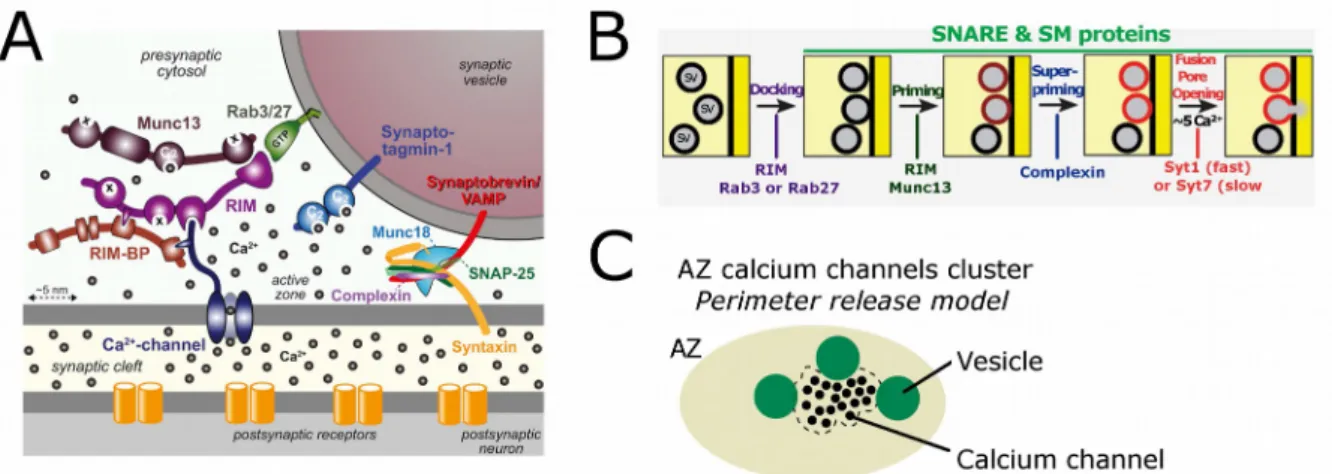
To keep our vesicle anchored at the AZ membrane, specific interactions would be necessary between proteins located at the vesicular membrane, at the AZ membrane and in the cytomatrix of the AZ. One of the assembly complexes made by the interactions of proteins located in these three regions is the SNARE/SM complex which triggers fusion. Synaptobrevin, a vesicular SNARE protein, forms a complex with its counterparts at the synaptic membrane, the SNARE proteins Syntaxin-1 and
SNAP-25. The assembling of this complex is tightly controlled by Munc18, which is a
cytosolic protein involved in vesicular priming (Figure 7A; Südhof, 2013; Haucke, Neher, & Sigrist, 2011). This SNARE/SM complex is one example of direct protein interaction anchoring the vesicle to the AZ membrane.
At this first stage, we have held the vesicle to the membrane, however we still need to address an important question: How is it possible that, after the inflow of calcium, a vesicle can be released in less than 200µs? There are still two important steps missing to gain this battle in favor of speed. One is a vesicular calcium sensor protein, as Katz already predicted; and second, an anchoring of that calcium sensor to calcium channels and to the rest of protein complexes. This tight assembling ensures that each calcium ion passing trough the channels is immediately captured by the sensor, triggering all the molecular machinery involved in vesicular membrane fusion.
Synaptotagmins are vesicular proteins with two cytoplasmic C2 domains that bind
calcium. By the use of point mutations on the endogenous Synaptotagmin-1 gene, it has been shown that a decrease in their calcium binding affinity by about 2-fold, also translates into a decrease in the calcium affinity of neurotransmitter release by a factor of 2-fold, making synaptotagmins good candidates for the calcium sensors (Südhof, 2013).
Figure 7. Molecular organization of the active zone. (A) Molecular model of the active zone protein complex coupled with calcium channels and its relation to the synaptic vesicle fusion machinery by calcium influx triggering. (B) Schematic representation of synaptic calcium-controlled neurotransmitter release. Presumably, partial SNARE complex assembly (from docking up to super-priming state) may precede calcium triggering of exocytosis. (C) Cartoon showing a possible active zone topography for calcium channels and synaptic vesicles released by a single AP in the calyx of Held. Releasable synaptic vesicles are positioned at the perimeter of calcium channel cluster. Nakamura et al. speculate that there are more than one releasable vesicle per active zone (Nakamura et al, 2015). (A) and (B) adapted from (Südhof, 2013).
Molecular nature of the building block of the synapses.
On the other hand, there is another large protein complex involving three multidomains proteins, called: RIM, RIM-BP and Munc13. This complex apart of mediating the docking of the vesicle at the active zone, is also important in recruiting calcium channels, placing the calcium sensor in the vicinity of the channels. RIM proteins are cytosolic proteins that bind to Munc13, calcium channels and a vesicular trans-membrane protein, Rab3 (Südhof, 2012). RIM and RIM-BP bind to each other, and both bind to calcium channels. Electrophysiological recordings from cultured hippocampal neurons and from the calyx of Held have suggested that the deletion of RIM-BP increases the average distance between calcium channels and synaptic vesicles (Acuna et al., 2015).
Let us try to put all the process together. Our vesicle, which is coming from the reserve pool, is guided from the backfield of the synapse up to the active zone by a series of interactions mediated by Piccolo and Bassoon, proteins specific to vertebrates that have been related with vesicle clustering and tethering (Hallermann et al., 2010). The vesicular anchoring begins with the action of RIM that interacts with Rab3, starting the pulling of the vesicle to the proximity of calcium channels (Figure 7B). A second state is priming, which involves the binding of Munc13 to RIM, and the switching of syntaxin-1 from close to open conformation, starting the assembly of the SNARE/SM complex. Further, vesicles can reach a super-primed state by the action of complexin, an universal cofactor for synaptotagmin and a fundamental component in the assembling of SNARE complexes. Actually, it has been shown that loss of function of complexin could cause a decrease in calcium-triggered release, an increase of spontaneous release, and a decrease in the number of vesicles ready to be released. Finally, this super-primed vesicle is now the substrate of synaptotagmin, the synaptic calcium sensor candidate, which is positioned less than 100nm away from calcium channels. This architecture allows direct influx of calcium trough channels to synaptotagmin, which triggers vesicular fusion. All these proteins form a single complex, the so-called Docking Site (Südhof, 2013).
3.3 Distribution of calcium channels along the active zone
I have been describing proteins sensitive to calcium that are close to calcium channels. The efficiency of this coupling is of special importance for securing