HAL Id: tel-01089002
https://tel.archives-ouvertes.fr/tel-01089002
Submitted on 30 Nov 2014HAL is a multi-disciplinary open access
archive for the deposit and dissemination of sci-entific research documents, whether they are pub-lished or not. The documents may come from teaching and research institutions in France or abroad, or from public or private research centers.
L’archive ouverte pluridisciplinaire HAL, est destinée au dépôt et à la diffusion de documents scientifiques de niveau recherche, publiés ou non, émanant des établissements d’enseignement et de recherche français ou étrangers, des laboratoires publics ou privés.
Differential Effects of the Cytokine Thymic Stromal
Lymphopoietin on Human Dendritic Cell Subsets
Carolina Martinez Cingolani
To cite this version:
Carolina Martinez Cingolani. Differential Effects of the Cytokine Thymic Stromal Lymphopoietin on Human Dendritic Cell Subsets. Immunology. Université Paris Sud - Paris XI, 2013. English. �NNT : 2013PA11T083�. �tel-01089002�
UNIVERSITE PARIS-SUD
École Doctorale de Cancérologie Discipline: Immunologie
THESE DE DOCTORAT
Intitulée:
Differential effects of the cytokine Thymic Stromal Lymphopoietin on
human dendritic cell subsets
Soutenue par
Carolina MARTINEZ CINGOLANI
Le 29 novembre 2013
Directeur de Thèse : Vassili SOUMELIS
Composition du jury :
Président du jury : Annelise BENNACEUR
Table of Contents
PREAMBLE ... 5
LIST OF ABBREVIATIONS ... 7
1 INTRODUCTION ... 9
1.1 DENDRITICCELLS:HISTORYOFAMAJORDISCOVERY ... 11
1.2 ORIGINOFHUMANDENDRITICCELLS ... 12
1.2.1 FROMMICETOHUMANS ... 12
1.2.2 LANGERHANSCELLORIGINS ... 15
1.2.3 TRANSCRIPTIONFACTORSINVOLVEDINDENDRITICCELLDEVELOPMENT ... 16
1.3 LYFECYCLEOFHUMANDENDRITICCELLS ... 17
1.3.1 DENDRITICCELLCHARACTERISTICSANDFUNCTION ... 17
1.3.2 TCELLPOLARIZATIONBYDENDRITICCELLS ... 19
1.3.3 HUMANDENDRITICCELLMIGRATION ... 20
1.4 HUMANDENDRITICCELLSUBSETS ... 23
1.4.1 BLOODDENDRITICCELLS ... 24
1.4.2 DENDRITICCELLSUBSETSINTHESECONDARYLYMPHOIDORGANS ... 26
1.4.3 DENDRITICCELLSINTHETHYMUS ... 28
1.4.4 DENDRITICCELLSUBSETSINPERIPHERALTISSUES ... 28
1.4.5 DIFFERENTSUBSETSSUGGESTDIFFERENTFUNCTIONS ... 32
1.5 THYMICSTROMALLYMPHOPOIETINBIOLOGYINHUMANS ... 34
1.5.1 TSLPANDTSLPRECEPTOR ... 34
1.5.2 TSLPEFFECTSONHUMANDENDRITICCELLFUNCTION ... 35
1.5.3 TSLPANDALLERGICDISORDERS ... 36
1.5.4 TSLPANDIMMUNEHOMEOSTASIS ... 39
1.5.5 TSLPANDDENDRITICCELLSUBSETS ... 39
2 OBJECTIVES ... 43
3 MATERIALS AND METHODS ... 45
4 RESULTS ... 53
4.1 PUBLICATION1 ... 55 Human blood BDCA-1+ dendritic cells differentiate into bona fide Langerhans cells with Thymic Stromal Lymphopoietin and TGF-β.
4
4.3 PUBLICATION3 ... 101
Molecular mechanisms implicated in TSLP induction of dendritic cell migration. 5 GENERAL DISCUSSION AND PERSPECTIVES ... 119
5.1 HUMAN BLOOD DC SUBSETS AS DC PRECURSORS. ... 121
5.2 RELEVANCE OF TSLP+TGFΒ -DERIVED LCS TO HUMAN PATHOLOGY ... 122
5.3 DIFFERENTIAL MIGRATION OF TSLP BLOOD BDCA-1+ AND BDCA-3+DCS... 124
6 REFERENCES ... 127
7 APPENDIX ... 139
7.1 APPENDIX1 ... 141
Other effects of TSLP on blood dendritic cell subsets. 7.2 APPENDIX2 ... 145
Telomere crisis in kidney epithelial cells promotes the acquisition of a microRNA signature retrieved in aggressive renal cell carcinomas. AKNOWLEDGEMENTS ... 155
PREAMBLE
The human body is in permanent contact with millions of microbes that live around it, on it or within it. The immune system, in all its complexity ensures the maintenance of our internal homeostasis. Numerous cellular and molecular actors participate in time and space in the orchestration of the immune response. These actors have been classified as part of the innate or adaptive immune systems. The innate system is characterized by an immediate antigen non-specific response. It is constituted by cells at the barrier surfaces and several types of immune cells such as macrophages, granulocytes, mast cells, eosinophils and natural killer cells. The adaptive immune system is characterized by a response that is specific to the pathogen. This specific response is provided by B and T lymphocytes and generates a long-term immunological memory.
At the interface between these two systems we find dendritic cells (DCs). These cells detect when the tissue microenvironment equilibrium is perturbed, and they sense, capture and process foreign antigens. First, activated DCs help in the recruitment of innate actors to the tissue. Then they migrate to the lymph nodes where they activate naïve T cells in an antigen-dependent manner, activating the adaptive immune response. To link a specific T cell response to the type of inflammation, DCs integrate multiple signals provided by the inflammatory milieu. As a first level of complexity, the type of T cell antigen-specific response depends on the type of antigen and molecules that activated the DCs. A second level of complexity is added by the fact that DCs constitute a heterogeneous and dynamic population and different DC subsets are associated with specific T cell outcomes.
In the context of allergic inflammation, DCs are activated by tissue factors that instruct them to induce an excessive immune response to certain non-pathogenic antigens called allergens. One of these factors is Thymic Stromal Lymphopoietin (TSLP), a cytokine produced by the skin keratinocytes that activates DCs. TSLP-activated DCs mediate the recruitment and activation of innate cells such as basophils and eosinophils and induce the differentiation of naïve T cells into effector cells with a pro-allergic phenotype, called inflammatory Th2. To induce a Th2 polarization, TSLP-activated DCs need to get in contact with the naïve T cells in the lymph nodes, yet the mechanism by which TSLP-treated DCs migrate is unknown. Although TSLP has been shown to stimulate several immune cells in the murine system, interestingly in humans, TSLP preferentially targets primary DCs. The DC subsets activated by TSLP may have differential implications in TSLP-linked allergic disorders. Nevertheless, human DC diversity and the potential differential effects of TSLP on human DC subsets remain unexplored. In this context, I dedicated my PhD work to the study of TSLP effects on human DC subsets. I assessed the differential response of human DC subsets to TSLP and studied the effects of TSLP s on DC migration.
6
My results will be presented in three chapters. First, I will present a submitted article showing
that TSLP and TGF-β synergize to induce Langerhans cell differentiation from BDCA1+ but
not BDCA3+ blood DCs. Then I will present our published results showing that TSLP induces
DC migration. Finally I will present a manuscript in preparation assessing the molecular mechanisms implicated in TSLP-induced DC migration.
In the discussion section at the end of this manuscript I will put my results in perspective to published studies in related topics.
In the appendix, I will show ongoing work on the study of TSLP effects on DC subsets and other projects in which I collaborated during my PhD thesis.
LIST OF ABBREVIATIONS
DCs: Dendritic Cells
CBA: Cytometric bead Array
CDPs: Common Dendritic cell Precursors
CLA: Cutaneous Lymphocyte Associated protein
CLPs: Common Lymphoid Precursors
CMPs: Common Myeloid Precursors
Flt3L: Fms-like tyrosine kinase 3 Ligand
Flu: Influenza virus
GATA2: GATA-binding factor 2
Giα: Small G inhibitory protein α
GM-CSF: Granulocyte-Macrophage Colony-Stimulating Factor
GMPs: Granulocyte-Macrophage Precursors
GPCR: G-Protein-Coupled Receptor
HSCs: Hematopoietic Stem Cells
IFN: Interferon
IL: Interleukin
LCs: Langerhans Cells
LCH: Langerhans Cell Histiocytosis
LPS: Lipopolysaccharide
M-CSF: Macrophage Colony-Stimulating Factor
M-CSFR: Macrophage Colony-Stimulating Factor Receptor
MDPs: Macrophage/Dendritic cell Progenitors
MHC: Major Histocompatibility Complex
MLPs: Multi-Lymphoid Progenitors
MMPs: Matrix Metalloproteases
MTOC: Microtubule-Organizing Center
PAMPs: Pathogen-Associated Molecular Patterns
PBMCs: Peripheral Blood Mononuclear Cells
PDCs: Plasmacytoid Dendritic Cells
Pre-DCs: Precursors for Dendritic Cells
PRRs: Pattern Recognition Receptors
PTX: Pertussis Toxin
TGF: Transforming Growth Factor
TLRs: Toll-like receptors
TNF: Tumor Necrosis Factor
TSLP-DCs: Thymic Stromal Lymphopoietin-Primed Dendritic Cells
TSLP-PDCs: Thymic Stromal Lymphopoietin Primed Plasmacytoid Dendritic Cells
INTRODUCTION
1.1 DENDRITIC CELLS: HISTORY OF A MAJOR DISCOVERY
The first evidence of the existence of DCs was made by a medical student called Paul Langerhans in 1868 [1, 2]. He had discovered in epidermal tissue sections, what he thought was a new cell type of the nervous system that was named then Langerhans cells (LCs) (Figure 1-1). The origin of LCs and their link to immunology was not known. The major discovery in the history of DCs was made in 1973 when Ralph Steinman and Zanvil Cohn identified a new cell type in the spleens of mice [3]. They found that these cells were different from macrophages and other leukocytes and because they had tree-like cytoplasmic extensions they decided to name them dendritic cells (Figure 1-1). Within few years they enriched this spleen population and did functional studies revealing the potent T cell-activating capacity of DCs [4]. In the meanwhile, LCs were ontogenically linked to melanocytes [5] and to keratinocytes [6]. They were also linked to histiocytes when they were found within the bone and lung lesions of patients suffering from a disease called first “histiocytosis X” (later on called LC histiocytosis) [7]. The demonstration that LCs were bone marrow-derived leukocytes, and several studies showing their immunological role [8, 9], led to the final recognition of LCs as DCs in 1985 by Gerold Schuler and Ralph Steinman [10]. This last study showed that LCs, after several days in culture, acquired a mature DC phenotype and induced a strong response in naïve T cells.
The three major criteria defining DCs were proposed by Steinman in 1991 [11]: (1) dendritic morphology, (2) constitutive expression of high levels of major histocompatibility complex (MHC) class two molecules and (3) capacity to induce proliferation of naïve CD4 T cells in a mixed leukocyte reaction.
In the last 30 years, the studies of DC biology have multiplied and have rapidly shed new light on their origin and function. Today, DCs are still considered to be the most powerful stimulators of naïve T cells and the key cells initiating and shaping the immune cell response.
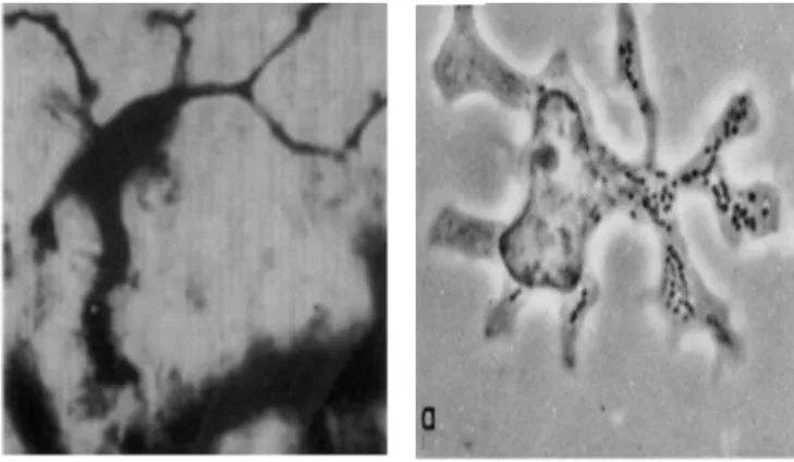
Figure 1-1: Stellar morphology of dendritic cells.
Left, a Langerhans cell seen by Paul Langerhans in 1686, adapted from Jolles, S., 2002. Right, a dendritic cell seen by Ralph Steinman in 1973, Steinman, R. 1973.
12
1.2 ORIGIN OF HUMAN DENDRITIC CELLS
The DC pool needs to be continuously maintained in the organism. Different factors and cells participate in this process. DC requirements and sources differ in steady-state and inflammatory conditions. The purpose of this chapter is to review our current knowledge on the origins of human DCs and the missing links in DC generation during inflammation. Due to their particular development, a separated paragraph will be dedicated to LC ontogeny. Finally a brief description of the transcription factors that are involved in DC development will be given.
1.2.1 FROM MICE TO HUMANS
Human DCs are generated on a regular basis from hematopoietic stem cells (HSCs) located in the bone marrow. Several experiments in mice, of isolation and further transplantation of bone marrow precursor cells, have helped to clarify the ontogeny of DC and monocyte-macrophage lineages [12, 13].
The current models propose that bone marrow HSCs, characterized by the expression of the surface molecule CD34, give rise to non-self renewing multipotent progenitors which give rise to proliferating progenitors that gradually become lineage-restricted. Two early committed progenitors have been identified in mice and human, the common lymphoid precursors (CLPs) and the common myeloid precursors (CMPs) [14]. For a long time it was believed that the CLPs gave rise to the lymphoid lineages and plasmacytoid dendritic cells (PDCs) whereas the CMPs gave rise to monocytes, macrophages and DCs. Today, we know that both precursors maintain the capacity to generate DCs and PDCs [15, 16] depending mainly on the expression of Fms-like tyrosine kinase 3 ligand (Flt3L) [17-19]. Indeed, Flt3L injection, in humans leads to massive expansion of blood PDCs and DCs [20].
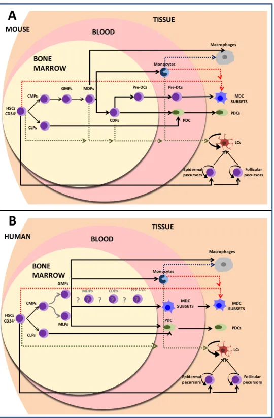
A scheme showing all the intermediate precursor populations that give rise to DCs in mice and humans is depicted in Figure 1-2.
In mice it has been proposed that in addition to CMPs there are also granulocyte-macrophage precursors (GMPs) and macrophage/DC progenitors (MDPs) (Figure 1-2A). The MDPs give rise to monocytes and to common DC precursors (CDPs). The CDPs further develop into PDCs and precursors for DCs (pre-DCs) that do not have anymore the potential to give rise to monocytes. At a steady state, pre-DCs are found in the bone marrow, the blood and the spleen. They acquire further surface phenotype and morphology of DCs and enter the peripheral tissues, including the lymphatic tissues [13, 21].
The equivalents of MDPs and CDPs have not yet been described in humans. Yet, it has been shown that there are progenitors with combined myeloid and lymphoid potentials [22]. GMPs and the multi-lymphoid progenitors (MLPs) appear to have DC potential. The circulating human blood precursors of DCs (equivalents of mice pre-DCs) must exist. However, a clear
INTRODUCTION
phenotype, distinct from those of the terminally differentiated DCs has not been identified (Figure 1-2B). I will detail the precursor capacity of human blood DC subsets, and the hypothesis that these cells may be the equivalents of the murine pre-DCs, in the “General discussion and perspectives” chapter of this manuscript.
During inflammatory conditions, other cellular precursors of DCs have been found. In vitro experiments with human cells have shown that exposure to cytokines such as granulocyte-macrophage colony-stimulating factor (GM-CSF) and Interleukin (IL)-4, induces the differentiation of human monocytes into immature DCs [23]. An addition of proinflammatory
cytokines such as Tumor Necrosis Factor (TNF)-α [24], microbial products such as
lipopolysaccharide (LPS) or T cell-derived CD40L further activates them into mature DCs whereas exposure to macrophage colony-stimulating factor (M-CSF) induces monocytes to differentiate into macrophages. This confirms mice studies showing that under inflammatory conditions, blood monocytes give rise to macrophages and DCs [21]. DCs can also be
generated in vitro from CD34+ HSCs isolated from human peripheral blood or cord blood.
This differentiation is dependent on Flt3L and further GM-CSF and TNF-α stimuli [25, 26].
This suggests that under inflammatory conditions, the CD34+ HSC circulating in human blood
could be a new source of DCs besides monocytes. Finally, the possibility that inflammatory tissue signals activate the potential precursor role of blood DCs remains elusive and will be discussed later in this manuscript.
14
Figure 1-2 : Dendritic cell ontogeny.
A. Dendritic cell development in mice. B. Dendritic cell development in human. Grey arrows show
undetermined relationships. Pointed arrows show the precursor-cell relationships under inflammatory conditions. (HSCs, hematopoietic stem cells; CMPs, common myeloid precursors; CLPs, common lymphoid precursors, GMPs, granulocyte-macrophage precursors; MDPs, macrophage/DC progenitors; CDPs, common dendritic cell precursors; pre-DCs, precursors for dendritic cells; MDCs myeloid dendritic cells, PDCs, plasmacytoid dendritic cells; MLPs, multi-Lymphoid progenitors, LCs Langerhans cells). HSCs CD34+ CMPs CLPs GMPs MDPs CDPs Pre-DCs Monocytes PDC Pre-DCs BLOOD BONE MARROW MDC SUBSETS Macrophages PDCs Epidermal
pecursors pecursorsFollicular LCs TISSUE MOUSE HSCs CD34+ CMPs CLPs GMPs MDPs CDPs Pre-DCs Monocytes PDC BLOOD BONE MARROW MDC SUBSETS Macrophages PDCs Epidermal
pecursors pecursorsFollicular LCs TISSUE HUMAN ? ? ? ? ? ? MDC SUBSETS MLPs
A
B
INTRODUCTION
1.2.2 LANGERHANS CELL ORIGINS
LCs have been shown to have a particular ontogeny. Recent experiments in mice showed that in contrast to other DCs, LCs develop from an embryonic precursor that colonizes the epidermis before birth. Then, in the adult mouse, LCs self-renew from unknown local precursors in the epidermis [27] (Figure 1-2).
In human, these findings are confirmed by the fact that LCs proliferate in situ [28-30] and donor LCs were shown to persist in human skin that was transplanted into immunodeficient mice [31]. Moreover, human limb transplants showed that donor LCs persist for years [32, 33]. Finally, several reports have shown that there are proliferating cells in the bulge region of the hair follicle that could contribute to LC generation [34].
The fact that mice lacking the transforming growth factor (TGF)-β1 do not have LCs [35]
shows that this factor is essential for LC development. Studies in humans confirmed this
finding; TGF-β1 is required for the in vitro generation of LCs from human CD34+ precursors
[36, 37] or CD14+ monocytes [38] (Figure 1-2). In addition it was recently shown that the
macrophage colony-stimulating factor receptor (M-CSFR) is required for the development of LCs in mice. The high affinity ligand of this receptor, IL-34, might be essential for the development of LCs precursors, LCs differentiation, survival or proliferation [28]. Further experiments on IL-34 and human LC development are needed to translate this finding into human biology.
The in vitro generation of LCs from CD34+ precursors and CD14+ monocytes confirmed that
in humans, just as in the mouse model, blood precursors are recruited to renew LCs in
inflammatory conditions [39]. Blood CD34+ cells stimulated with a combination of Fl3L,
GM-CSF, TNF-α and TGF-β, as well as CD14+ monocytes stimulated with GM-CSF, IL-4
and TGF-β differentiate into bona fide LCs [23, 37, 38]. This shows that as opposed to steady
state conditions, in inflammatory contexts tissue –derived cytokines can trigger the differentiation of circulating precursors into LCs.
Whether blood DCs can differentiate into LCs under inflammatory conditions remains
controversial. Ito, T et al. showed that a fraction of human blood CD1a+ DCs stimulated with
GM-CSF, IL-4, and TGF-β can give rise to LCs in vitro [40]. However in my experience and
also as published by other groups [41], human blood DCs do not express CD1a at the cell surface or at the gene transcriptional level [42]. This discrepancy might be due to the use of
different cell isolation methods. To obtain the CD1a+ DCs, Ito, T. et al. enriched the DC
fraction of peripheral mononuclear cells, by magnetically depleting CD3+ (Lymphocytes) and
CD14+ (monocytes) cells. This depletion was followed by a positive selection of CD4+ cells
(DCs) and further sorting of lineage (CD3, CD7, CD14, CD16, CD19) –negative and CD1a+
cells. Monocytes can express CD4 marker [43]. And CD14+ monocytes that may have
16
To my knowledge, Ito, T. et al study is the only published work that assesses human DCs capacity to differentiate into LCs. The possibility that inflammatory cytokines from the tissues
can instruct human blood CD1a- DCs to differentiate into LCs remained unknown. The data
presented in the first section of the “Results” chapter reveal the capacity of blood DCs to differentiate into LCs.
1.2.3 TRANSCRIPTION FACTORS INVOLVED IN DENDRITIC CELL DEVELOPMENT
Recent descriptions of human DC deficiency have been helpful to identify several transcription factors implicated in DC development [44-46].
By analogy to mice, it seems that Ikaros, PU.1, Gfi1 and Id2 are involved in DC development in humans. These transcription factors regulate genes that encode proteins of the early hematopoiesis (such as Flt3, Il7r, and Stat3); therefore their mutations lead to severe defects in global hematopoiesis [45].
More specific transcription factors are linked to DC ontogeny. This is for example the case of E2-2; indeed, human deficiency on E2-2 leads to a PDC impaired function [47].
Moreover, mutation of GATA-binding factor 2 (GATA2) is the cause of a DC deficiency called DC, monocyte, B and natural killer lymphoid deficiency [48]. The loss of GATA2 is characterized also by the absence of MLPs and diminished GMPs numbers. The transcription factor IRF8 was found to compromise DC and monocyte development as well. But in contrast to the GATA2 mutation, the IRF8 mutation is associated to an expansion of all the progenitor compartments [49].
A transcription factor mutation affecting exclusively DC development has not yet been identified and could have important implications in our comprehension of DC generation under inflammatory conditions. A common transcription factor to all human DC subtypes has been found: zbtb46 [50, 51], but its contribution to human DC development has not been clarified.
INTRODUCTION
1.3 LYFE CYCLE OF HUMAN DENDRITIC CELLS
It is important to understand that a single DC may induce different T cell outcomes depending on the combination of signals that the DC receives. The purpose of this chapter is to highlight the special features that allow DCs to sense a wide range of signals from the tissue microenvironment and differentially induce particular T cell responses. In a separated paragraph, I will present the model of DC and DC precursor migration to the lymph nodes and the tissues. The particular effects of TSLP on human DC biology will be addressed later in the introduction.
1.3.1 DENDRITIC CELL CHARACTERISTICS AND FUNCTION
Fully differentiated, patrolling DCs are found in almost all the peripheral tissues at steady state. They get activated when they sense a particular threat.
Indeed, DCs express a broad set of receptors that allow them to recognize danger signals. Through different cytokine and chemokine receptors, they recognize microenvironment signals secreted by the neighboring cells in response to microbes. Through the pattern recognition receptors (PRRs) they recognize conserved molecules from microbes called pathogen-associated molecular patterns (PAMPs) [52]. These receptors include the Toll-like receptors (TLRs), C-lectins, NOD like receptors and RIG-I like receptors. These recognize different molecules from glycoproteins and polysaccharides to double or single stranded nucleic acids. Finally the Fc and complement receptors allow DCs to capture antigens from microbes and apoptotic cells.
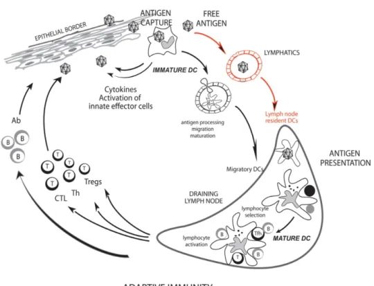
Once they get activated, DCs change their morphology, and phenotype. They up-regulate major histocompatibility molecules, costimulatory molecules (CD80, CD83, CD86 and CD40) and chemokine receptors that trigger migration [53]. All these changes enable them to interact with T cells. Nevertheless they first secrete inflammatory mediators, cytokines and chemokines that induce the recruitment and activation of different actors of the innate immune response such as granulocytes, natural killer cells, and other DCs (Figure 1-3) [10, 48].
Next, they enter the afferent lymphatics and reach the draining lymph nodes were they present the captured antigens to T cells. The endogenous proteic antigens are loaded onto MHC molecules of class I to be presented to cytotoxic T cells expressing the surface marker CD8. This response leads to cell apoptosis. The exogenous proteic antigens are bound to MHC class II molecules and presented to T cells expressing the surface marker CD4 leading to a T helper cell response [54]. The exogenous molecules can also be presented in the context of MHC class I molecules to CD8 T cells, a process called presentation. DCs indeed,
cross-18
cell. This mechanism is also required for antitumoral responses even if the mechanisms are not well understood yet [55]. Finally the lipidic antigens are presented through the CD1 (a-d) molecules to T cells and natural killer T cells.
In conclusion, DCs interact directly and indirectly with all the cell types responsible for the regulation of the innate and adaptive immune systems. The way DCs induce particular immunological responses that are adapted to the requirements of the inflammatory milieu, will be presented in the next chapter.
Figure 1-3: Dendritic cells at the interface between innate and immune systems. Adapted from Ueno H. et al., 2010.
Immature dendritic cells capture antigens at the periphery and become activated. They release inflammatory mediators in situ. They process the captured antigen and in parallel acquire a mature phenotype. They migrate through the afferent lymphatics to the lymph nodes to present the antigens to the CD4 T and CD8 T cells. T helper cells develop into follicular T helper cells that induce B cell activation to antibody secreting cells, T regulatory cells that mediate tolerance and T helper cells inducing Th1, Th2 and Th17 responses. T helper cells activate the cytotoxic CD8 T cells to induce clearance of infected cells. (DC, dendritic cells; Tfh, T follicular helper cells; B, B lymphocyte; Tregs, T regulatory cells; Th, T helper cells; CTL, cytotoxic T lymphocytes; Ab, antibody).
INTRODUCTION
1.3.2 T CELL POLARIZATION BY DENDRITIC CELLS
The special feature of DCs in comparison with other antigen-presenting cells is their unique capacity to prime naïve CD4 T cells leading to the generation and proliferation of antigen-specific helper and memory T cells [54].
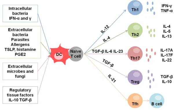
DCs and T cells interactions in the lymph nodes are complex and give rise to different types of T cell responses that depend on the priming of the DCs themselves. Therefore depending on the activating stimuli (tissue factors), DCs express one or another combination of what can be called DC factors, cytokines and surface molecules, which are sensed by naïve T cells. Then, the naïve T cells get differentiated into different types of T helper cells. The T helper cells secrete T cell factors that define the type of T cell response (Figure 1-3).
In summary, DCs activated by intracellular bacteria and viruses, can produce large amounts of
IL-12, and induce T helper cells that secrete IFNγ and TNF-α. This T helper response is called
Th1 (Figure 1-4).
DCs activated by extracellular microbes such as helminthes induce T cells that produce IL-4, IL-5, and IL-13. This immune response is called a Th2 response. It characterizes the allergic responses (Figure 1-4).
DCs secreting variables amounts of IL-1b, IL-23, IL-6 and TGF-β induce a Th17 response.
The T helper cells induced are characterized by the secretion of IL-17A, IL-17F and IL-22 (Figure 1-4). They are responsible for immunity against extracellular microbes and fungi. They promote an important neutrophil response and are often linked to autoimmune diseases such as psoriasis.
The Th1, Th2 and Th17 are not the only T cells that result from the DC-T interaction. In fact, DCs can also induce regulatory T cells that have an important role in the immunological tolerance [56]. The priming of T regulatory cells seems to be achieved at the steady state, by the constant presentation of self antigens in the context of MHC class I, or when DCs stimulate T cells while expressing low amounts of costimulatory molecules. T regulatory cells
regulate the immune response through the secretion of IL-10 and TGF-β and play an
important role in the responses towards human microbiota (Figure 1-4).
Finally, concerning B cell activation, it is known that a group of T helper cells, present in the B follicular zones induce the B cell activation. It has been proposed that there are different T follicular helper cells depending on the DC-T cell priming (Figure 1-4). Moreover, the DCs also directly induce the activation of B cells and their differentiation to antibody-secreting cells [57] (Figure 1-3).
20
Figure 1-4: Dendritic cells induce different T cell responses.
Immature dendritic cells become activated by different microbes and tissue factors. Differentially activated DCs induce the polarization of naïve T cells into Th1, Th2, Th17 Tregs (regulatory T cells) and Tfh (T follicular helper cells). The different T cell profiles are defined by the cytokines secreted.
In order to prime naïve T cell differentiation, tissue-residing DCs must travel considerable distances, from the inflammation sites, to lymph nodes. Moreover, the peripheral pools must be replenished with new DCs. In the next chapter, the different molecules that are implicated in the process of DC migration will be reviewed.
1.3.3 HUMAN DENDRITIC CELL MIGRATION
Although free antigen can enter the afferent lymphatics and be presented by resident DCs in the lymph nodes [58] (Figure 1-3), DCs get activated in the periphery and migrate themselves to the lymph nodes to present the antigens. Again, most of our knowledge about DC trafficking comes from studies in mice mainly on LC migration.
LCs are retained in the epidermis through the expression of E-cadherin that mediates their binding to keratinocytes [59]. During inflammation, the DCs themselves and the surrounding
cells secrete IL-1β and TNF-α that induce the down-regulation of E-cadherin and the
up-regulation of the chemokine receptor CCR7 that mediates the lymph node homing [60-62]. Indeed, CCR7 has been shown to mediate migration of mouse skin DCs to lymph nodes under inflammatory and even steady state conditions [63]. Although in this last case, much less is known.
INTRODUCTION
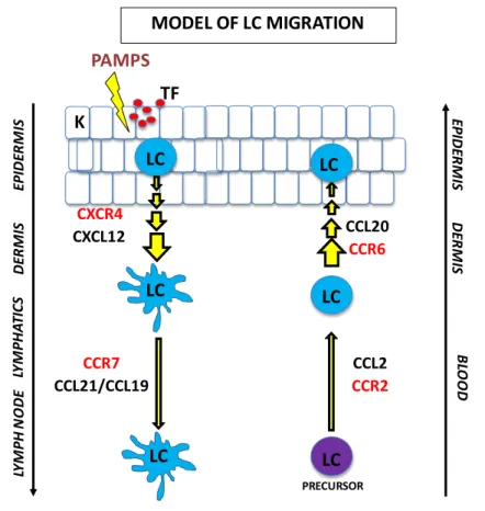
The two ligands of CCR7, CCL19 and CCL21 are expressed by the endothelial cells of the lymphatic vessels in the dermis. This means that first LCs need to migrate from the epidermis to the dermis. This seems to be mediated by the interaction between the chemokine receptor CXCR4 present in mature LCs and its ligand CXCL12 present in the dermis [53, 64-66] (Figure 1-5).
Once in the dermis, non-soluble CCL21 gradients guide the LCs into the lymphatic vessels [67, 68]. LCs squeeze through the gaps present in the basement membrane and are driven to the lymph nodes by a passive flow [69].
Finally, once in the lymph nodes, gradients of CCL19 and CCL21 will further guide the DCs to the T cell zone [70].
Besides chemokine – chemokine receptor interactions, there are other molecules implicated in the entrance of DCs to the lymphatics. Mature DCs also upregulate the receptor for sphingosine-1-phosphate, an inflammatory molecule implicated in leukocyte migration. Nevertheless its role in DC trafficking is still not defined [71].
Proteases such as the matrix metalloproteases (MMPs) are expressed by mature DCs. Activated LCs secrete MMP-2 and MMP-9 that help them to degrade the surrounding collagen matrix and trespass epidermal basal membrane [72]. The matrix metalloproteases can also act in DC migration via their action on chemokines, indeed they can mediate chemokine proteolysis into agonistic or antagonistic chemotactic fragments [73].
Finally during inflammation the lymphatic endothelial cells upregulate adhesion molecules such as E-selectin, ICAM-1 and Vcam-1 that interact with integrins expressed by mature DCs [74]. Nevertheless migration of DCs into the lymph nodes seems to be a process independent from integrins [75].
DC precursor migration from blood to the peripheral organs is less well understood. CCR6 is a skin-homing receptor that is mainly expressed on differentiated LCs. It binds to the CCL20
chemokine present in the epidermis. The LC precursors (monocytes and CD34+ HSC) express
low levels of CCR6; instead they highly express CCR2 that allows them to bind the CCL2 present in the dermis. Under inflammatory conditions, both CCL2 and CCL20 are secreted, and LC precursors can be recruited to the dermis through CCR2 and give rise to LCs that will be attracted to the epidermis through CCR6 [27, 76].
The model of LC migration is depicted in Figure 1-5. Excluding the dermis-epidermis passage, this model can be applied to DCs in general. Indeed monocytes and immature DCs express CCR1, CCR2, CCR3 and CCR5 that allow them to reach the inflamed tissues [53]. These chemokine receptors are down-regulated upon activation and replaced by high levels of CCR7 that allow them to reach the lymph node.
22
Figure 1-5 : Model of Langerhans cell migration.
First, Langerhans cells sense a threat in the epidermal layer, they get activated and upregulate the chemokine receptor CXCR4. This receptor allows LCs to sense a gradient (light blue background) of its ligand, CXCL12 that directs them to the dermis. Once in the dermis the expression of CCR7 receptor allows them to bind CCL19 and CCL21 expressed by the afferent lymphatic epithelial cells and in the lymph nodes. On the other hand, Langerhans cells precursors in the blood express CCR2 that mediates their homing to the dermis. There, the expression of CCR6 allows them to sense CCL20 gradients that will position them in the epidermis. (PAMPS, pathogen-associated molecular patterns; TF, tissue factors; K, keratinocytes; LC, Langerhans cells).
LC MODEL OF LC MIGRATION LC K EP ID ER M IS DER M IS LC LY M PH AT IC S LY M PH N O DE PAMPS TF CXCR4 CCR7 CCL21/CCL19 CXCL12 LC PRECURSOR LC LC CCL2 CCR2 CCR6 BL OOD CCL20 EP ID ER M IS DER M IS
INTRODUCTION
1.4 HUMAN DENDRITIC CELL SUBSETS
During my PhD thesis, in 2010, several publications appeared showing functional differences between human blood DC subsets. These functional differences represent a second level of complexity in the induction of T cell responses. I was studying TSLP effects on DC migration and my interest in the function of DC subsets led me to address the differential response of DC subsets to TSLP stimulation. The DC population is heterogeneous and the different subsets have overlapping phenotypes, therefore the purpose of this chapter is to present the DC diversity in human blood, lymphoid organs and tissues in a comprehensive way. After defining the nature and phenotype of the DC subsets, I will dedicate a paragraph to the studies that have assessed the functional differences between DC subsets.
Increasing evidence has highlighted the fact that the DC population is heterogeneous. Initially, LCs and PDCs were defined as different types of DCs [10, 77]. Then, the study of human blood, skin, lymph nodes, thymus and other tissues has confirmed the presence of several dynamic subsets of DCs in different maturation states [41]. Today, gene transcriptomic data on these different subsets are available. The comparison between the genes expressed in one or another sub-population has allowed defining their correspondent gene signatures. Statistical tools such as the hierarchical clustering and principal component analysis allow the unbiased and simultaneous analysis of all the genes (variables) from the different sub-populations and, preserving the variance of the data, provide a summary of the information were the similarities and differences between the datasets is easier to interpret. Furthermore the different signatures can be confronted to several public databases and translated as profiles enriched for different biological relevant gene sets (gene enrichment analysis). These types of analysis have further highlighted the different gene expression profiles of DC subsets and the relationships between them [42, 78-81].
In general the DC subsets can be defined according to the expression of different combinations of surface markers that allow their isolation from different human samples. Several studies attempted to correlate these different phenotypes to distinct functions in the immune response in steady-state and pathological conditions [25, 78, 82]. Nevertheless, no exclusive surface markers or functions can be attributed to the distinct subsets and their clearest classification may be the one linked to their location. Therefore I will present the different DC subsets according to their anatomical distribution based on the latest review of Collin et al on human DC subsets [83]. A summary of the different DC subsets and their phenotypes is shown in Figure 1-7 and Table 1-1.
24
1.4.1 BLOOD DENDRITIC CELLS
The blood DC subsets are very well characterized in humans and seem to be the ones that replenish the tissue and lymphoid organs pools. The first study in 1993, characterizing blood DC compartment, defined three initial subsets according to the surface markers CD33 and
CD14 [84]. This study included a population of monocytes (CD33+CD14bright) and two
populations of DCs, (CD33dimCD14dimCD16- or CD33brightCD14dimCD16-).
Later on in 2000, Dzionek et al, described blood DC subsets as we know them today; they used the surface markers BDCA-2 and BDCA-4 to identify PDCs, BDCA-1, and BDCA-3 to identify two subsets of myeloid DCs [85].
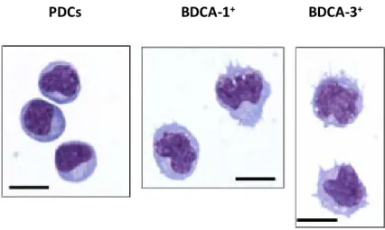
By inmmunohistological techniques like the Giemsa/May Grunwald staining, PDCs have a smooth, round, plasma-cell morphology; they display an eccentric kidney-shaped nucleus and
have a violet basophilic cytoplasm that contains a pale Golgi zone. Myeloid BDCA-1+ and
BDCA-3+ DCs are less rounded cells, with numerous short processes and more
hyperlobulated nuclei (Figure 1-6).
The three blood DC subsets express high levels of major histocompatibility complex class II molecules (HLA-DR) and lack typical lineage markers such as CD3 (T cells), CD19/CD20 (B cells) and CD56 (natural killer cells) [86].
Figure 1-6: Blood and tonsillar dendritic cell morphology. Adapted from Segura et al., 2013.
These images correspond to Giemsa/May-Grunwald staining of sorted DC subsets from the tonsils. PDCs are round-shaped and have a lymphoid-like morphology. The myeloid BDCA-1+ and BDCA-3+ subsets present short cytoplasmic processes and lobulated nuclei. (Bar corresponds to 10 μm).
BDCA-1+
INTRODUCTION
1.4.1.1 PLASMACYTOID DENDRITIC CELLS
PDCs constitute between 1 to 2 % of peripheral blood mononuclear cells (PBMCs). They are very different from DCs. They have round plasma-cell morphology and the capacity to secret large amounts of type-1 interferons after viral encounter [77, 87, 88].
In terms of surface markers, they are distinguished by the expression of CD123 (IL-3 receptor), BDCA-2 and BDCA-4 and the lack of myeloid surface markers (CD11c and CD11b). They express the endosomal nucleic acid–sensing TLR7 and TLR9 that allow them to recognize single-stranded RNA, and unmethylated CpG-containing DNA respectively [89]. The response to these stimuli is rapid IFN secretion, which helps the activation of natural killer and B cells, establishing PDCs as a key link between innate and adaptive immunity. PDCs circulate in the blood and enter the T cell rich areas of lymphoid organs through the high endothelial venules. Under steady-state conditions they are hardly found in peripheral tissues.
1.4.1.2 MYELOID DENDRITIC CELLS
DCs are characterized by the expression of the myeloid surface markers CD11c, CD13, CD33 and CD11b. As PDCs, they also express CD4. They do not express at the steady state CD14 or CD16 which characterize human blood monocyte subsets.
They are separated into two subsets, the BDCA-1+ and the BDCA-3+ subset (also called CD1c
and CD141, respectively). In the last two years, several studies including cross-species comparisons of transcriptomic profiles, have shown that these two subsets are the equivalent
of the mice myeloid subsets CD8α- and CD8α+ respectively [90-93]. Nevertheless these two
subsets in mice show clear functional differences that are less evident in their human counterparts.
BDCA-1+ DENDRITIC CELLS
BDCA-1+ DCs are the major population of human DCs. They are approximately 1% of
PBMCs. Besides the typical myeloid antigens, they also express CD172 (also named SIRPα).
The expression of the TLRs 1 to 8 allows them to recognize several pathogen associated molecules as LPS, flagellin and double stranded RNAs. They express dectins 1 (CLEC7a) and 2 (CLEC6A) that account for fungi recognition, DEC205 and macrophage mannose receptor (CD206) [42]. Once activated, they are very good stimulators of naïve CD4 T cells, and are
26
Depending on the antigen and TLR involved, they prime different types of T cell responses,
owing to their capacity to secrete different combinations of the cytokines TNF-α, IL-8, IL-10,
IL-12 and IL-23 [95].
BDCA-3+ DENDRITIC CELLS
This is the minor subset in human blood; it corresponds to 0.1% of PBMCs. The expression of the lectin CLEC9A, the chemokine receptor XCR1, the cellular adhesion molecule Necl2, and
high TLR3 have identified them as the human counterpart of the CD8α+ mouse subset [90-93,
96, 97]. Via CLEC9A, these cells have a good capacity to take up necrotic material from dead
cells, and via TLR3 and 8 they can sense viral nucleic acids. Mice CD8α+ cells have been
functionally defined as “cross-presenting” cells in opposition to the CD8α- cells. Human
blood BDCA-3+ cells can indeed cross-present antigens to CD8 T cells, but as opposed to
mice, they cannot be defined as the main cross-presenting subset as the BDCA-1+ has a
similar capacity [55, 94]. They can secrete the cytokines TNF-α, CXCL10, and IFNλ and
IL-12 [41, 92, 95].
Careful attention must be taken for their isolation, as BDCA-3 surface marker can be up-regulated upon maturation by other DC subset as well [85].
1.4.2 DENDRITIC CELL SUBSETS IN THE SECONDARY LYMPHOID ORGANS
The spleen and tonsils do not receive lymphatic flux. Therefore DC subsets in these organs are more likely resident cells coming from blood. In contrast, in the lymph nodes there are resident cells coming probably form blood and cells that have migrated from the peripheral organs.
1.4.2.1 TONSILS
In human tonsils, DCs are found mainly in the T cell zones and also in the germinal centers. The phenotype of DC populations was first described based on the expression of HLA-DR,
CD11c, CD13 and CD123 by Summers et al. [98]. The initially identified HLA-DRmodCD11c
-CD123+, HLA-DR highCD11c+ and HLA-DRmod CD11c+CD13+ cells turned out to be the
PDCs, and the DC subsets BDCA-1+ and BDCA3+ respectively [80]. These subsets are
present at frequencies of 62.9%, 32.5% and 4.6% of lineage negative mononuclear cells
respectively [80]. There is also a population of CD14+ cells that has been shown to have a
macrophage-like morphology [99]. Tonsillar human subsets express higher amounts of TLR, C-lectin, cytokine and chemokine receptors than their blood counterparts, but they still present an unactivated phenotype at the steady-state [80, 94]. The immune-cytological
INTRODUCTION
morphology of tonsillar DCs is equivalent to the one of blood DCs (Figure 1-6). In contrast to blood subsets, the tonsillar subsets do not cycle, confirming the fact that they are terminally differentiated cells [99].
1.4.2.2 SPLEEN
In the human spleen, DCs correspond to less than 1% of mononuclear cells. They are found in the periphery of the white pulp, the T cell zone and also the B cell zone [100]. Due to human tissue sample availability, tonsils are better characterized than spleen. Nevertheless we know that both organs contain the same subsets of DCs and that in general, in both cases DCs are found mainly in an unactivated state and do not express costimulatory molecules [94, 99, 100].
1.4.2.3 LYMPH NODES
Human lymph nodes contain PDCs and different DC subsets in the T cell zones. Angel C. et
al showed in 2009 that in human lymph nodes there were CD209+ cells expressing different
combinations of CD206, CD14 and CD68, and cells expressing CD1a, CD207 and CD208. He suggested that the populations expressing CD209 were resident cells in the lymph nodes and the populations expressing CD1a, CD207 and CD208 came from the periphery [101]. Therefore, this suggested that we could find lymph nodes resident DCs and DC that had emigrated from distant sites. Van de Ven et al also studied the presence of DC subsets in
human lymph nodes finding two subsets (CD1ahi and CD1aint) that would come from the skin
and one subset (CD1a-CD14-) of resident cells [82]. A fourth subset (CD1a-CD14+) would
correspond to macrophages and not to DCs.
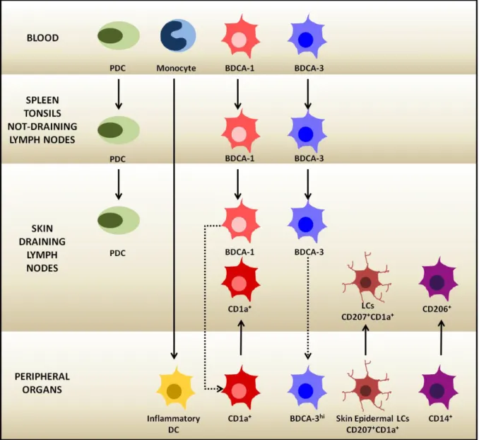
Finally a recent work from Segura et al, showed that human lymph nodes contain six different populations of DCs [99]. The largest population corresponds to PDCs (around 80% of lineage
negative and HLADR+ cells). Then the CD11c+CD14- cells (30%) can be further divided into
BDCA-1+, BDCA-3+ (or Clec9A), CD1a+, CD206+ and LCs (30%, 6%, 15%, 20% and 5% of
CD11c+CD14- cells respectively). They also found CD14+ cells that correspond to
macrophages.
This study showed that while PDCs, BDCA-1+ and BDCA-3+ DCs are also found in several
lymph nodes, the CD1a+, CD206+ and LC populations are found only in the skin-draining
lymph nodes, showing that the first three populations are resident cells, and the other three correspond to populations that have migrated from the skin. They also suggested that the
28
The resident populations have an unactivated phenotype as in the spleen and tonsils. It has been shown that these populations can capture, process and present soluble free antigens that reach the lymph nodes through the afferent lymphatics [58]. The populations coming from the skin have a more activated phenotype, they express several costimulatory molecules and the lymph node homing chemokine receptor CCR7.
1.4.3 DENDRITIC CELLS IN THE THYMUS
Little is known today about the different DC subsets present in human thymus. In 2001, Bendriss-vermare et al and Vandenabeele et al showed that human thymus contains PDCs and
two subsets of mature DCs, CD11c+CD11b-CD45ROlow and CD11c+CD11b+CD45ROhigh DCs
[102, 103]. The equivalent of these subsets to the ones found in other locations is not known. The thymic DCs derive from a thymic precursor, they are present in the cortex and the medulla of the thymus where they positively and negatively select the thymocytes [104]. Therefore they are more importantly involved in the presentation of self-antigens and the induction of central tolerance.
1.4.4 DENDRITIC CELL SUBSETS IN PERIPHERAL TISSUES
DCs are present in almost all the peripheral tissues except the brain [105]. Excluding LCs, all contain the same type of DC subsets [41]. Nevertheless the best characterized peripheral tissue in humans is still the skin.
For many years, LCs were considered as the only DCs present in the skin and mucosae, until
1993 when Nestle et al defined two additional DC subsets in the dermis, the CD1a+ and the
CD14+ dermal DCs [106]. In mice there is a population of Langerin+ dermal cells, but these
have not been identified in humans [107]. In steady-state conditions no PDCs are found in the peripheral tissues.
Langerhans cells are found in the basal and supra-basal layers of the epidermis and in the respiratory, gastric and vaginal tracts mucosae. They are characterized by a high expression of CD1a, the expression of CD207 (langerin) and the absence of CD14 [108].
The hallmarks of LCs are the Birbeck granules. These rod-shaped compartments contain langerin and are part of the endosomal pathway [109] [110]. No essential specific function has been attributed to these granules. Indeed, a single individual reported to lack Birbeck granules, owing to a heterozygous point mutation in langerin gene, showed no particular functional phenotype [111, 112]. Moreover some reports claimed that pig’s skin epidermal LCs do not have Birbeck granules at all [113].
INTRODUCTION
LCs also express adhesion molecules like CLA (cutaneous lymphocyte associated protein), Epcam and E-cadherin that mediate their adhesion to keratinocytes. They express the chemokine skin-homing receptor CCR6 and upon activation they upregulate CCR7 to migrate to the lymph nodes [114].
Dermal DCs are divided in two HLADR+ subsets. The dermal CD1a-CD14+ and the
CD1adimCD14- referred as CD14+ and CD1a+ cells respectively [25]. The dermal CD1a+ are
more or less 40% of CD45+ cells in digested skin, whereas, CD14+ are less abundant (less
than 10%)[115].
It has been difficult to relate these dermal subsets to the BDCA-1+ and BDCA-3+ subsets
identified in blood and in the lymphoid organs. Haniffa et al have recently tried to find these
equivalences, finally describing three dermal DC subpopulations [41]. Within the Lineage
-and HLA-DR+ populations of the skin, they defined first a CD14+subset. Then within the
CD14- cells, a subset of CD11c+BDCA-1+CD1a+ BDCA-3 +and- cells and a new subset of
CD11cloBDCA-1+CD1aloBDCA-3hi cells. In this study they suggested that these two last
subsets are related to the BDCA-1+ and BDCA-3+ blood subsets respectively, and that the
new BDCA-3hi was ignored until now due to the fact that the other subsets might upregulate
BDCA-3 marker as well. They found that the CD14+ subset is more similar to monocytes and
therefore would probably arise from this blood population.
In general, DC subsets in the tissue seem to be more activated than their blood counterparts, nevertheless it is difficult to identify if this happens in vivo or whether it is related to the different isolation protocols used.
Finally, under inflammatory conditions, the blood monocytes have been implicated in the generation of infiltrating inflammatory DCs [116]. These cells express BDCA-1, CD1a, CD206, FcεR1, SIRPα and no CD209 or CD16. They stimulate TH17 cells. Inflammatory DCs were before identified as inflammatory epidermal and iNos producing DCs (IDECS and TiPs) in atopic dermatitis and psoriasis respectively [117, 118]. They prime completely different T cell responses and therefore prove that different inflammatory DCs rise out from monocytes in different pathological conditions [119].
In summary, the final global characterization of the different human subsets of DCs has highlighted the relationship between them. Today, more than a classification that depends on an activation state, different combinations of surface markers have been defined to identify different sub-populations of DCs that are repeatedly found in several tissues. We are now able to distinguish between populations that reside in the different sites from populations that are migrating. The existence of DC subsets opens up a new field of research concerning their functional differences and their differential implications under inflammatory conditions.
30
Figure 1-7: Human dendritic cell subset distribution in different anatomical sites.
Plain arrows represent direct relationships. Excepting inflammatory DCs, which have a developmental relationship with monocytes, all the other plain arrows represent relationships based on migration and homing to the different locations. Pointed arrows represent only suggested relationships.
INTRODUCTION
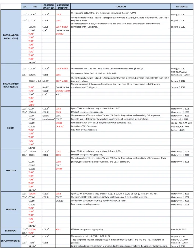
Table 1-1 : Dendritic cell subset phenotypes at steady state.
Transcriptomic profiles have given rise to complete phenotypes of the human DC subsets. In this table we present the major characteristic CD1, PPRs, adhesion molecules and chemokine receptors of each subset. The black color means protein expression, red color means RNA expression. (PRR, pattern recognition receptors; SLO, secondary lymphoid organs).
CD1 PRRs MOLECULESADHESION CHEMOKINE RECEPTORS FUNCTION REFERENCES
CD1a-CLEC6a+ CD11chi CCR2+ They secrete CCL3, TNFα, and IL-12 when stimulated through TLR7/8. Mittag, D. 2011
CD1c+CLEC7a+ CD11b+ CCR5+ They efficiently induce Th1 and Th2 responses if they are in tonsils, but more efficiently Th1 than Th2 if they are in blood. Segura, E. 2012
DEC205+ CD62L+ CCR7+ in SLO They crosspresent if they come from tissue, the ones from blood crosspresent only if they are stimulated with TLR ligands. Segura, E. 2012
CD206+ CLA+ CXCR4+ in SLO TLR1+ CX3CR1+ TLR2+ TLR3+ TLR4+ TLR5+ TLR6+ TLR7+ TLR8+ SIRP1a+
CD1a-CLEC9a+ CD11clo CCR2+ in SLO They secrete low CCL3 and TNFα, and IL-12 when stimulated through TLR7/8 Mittag, D. 2011
CD1c- DEC205+ CD11b- CCR5+ They secrete TNFα, CXCL10, IFNλ and little IL-12. Haniffa, M. 2012; Lauterbach, H. 2010
CD206+ in SLO MRC2+ CCR7+ in SLO They efficiently induce Th1 and Th2 responses if they are in tonsils, but more efficiently Th1 than Th2 if they are in blood. Segura, E. 2012
TLR1+ Necl2+ CXCR4+ in SLO They crosspresent if they come from tissue, the ones from blood crosspresent only if they are stimulated with TLR ligands. Segura, E. 2012
TLR2+ CD62L+ CX3CR1+ in SLO
TLR3+ CLA+ XCR1+
TLR6+
TLR8+
MRC2+
CD1a+CD207+ CD11clo CCR2- Upon CD40L stimulation, they produce IL-8 and IL-15. Kletchevsy, E. 2008
CD1c+DEC205+ CD11b- CCR5- Efficient crosspresenting capacity. Kletchevsy, E. 2008
CD206- Epcam + CCR6+ They stimulate effitiently naïve CD4 and CD8 T cells. They induce preferentially Th2 responses. Kletchevsy, E. 2008
CD208+ E-cadherine+CCR7lo Possible role in tolerance. They induce proliferation of autologous memory Tregs. Seneschal, J. 2012
CD209- CXCR4+ When stimulated with VitD3 they induce TGF-β -secreting Tregs. van der Aar, A.M. 2011
CLEC9a- CX3CR1+ Induction of TH17 response Mathers, A.R. 2009
TLR1+ Induction of Th22 response Fujita, H. 2009
TLR2+
TLR3+
TLR6+
TLR7+
SIRP1a+
CD1a+DEC205+ CD11c+ CCR2- Upon CD40L stimulation, they produce IL-8 and IL-15. Kletchevsy, E. 2008
CD1c+CD206+ CD11b+ CCR5+ Efficient crosspresenting capacity. Kletchevsy, E. 2008
CD208+ CCR6
-They stimulate effitiently naïve CD4 and CD8 T cells. -They induce preferentially a Th2 reponse. Their
phenotype is intermediate between LCs and CD14+ dermal DC. Kletchevsy, E. 2008
CD209- CCR7+ CLEC9a- CXCR4+ TLR1+ TLR2+ TLR3+ TLR6+ TLR7+ SIRP1a+
CD1a-DEC205+ CD11c+ CCR2- Upon CD40L stimulation, they produce IL-1β, IL-6, IL-8, IL-10, IL-12, TGF-β, TNFα and GM-CSF. Kletchevsy, E. 2008
CD1c+CD206+ CD11b+ CCR7lo They prime CD4 T cells to induce isotype switch on naïve B cells and Igs secretion. Kletchevsy, E. 2008
CD209+ CX3CR1+ They do not stimulate efficiently naïve CD4 and CD8 T cells Kletchevsy, E. 2008
CLEC9a- Poor crosspresenting capacity. Kletchevsy, E. 2008
TLR1+ TLR2+ TLR3+ TLR6+ TLR7+ SIRP1a+
CD1aloCLEC9A+ CD11clo XCR1+ Efficient crosspresenting capacity. Haniffa, M. 2012
CD1c+TLR3+
CD1a+CD206+ CD11c+ They produce IL-1, IL-6, TNFα, IL-12, IL-23. Segura, E. 2013
CD1c+FcεR1+ CD11b+ They can prime Th1 and Th2 responses in atopic dermatitis (IDECS) and Th1 and Th17 responses in psoriasis. Nakano, K.L. 2009; Hammad, H. 2010
SIRP1a+ In sinovial and ascitis fluids from reumathoid arthritis and cancer patiens they induce Th17 responses. Segura, E. 2013
SKIN BDCA3
INFLAMMATORY DC BLOOD AND SLO
BDCA-1 (CD1c)
BLOOD AND SLO BDCA-3 (CD141)
SKIN LC
SKIN CD1A
32
1.4.5 DIFFERENT SUBSETS SUGGEST DIFFERENT FUNCTIONS
As the study of different subsets of DCs in human blood and tissues samples evolved, several attempts to link the phenotypic differences to different functions and T cell profile induction have been made.
In mice, functional specializations have been attributed to the different DC subsets. For
example CD8α+ and CD103+ subsets are specialized in the activation of CD8+ T cells [120].
These two subsets are also enabled with the capacity to cross-present antigens. In the case of
CD4+ T cell priming, the different mice subsets CD8α+, CD8α-, CD103+ and LCs show
different behaviors depending on the signal triggering the DC activation [120]. Nevertheless
it has been shown that adoptive transfer of antigen-pulsed CD8α+ versus CD8α- DCs
differentially induces Th2 versus Th1 responses [121, 122]. Moreover, it has been shown that
targeted CD8α+and CD8α
DCs induce CD8+T and CD4+T cell responses respectively [123].
In humans, it is clear that PDCs are more involved than DCs in antiviral responses. Moreover, already in 1999, Rissoan et al. established, for the first time, a direct link between different sup-populations of DCs (DC1 and DC2) and different T helper cell profiles (Th1 and Th2) [124]. Nevertheless the DC subsets have overlapping functions concerning cross-presentation, tolerance induction and Th1, Th2, Treg, and Th17 primings. The functional differences between human DC subsets are less clear than in murine model. A summary of the overlapping functions of human DC subsets is shown in Table 1-1.
In the case of skin DCs, there is evidence that functional differences between LCs and dermal
CD14+ cells exist. For example, Kletchevsky et al, compared functionally human skin subsets
[25]. They found that CD14+ dermal DCs stimulated with CD40 secrete IL-1α, 6, 8,
IL-10, IL-12 GM-CSF and TGF-β, whereas LCs stimulated with CD40 only secrete IL-15 and
small amounts of IL-8. Even if LCs and CD14+ DCs were both able to induce naive B cell
production of IgM, CD14+ and not LCs were able to induce isotype switching to IgG and IgA.
On the other hand LCs stimulated better naïve CD4+T cell proliferation than CD14+ cells, and
primed a Th2 cell response. They were also found to be more efficient in the induction of
antigen specific CD8+Tcells and cross-presentation. Moreover, it was later shown by the same
authors that LCs stimulated a better cytotoxic response through the secretion of IL-15 while
IL-10 and TGF-β secreted by dermal CD14+ inhibited this response [125]. These studies
suggest that dermal CD14+ cells preferentially activate B cells while LCs preferentially prime
a Th2 cell response.
In tolerance induction, LCs and dermal cells were also found to behave differently. LCs were shown to induce a higher proliferation of autologous memory Treg cells than the dermal DCs [126]. Concerning Th17 differentiation controversial results were found. Mathers et al showed that LCs had a superior capacity than dermal DCs in the generation of Th17 cells [127], yet, Fujita et al found no Th17 induction by LCs but the induction of IL-22 secreting T cells [128]. In general several functions have been attributed to LCs [127-129]. Controversial results are often attributed to different protocols of isolation of the skin DCs.
INTRODUCTION
In the case of blood BDCA-1+ and BDCA-3+ subsets, the cross-presenting capacities have
been extensively evaluated giving rise to controversial results. First, the comparison between
human blood BDCA-1+ and BDCA-3+ subsets let to the conclusion that after activation with a
TLR3 ligand, poly-IC, BDCA3+ cells were specialized in antigen cross-presentation [41, 90,
91, 130]. Secondly, tonsillar BDCA3+ cells were also found to be more efficient at
cross-presenting death associated antigens than the BDCA-1+ counterpart. These results led to the
conclusion that BDCA-3+ cells as the murine CD8α+ cells were specialized in
cross-presentation. As non activated DCs are have a poor cross-presentation capacity [94], these results were obtained activating DC subsets with TLR3 ligands that preferentially stimulate
BDCA-3+ cells. Therefore, Segura et al, used tonsillar BDCA-1+and BDCA-3+ subsets (which
are per se more activated than the blood counterparts) and showed that both cell subsets are equally able to cross-present soluble antigens in the absence of additional activation stimuli [94]. This result was further confirmed by two other studies showing that these subsets have the same cross-presenting capacities but different TLR stimulation requirements [131, 132]. These studies infer that human DC subsets overlap in their functions, and that the functional specialization of DC subsets can be in part attributed to their differential capacities to sense single stimuli. Uncoupling differential activation of DC subsets induced by the signals of the microenvironment (e.g. TLR activation requirements) from their functional specializations remains a big challenge. Another factor to take into account is the isolation procedure. Different protocols of DC isolation result often in controversies as it is the case of Th17 induction by LCs. DC functional differences are mainly evaluated in the context of the T cell responses. However differential DC subset secretion of cytokines and chemokines may mediate different innate immune responses.
To study the functional differences between human DCs cell subsets stimulatory signals that activate the DC subsets to the same extent must be used. One of these stimulatory signals
could be the cytokine TSLP. To address functional differences between blood BDCA-1+ and
BDCA-3+ DCs, I used TSLP as a model of inflammatory tissue factors. Therefore the
following chapters of my introduction will address our current knowledge on human TSLP biology, and its role in the immune system through the activation of DCs.
34
1.5 THYMIC STROMAL LYMPHOPOIETIN BIOLOGY IN HUMANS
TSLP is a cytokine secreted by epithelial cells and keratinocytes at the barrier surfaces like the skin and the mucosa. It is detected in atopic dermatitis lesional skin and not in healthy skin or non-lesional skin; this highlights its important role in allergic disorders [133]. In humans the main responders to this cytokine are the DCs [134]. TSLP primed DCs (TSLP-DCs) induce Th2 cells and are closely linked to the initiation of the allergic immune responses [133]. Nevertheless a homeostatic role has been attributed to TSLP -DCs in the thymus and the periphery [135, 136]. Although mice studies have greatly helped us to understand TSLP role in the immune system, TSLP biology in humans differs in comparison to the mouse model.
The purpose of this chapter is to review the knowledge gathered until now about human TSLP and its implications in health and disease. I will present first TSLP and TSLP receptor characteristics followed by TSLP effects on DCs and its role in allergic disorders and immune tolerance. At the end I will present in depth two main studies addressing TSLP –treated PDC and DC functions in immune tolerance and TSLP-treated LC implication in allergy.
1.5.1 TSLP AND TSLP RECEPTOR
TSLP was first identified in the supernatants of a murine thymic stromal cell line. It was described as a factor supporting B cell growth and thymocyte survival [137]. Later on, this factor was cloned in mice and humans and defined as a member of the hematopoietic cytokines family [134, 138, 139]. The homology between the human and mouse protein was found to be poor (43% of the amino acid sequence) [134]. In humans, TSLP messenger RNA was identified in skin keratinocytes, lung fibroblasts, bronchial epithelial cells, mammary epithelial cells, smooth muscle cells and activated mast cells [133]. By inmmunohistological techniques, TSLP has been found in the crypts of the tonsillar epithelium, the Hassals corpuscles in the thymic stroma and the apical layers of lesional skin in atopic dermatitis [133, 135].
The TSLP receptor was cloned in mice and humans as a heterodimeric receptor constituted by
the TSLPR chain and the IL-7Rα chain [140, 141]. It was found that TSLP binds to its
receptor with high affinity only if the two chains are expressed. Moreover, the IL-7Rα chain
is needed to trigger an intracellular signal [141]. In the case of TSLPR the homology between mice and humans was also found to be poor (39% aminoacid sequence).
The TSLP receptor complex has a restricted expression. Both chains of the receptor are expressed mainly by dendritic cells [134]. It has been reported that CD4T cells can also respond to TSLP in vitro [142]. Although this has been well established in mice, in humans CD4T cells do not seem to co-express both chains of the receptor, or do it very poorly considering the levels reached by DCs [134].
INTRODUCTION
TSLP signaling pathway implicates the phosphorilation of STAT3 and STAT 5 [143]. These factors are activated by several other cytokines. In the case of human DCs, TSLP activates the JAK-STAT pathway by inducing the phosphorilation of janus activating kinases 1 and 2. It was shown that it also induces the phosphorilation of STAT 1, 4 and 6, AKT, and the MAPKs
ERK and JNK. Finally it triggers the nuclear translocation of the NF-κB members, p50, p52
and RelB [143]. However the study of TSLP receptor signaling on human primary DCs is very difficult due to the scarcity of these cells and the detailed signaling pathway is still unknown. A recent study by Pandey et al on a murine pro-B cell line (Ba/F3) transfected with the human TSLPR complex implicates additional molecules in the signaling pathway of TSLP [144]. This last study suggest that different members of the Src and Tec kinases families (Btk, Lyn and Tec), and the protein phosphatases Ptpn6 (Shp-1) and Ptpn11 (Shp2) participate in the protein complex activated downstream TSLP signaling.
1.5.2 TSLP EFFECTS ON HUMAN DENDRITIC CELL FUNCTION
TSLP strongly activates DCs in vitro. It triggers the up regulation of MHC class I molecules and of costimulatory molecules CD80, CD83, CD86 and CD40 [133]. It also enhances DC survival in culture. TSLP-DCs, as opposed to LPS, CD40L or IL-7 treated DCs do not secrete
IL-12, IL-6 or IL-1 α/β, cytokines described as Th1 polarizing signals. Instead, they were
shown to produce large amounts of the chemokines CCL17 (TARC) and CCL22 (MDC), IL-8 and eotaxin-2 known to recruit Th2 cells, neutrophils and eosinophils respectively [133]. Moreover, TSLP-DCs induce a potent proliferation of allogenic naïve CD4 T cells in vitro and the primed T cells produce high levels of IL-4, IL-5 and IL-13 compared to DCs stimulated with LPS, CD40L or IL7 [133]. These T cell cytokines are characteristic of the Th2 profile. Nevertheless, this Th2 profile is unconventional as CD4 T cells primed by
TSLP-DCs do not secrete IL-10 but considerable levels of TNF-α. Therefore, as opposed to a
conventional Th2 profile it has been named “inflammatory Th2 profile” [133]. In the context of allergic inflammation, IL-10 is recognized as an anti-inflammatory cytokine whereas TNF-α is one of the most potent pro-inflammatory cytokines involved [145, 146]. Thus the inflammatory Th2 cells primed by TSLP-DCs have a critical role in the development of uncontrolled allergic inflammation.
It has been shown that TSLP-DCs capacity to induce the inflammatory Th2 profile is mediated by the up-regulation of the surface molecule OX40L [147] (Figure 1-8). Indeed, the use of a neutralizing antibody against OX40L changed the T cell profile induced by
TSLP-DCs to a profile characterized by the absence of Th2 cytokines and TNF-α and the secretion
of IL-10. The current model proposes that the OX40L expression by human TSLP-DCs in the
absence of IL-12 secretion, induces the secretion of the Th2 cytokines and TNF-α, whereas,
the presence of IL-12 induces a Th1 profile and the subsequent production of IL-10 [143]. The IL-12 production is dependent on the activation of STAT4 and interferon regulatory
36
sites, and it is known now, that is the p50 unit that mediates its transcription upon TSLP binding [143](Figure 1-8).
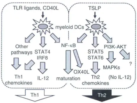
In conclusion, TSLP generates an environment for Th2 response development, first, by inducing Th2-attracting chemokines, and second, by inducing Th2 cells through the up-regulation of OX40L in the absence of IL-12.
Figure 1-8 : TSLP induces a Th2 profile. Adapted from Ito, T. et al 2012.
TLR ligands and CD40L induce the activation of STAT4 and IRF8 leading to the secretion of IL-12 and the priming of a Th1 response. By contrast TSLP does not induce STAT4 and IRF8 activation. It induces OX40L in an Nf-κB dependent way; in the absence of IL-12, OX40L triggers a Th2 response. Through STAT5 and 6 activation, TSLP triggers the secretion of CCL17 and CCL22 chemokines, characterizing further the Th2 response.
1.5.3 TSLP AND ALLERGIC DISORDERS
The different allergic states are characterized by an exaggerated immune response against a harmless antigen. The type of T cell response linked to allergy is the previously mentioned Th2 response.
TSLP has been linked for a long time to different allergic disorders. First, because it was found to be present in the skin lesions of patients suffering from atopic dermatitis and second because it induces an inflammatory Th2 response [133]. Moreover, the human TSLP gene is found close by the Th2 cytokine gene cluster locus [134].