Any correspondence concerning this service should be sent to the repository administrator:
[email protected]
To link to this article:
DOI:10.3727/096368910X514297
URL : http://dx.doi.org/10.3727/096368910X514297
This is an author-deposited version published in:
http://oatao.univ-toulouse.fr/
Eprints ID: 8681
To cite this version:
Trouche, Elodie and Girod Fullana, Sophie and Mias,
Céline and Ceccaldi, Caroline and Tortosa, Florence and Seguelas, Marie
Hélène and Calise, Denis and Parini, Angelo and Cussac, Daniel and Sallerin,
Brigitte Evaluation of alginate microspheres for mesenchymal stem cell
engraftment on solid organ. (2010) Cell Transplantation, vol. 19 (n° 12). pp.
1623-1633. ISSN 0963-6897
O
pen
A
rchive
T
oulouse
A
rchive
O
uverte (
OATAO
)
OATAO is an open access repository that collects the work of Toulouse researchers and
makes it freely available over the web where possible.
Copyright 2010 Cognizant Comm. Corp. E-ISSN 1555-3892 www.cognizantcommunication.com
Evaluation of Alginate Microspheres for Mesenchymal
Stem Cell Engraftment on Solid Organ
E. Trouche,* S. Girod Fullana,†‡ C. Mias,* C. Ceccaldi,†‡ F. Tortosa,*†
M. H. Seguelas,* D. Calise,* A. Parini,*† D. Cussac,*†
1and B. Sallerin*†
1*INSERM U858, Toulouse, France
†Universite´ Toulouse, UPS, Faculte´ des Sciences Pharmaceutiques, Toulouse, France ‡CIRIMAT, UPS-INPT-CNRS 5085, Toulouse, France
Mesenchymal stem cells (MSCs) may be used as a cell source for cell therapy of solid organs due to their differentiation potential and paracrine effect. Nevertheless, optimization of MSC-based therapy needs to develop alternative strategies to improve cell administration and efficiency. One option is the use of alginate microencapsulation, which presents an excellent biocompatibility and an in vivo stability. As MSCs are hypoimmunogenic, it was conceivable to produce microparticles with [alginate-poly-L-lysine-alginate (APA) microcapsules] or without (alginate microspheres) a surrounding protective membrane. Therefore, the aim of this study was to determine the most suitable microparticles to encapsulate MSCs for engraftment on solid organ. First, we compared the two types of microparticles with 4× 10⁶MSCs/ml of alginate. Results showed that each microparticle has distinct morphology and mechanical resistance but both remained stable over time. However, as MSCs exhibited a better viability in microspheres than in microcapsules, the study was pursued with microspheres. We demonstrated that viable MSCs were still able to produce the paracrine factor bFGF and did not present any chondrogenic or osteogenic differentiation, processes sometimes re-ported with the use of polymers. We then proved that microspheres could be implanted under the renal capsule without degradation with time or inducing impairment of renal function. In conclusion, these micro-spheres behave as an implantable scaffold whose biological and functional properties could be adapted to fit with clinical applications.
Key words: Mesenchymal stem cells; Alginate; Microspheres
INTRODUCTION A way to protect MSCs and take advantage of their paracrine properties could be their inclusion in a bioma-trix. This could permit to 1) prevent mechanical stress, Mesenchymal stem cells (MSCs) are multipotent
stem cells derived from bone marrow stroma. They have 2) diminish the negative influence of the injured envi-ronment on grafted cells, 3) easily manipulate and de-the ability to form a variety of mesenchymal tissues
in-cluding bone, fat, and cartilage, and non-mesenchymal liver the cells near the organ, and 4) better follow the phenotype of the grafted cells. This goal may be tissues like neuron, kidney, and heart (13,15,27,36,55).
Recent studies have shown that most of the therapeutic achieved through the microencapsulation of MSCs in biomatrices and implantation of this scaffold. The de-effects of MSCs are related to the secretion of paracrine
factors (17,47). Systemic administration is easy but there sign of the microparticles has to be optimized for perme-ability, stperme-ability, biocompatibility, and easiness of injec-is little quantity of cells reaching the organ. These
find-ings supported the idea that direct intraparenchymal in- tion and integration. The advantage of natural polymers in comparison to synthetic polymers is that they induce jection would allow to concentrate the paracrine factors
produced by MSCs within the injured organ. However, less cytotoxicity or inflammatory reactions (21). Alginate is the most commonly employed polymer the intraparenchymal administration is characterized by
early death of grafted cells (23,29,38,44,48) and in the for cell encapsulation because of its excellent biocom-patibility and an in vivo stability (6,40,53). Alginate is case of their survival they potentially differentiate in
un-suitable phenotypes. a polysaccharide isolated from brown algae found in
Received November 19, 2009; final acceptance June 8, 2010. Online prepub date: August 17, 2010.
1These authors provided equal contribution to this work.
Address correspondence to Brigitte Sallerin, I2MR, INSERM U858, CHU Rangueil, BP 84225, 31432 Toulouse Cedex 4, France. Tel: 33-5-65-25-68-43; Fax: 33-5-65-25-98-16; E-mail: [email protected]
coastal waters around the globe (19). It is a linear copol- were able to differentiate into cells from mesodermal lineage: osteoblasts, chondrocytes, and adipocytes (data ymer composed ofβ-D-mannuronic acid andα-L
-gulur-onic acid that can be easily transformed into a gel by not show). binding the guluronic acids with a divalent cation such
MSC Encapsulation
as calcium (41). Classically, cells are encapsulated in
alginate microcapsules with a semipermeable mem- Rat MSCs (passage 3) were lifted with trypsin, coun-ted, and centrifuged. Microspheres and microcapsules brane, generally polylysine (PLL), which allows
nonau-tologous cells to be implanted. This membrane protects were produced by the method of Goosen et al. with modifications (10) under sterile conditions. We used the cells from immune mediators and allows the release
of beneficial factors from the microcapsule. sterile ultrapur sodium alginate with 54%β-D
-mannuro-nic acid (type M alginate) with an apparent viscosity of Previous results from our laboratory have shown that
adrenal medullary bovine chromaffin cells could be en- 141 mPas.s (Pronova, SLM 100, Novamatrix, Norway). Briefly, a 1.4% w/v sodium alginate solution was pre-capsulated in microcapsules of alginate-poly-L-lysine
(APA) with a liquefied inner core (28). This procedure pared by dispersing alginate in NaCl 150 mM, buffered to pH 7.4 with 12.5 mM HEPES. Cells were resus-was associated to an increase in the viability of
func-tional cells. However, in clinical applications of MSCs, pended in this sodium alginate solution at a density of 2.5× 10⁶ or 4× 10⁶ cells/ml alginate. Homogeneous a semipermeable membrane may not be necessary as
these cells are poorly immunogenic (1,7,16,49). We hy- alginate microspheres were produced by extruding through an encapsulator (Inotech IE-50R, Switzerland), pothesized that alginate microspheres could also be
suit-able for MSC encapsulation. However, little is known equipped with a 300-µm vibrating nozzle, the alginate– cell suspension into a solution of 1% CaCl2, 2H2O, 0.4% concerning viability and functionality of MSCs in
mi-crospheres. In addition, the use of alginate microsphere NaCl, 12.5 mM HEPES, pH 7.4, which was continu-ously swirled. Microspheres were gelled for at least 20 graft for cell therapy of solids organs has not been
ex-tensively investigated. Indeed, microspheres present dif- min and then rinsed with HEPES buffer and either trans-ferred into culture medium (case of alginate ferent permeability and mechanical behavior than
micro-capsules that could consequently impact the implantation spheres) or coated by incubating in 0.1% w/v PLL, 150 mM NaCl, 12.5 mM HEPES, pH 7.4, under gentle agita-procedure and in vivo scaffold integrity.
Thus, we designed a strategy based on the encapsula- tion. In the latter case, an outer alginate layer was subse-quently applied by 10-min incubation in a dilute (0.1% tion of MSCs in alginate microspheres or microcapsules
in order to determine which one was the most appro- w/v) alginate solution in saline buffer under gentle agita-tion (case of APA microcapsules). Finally, microcap-priated for cell therapy. We first evaluated the stability
and mechanical parameters of microparticles. We then sules were treated with 55 mM sodium citrate to liquefy the inner alginate core, and washed extensively with sa-tested MSC viability and functionality in microparticles.
We also determined the effect of alginate on differentia- line buffer. Both types of microparticles—microspheres and microcapsules—were studied.
tion of MSCs. Finally, we studied the feasibility of
graft-ing alginate-encapsulated MSCs under the rat renal cap- Microspheres and microcapsules were incubated in culture medium at 37°C in 5% CO2 and 95% humidity. sule.
At specified days postencapsulation, they were collected
MATERIALS AND METHODS and further analyzed. In order to characterize their
mor-Cell Culture phology and properties, control microparticles (without
cells) were also prepared according to the same protocol. Marrow aspirate was obtained from femurs cavity of
Lewis rats (Harlan, France) weighing 180–200 g. Bone
Morphology and Stability of Particles Over Time
marrow from the femur cavity was flushed with MEM
medium (ABCYs, France) containing 10% FCS and 1% Size and morphology of microparticles were rou-tinely examined with a light microscope. The stability penicillin/streptomycin (Invitrogen, USA) and the cell
suspension was centrifuged (400× g, 5 min). Then, cells of control microparticles was studied for 35 days at 37°C in saline buffer both visually and by light micro-were plated in culture flasks (200,000 cells/cm2).
Nonad-herent cells were removed after 72 h and MSCs were scopic observation. In the case of microcapsules, their integrity was assessed.
recovered by their capacity to adhere highly to plastic culture dishes. MSCs were then routinely cultured and
Scanning Electron Microscopy (SEM)
were used for the experiments. Most adherent cells
ex-pressed MSC markers CD90, CD29, and CD106 and SEM analysis of the surface and cross section of dried control microparticles was performed with a scan-were negative for CD34 (hematopoietic marker), CD45
microparti-cle samples were mounted on an aluminum sample branes were incubated to horseradish peroxidase-conju-gated anti-rabbit secondary antibody (1:10,000; Santa mount and sputter coated with silver. The specimens
were observed at a 10 kV accelerating voltage. Cruz Biotechnology). Expression of bFGF is related to the expression of ERK2.
Mechanical Resistance
The mechanical resistance of the microparticles was Chondrogenic Phenotype of Encapsulated MSCs evaluated on microspheres or microcapsules obtained
MSCs encapsulated in microspheres where cultured with a 0.8-mm needle according to the same protocol.
in high-glucose Dulbecco’s modified Eagle’s medium At defined time intervals, the microparticles were
sub-(ABCYs, France) with 1% fetal bovine serum (In-mitted to a standardized compression test in a TA-XT2
vitrogen, USA), antibiotics, modified or not with ascor-texture analyzer (Stable Microsystems, UK). Briefly, the
bate-2-phosphate (50µg/ml, Sigma, France), proline (40 compression resistance of the microparticles was deter- µg/ml, Sigma), pyruvate (3 mM, Sigma), ITS+ (1×, BD mined as the main force (g) required to generate a 30%
Biosciences, USA), dexamethasone (100 mM, Sigma), compression of a sample of microparticles. The appara- transforming growth factor-β3 (10 ng/ml, TGF-β3, tus consisted of a mobile probe moving vertically, up
R&D Systems, USA), and recombinant bone morpho-and down at constant morpho-and predefined velocity (0.5 mm/
genic protein-2 (200 ng/ml, R&D Systems). Medium s). The force exerted by the probe on the microparticles
was replaced twice weekly. On day 21, 31, 51, 57, and was recorded as a function of the displacement, leading
61, MSCs were harvested from microspheres after incu-to a force versus strain curve. The results are expressed
bation with citrate (50 µM). Cells were then centrifu-as the average maximal mechanical force in grams from
gated, resuspended with cultured medium, and then were at least five independent observations. The integrity of
seeded on slices to perform immunocytology. the microparticles after the test was assessed by
micro-scopic observation.
Immunocytology
Cell Viability Assay For in vitro differentiation, cells where stained with
alizarin red or alcian blue coloration using standard The viability of encapsulated cells was assessed using
a LIVE/DEAD Viability/Cytotoxicity kit (FluoProbes, methods. For chondrocyte detection, cells were incu-bated (1 h, RT) with an anti-PS100 rabbit antibody (1: France) for 35 days following microencapsulation.
Briefly, microparticles were rinsed twice with phos- 200, ABCYs, France). phate-buffered saline (PBS) and MEM (v/v).
Microparti-cles were then incubated for 30 min with a solution con- Animal Transplantation and Histology
taining 2µM ethidium homodimer-3 and 1 µM calcein
Experimental animals were handled in accordance AM. Microparticles were then rinsed with PBS and
ob-with the European animal care guidelines. Four Lewis served using a confocal microscope (Leica
Microsys-rats (Harlan, France) weighing 180–200 g were used for tems, Germany). Cell viability was determined from
allogenic recipients of MSC-alginate. For transplanta-confocal images as previously described (26). After
tion, rats were anesthetized with isoflurane/oxygen inha-staining, cells appear with red nuclei and lived cells with
lation (3/97). A total of 18 microspheres containing 4× green cytoplasm. Some population of MSCs exhibited
106 MSCs were transplanted under the renal capsule. apoptotic transformation so their nuclei looked
yellow-Sham-operated animals were subjected to the same sur-orange, revealing the colocalization of fluorescence in
gical procedure without transplantation. Kidney sections green and red spectral regions. The cell viability was
were collected 25 days after MSC injection. estimated by the ratio of green pixels to the total number
After euthanasia, kidneys were collected, fixed in of lightened pixels.
paraformaldehyde (4%), dehydrated, and then embedded in paraffin. Paraffin sections (10µm) were stained with
Western Blot
hematoxylin/eosin. For Western blot (WB), MSCs were extracted of
mi-crospheres through an incubation with citrate (50 µM)
Statistical Analysis
and centrifugation. Then MSC proteins were extracted
from pelleted MSCs. WB analyses were performed with Results are expressed as mean± SEM. Statistical comparison of the data was performed using the t-test samples normalized for protein concentration.
Mem-branes were probed with anti-bFGF (1:500; Santa Cruz for comparison between two groups or one-way ANOVA and post hoc Tukey’s test for comparison of more than Biotechnology, USA) or anti-ERK2 (1:1,000; Santa
Cruz Biotechnology, USA) antibodies. Following sev- two groups. A value of p< 0.05 was considered signifi-cant.
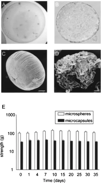
mem-RESULTS cles present an average diameter between 500 and 710 µm with alginate microspheres slightly bigger than
mi-Morphology and Stability of the
crocapsules. The examination showed that MSCs
ap-Alginate Microparticles
peared evenly distributed throughout the microparticles, Morphology and stability of two types of
microparti-which remained stable in saline buffer and in culture cles, microspheres and microcapsules, were examined
media over the study. using optical and electron microscopy. Under optical
In order to demonstrate that the microcapsules had a microscopy, all cell-loaded microparticles had a uniform
liquefied core, intact microparticles were dried and then and spherical morphology (Fig. 1A, B). Concerning
mi-observed by electron microscopy (Fig. 1C, D). In these crocapsules (Fig. 1B), a smooth refringent ring,
corre-conditions, microcapsules appeared no longer spherical, sponding to the continuous transparent alginate-PLL
as they would appear if they had a solid core, but flat-membrane, could be observed. All of these
microparti-tened with a highly wrinkled surface (Fig. 1D). Because they became fragile upon drying, some of them were broken, clearly showing that initial capsules were hol-low and only constituted of a membrane surrounding a liquefied core.
Mechanical Resistance
In order to complete the study of microparticle mor-phology and stability, we determined their mechanical resistance. The resistance and durability of the micropar-ticles were studied by submitting them to a standardized mechanical stress (28). This resistance to compression protocol is derived from the method of Orive et al. (3,31). The authors demonstrated that this technique per-mits differentiation among solid particles in order to choose the most resistant internal configuration. More-over, it can be applied to both microcapsules and micro-spheres.
Figure 1E shows the evolution of the maximal me-chanical force (g) required to compress the beads of 30% of their height with time. In all cases, until day 35 postencapsulation, resistance to compression of micro-particles remained stable. As expected, forces required to compress microspheres are higher than microcap-sules, although the latter exhibited a more elastic be-havior.
These results show that mechanical resistance of the two types of microparticles was unchanged with time (until day 35). In contrast, their mechanical behavior ap-peared quite different and could play a role in micropar-ticle behavior postimplantation. Although they support small deformations, microcapsules remain prone to membrane disruption, while microspheres are mechani-cally stronger: their use would obviously increase the durability of the transplant and improve the feasibility of its retrieval.
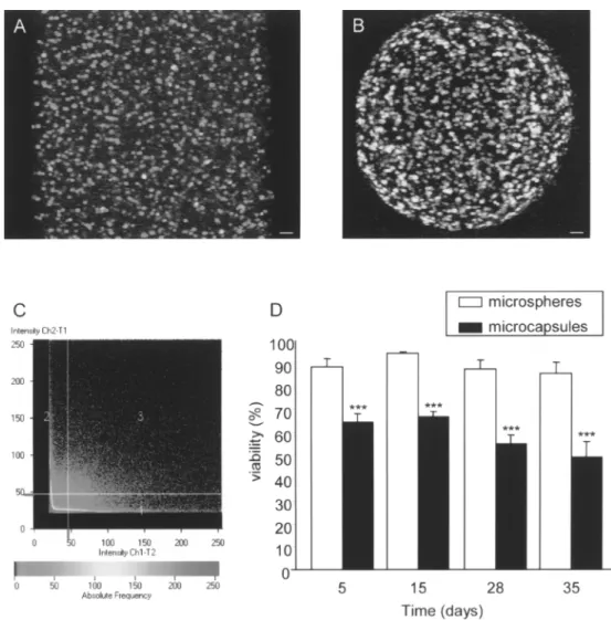
In Vitro Viability of Encapsulated MSCs Figure 1. Characterization of alginate microparticles. (A, B)
Microsphere (A) and microcapsule (B) size and morphology In order to evaluate the effect of the different micro-examined with a light microscope (objective 2× and 10×, re- particles on the viability of MSCs, microspheres and mi-spectively). (C, D) Scanning electron microscopy of
micro-crocapsules were followed for 35 days postencapsula-spheres (C) and microcapsules (D) Scale bars: 100 µm. (E)
tion after loading with 4× 10⁶ MSCs/ml of alginate. Measurement of the mechanical force needed to compress the
homodimer-3 and calcein followed by homodimer-3D reconstitutions performed have shown that bFGF mediates part of the paracrine effects of MSCs (25 ). We examined the production of using confocal microscopy. The viability of MSCs was
quantified by automatic cell counting. This quantifica- bFGF in microencapsulated MSCs 7 days postencapsu-lation (Fig. 3). Results showed that bFGF remains de-tion shows that viability of MSCs was poorly affected
in microspheres (Fig. 2A, D, green cytoplasm) until day tectable in microencapsulated MSCs and the amount of protein was not significantly different from that of non-35 (85.6± 4.8%) whereas it significantly decreased
(64.6± 3.4%, 49.3 ± 4.9% on day 5 and 35, respec- encapsulated MSCs. These results indicated that, as showed for cultured MSCs, viable microencapsulated tively) in microcapsules (Fig. 2B, D). These results led
us to select microspheres rather than microcapsules for MSCs are able to produce bFGF. the next steps of our study.
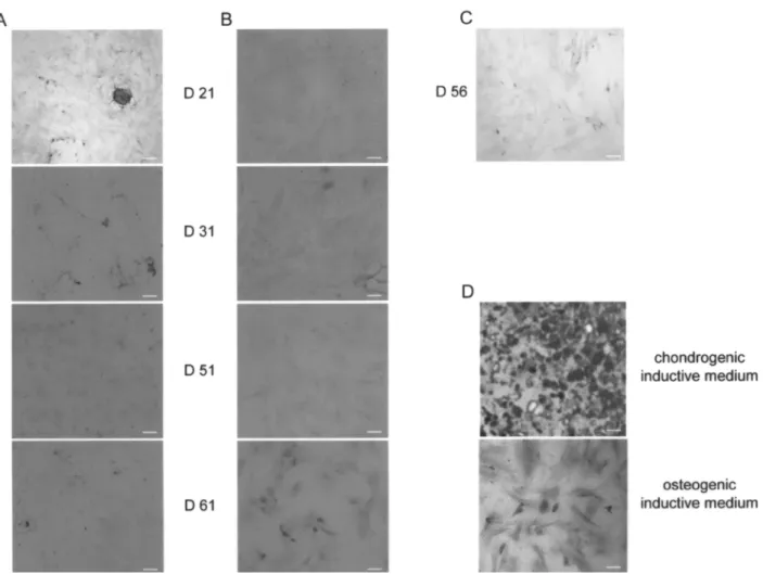
In Vitro Differentiation of Microencapsulated MSCs In Vitro Functionality of Encapsulated MSCs Some studies have investigated the possible
differen-in Microspheres tiation of matrix encapsulated MSCs into mesodermal
lineages and more particularly into the chondrogenic and Several studies have shown that MSCs are able to
secrete a number of cytokines, which may explain their osteogenic phenotypes. We compared the degree of chondrogenic differentiation of MSCs harvested from beneficial effects. Among these factors, we and others
Figure 2. In vitro viability of MSCs in alginate microparticles. (A, B) Resulting confocal image of encapsulated MSCs in
micro-spheres (A) and in microcapsules (B) after 2µM ethidium homodimer-3 (red fluorescence) and 1 µM calcein (green fluorescence) labeling. Scale bars: 50µm. (C) Representative correlation plot for images presented in (A) and (B). Pixels were counted to evaluate cell viability in gate 3. (D) Quantitative analysis of confocal images. ***p< 0.001 versus microspheres.
cell densities. Nevertheless, bFGF production was in-creased in higher concentrated microspheres. These re-sults led us to choose higher concentrated microspheres for our study.
Implantation of Microencapsulated MSCs in Rats
As we have observed that microspheres are less elas-tic than microcapsules, we have evaluated the feasibility of manipulating and implanting microspheres at the sur-face of solid organ without degradation. Therefore, we have developed a model based on a deposition of micro-spheres under the renal capsule (Fig. 6A). Optical obser-vations showed that microspheres are still intact and did not migrate out of the renal capsule 25 days after graft (Fig. 6B). Analysis of histological preparations stained with hematoxilin/eosin coloration showed detectable MSCs in microspheres 25 days after graft (Fig. 6C, dark arrow). No evidence of malignant invasion or inflamma-tion was found in any of the specimens; however, long-term follow-up data are missing. We observed an accu-mulation of cells around microspheres with histological characteristics of a scar formation in the area under the renal capsule, but not in the cortical zone. This is in agreement with others results (22) and suggests that morphological characteristics of the renal parenchyma were not altered. Moreover, no fibrosis formation was observed around the microspheres (data not shown). In order to evaluate the effect of the graft on renal function,
Figure 3. Production of cytokines by MSCs in alginate
micro-spheres. In vitro expression of bFGF by the MSCs entrapped we compared plasma urea and creatinine of grafted and in microspheres. The same quantity of protein was loaded and sham rats (Fig. 6D). Results indicated that no significant the histogram shows the ratio of bFGF/ERK2.
differences could be observed between the two groups until day 25 postgraft. Altogether, these results indicate that microspheres may be manipulated and grafted onto a solid organ, for a long period, without any significant microspheres (4× 10⁶ million MSCs/ml alginate) to
MSC growth in chondroinductive medium. Results degradation or impairment of the renal function. showed that in all conditions, MSCs extracted from
mi-DISCUSSION
crospheres at 21, 31, 51, 56, and 61 days after
encapsu-lation and replaced in culture were negative for alcian Microencapsulation of cells could be a promising strategy to improve cell survival after graft on solid or-blue staining (Fig. 4A) nor labeled for PS100 (Fig. 4B).
Furthermore, 56 days after encapsulation and growth gan. Indeed, it allows avoiding intraparenchymal injec-tion of cells, protecting them from mechanical stress, into standard medium, MSCs extracted from
micro-spheres were also negative for alizarin red (Fig. 4C), a oxidative and inflammatory environment. Various poly-mers have been tested for microencapsulation such as marker of osteogenic differentiation.
alginate, agarose, fibrin, or collagen (12). Among all,
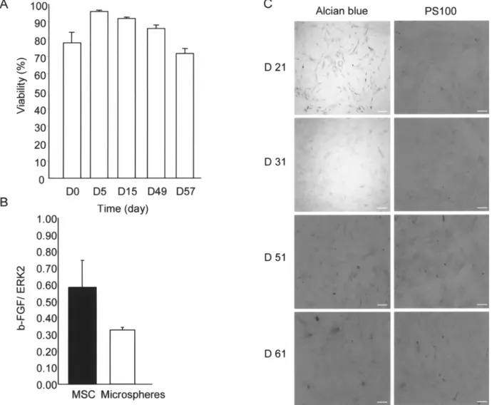
Impact of Cell Density on MSC Fate alginate remains the most dominantly applied because
of its well-documented biocompatibility, depending of Recent studies demonstrated that variations of cell
density in various scaffold may influence chondrogenic its purity and composition (32). We have chosen ultra-pure type M alginate as De Vos et al. observed that mi-differentiation of MSCs (14,42). According with this
ob-servation, we have verified whether cell density has an crocapsules prepared with purified M alginate remained free of any significant foreign body response for pro-impact on MSC viability, functionality, and
differentia-tion in alginate microspheres. In a first step, we pro- longed periods of time after implantation (6), while mi-crocapsules prepared with high-G alginate were consis-duced alginate microspheres with 2.5× 10⁶ or 4× 10⁶
MSCs/ml of alginate (Fig. 5). Results showed no differ- tently associated with low recovery rates and extensive overgrowth of inflammatory cells.
Figure 4. In vitro differentiation of MSCs in microspheres. Immnunocytochemistry on MSCs extracted from microspheres and
replaced in culture. (A) Alcian blue staining at day 21, 31, 51, 61. (B) PS100 immunostaining at the same days. (C) Alizarin red staining at day 56 postencapsulation. (D) MSCs cultivated in a chondrogenic (upper panel) or osteogenic (lower panel) medium were used as a positive control for alcian blue and alizarin red staining. Scale bars: 50µm.
Another parameter that could be modulated is the ity that can be applied to cell immobilization. The challenge is therefore not to increase stability but to de-type of microparticle used. Depending on encapsulation
process, various mechanical and mass transport proper- termine what mechanical resistance is required for a spe-cific application without changing other relevant particle ties of alginate microparticles should be obtained, both
influencing scaffold integrity upon implantation and characteristics (5,46). According to microparticle perme-ability, the relation between tissular environment and cells viability and functionality. APA microcapsules
with a liquefied inner core, pioneered by Lim and Sun encapsulated cells could be modified (50). However, APA microcapsule cut-off is generally reported to be (20), have been the most frequently employed devices
to transplant biomaterials to the host in the absence of between 60 and 90 kDa (4), while the absence of mem-brane in microspheres enables an open macroporosity, immunosuppression. However the long-term in vivo
success of APA microcapsules has been limited, princi- permitting higher molecular weight molecules to diffuse (45).
pally due to the mechanical fragility of the membrane
complex and to the difficult handling. Alginate micro- In this study, we investigated both microcapsules and microspheres in order to define the most suitable for en-spheres are expected to be stronger, obviously
increas-ing the durability of the transplant and improvincreas-ing the capsulation of MSCs before grafting. Our results showed that they presented differences in their morphologies and feasibility of its retrieval. But there is still an open
ques-tion related to the optimal condiques-tions during the forma- their mechanical resistance but both remained stable over time. Moreover, our results indicated that micro-tion of mechanically stable particles with defined
poros-spheres were associated with a higher survival rate of Altogether these results present an interest for future clinical approaches as microspheres are easier to obtain MSCs when compared to microcapsules. The higher
sur-vival of MSCs in microspheres could be explained by than microcapsules and would consequently facilitate a patient’s graft. In order to evaluate functionality of en-their need of adherence, as they are immobilized in the
alginate matrix while they are in suspension in micro- capsulated MSCs we measured MSC viability and the expression of bFGF, which is one of most abundant cy-capsules. However, a deleterious influence of capsule
membrane cut-off cannot be excluded. Furthermore, tokines secreted by MCSs (25). Indeed, bFGF is an angi-ogenic and mitangi-ogenic factor that has been previously in-Tam et al. have showed that the extent of
immunoglobu-lin adsorption on alginate microcapsules was highly de- volved in renal protection and repair, mainly by stimulating angiogenesis and regeneration of renal cells pendent on the presence of the polylysine membrane,
indicating that positive charges of the polycation are (9,47). The fact that encapsulated MSCs are still func-tional in these microspheres is important because benefi-mainly responsible for the binding of immunoglobulin
(35,43). Because IgG, IgM, and IgA are known to be cial effects of MSCs are mainly due to their paracrine activity (54).
opsonizing proteins that lead to complement activation
upon their adsorption to foreign surfaces, we decided to The last unknown data remained their ability to be grafted on solid organs. In a recent study, RGD-alginate proceed with alginate microspheres.
Figure 5. Effects of cell density on MSCs behavior in microspheres. Microspheres were produced with a density of 2.5× 10⁶MSCs/
ml. (A) Evaluation of MSC viability in microspheres. (B) In vitro expression of bFGF by the MSCs entrapped in microspheres. (C) Alcian blue staining and PS100 immunostaining at day 21, 31, 51, 61 postencapsulation. Scale bars: 50µm.
Figure 6. In vivo implantation of microspheres. (A) Injection of MSC microspheres (white arrow) under the renal capsule. (B)
Localization of MSC microspheres (white arrow) under the renal capsules 25 days after graft. Scale bars: 5 mm. (C) Hematoxylin/ eosin staining of renal histological sections showing microsphere (MSCs, arrow) 25 days after graft. Scale bars: 100 µm. (D) Plasmatic urea and creatinine concentrations in rats grafted with microparticles.
microspheres were grafted in bone (11) but nothing is nate molecular weight and consecutive biodegradability to obtain cells release in the purpose of tissue engi-known about their graft at the surface of solid organs
like the heart or kidney. Another study has shown that neering.
Encapsulating MSCs in alginate microspheres would intramyocardial injection of microcapsules could be
done (52,56), but we have preferred avoiding intrapar- be a promising approach to improve cell survival after their graft in a solid organ. However, concerning the enchymal graft and the consecutive mechanical stress
imposed to cells. So, we developed a protocol based heart, it could be difficult to implant microspheres. Re-cent studies have proposed to use patches to repair in-on the graft of microspheres under the renal capsule
(37). This site has been chosen as we have previously farcted cardiac tissue (2,18,30,34,39,51). In our labora-tory, we have already showed that intramyocardial graft showed that MSC administration could have beneficial
effects on kidney recovery, in a model of renal ische- of pharmacologically modified MSCs improves cardiac function (24). To further enhance MSC viability after mia-reperfusion, through a paracrine activity (25).
Fur-thermore, this graft site has already been validated for graft and consequently MSC efficiency it could be inter-esting to combine the easiness of engineering highly the transplantation of encapsulated pig islets with a
better biocompatibility than for the intraperitoneal biocompatible alginate scaffolds with beneficial effects of MSCs.
route (8).
Our in vivo approach shows that it is possible to graft
microspheres and to follow them during several weeks ACKNOWLEDGMENTS: We thank Serge Estaque (Service d’anatomie et cytologie pathologiques, Centre Hospitalo-without any microsphere degradation and impact on
re-Universitaire Rangueil, Toulouse, France) for assistance in nal function. The small and uniform microspheres tested
tissue embedding and processing, the Service de Zootechnie offer many advantages when compared to intraparenchy- (INSERM, IFR31, Toulouse, France), and the Cellular Im-mal injection or bigger scaffolds: a higher degree of bio- aging Facility IFR150-Rangueil. This work was supported in compatibility, a reduced total implant volume, a better part by the INSERM [European Community’s Seventh Frame-work Programme (FP7/2007-2013) under grant agreement diffusion of cellular paracrine secretion, an improved
No. HEALTH-F5-2008-223007 STAR-T REK and grants from cell oxygenation, a potential access to different
implan-the National Research Agency (ANR) (Grant under program tation site (32,33), and a concentration of the grafted Physiopathologie des Maladies Humaines, project SYNMESC cells in a dedicated zone. Moreover, it would be possible ARI)] and by CNRS (CNRS grant for exploratory projects “In-to retrieve microspheres after graft or “In-to play with algi- terface Mate´riau/Vivant”).
Krause, D. S.; Cantley, L. G. Bone marrow stem cells
REFERENCES
contribute to repair of the ischemically injured renal tu-bule. J. Clin. Invest. 112(1):42–49; 2003.
1. Aggarwal, S.; Pittenger, M. F. Human mesenchymal stem
cells modulate allogeneic immune cell responses. Blood 16. Keyser, K. A.; Beagles, K. E.; Kiem, H. P. Comparison of mesenchymal stem cells from different tissues to sup-105(4):1815–1822; 2005.
2. Amir, G.; Miller, L.; Shachar, M.; Feinberg, M. S.; press T-cell activation. Cell Transplant. 16(5):555–562; 2007.
Holbova, R.; Cohen, S.; Leor, J. Evaluation of a
perito-neal-generated cardiac patch in a rat model of heterotopic 17. Kinnaird, T.; Stabile, E.; Burnett, M. S.; Lee, C. W.; Barr, S.; Fuchs, S.; Epstein, S. E. Marrow-derived stromal cells heart transplantation. Cell Transplant. 18(3):275–282;
2009. express genes encoding a broad spectrum of arteriogenic cytokines and promote in vitro and in vivo arteriogenesis 3. Bartkowiak, A.; Lisiecki, S.; Orive, G.; Pedraz, J. L. The
effect of selected parameters of formation on properties of through paracrine mechanisms. Circ. Res. 94(5):678–685; 2004.
alginate/Ca2+/oligochitosan capsules. J. Chem. Technol.
Biotechnol. 81(4):511–518; 2006. 18. Kochupura, P. V.; Azeloglu, E. U.; Kelly, D. J.; Doronin, S. V.; Badylak, S. F.; Krukenkamp, I. B.; Cohen, I. S.; 4. Chen, H.; Ouyang, W.; Lawuyi, B.; Lim, T.; Prakash, S.
A new method for microcapsule characterization: use of Gaudette, G. R. Tissue-engineered myocardial patch de-rived from extracellular matrix provides regional mechani-fluorogenic genipin to characterize polymeric
microcap-sule membranes. Appl. Biochem. Biotechnol. 134(3):207– cal function. Circulation 112(9 Suppl.):I144–149; 2005. 19. Leal, D.; Matsuhiro, B.; Rossi, M.; Caruso, F. FT-IR spec-222; 2006.
5. de Vos, P.; Bucko, M.; Gemeiner, P.; Navratil, M.; Svitel, tra of alginic acid block fractions in three species of brown seaweeds. Carbohydr. Res. 343(2):308–316; 2008. J.; Faas, M.; Strand, B. L.; Skjak-Braek, G.; Morch,
Y. A.; Vikartovska, A.; Lacik, I.; Kollarikova, G.; Orive, 20. Lim, F.; Sun, A. M. Microencapsulated islets as bioartifi-cial endocrine pancreas. Science 210(4472):908–910; G.; Poncelet, D.; Pedraz, J. L.; Ansorge-Schumacher,
M. B. Multiscale requirements for bioencapsulation in 1980.
21. Lubiatowski, P.; Kruczynski, J.; Gradys, A.; Trzeciak, T.; medicine and biotechnology. Biomaterials 30(13):2559–
2570; 2009. Jaroszewski, J. Articular cartilage repair by means of bio-degradable scaffolds. Transplant. Proc. 38(1):320–322; 6. de Vos, P.; Hoogmoed, C. G.; Busscher, H. J. Chemistry
and biocompatibility of alginate-PLL capsules for immu- 2006.
22. Ma, H. L.; Chen, T. H.; Low-Tone Ho, L.; Hung, S. C. noprotection of mammalian cells. J. Biomed. Mater. Res.
60(2):252–259; 2002. Neocartilage from human mesenchymal stem cells in algi-nate: Implied timing of transplantation. J. Biomed. Mater. 7. Di Nicola, M.; Carlo-Stella, C.; Magni, M.; Milanesi, M.;
Longoni, P. D.; Matteucci, P.; Grisanti, S.; Gianni, A. M. Res. A 74(3):439–446; 2005.
23. Maurel, A.; Hernandez, C.; Kunduzova, O.; Bompart, G.; Human bone marrow stromal cells suppress T-lymphocyte
proliferation induced by cellular or nonspecific mitogenic Cambon, C.; Parini, A.; Frances, B. Age-dependent in-crease in hydrogen peroxide production by cardiac mono-stimuli. Blood 99(10):3838–3843; 2002.
8. Dufrane, D.; Steenberghe, M.; Goebbels, R. M.; Saliez, amine oxidase A in rats. Am. J. Physiol. Heart Circ. Phys-iol. 284(4):H1460–1467; 2003.
A.; Guiot, Y.; Gianello, P. The influence of implantation
site on the biocompatibility and survival of alginate en- 24. Mias, C.; Lairez, O.; Trouche, E.; Roncalli, J.; Calise, D.; Seguelas, M. H.; Ordener, C.; Piercecchi-Marti, M. D.; capsulated pig islets in rats. Biomaterials 27(17):3201–
3208; 2006. Auge, N.; Salvayre, A. N.; Bourin, P.; Parini, A.; Cussac, D. Mesenchymal stem cells promote matrix metalloprotei-9. Efthimiadou, A.; Lambropoulou, M.; Pagonopoulou, O.;
Vakalopoulos, I.; Papadopoulos, N.; Nikolettos, N. The nase secretion by cardiac fibroblasts and reduce cardiac ventricular fibrosis after myocardial infarction. Stem Cells role of basic-fibroblast growth factor (b-FGF) in
cyclosporine-induced nephrotoxicity. In Vivo 20(2):265– 27(11):2734–2743; 2009.
25. Mias, C.; Trouche, E.; Seguelas, M. H.; Calcagno, F.; 269; 2006.
10. Goosen, M. F. A.; O’shea, G. M.; Gharapetian, H. M.; Dignat-George, F.; Sabatier, F.; Piercecchi-Marti, M. D.; Daniel, L.; Bianchi, P.; Calise, D.; Bourin, P.; Parini, A.; Chous, S.; Sun, A. M. Optimization of microencapsulation
parameters: Semipermeable microcapsules as a bioartifi- Cussac, D. Ex vivo pretreatment with melatonin improves survival, proangiogenic/mitogenic activity, and efficiency cial pancreas. Biotechnol. Bioeng. 27:146–150; 1985.
11. Grellier, M.; Granja, P. L.; Fricain, J. C.; Bidarra, S. J.; of mesenchymal stem cells injected into ischemic kidney. Stem Cells 26(7):1749–1757; 2008.
Renard, M.; Bareille, R.; Bourget, C.; Amedee, J.;
Barbosa, M. A. The effect of the co-immobilization of hu- 26. Mironova, E. V.; Evstratova, A. A.; Antonov, S. M. A fluorescence vital assay for the recognition and quantifica-man osteoprogenitors and endothelial cells within alginate
microspheres on mineralization in a bone defect. Biomate- tion of excitotoxic cell death by necrosis and apoptosis using confocal microscopy on neurons in culture. J. Neu-rials 30(19):3271–3278; 2009.
12. Hauser, O.; Prieschl-Grassauer, E.; Salmons, B. Encapsu- rosci. Methods 163(1):1–8; 2007.
27. Moscoso, I.; Centeno, A.; Lopez, E.; Rodriguez-Barbosa, lated, genetically modified cells producing in vivo
thera-peutics. Curr. Opin. Mol. Ther. 6(4):412–420; 2004. J. I.; Santamarina, I.; Filgueira, P.; Sanchez, M. J.; Dominguez-Perles, R.; Penuelas-Rivas, G.; Domenech, N. 13. Herzog, E. L.; Chai, L.; Krause, D. S. Plasticity of
mar-row-derived stem cells. Blood 102(10):3483–3493; 2003. Differentiation “in vitro” of primary and immortalized porcine mesenchymal stem cells into cardiomyocytes for 14. Hui, T. Y.; Cheung, K. M.; Cheung, W. L.; Chan, D.;
Chan, B. P. In vitro chondrogenic differentiation of human cell transplantation. Transplant. Proc. 37(1):481–482; 2005.
mesenchymal stem cells in collagen microspheres:
Influ-ence of cell seeding density and collagen concentration. 28. Moustafa, T.; Girod, S.; Tortosa, F.; Li, R.; Sol, J. C.; Biomaterials 29(22):3201–3212; 2008. Rodriguez, F.; Bastide, R.; Lazorthes, Y.; Sallerin, B. Via-bility and functionality of bovine chromaffin cells encap-15. Kale, S.; Karihaloo, A.; Clark, P. R.; Kashgarian, M.;
sulated into alginate-PLL microcapsules with a liquefied Yahia, L.; de Vos, P. Adsorption of human immunoglobu-lin to implantable alginate-poly-L-lysine microcapsules: inner core. Cell Transplant. 15(2):121–133; 2006.
29. Muller-Ehmsen, J.; Whittaker, P.; Kloner, R. A.; Dow, Effect of microcapsule composition. J. Biomed. Mater. Res. A 89(3):609–615; 2009.
J. S.; Sakoda, T.; Long, T. I.; Laird, P. W.; Kedes, L.
Survival and development of neonatal rat cardiomyocytes 44. Tambara, K.; Sakakibara, Y.; Sakaguchi, G.; Lu, F.; Premaratne, G. U.; Lin, X.; Nishimura, K.; Komeda, M. transplanted into adult myocardium. J. Mol. Cell. Cardiol.
34(2):107–116; 2002. Transplanted skeletal myoblasts can fully replace the in-farcted myocardium when they survive in the host in large 30. Nasseri, B. A.; Kukucka, M.; Dandel, M.; Knosalla, C.;
Potapov, E.; Lehmkuhl, H. B.; Meyer, R.; Ebell, W.; numbers. Circulation 108(Suppl. 1):II259–263; 2003. 45. Tanaka, H.; Matsumura, M.; Veliky, I. A. Diffusion char-Stamm, C.; Hetzer, R. Intramyocardial delivery of bone
marrow mononuclear cells and mechanical assist device acteristics of substrates in Ca-alginate gel beads. Biotech-nol. Bioeng. 26(1):53–58; 1984.
implantation in patients with end-stage cardiomyopathy.
Cell Transplant. 16(9):941–949; 2007. 46. Thanos, C. G.; Calafiore, R.; Basta, G.; Bintz, B. E.; Bell, W. J.; Hudak, J.; Vasconcellos, A.; Schneider, P.; Skinner, 31. Orive, G.; Hernandez, R. M.; Gascon, A. R.; Igartua, M.;
Pedraz, J. L. Development and optimisation of alginate- S. J.; Geaney, M.; Tan, P.; Elliot, R. B.; Tatnell, M.; Esco-bar, L.; Qian, H.; Mathiowitz, E.; Emerich, D. F. Formu-PMCG-alginate microcapsules for cell immobilisation.
Int. J. Pharm. 259(1–2):57–68; 2003. lating the alginate-polyornithine biocapsule for prolonged stability: Evaluation of composition and manufacturing 32. Orive, G.; Ponce, S.; Hernandez, R. M.; Gascon, A. R.;
Igartua, M.; Pedraz, J. L. Biocompatibility of microcap- technique. J. Biomed. Mater. Res. A 83(1):216–224; 2007.
sules for cell immobilization elaborated with different
type of alginates. Biomaterials 23(18):3825–3831; 2002. 47. Togel, F.; Weiss, K.; Yang, Y.; Hu, Z.; Zhang, P.; Westenfelder, C. Vasculotropic, paracrine actions of in-33. Orive, G.; Tam, S. K.; Pedraz, J. L.; Halle, J. P.
Biocom-patibility of alginate-poly-L-lysine microcapsules for cell fused mesenchymal stem cells are important to the recov-ery from acute kidney injury. Am. J. Physiol. Renal Phys-therapy. Biomaterials 27(20):3691–3700; 2006.
34. Piao, H.; Kwon, J. S.; Piao, S.; Sohn, J. H.; Lee, Y. S.; iol. 292(5):F1626–1635; 2007.
48. Toma, C.; Pittenger, M. F.; Cahill, K. S.; Byrne, B. J.; Bae, J. W.; Hwang, K. K.; Kim, D. W.; Jeon, O.; Kim,
B. S.; Park, Y. B.; Cho, M. C. Effects of cardiac patches Kessler, P. D. Human mesenchymal stem cells differenti-ate to a cardiomyocyte phenotype in the adult murine engineered with bone marrow-derived mononuclear cells
and PGCL scaffolds in a rat myocardial infarction model. heart. Circulation 105(1):93–98; 2002.
49. Tse, W. T.; Pendleton, J. D.; Beyer, W. M.; Egalka, Biomaterials 28(4):641–649; 2007.
35. Ponce, S.; Orive, G.; Hernandez, R.; Gascon, A. R.; M. C.; Guinan, E. C. Suppression of allogeneic T-cell pro-liferation by human marrow stromal cells: Implications in Pedraz, J. L.; de Haan, B. J.; Faas, M. M.; Mathieu, H. J.;
de Vos, P. Chemistry and the biological response against transplantation. Transplantation 75(3):389–397; 2003. 50. Uludag, H.; De Vos, P.; Tresco, P. A. Technology of immunoisolating alginate-polycation capsules of different
composition. Biomaterials 27(28):4831–4839; 2006. mammalian cell encapsulation. Adv. Drug Deliv. Rev. 42(1–2):29–64; 2000.
36. Poulsom, R.; Forbes, S. J.; Hodivala-Dilke, K.; Ryan, E.;
Wyles, S.; Navaratnarasah, S.; Jeffery, R.; Hunt, T.; 51. Wei, H. J.; Chen, C. H.; Lee, W. Y.; Chiu, I.; Hwang, S. M.; Lin, W. W.; Huang, C. C.; Yeh, Y. C.; Chang, Y.; Alison, M.; Cook, T.; Pusey, C.; Wright, N. A. Bone
mar-row contributes to renal parenchymal turnover and regen- Sung, H. W. Bioengineered cardiac patch constructed from multilayered mesenchymal stem cells for myocardial eration. J. Pathol. 195(2):229–235; 2001.
37. Rajab, A.; Buss, J.; Diakoff, E.; Hadley, G. A.; Osei, K.; repair. Biomaterials 29(26):3547–3556; 2008.
52. Wei, H. J.; Yang, H. H.; Chen, C. H.; Lin, W. W.; Chen, Ferguson, R. M. Comparison of the portal vein and kidney
subcapsule as sites for primate islet autotransplantation. S. C.; Lai, P. H.; Chang, Y.; Sung, H. W. Gelatin micro-spheres encapsulated with a nonpeptide angiogenic agent, Cell Transplant. 17(9):1015–1023; 2008.
38. Reinecke, H.; Zhang, M.; Bartosek, T.; Murry, C. E. Sur- ginsenoside Rg1, for intramyocardial injection in a rat model with infarcted myocardium. J. Control. Release vival, integration, and differentiation of cardiomyocyte
grafts: A study in normal and injured rat hearts. Circula- 120(1–2):27–34; 2007.
53. Wu, T. J.; Huang, H. H.; Hsu, Y. M.; Lyu, S. R.; Wang, tion 100(2):193–202; 1999.
39. Rosellini, E.; Cristallini, C.; Barbani, N.; Vozzi, G.; Y. J. A novel method of encapsulating and cultivating ad-herent mammalian cells within collagen microcarriers. Giusti, P. Preparation and characterization of
alginate/gel-atin blend films for cardiac tissue engineering. J. Biomed. Biotechnol. Bioeng. 98(3):578–585; 2007.
54. Xiang, M. X.; He, A. N.; Wang, J. A.; Gui, C. Protective Mater. Res. A 91(2):447–453; 2009.
40. Shoichet, M. S.; Rein, D. H. In vivo biostability of a poly- paracrine effect of mesenchymal stem cells on cardiomyo-cytes. J. Zhejiang Univ. Sci. B 10(8):619–624; 2009. meric hollow fibre membrane for cell encapsulation.
Bio-materials 17(3):285–290; 1996. 55. Xu, W.; Zhang, X.; Qian, H.; Zhu, W.; Sun, X.; Hu, J.; Zhou, H.; Chen, Y. Mesenchymal stem cells from adult 41. Silva, C. M.; Ribeiro, A. J.; Ferreira, D.; Veiga, F. Insulin
encapsulation in reinforced alginate microspheres pre- human bone marrow differentiate into a cardiomyocyte phenotype in vitro. Exp. Biol. Med. (Maywood) 229(7): pared by internal gelation. Eur. J. Pharm. Sci. 29(2):148–
159; 2006. 623–631; 2004.
56. Zhang, H.; Zhu, S. J.; Wang, W.; Wei, Y. J.; Hu, S. S. 42. Takagi, M.; Umetsu, Y.; Fujiwara, M.; Wakitani, S. High
inoculation cell density could accelerate the differentiation Transplantation of microencapsulated genetically modi-fied xenogeneic cells augments angiogenesis and im-of human bone marrow mesenchymal stem cells to
chon-drocyte cells. J. Biosci. Bioeng. 103(1):98–100; 2007. proves heart function. Gene Ther. 15(1):40–48; 2008. 43. Tam, S. K.; de Haan, B. J.; Faas, M. M.; Halle, J. P.;