Biochem. J.(1993)292,563-570(Printed inGreatBritain)
Characterization of
the
sporulation-related
y-D-glutamyl-(L)meso-diaminopimelic-acid-hydrolysing
peptidase
I of
Bacillus
sphaericus
NCTC
9602
as a
member of the metallo(zinc)
carboxypeptidase
A
family
Modular
design of
the
protein
Marie-Laure
HOURDOU,* Micheline
GUINAND,t
Marie-Jeanne
VACHERON,t Georges MICHEL,t
LucDENOROY,$
ColetteDUEZ,*
Serge
ENGLEBERT,*
Bernard
JORIS,* Georges
WEBER§
and Jean-MarieGHUYSEN*II
*Centred'lngenierie des Proteines, Universit6 deLiege, Institut deChimie, B6, B-4000 Sart Tilman(Liege 1), Belgium,
tLaboratoire
deBiochimie Microbienne, Universite Claude BernardLyon
1, 43 Bd du 11 Novembre1918, F-69622 VilleurbanneCedex,France,
tService
Centrald'Analyses
du CentreNational de la RechercheScientifique,
BP22,F-69390 Vernaison, France,and §1nstitut dePhysique Nucl6aire Exp6rimentale (IPNE), Universite de Liege, B15, B-4000Sart Tilman (Liege 1), Belgium
The sporulation-related
y-D-glutamyl-(L)meso-diaminopimelic-acid-hydrolysing peptidase I of Bacillus sphaericus NCTC9602 hasbeen analysed by proton-inducedX-rayemission.Itcontains 1 equivalent Zn2+ per mol of protein. As derived from gene cloningandsequencing, theB.sphaericusZnpeptidaseIis a two-module protein. A 100-amino-acid-residue N-terminal domain consisting oftwotandem segments ofsimilar sequences,isfused to a 296-amino-acid-residue C-terminal catalytic domain. The catalyticdomain belongsto theZncarboxypeptidaseA family, the closest match being observedwith theStreptomycesgriseuscarboxypeptidase
[Narahashi (1990) J. Biochem. 107, 879-886] andwith thefamily prototype, bovine carboxypeptidaseA.The catalytic domain of the B. sphaericus peptidase I possesses,INTRODUCTION
Bacillus sphaericus NCTC 9602 produces two sporulation-related,
y-D-glutamyl-(L)diamino-acid-hydrolysing
peptidases, known asendopeptidases I and II. They differ with respect tocellular localization (Guinand et al., 1979; Vacheron et al., 1979), molecularmassandcatalytic mechanism (Gamieretal., 1985; Bourgogneetal.,1992,Hourdouetal., 1992) andspecificity profile(Arminjon etal., 1977; Valentinetal., 1983).
EndopeptidaseI,a45kDaprotein,isproducedatstageIVof sporulation (Guinand et al., 1974; Tipper et al., 1977). It has been purified in trace amounts from the sporulation medium (Garnieretal.,1985)and inlargeramountsfrom theinteguments ofthe forespores and spores in the presence ofBrij 58
(Baji-Kourdaetal., 1989). EndopeptidaseI,referredtoaspeptidaseI
throughout this paper, is a carboxypeptidase/peptidyl
di-peptide hydrolase. It hydrolyses the y-D-glutamyl-(L)meso-diaminopimelic acid (msA2pm) bond of L-Ala-y-D-Glu-(L)msA2pm and L-Ala-y-D-Glu-(L)msA2pm(L)-D-Ala peptides. The L-alanine amino group may be free or substituted
by an N-acetylmuramoyl or
N-acetylglucosaminyl-fl-1,4-N-acetylmuramoylmoiety. Conversely, thepresenceofamsA2pm
residue with free e-NH2 and 6-COOH groups is a strict
re-quirement for activity. Peptides containing L-lysine instead of msA2pm, peptidesterminating withthesequence
msA2pm(L)-D-Ala-D-Ala and cross-linkedpeptide dimers(in which the amino
distributed alongthe amino-acid sequence,peptide segments, a
triad His162-Glu165-His307 and a dyad Tyr347-Glu366 that are
equivalent
to secondary structures, the zinc-binding triad His69-Glu72-His'96and thecatalytic dyad Tyr248-Glu270of bovine carboxypeptidaseArespectively. TheN-terminalrepeatsof the B. sphaericus peptidase I have similarity with the C-terminal repeatsofthe Enterococcus hirae muramidase 2, theStreptococcus(now
Enterococcus)faecalisautolysinand the BacillusqSPZAand q529 lysozymes,towhicharolein therecognitionof aparticular
moiety of the bacterial cell envelope has been tentativelyassigned.
Detergents enhance considerablythespecificactivity of the B.sphaericus peptidase I.groupontheD-centreofmsA2pmofonepeptideislinked tothe carbonyl group on the L-centreofmsA2pm of another
peptide
via an intervening D-Ala residue) have no substrateactivity.
Peptidase I is inactivated by EDTA and reactivated by zinc, cobalt and manganese ions, suggesting that it may be ametallo(zinc)peptidase (Gamieretal., 1985).
In order to gain some understanding of the molecular organization of the B. sphaericus peptidase I, the protein has been analysed byproton-induced X-ray emission(p.i.x.e.), the encoding gene has been cloned and sequenced and the derived amino-acid sequence has beensubmitted tohydrophobic-cluster analysis. The results of these investigations are presented and discussedbelow.
MATERIALS
ANDMETHODS
Enzyme, substrate and enzyme assay
The B. sphaericus peptidase I (purified from the spore integuments) (Baji-Kourda et al., 1989) was at a0.1 % (w/v) concentration in 20 mM Tris/HCl, pH 8.0/10 mM MgCl2 (re-ferred to as buffer) containing 0.2 % (w/v) Brij 58. The enzyme (diluted to the extent that the samples contained less than 0.0002 % Brij 58 before each experiment) and 0.4 mM
N-acetylmuramoyl-L-Ala-y-D-Glu-(L)msA2pm(L)-D-[14C]Ala
(1.1x 10⁶c.p.m./,umol)wereincubated at 37°Cin 10,1 (finalvolume) of buffer. Under these conditions, i.e. in thevirtualabsence ofAbbreviations used: p.i.x.e., proton-inducedX-rayemission; msA2pm, meso-diaminopimelic acid; HPC1, 1-hexadecylpyridinium chloride;CHAPS,
3-[(3-cholamidopropyl)dimethylammonio]-1-propanesulphonate; ORF,open readingframe. 11 Towhomcorrespondenceshould be addressed.
564
M.-L.
Hourdou and others
detergent, the specificenzymeactivitywas 14,tmol of
ms-A2pm-D-['4C]Ala
released/min (i.e. 14units) per mg of protein (Baji-Kourdaetal., 1989).Detergents
Brij 58 (polyoxyethylene-20 cetyl ether), Sarkosyl (sodium do-decanoylsarcosinate), HPC1 (1-hexadecylpyridinium chloride), Cetavlon(hexadecyltrimethylammoniumbromide), TritonX-
l00
(octylphenoxypolyethoxyethanol), n-octylglucoside (n-octyl f-D-glucopyranoside), SDS andCHAPS {3-[(3-cholamidopropyl)di-methyl ammonio]-1-propanesulphonate}were tested as modifiers of the hydrolytic activity of the B. sphaericus peptidase I. Theywereusedat afinal concentrationof0.4 %(w/v) in buffer.P.i.x.e.
TheB.sphaericuspeptidaseI(inbuffer containing0.2 % Brij 58) wasdialysed extensively against2.5 mM
Tris/HCl,
pH 8.0 and lyophilized. Thesame volumeof buffercontaining 0.2 % Brij 58 wastreated similarly and servedasthecontrol. The pelletsfrom theprotein and the controlweresuspended inwaterand samples (20Iu)
(i.e. 11.25nmol ofpeptidase I)weredeposited on 4,um-thickpropylene films togetherwith20,1
ofaliposome solution (in order to ensure a good homogeneity ofthe samples) and 7.7,ug of yttrium nitrate (used as internal standard). After evaporation ofthe solvent, the filmswerestretchedoncommercial 24 mmx36 mmslide frames and irradiated undervacuumbya 2.5MeV protonbeam(40 nAintensity) usingthefacilities of the Liege University Cyclotron Research Centre. The beam, of 10 mmdiam, covered the entire sample, thus avoiding possible problems of non-uniformity of the target. The measurements were made in duplicate. The protein-bound cations were estimated by subtracting the amounts found in the protein samples from those found in the controls. The values thus obtaineddepended criticallyontheamountsofprotein used for theexperiments. The proteincontent wasestimatedonthe basis ofthe A230/A260 value(Kalb
andBernlohr, 1977) and by using the method of Bradford (1976) with ovalbumin as standard. Both methods gaveidentical results.Papain
degradation and amino-acid sequencing
Beforeproteolysis,the B.sphaericuspeptidaseIwastreatedwith 100 mM EDTA and dialysed against 2 mM
Tris/HCl,
pH 8.0. Proteolysis wascarriedoutaccordingto Clevelandetal. (1977) in the presenceof1%
(w/v)SDS,at apapain (Sigma)/peptidase Iratioof1: 10(w/w),in 125 mMTris/HCl,
pH6.8, for20 min at 37'C. The reaction was stopped by heating the solution at100'C for 5min. Samples of untreated and papain-degraded peptidase1(25,g)weresubmittedtoreverse-phase h.p.l.c. ona
4.6 mmx220 mm Vydac C4 column. Elution was carried out withalinearly increasing gradient of acetonitrile madein0.1 % trifluoroaceticacid at aflowrateof1 ml per min.PeptidaseIand the h.p.l.c.-isolated peptides were analysed by SDS/PAGE on 12.5% (w/v) gels
(Laemmli
and Favre, 1973) and stained with Coomassie Brilliant Blue. They were submitted to amino-acid microsequencing by using anApplied Biosystems model 470A. DNArecombinant techniques
The B. sphaericusgenomicDNA was preparedas describedby
Hopwood
etal.(1985) from cellsintheearly sporulation phase.
Escherichia coli strains HB1I1 (Boyer et al., 1969) and DHSaMCR (Jessee and Bloom, 1988), grown at 37°C in Luria-Bertani medium,ashosts. The librarieswereconstructed
using dephosphorylated vectors. E. coli DH5aMCR was
transformedby electroporation (Doweretal., 1988) usinga
Bio-Rad Gene Pulser apparatus. Transformants were selected on
agar plates containing ampicillin (50 ,g/ml) or tetracycline
(25 ,ug/ml). Genomic librarieswerescreenedby hybridization (at
tm -5 C, for 18 h) with radioactive synthetic oligonucleotides (Eurogentec, Liege, Belgium) using a modified Southern-blot
procedure (Wallace et al., 1981; Woods, 1984). Other DNA-manipulation experiments were carried out as described
(Maniatisetal., 1982).
DNA segmentscloned into plasmid pSLI 190 (Brosius, 1989) and double-stranded DNA segments were sequenced by the
dideoxynucleotide chain termination method (Sangeretal.,1977) using the T7 sequencing kit (Pharmacia LKB BiotechnologyAB, Uppsala, Sweden). Denaturation of double-stranded DNAwas
performedasdescribed(Zhangetal., 1988) and thesequencing reactions wereinitiated with synthetic oligonucleotides.
Sequence
identity
searchesSearchesthrough the nucleic acid (EMBL version 30) and protein (PIR version 32) sequence databases were performed byusing
the procedure of Pearson and Lipman (1988) (FASTA and TFASTA softwares, GCG package). Alignments of pairs of proteinsweremadeby using the Goad and Kanehisa procedure
(1982) andauniformgappenalty of +8. Thesignificance of the comparison between pairs of alignedsequenceswasassessedby
using theSEQDPprogram(Kanehisa, 1982). Thisprogramgives
thescoreof the bestalignment oftwosequences.Thesignificance of the score is expressed by the S.D. unit of the scores of 20
randomsequencesofthesamecomposition. A S.D. unit value of
5 or higher indicates a statistically significant similarity. The
theoretical pl value of the proteinwas computed by using the
GCGpackage (Devereuxetal., 1984).
Hydrophobic-cluster analysis
Hydrophobic-cluster analysis (Gaboriaudetal., 1987; Henrissat
etal.,1990) isapowerful method for comparing proteins thatare
weakly related in their primary structures. It rests upon a
duplicated representation of the amino-acidsequenceson an
a-helical two-dimensional pattern (in which the hydrophobic residues tendtoformclusters) andcomparesthe distribution of
the clusters along the sequences. The shapes of the clustersare
usually associated with definite secondarystructures.Clusters of similar shapes, sizes and relative positionsexpress similarity in
thepolypeptide foldings.
Zinc
peptidases
Bovine carboxypeptidase A of known primary and three-dimensional structure(KimandLipscomb, 1990; Le Huerouet
al., 1991) servedas astandard of reference.CarboxypeptidaseA
is a one domain protein the dominant feature of which is an
eight-stranded f-sheet that constitutes thecoreof the molecule.
The Streptomyces griseus carboxypeptidase of known primary
structure(Narahashi, 1990)wasalso included in thecomparison.
Nucleotide sequence accession number
The EMBL accession number for the nucleotide sequence
encoding
theB.sphaericus peptidase
I is X69507. Genecloning
wasperformed
using pBR322
(BoUivar
etal.,
1977)
Bacillus
sphaericus
y-D-glutamyl-(L)meso-diaminopimelic-acid-hydrolysing
peptidase
RESULTS
Amino-acid
sequencedata and nucleotide
probes
Figure 1 shows the h.p.l.c. fractionation profile of the papain
digest of B. sphaericus peptidase I and, as an insert, the
SDS/PAGE profile of the untreatedenzyme(E)and theisolated peptides 6 (28kDa), 7 (43 kDa) and 8 (32 kDa). Peptides2and
4(< 14kDa)ranoutofthe gels. Figure 2 gives the results of the
0.3
0.2~
0.1
0 10 20 30 40 50 60
Volume (ml)
Figure1 Isolation by reverse-phase h.p.l.c. chromatography on a
Vydac C4 column of the peptides produced by papain hydrolysis of the
B. sphaericuspeptdase I
Continuous line:linearlyincreasing gradientofacetonitrilemadebymixingataconstantflow rate(1ml/min) solution B (acetonitrile/H20/trifluoroacetic acid;90:10:0.1,by vol.)insolution A (0.1% trifluoroacetic acid in water).SDS/PAGE of the untreated peptidase (E) and the isolatedpeptides 6,7and 8:S: proteins of standard molecularmass(shown ininsert).
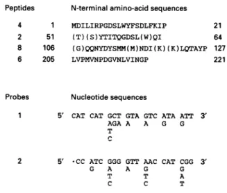
Peptides N-terminalamino-acidsequences
4 1 MDILIRPGDSLWYFSDLFKIP 2 51 (T)(S)YTITQGDSL(W)QI 8 106 (G)QQNYDYSMM(M)NDI(K)(K)LQTAYP 6 205 LVPMVNPDGVNLVINGP Probes 21 64 127 221 Nucleotidesequences
1 5' CAT CAT GCT GTA GTC ATA ATT 3'
AGA A A G G
T C
2 5' -CC ATC GGG GTT AAC CAT CGG 3'
G A A G G
T T A
C C T
Figure 2 N-terminal amino-acid sequences of peptides 4, 2, 8 and 6
produced by papain hydrolysis of the B. sphaericus peptidase I and
nucleotide sequences ofprobes 1 and 2
The isolatedpeptidesarethose shown inFigure1.Theamino-acidnumberingisattributedon
thebasis of theprimarystructureof the B.sphaericus peptidase (Figure 4).Theamino-acid residues inparentheseswerenotdetermined withcertainty bychemical sequencing.The two
degeneratednucleotides weresynthesizedonthebasis of thesequencesofpeptide8(probe 1)and peptide6(probe 2).
amino-acidsequencingperformedonthe B.sphaericuspeptidase Iand the isolatedpeptides4, 2,8 and 6.PeptidaseI andpeptide 4 gaveidentical results. The amino-acidnumberingwasattributed on the basis of the primary structure of the protein asderived from genecloning andsequencing(seebelow).
These structural data were used to synthesize the two
degenerated nucleotides shown in Figure 2. Nucleotide 1 was
complementary to the nucleotide sequence encoding the NYDYSMM amino-acid sequence of peptide 8. Nucleotide 2 was complementary to the nucleotide sequence encoding the PMVNPD(G) amino-acid sequence of peptide 6.
Gene
cloning
Attempts to clone the B. sphaericus peptidase-I-encoding gene were first carried out with BamHI, EcoRI, HindIII, PstI, Sall
andSphIlibraries made in pBR322 and pSP73, by using
CaCl2-treated E. coliHB1O1 ashost and the 32P-labelled nucleotides 1
and 2 as probes. Of the 50000 recombinant clones analysed,
none gave ahybridization signal.
Given that B.sphaericusproducesthe restriction endonuclease Bspl286, its DNA isprobably methylated (Raleighetal., 1988), making it difficulttoclonethe desiredgeneinanE.coli host that
contains the Mcr/Mrr restriction enzyme (Blumenthal, 1986;
Woodcock et al., 1988). Consequently, samples containing 2.5x101cells of E. coli DH5aMCR(recA-, mcrA-, mcrBJ and
mrr-) were electroporated with 5ng of purified recombinant
pBR322 plasmids prepared from HindIll, EcoRI and SphI libraries.The yieldwas5x105transformed cellsper,ug of DNA,
with a survival rate ofabout 1
%.
Of the 6000 recombinantclones analysed, two originating from the HindIll library, hybridized with probes 1 and 2. The corresponding plasmids,
pDML205 and pDML206, contained an insert of 6.6 kb and
7.8 kb respectively. Upon restriction, each plasmid yielded a
smallSpeI-SpeI 461 bp DNA fragment which, after separation by agarose-gelelectrophoresisandtransfertoanylon membrane,
gave a strong hybridization signal with probe 1. This DNA
segment was subcloned in pSL1190. Sequencing, using the
universalandreverseprimers, showed that it had the information
for a 153-amino-acid polypeptide and that this polypeptide
contained the N-terminal sequenceofpeptide 8 at aninternal
position.
Gene
sequencing
andprimary
structure of B.sphaericus
peptidase
INucleotide sequencing of pDML205 was performed in both
directionsupstreamanddownstreamfrom the SpeI-SpeI DNA fragment, by using thestrategyshown inFigure 3. The sequenced 1710bp DNAsegmentcontainedanopenreading frame (ORF),
1188nucleotides long, which started withaGTG triplet coding
for methionine at position 220 (itself preceded by a putative
Shine-Dalgarno sequence) and terminated withaTAAcodonat
position 1408 (Figure 4). It translated into a 396-amino-acid
protein (Figure 4). This protein had no signal peptide. Its
theoretical molecularmass, 44724Da,coincidedperfectly with
the 45 kDa value attributed to the isolated peptidase I on the
basis ofitsmigration by SDS/PAGE (Figure 1).Its theoretical
5.46pl value also coincided with the experimental 5.40 pl value (Garnieretal., 1985). Peptides 4, 2, 8 and 6obtainedby papain hydrolysis of peptidaseIandof established N-terminalsequence
(Figure 2) wereeasily identified along the primary structureof
theprotein (Figure 4). Downstream from thestopcodon of the B. sphaericus peptidase-I-encoding gene, a second ORF was
identified which started with an ATG codon at position 1594
I S 7 E 8 6 kDa 4 94 -_ 67-_ 8 43- _ -2Q. -K; 4 1 14.4--0
i10
.A565
M.-L.
Hourdou and others
0 ---0 0 00 0* & 5 500ta
1000 II I 0 CD (1 1500 bp -- Peptidase I-encodinggene---ORF2
Figure 3 Strategy used for sequencing the B. sphaericus
peptidase-l-encodinggene.Detectionofan additionalORF
Keyto symbols: 0, sequences initiated with the M13 universal orreverse primer; 0, sequencesinitiatedwithsynthetic nucleotides;arrows,orientation andlengthofthesequenced
DNAsegments.Abbreviation: ORF,openreadingframe.
(Figure 4). This ORF extended downstream from the 3' end of the sequenced 1710 bp DNAsegment.
P.i.x.e.
P.i.x.e. of the B. sphaericus peptidase I (see the Materials and methodssection) detected thepresenceof 10.9equivalent Zn2+
and 107 equivalent K+ for 11.25 nmol of protein, i.e. almost
exactly 1 equivalent Zn2+ and 10 equivalent K+ per mol of
protein.
Sequence
Identity searches,
hydrophobic-cluster analysis
and amino-acidalignments
Provided that the 100-amino-acid-residue N-terminal region of the protein was excluded from the analysis, sequence identity
searches (see the Materials and methods section) led to the
conclusion that theB. sphaericuspeptidaseI hadsimilarity with the zinc peptidases of the carboxypeptidase A family. Among them, the S. griseuscarboxypeptidase(Narahashi, 1990) and the prototypic bovine carboxypeptidase A exhibited the closest match.
The amino-acidsequenceof the B.sphaericuspeptidaseIand
that of the S. griseus carboxypeptidase were submitted to
hydrophobic-cluster analysis by reference to bovine carboxypeptidaseA(Figure 5).Inthisgraphical representation, the hydrophobic residues Phe, Ile, Leu, Met, Val, TrpandTyr
are encircled; thehydrophobicclustersare alsodelineated; the
hydrophobic residuesandclustersoccurringatequivalent places inthe threesequencesareinbold;the other amino-acidresidues
occurringasstrictidentitiesaremarkedby scattered points;and thesecondary structures ofcarboxypeptidaseA areidentified.
The linear amino-acid alignments derived from the data of Figure 5aregiveninFigure 6.Thealignmentsbetween thepair
B. sphaericus peptidase I and bovine carboxypeptidase A, the pairB.sphaericus peptidase I and S. griseuscarboxypeptidase,
and the pair S. griseus carboxypeptidase and bovine
carboxypeptidase Ahad scores whichwere 16, 18 and 46 S.D.
units respectively, above those expected from a run of 20
,randomizedpairs of proteins havingthesameamino-acid
com-position as the pairs under comparison (seethe Materials and
methods section).The threepeptidasesboreasimilar signature
inthe form of conserved amino-acid residues (Figure 6).
Peptide(
N D I L I I P G D S L 9 Y f S D L ATW!.AA TLQATAGG0AA9TSAS1!ATATCA?TAG0-TQ__ _ GAwAG0ATT
SD 2D 30 0 f 9 I PL Q L L L D S I I I I I P Q L L Q V G Q I I Q I P G so Peptide
(2
60 70 Y V T S Y T I T Q G D S LUQV I A Q VIKV L P L I A I L L Spei 80 90 100 V I P I I Q P S I L I I G Q T I Q V P Q R L T V I L V G Q n1o Peptide .0 120 130 Q I Y D Y S N N N I D I K K L Q T k Y 'Pf L Q G S P I G I SCAAAATATQTTACAG CATG8GT CGTTAMAAATACAA0TAT0CAITC?CAGA60&9GA
140 150 160 * * V L k Q P I P I I L I G I G S K R I I Y K k S I I A I I 0 I 170 190 190 T T P I I N T f L I D Y L L k L t J Q T T I I G L S N G P L 20 210 Peptide( * 22_sp1286 Y IQ T T L S V P N V I P D G V I L V I I G P Ak N A L
TATAlAolCAAAANLAClcTAcw =5GwAA _G0cAAwTArc~ACATAoAT
* *SpieI 230 240 250 K I K L I A V I I I S Q N E S G N K A I I I C V D L N D Q F 260 270 290 P A K V e L I I k I I P Q T P G P I D Y G G e A P L T Q P I 290 300 * 310 A I A N k D L T I S I I P k V V L A f I T Q G R V I Y V G I OCACTATATGC007YTACAAGGAQCOGOU CFGTFACTY&A0CA0>CY?T0GA 320 330 340 1 J L I P P E s Q t N V I I f S I V S G Y E P I Q S k I S Y . .* .8spl1286 350 360 370 A G Y K D V f I Q D V RI P G f T V I L G S G t I P L P I S 380 390 396 I f D T I Y Q I A L G I I L A G L Y L GTA&TA__TT¶CAATAC AVCATCAGAAACAATTMTGTMTTGTGATAATTcTG N V k S D I I I G 10 * *ORF2 10 20 30 1 L D S I I L R L I L E K D R Y G Y E I S Q E I S N R T I N ILSILOILKDYGEIQESITI
.TAITO .ATITGGAO& .GTTGTM .A?!CC .GAAAG .ACOAAATA 90 190 270 360 450 540 630 810 990 I060 1170 1260 1350 1440 1530 1620 1710
Figure4 Nucleotidesequence ofthe B. sphaericuspeptidase-l-encoding
geneand the deduced amino-acidsequence of theprotein
TheShine-Dalgarnosequence(SD)and theSpelandBspl 286 restriction sitesoccurringalong thenucleotidesequenceofthepeptidase-l-encodinggeneareindicated.The N-terminalregions of peptides 4, 2,8and 6 and the zincligands(*)arealso indicated.Also shown isthe5'region ofasecond ORF located downstreamfromthe peptidase-l-encodinggene.
The 100-amino-acid-residue N-terminal domain of the B.
sphaericuspeptidase I consisted oftwopeptide segments, from
Ile5toThr50and fromIle55toThr100, having similaritywitheach
other(Figure 7).Thesesegments also hadsimilaritywithpeptide
segmentsknowntooccur asrepeats in the C-terminalregionsof
variouswallpeptidoglycan hydrolases (Jorisetal., 1992), namely the Enterococcus hirae muramidase 2 (six repeats) (Chuet al., 1992), the Streptococcus (Enterococcus) faecalis autolysin (four repeats) (Beliveauetal., 1991) and theBacillus qPZA and
029
lysozymes (tworepeats) (Garveyetal.,1986;Paces etal., 1986). 566
Bacillus
sphaericusy-D-glutamyl-(L)meso-diaminopimelic-acid-hydrolysing
peptidase 567 6 - O E E o ct 3E ' _ o~ ts a E -C) '0 I w Yw W _~oo
@ :t = ~~~cn *0 C#i _ = ffi cn O 0) -.I. c* cmC CD t ; -= - ~C (4 -)~C 0 CD *0 * Go K U cn 0_ > cn-u O C .0 . O . C-° * >* co * Qc _ cn _C O Cu -o o c = ._ CJ w 21> 2 > UCD . o (C)C CC I C-O C0 = CD ('4.5- O' (C) W -q 14D° >' Cu: 0, 0 Ct) 00 CCJ CC) (I) -CD 0) CC) -jI CD co CC) wL ._
Co I ein '('4 rlg -Jo 'r -.4. 0 CV) Ln m- ctxM.-L. Hourdou and
others101 WRLVNGQQNYDYSMMMN DIKKLQTAYP FLQGT... .PIGNSVLAQ 141 PIPEILIGNG .SKRIHYKAS FHANEWITTP IIMTFLNDYL LALTNQTTIR 190
*~~ * ~ ~~~* la . * see 0
1 ARSTNTFNA TYHTLDEIYD FMDLLVAEHP QLVSKLQIGR SYEGRPIYVL 50 KFSTG.. .GS NRPAIWIDLG IHSREWITQA TGVWFAKK.FT EDYGQ....92 a 0 * so* 0 0**0 0 a.... * - *
1 ...DFPPADS
RYHNYAE.MNA
AIDARIAANP SIMSKRVIGK( TYQGRDVIAV 47 KVSDNVAADE AEPEVLFTAH QHAREHLTVE MALYLLRELG OGYGS....92--- --- ---P --- ---G--- --- --- -H--E--T-- ---
---GLSMGPLYNQ TTLSLVPMVN PDGVNLVING PPANEALKNK LIAWNHNSQN 240 FSGWKANING VDLNDQFPAK WELENARNPQ TPGPRDYGGE APLTQPEAIA 290
* * *0 * *0 - * . s. . * 0
DPSFTAILDS MDIFLEIVTN PDGFAFT..H SQNRL... ...WRKTRSV 132 . .TSSSLCVG VDANRNWDAG FGKAGA..SS SPCSETYHGK YANSEVEVKS 178
* se a .. . .... . 0 . * *0 -- *
DSRITQAVNG RELWIVPDMN PDGGEYDIAS GSYRS. WRKNRQP 134 NAGSSAVG TDLNRNWAYK WGCCGG.SSS SPSSETYRGA AAESAPETKV 181
- .--- ---.N PDG--- --- --- ---G -D-N--- --- -P----Y-G-
---E---MADLTRS...RNFAWVL AFHTQGRVIY WGFENLEPPE SQT..MVEEF 332 SRVSGYEP...I QSANSY...A
IVDFVKDHG. ....NFKAFL SIHSYSQLLL YPYGYTTQSI PDKTELNQVA 223 KSAVEALKSL YGTSYKYGSI I.TTIYQASG VADFVRSRW GGKQQITAAI DFHTYSELVL WPFGYTYNDT APG..MTADD 229 RNAFAAVGQK MAASNGYTAE QSSDLYITDG
--D --- -H--- --- --- ---y --GYKDWFIQDW RRPGFTVELG 368 * * . . GSIDWSYNQG IKYSFTFELR 272 SIDDWLWGSQ KIFGYTFEMY 279 ---DW--- --- T-E--SGTNPLPISE FDTIYQEALG IFLAGLYL.. ... ... 396.
* 0
DT. G
PRSAS...
RYGFLLPASQ IIPTAQETWL GVLTIMEHTL NNLY... 309
GGGFYPPDEV IERETSRNRD AVLQLIENAD CMYRSIGKEA 324 QYCS...
--- I--- .-.--_
---Hi
62(X2)El65(X89)D255 (X51)H307(X39)Y347 (X I8)E366 Bov. H69(X2)E72 (X72)R145(X50)H196(X51)Y248(X21)E270 S.g. H69(X2)E72(X74)R147(X56)H204(X50)Y255(X21)E277Figure 6 Linearamino-acid
alignments
and signature of the S.griseuscarboxypeptidase(S.g),
bovinecarboxypeptidase A (Bov.) and thecatalytic domain oftheB. sphaericuspeptidase
I (B.s.)Theproposed alignments derive from the data shown in Figure 5. The catalytic domain of the B. sphaericus peptidase starts atTrp'01. Identities: the pairS. griseus carboxypeptidase and carboxypeptidaseAhas 87 identities (28%) (black dots), the pairS. griseuscarboxypeptidase and B. sphaericus peptidase has 58 identities (19%) (not indicated), the pair B. sphaericus peptidase andcarboxypeptidaseA has45 identities(15%)(blackdots) and the triadS.griseus carboxypeptidase,carboxypeptidase A and B. sphaericus peptidase has 25 identities (8%) (not indicated). For the roles assigned to theamino-acid residues thatconstitutethesignatureof bovinecarboxypeptidase A, see the Discussion section.
B.s. 5IRPGDSLWYFSDLFKIPLOLLLDSNRNI-NPOLLQVGQRIQIPGYVTTSYT 54 . . * . * * 55 ITQGDSLWQIAQONKNLPLNAILLVNPEIO-QPSRLHIGOTIQVPQRLT 100 OPZA VKSGDNLTKIAKKHNTVATLLKLNPSIKDPMIRVGQTINV Consensus T S VDNK SR MS ETN H K RL VKSGDTLNKIAAQYGVSVANLRSWN-GIS-GDLIFAGQKLIVKKGTS I S F T K V TI A E.h. VKAGESVWGVANKHGISMNOLIEWN-NIKN-NFIYPGQKLIVKKG Consensus SD KISHSFH T A Q V QVVISG
S DDN D E T
General IK-GDSL--IA---I---N--I---I--GQ-1-I
Consensus VR ETV VS L L LL
(T) (N) F V V V
Figure7 Amino-acid alignments of the N-terminal tandem segments
(llek-Thrm;
lle5q-Thr19)
ofthe B.sphaericuspeptidase I(B.s.)andtheC-terminal repeats of the Bacillus sp. PZA lysozyme (two repeats), the
Streptococcus(Enterococcus) tawcalis autolysin(S.1.;fourrepeats)and the Enterococcushirae muramidase2 (E.h.;six repeats)
0:Identities(19for 46alignedamino-acidresidues; i.e. 41%)present in thetwoN-terminal repeats of B.sphaericus peptidase 1.Theotheralignedsequencesareconsensusof:(i)the two segments 165-206 and 216-257ofthe0PZAlysozyme;(ii)thefoursegments 365-409, 433-477, 501-545 and 574-618 of the S. (E)faecalis autolysin; (iii) the sixsegments
259-301,340-382, 415-457, 490-532,565-607 and 624-666of the Ehiraemuramidase 2; and(iv)the 14peptidesegmentsundercomparison.Forreferences,seethemain text.
Effects ofdetergents
The activity of the B. sphaericus peptidase I was measured in
buffer under conditions of initial velocity in the absence and
presence ofvarious detergents (0.4%, final concentration; see theMaterials and methods section).At 37°C, thespecificenzyme activity, 14unitsper mgof protein in the absence of detergent (seetheMaterialsand methodssection), increasedto85and140 units per mg of
protein
in the presence of the anionic SDS and Sarkosyl respectively, to 200 units per mg ofprotein in thepresence of the cationicHPC1
and Cetavlon and the non-ionicn-octylglucoside
and Triton X-100, and to 265 units per mgofprotein in thepresenceof the zwitterionic CHAPS. After a24 hincubationat37°Cin thepresenceofSDSandat 60°C in thepresenceofCHAPS, theenzymeretained20 % and75% of theoriginal activityrespectively.
DISCUSSION
The
y-D-Glu-(L)msA2pm-hydrolysing
peptidaseIofB.sphaericus has similarity in the primary structure with the metallo (Zn) bovinecarboxypeptidase
A and S. griseus carboxypeptidase except that theB.sphaericus peptidaseIis a two-moduleprotein. The 296-amino-acid-residue C-terminal catalytic domain(Trp101-Leu396)
bears,fused at itsN-terminalend,a 100-amino-acid-residue polypeptide extension. Hence, similarity with carboxypeptidase A and the S. griseuscarboxypeptidase
is restrictedtotheC-terminalcatalytic domainof theB. sphaericuspeptidase
I. These conclusions rest upon thefollowing
observations.Thecatalytic domainof the B. sphaericuspeptidase
I,
the S. griseuscarboxypeptidase
andcarboxypeptidase
Ahaveasimilarpattern
of distributionofhydrophobic
clusters andofhydrophilic
568
B.s. Bov. S.g. B.s. Bov. S.g. B.s. Bov. S.g. B.s. Bov. S.g. Signature B.s. S.f. ConsensusBacillussphaericus
y-D-glutamyl-(L)meso-diaminopimelic-acid-hydrolysing
peptidase
residues between conserved hydrophobic clusters (Figure 5).The alignment requires few deletions/insertions to be made in the
sequences. Peptide segmentsequivalentto strands
fll
to /38 andtoseverala-helices ofcarboxypeptidase Aareeasilyidentified in
the sequences of the B. sphaericus peptidase I and S. griseus
carboxypeptidase. These twopeptidases, however, maylack a5
andtheB. sphaericuspeptidase I also lacks the C-terminal a8. The family prototypic bovine carboxypeptidase A bears a
unique signature in the form of several amino-acid residues (Figure 6). These residuesarebrought together within the active
sitesas a result of the polypeptide folding. They play essential
roles in zinc binding (His69, Glu72, His196), substrate binding (Arg145) and catalysis (Tyr248, Glu270) (Argos et al., 1978; Kim andLipscomb, 1990;Vallee and Auld, 1990; Le Huerouetal., 1991). The triad His'62-Glu165-His307 of the B. sphaericus
pep-tidase I and the triad His69-GIu72-His204 of the S. griseus
carboxypeptidase align with the zinc-binding triad His69-Glu72-His'96 of carboxypeptidase A (Figures 5 and 6).
Simi-larly, the dyadTyr347-GIu366of theB.sphaericus peptidase I and thedyadTyr255-Glu277 ofthe S.griseuscarboxypeptidase align with the catalytic dyad Tyr248-Glu270 of carboxypeptidase A. However, the substrate-binding Arg145 ofcarboxypeptidase A, which aligns with Arg147 of the S. griseus carboxypeptidase, aligns withAsp255 of the B. sphaericus peptidase I. Consistently
with the specificity profile of the B. sphaericus peptidaseI(seethe Introduction), substrate binding might be mediated via a salt
linkage between the carboxylate of Asp255 and the free amino
group ontheD-centre of msA2pm inanepositiontothe scissile y-D-Glu-msA2pm peptide bond. Note also thatatvariance with carboxypeptidase A and the S.griseuscarboxypeptidase, the B.
sphaericus peptidase I hasno cysteine residues.
The N-terminal extension of the B. sphaericus peptidase I
consists oftwo peptide segments (Ile5-Thr50 and Ile55-Thr100) which have 41 % identities (Figure 7). Repeats are found
frequentlyamongcarbohydrate-bindingproteins, wall
peptido-glycan autolysins and lytic enzymes. They vary in number,
fromtwo tosix,perprotein molecule; theyoccuroften, butnot
always, as C-terminalextensions; they fall into several families
and, depending on the family to which they belong, they are
believedto participate in the recognition ofaparticular moiety
ofthe ligand. The repeats of B. sphaericus peptidase I do not
exhibit similarity to the repeats of the carbohydrate-binding proteins reviewed by Wren (1991), the surface proteins from Gram-positive cocci reviewed by Fischetti etal. (1991), and the pneumococcal peptidoglycan LYTA amidase and CPL-1, CPL-7 and CPL-9 muramidases (Sanz etal., 1992). They show similarity with therepeatsof other wallpeptidoglycan hydrolases suchastheE. hirae muramidase2, the S.
(E.)faecalis
autolysin,theBacillussubtilis sbPZA lysozyme and the homologous Bacillus
gene 15
0
29lysozyme (Figure 7) (Joriset al., 1992).TheN-terminalcatalytic domains of thepneumococcalLYTA N-acetylmuramoyl-L-alanineamidase andCPL-1 and CPL-7
N-acetylmuramidases have been expressed in E. coli (Sanz et al., 1992). Therepeat-free proteins, though able to adoptanactive
conformation, have nevertheless a lower enzyme activity than
thatof thecomplete proteins, suggesting that thepresence ofa
substrate-binding domain is an advantage for enzymes that
interact with polymeric substrates. The enhancing effects that detergents exerton theactivity of the B. sphaericus peptidase I areprobablydue tospecific interactionsbetween the repeats of theprotein molecule and the detergent micelles. Theysuggesta
closeinterplaybetween theN-terminalrepeatsand theC-terminal catalytic domain of the protein.- Deletion of the N-terminal
ligand-binding domain by genetic engineering and expression of
the truncatedproteinshould help clarifythepoint.
Thisworkwassupported in part by the Belgian programme on Interuniversity Poles ofAttraction initiated by the Belgian State, Prime Minister's Office, Science Policy Programming (PAI no. 19), the Fonds de la Recherche Scientifique Medicale
(contract no. 3.4531.92), the Centre National de la Recherche Scientifique and an ActionIntegree franco-belge (projects 90-20, 91-10 and 90-2-5). M. L. H. was a CEE fellow (Bridge programme S/BIOT-900022). C. D., B.J. and G.W. are chercheurs qualifies of the Fonds National de la Recherche Scientifique, Brussels.
REFERENCES
Argos, P., Garavito, R. M., Eventoff, N., Rossmann, M. andBrand6n, C.I.(1978) J. Mol. Biol. 126,141-158
Arminjon, F., Guinand, M., Vacheron, M. J. and Michel, G. (1977) Eur. J. Biochem. 73, 557-565
Baji-Kourda, F., Guinand, M., Vacheron, M. J. and Michel, G. (1989) Biotechnol. Appl. Biochem. 11, 169-175
Beliveau, C., Potvin,C., Trudel, J., Asselin, A. and Bellemare, G. (1991) J. Bacteriol. 173, 5619-5623
Blumenthal, R. M. (1986) Trends Biotechnol. 4, 302
Bolivar, F., Rodriguez, R. L., Greene, P. J., Betlach, M.C., Keyneker, H. L., Boyer, H. W., Crosa, H. J. and Falkow,S. (1977) Gene 2, 95-113
Bourgogne,T., Vacheron, M. J., Guinand, M. and Michel, G. (1992) Int. J. Biochem. 24, 471-476
Boyer, H. W.and Roulland-Dussoix, D. (1969) J. Mol. Biol. 41, 459-472 Bradford, M. M.(1976) Anal. Biochem. 72, 248-254
Brosius, J. (1989)DNA8, 759
Chu, C. P., Kariyama, R., Daneo-Moore, L. and Shockman, G. D. (1992) J. Bacteriol. 174, 1619-1625
Cleveland, D. W., Fischer, S. G., Kirschner, M. W. and Laemmli, U. K. (1977) J. Biol. Chem. 252, 1102-1106
Devereux, J.,Haeberli, P. and Smithies, 0. (1984) Nucleic Acids Res. 12, 387-395 Dower, W. J., Miller, F. J. and Ragsdale,C. W. (1988) Nucleic Acids Res. 16, 6127-6145 Fischetti, U. A., Pancholi, V. and Schneewind, 0. (1991) In Genetics and Molecular Biology
of Streptococci and Enterococci (Dunny, G. M., Cleary, P. P. and McKey, L. L., eds.), pp.290-294, American Society for Microbiology, Washington, DC
Gaboriaud, C., Bissery, V., Benchetrit, T. and Mornon, J. P. (1987) FEBSLett.224, 149-1 55
Garnier, M., Vacheron, M. J., Guinand, M. and Michel, G. (1985) Eur. J. Biochem. 148, 539-543
Garvey,K.J.,Sardi, M. S. and Ito, J. (1986) Nucleic Acids Res. 14, 10001-10008 Goad, N. B. and Kanehisa, M. T. (1982) Nucleic Acids Res. 10, 247-263 Guinand, M., Michel, G. and Tipper, D. J. (1974) J. Bacteriol. 120, 173-184 Guinand, M., Vacheron, M. J., Michel, G. and Tipper, D. J. (1979) J. Bacteriol. 138,
126-132
Henrissat, B.,Salohemo, M., Lavaitte,S. andKnowles, J. K. C. (1990) Proteins: Struct., Funct., Genet. 8, 251-257
Hopwood, D. A., Bibb, M. J., Chater, K.F., Kieser, T., Bruton, C. J.,Kieser, H. M., Lydiate, D. J., Smith,C. P., Ward, J. M. and Schrempf, H. (1985) Genetic Manipulation of Streptomyces: A Laboratory Manual, The John Innes Foundation, Norwich, U.K. Hourdou, M.L., Duez, C., Joris, B., Vacheron, M. J., Guinand, M., Michel, G. and Ghuysen,
J. M. (1992)FEMS Microbiol. Lett.91, 165-170
Joris, B.,Englebert,S., Chu, C. P., Kariyama, R., Daneo-Moore, L., Shockman, G. D. and Ghuysen, J. M. (1992) FEMS Microbiol. Left. 91, 257-264
Jessee,J. and Bloom, F. (1988) BRL Focus10, 69-70 Kalb,V.F. and Bernlohr,R. W.(1977)Anal. Biochem. 82, 362-371 Kanehisa, M.I.(1982) Nucleic Acids Res. 10, 183-195 Kim, H.and Lipscomb, W. N. (1990) Biochemistry 29, 5546-5555 Laemmli, U. K.and Favre, M. (1973) J. Mol. Biol. 80, 575-599
LeHuerou, I.,Guilloteau, P., Toullec, K., Puigserver, A. and Wicker, C. (1991) Biochem. Biophys. Res.Commun. 175, 110-116
Maniatis, T.,Fritsch, E. F. and Sambrook, J. (1982) Molecular Cloning. A Laboratory Manual, Cold Spring Harbor Laboratory, Cold Spring Harbor, NY
Narahashi,Y.(1990)J.Biochem. 107, 879-886
Paces, V., Uleek, C. and Urbane, P. (1986) Gene 14 107-114
Pearson, W. R. and Lipman, D. J. (1988) Proc. Natl. Acad.Sci. U.S.A. 85, 2444-2448 Raleigh, E. A., Murray,N.E., Revel, H., Blumenthal, R. M., Westaway, D., Reigh, A. D., Rigby,A.D., Rigby,P. W.J., Elhai, J.andHanakan, D.(1988) Nucleic Acids Res. 16, 1563-1 576
Sanger, F., Nicklen, S. and Coulson,A. R.(1977) Proc. Natl. Acad.Sci. U.S.A. 74, 5463-5467
Sanz,J.M.,Diaz, E.andGarcia,J.L.(1992) Mol. Microbiol. 6,921-931
Tipper, D.J., Pratt,I.,Guinand, M., Holt, S. C. and Linnett, P. E.(1977) Microbiology3, 50-68
Vacheron, M.J.,Guinand, M., Franon,A.and Michel, G.(1979)Eur. J. Biochem. 100, 189-196
570
M.-L.Hourdou and othersValentin, C., Vacheron, M. J., Martinez, C., Guinand, M. and Michel, G. (1983) Biochimie 65, 239-245
Vallee, B. L. and Auld, D. S. (1990) Biochemistry29,5647-5659
Wallace, R.B., Johnson, N.J., Hirose, T., Miyake, M., Kawashima, E. H. and Itakura, K.
(1981)Nucleic Acids Res.9,879-894
Woodcock, D. M., Crowther, P. J., Diver, W. P., Graham, M., Bateman, C., Baker,D. J. and Smith, S. S. (1988) Nucleic Acids Res.16, 4465
Woods,D. (1984) BRL Focus6, 1-3 Wren, B. W. (1991) Mol. Microbiol. 5, 797-803
Zhang, H., Scholl, R., Browse, J. and Somerville, C. (1988) Nucleic Acids Res.16, 1220