Université de Montréal
Studies on IL-18 in HIV infection: Effects of the cytokine
on intestinal integrity, and platelets as a new source of the
cytokine
par Ossama Allam
Département de Microbiologie, Immunologie et Infectiologie Faculté de Médecine
A été évaluée par un jury compose des personnes suivantes :
Dr Fernando Alvarez, Présidente rapporteur Dr Ali Ahmad, Directeur de recherche
Dr Hugo Soudeyns, Membre du jury Dr Subburaj Ilangumaran, Examinateur Externe Dr Daniel Lamarre, Représentant du Doyen de la FESP
Mars, 2015
Résumé
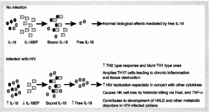
L'interleukine IL-18 (IL-18), un membre de la famille de l’IL-1, est une cytokine pro-inflammatoire multifonctionnelle. Elle est produite par les monocytes, les macrophages, les cellules dendritiques, les cellules épithéliales, les kératinocytes et le cortex surrénal dans le corps humain. Cette cytokine est d'abord produite comme une protéine précurseure inactive, qui est par la suite clivée en une forme mature par la caspase-1 activée. La caspase, en elle-même, existe comme précurseur inactif dans les cellules humaines et requiert l'assemblage d'inflammasomes pour son activation. L'IL-18 pour joue un rôle clé dans la médiation des conditions inflammatoires. Notre laboratoire et d'autres ont montré que l'infection par le VIH est accompagnée d'une augmentation des taux circulants d'IL-18 avec une diminution des niveaux de son antagoniste, l'interleukine-18 binding protein (IL-18BP).
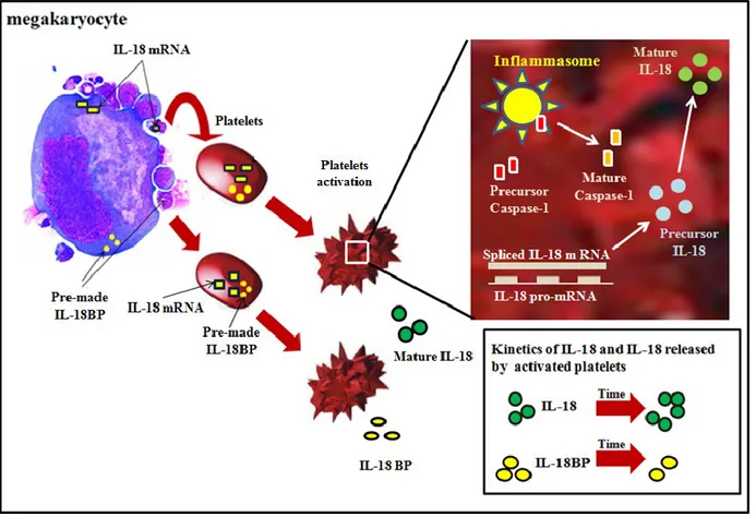
Dans cette thèse, nous démontrons pour que l'IL-18 est également produite et sécrétée par les plaquettes humaines lors de leur activation. Les plaquettes contiennent des composants de l'inflammasome. Ils assemblent et activent la caspase-1, qui ensuite traite le précurseur de l'IL-18 dans sa forme mature au cours du processus d'activation des plaquettes. La cytokine est synthétisée de novo lors de l'activation des plaquettes. Contrairement à l'IL-18, les plaquettes expriment constitutivement l’IL-18BP, et la libèrent de manière constitutive, ainsi que lors de l'activation. L'IL-18 et l'IL-18BP sont colocalisés avec CD63, un marqueur pour les granules α des plaquettes. L'IL-18 libéré des plaquettes constitue la source principale de cette cytokine dans la circulation humaine chez les individus sains. Nous avons identifié des concentrations faibles de cette cytokine dans les lysats de plaquettes chez les individus infectés par le VIH par rapport à ceux en santé. D'autre part, les concentrations ont été augmentées dans le sérum et le plasma pauvre en plaquettes chez les individus infectés. Des résultats similaires ont été obtenus avec l'IL-18BP dans les lysats de plaquettes d'individus sains et infectés par le VIH. Cependant, des quantités plus faibles de cet antagoniste ont été trouvées dans le sérum et le plasma pauvre en plaquettes d'individus infectés par le VIH par rapport à ceux en santé. Nos résultats ont des implications importantes pour les maladies inflammatoires chroniques dans laquelle une activité accrue de l'IL-18 joue un rôle pathogène.
Le VIH est également accompagné par une inflammation intestinale et une diminution de l'intégrité intestinale, mesurée par la réparation de la muqueuse, la régénération et la perméabilité. Cependant, on en sait peu sur la relation entre le niveau élevé de l'IL-18 associé à l'infection au VIH et la perméabilité intestinale: ceci n'a jamais été étudié. Dans cette thèse, nous démontrons le rôle du virus et sa protéine Tat à augmenter la production d'IL-18 chez deux lignées de cellules épithéliales intestinales (HT29 et Caco2) ainsi qu'une diminution de l'IL-18BP. L'IL-18 induit une hyperperméabilité de la barrière épithéliale en perturbant à la fois les jonctions serrées et adhérentes, et ce, en modulant l'expression et la distribution de l'occludine, de claudine-2 et de la bêta-caténine. Une désorganisation de l'actine F a également été observée dans les cellules lors de l'incubation avec l'IL-18. Les mêmes observations ont été faites avec la protéine Tat du VIH-1. Après une incubation prolongée, l'IL-18 a causé la mort des cellules intestinales et induit l'apoptose par l'activation de la caspase-1 et la caspase-3. Fait intéressant, les taux plasmatiques de lipopolysaccharides chez trois catégories différentes de patients au VIH (ART-naïf, ART-traitée et contrôleurs élite) sont en corrélation avec les niveaux plasmatiques de l'IL-18. Enfin, nous avons étudié la voie de signalisation à travers laquelle l'IL-18 induit une perméabilité intestinale accrue.
En bref, nos études identifient les plaquettes comme une source importante d'IL-18, et leur activation lors d'une infection à VIH contribue à des concentrations accrues de cette cytokine. Le virus entraine également l'augmentation de la production de cytokines par les cellules épithéliales intestinales. L'activité biologique accrue de ces cytokines contribue à la pathogenèse du sida en augmentant la perméabilité intestinale et en causant la mort des cellules intestinales. L'IL-18 pourrait servir de cible moléculaire pour retarder la progression du sida et réduire l'inflammation chronique dans un stade précoce d'une infection à VIH.
Mots-clés: Interleukine IL-18 (IL-18), protéine de liaison IL-18 (IL-18BP), VIH, plaquettes, perméabilité intestinale.
Abstract
Interleukin IL-18 (IL-18), a member of the IL-1 family, is a multifunctional pro-inflammatory cytokine. It is known to be produced by monocytes, macrophages, dendritic cells, keratinocytes and the adrenal cortex in the human body. This cytokine is produced as an inactive precursor protein, which is cleaved into mature form by activated caspase-1. The caspase itself exists as an inactive precursor in human cells and requires inflammasomes assembly for its activation. IL-18 has been shown to play a key role in mediating different inflammatory conditions. Our laboratory and others have shown that HIV infection is accompanied with increased circulating levels of IL-18 along with decreased levels of its antagonist IL-18 Binding Protein (IL-18BP).
In this thesis, we show that IL-18 is also produced and secreted by human platelets upon activation. The platelets also contain components of the inflammasome. They assemble and activate caspase-1 and process the precursor IL-18 into its mature form during the platelet activation process. The cytokine is synthesized in platelets de novo upon activation. Contrary to IL-18, the platelets constitutively express pre-formed IL-18BP, and release it constitutively as well as upon activation. Both IL-18 and IL-18BP colocalized with CD63, a marker for the platelet α granules. Platelet-released IL-18 constitutes the main source of this cytokine in the human circulation in healthy individuals. We found decreased amounts of this cytokine in the platelet lysates in HIV-infected individuals as compared to the healthy ones. On the other hand, its concentrations were increased in the serum and platelet-poor plasma in infected individuals. Similar findings were obtained with IL-18BP in platelet lysates from healthy and HIV-infected individuals. However, lower amounts of this IL-18 antagonist were found in the serum and platelet-poor plasma from HIV-infected individuals compared with the healthy ones. Our findings have important implications for chronic inflammatory disease conditions in which increased IL-18 activities play a pathogenic role.
HIV is also accompanied by intestinal inflammation and decreased intestinal integrity as measured by mucosal repair, regeneration and permeability. However, little is known concerning the relation between high level of IL-18 associated to HIV infection and intestinal
permeability. In this thesis, we demonstrate the role of HIV and its protein Tat in increasing IL-18 production in two intestinal epithelial cell lines (HT29 and Caco2) and decreasing IL-IL-18BP. IL-18 induces epithelial barrier hyperpermeability by disrupting both tight and adherens Junctions by modulating expression and distribution of occludin, claudin-2 and beta-catenin. Disorganization of F-actin was also observed within the cells upon incubation with treated by IL-18. Upon prolonged incubation, IL-18 caused intestinal cells death and induced apoptosis by activating caspase-1 and caspase-3. Interestingly, the plasma levels of lipopolysaccharide in three different categories of HIV-infected patients (ART-naïve, ART-treated and Elite controllers) correlated with their IL-18 plasma levels. Finally we investigated the signaling pathway through which IL-18 induces increased intestinal permeability.
Briefly, our studies identified platelets as an important source of IL-18, and their activation in HIV infection contributes to enhanced concentrations of the cytokine. The virus also induces increased production of the cytokine from intestinal epithelial cells. Increased biological activities of the cytokine contribute towards AIDS pathogenesis by increasing intestinal permeability and causing death of intestinal cells. The cytokine may serve as a molecular target for delaying AIDS progression and reducing low-grade chronic inflammation in HIV infection.
Keywords : Interleukin IL-18 (IL-18), 18 binding protein (IL-18BP), HIV infection, platelets, intestinal permeability.
Table des matières
Chapter 1: ... 1
Introduction ... 1
1.1. Human Immunodeficiency Virus ... 2
1.1.1 History... 2
1.1.2 Pathogenesis of HIV ... 3
1.1.2.1 Acute primary infection ... 3
1.1.2.2 Chronic asymptomatic phase ... 4
1.1.3 Structure of HIV ... 6
1.2. Intestinal Permeability ... 8
1.2.1 Structure of the Epithelial Barrier ... 8
1.2.2 HIV Infection and Intestinal Permeability ... 10
1.2.3 HIV Infection and the GI Immune System ... 11
1.2.4 Cytokines and Regulation of Intestinal Permeability ... 11
1.3. Platelets as Immune and Inflammatory Cells ... 15
1.3.1 Platelets as Immune Cells ... 15
1.3.2 Platelets and IL-1β Synthesis ... 16
1.3.3 Platelets and HIV Infection ... 16
1.4. Interleukin 18 ... 18
1.4.1 Interleukin 1 Family ... 18
1.4.2 IL-18 Production and Activation ... 23
1.4.3 IL-18 Receptors and Signaling ... 24
1.4.4 IL-18 Binding Protein ... 26
1.4.5 IL-18 as an Immunoregulatory Cytokine ... 26
1.4.6 IL-18 in Intestinal Inflammation ... 29
1.4.7 IL-18 and HIV... 30
1.4.8 The Role of IL-18 in HIV-associated Disorders ... 32
Chapter 2 ... 36
HYPOTHESES, RATIONALE, OBJECTIVES & AIMS ... 36
Differential synthesis and release of IL-18 and IL-18 Binding Protein in human platelets, and
their implications for HIV-infected AIDS patients ... 40
Chapter 4 ... 90
Manuscript 2 ... 91
HIV-induces IL-18 from intestinal epithelial cells that increases intestinal permeability and induces cell death ... 91
Chapter 5 ... 147
Discussion ... 147
1. Platelets produce IL-18 and release IL-18BP upon activation ... 148
2. Platelets synthesize IL-18 de novo from the gene transcripts upon activation ... 149
3. Production of mature IL-18 in platelets requires assembly of inflammasome and caspase-1 activation ... caspase-150
4. Platelets contribute to IL-18 and IL-18BP in the human circulation in activation dependent and independent manner, respectively ... 152
5. Implications for HIV-infected individuals ... 153
6. HIV infection increases IL-18 and decreases IL-18BP production in vitro from IEC ... 154
7. IL-18 induces intestinal cell death in a concentration- and time-dependent manner .... 155
8. IL-18-induced cell death involves caspase-1 and caspase-3 activation ... 156
9. IL-18 increases expression of MLCK and induces MLC phosphorylation via ROCK .. 160
10. IL-18 concentrations correlate with LPS levels in healthy and in HIV-infected individuals ... 162
Conclusions and future studies ... 165
Liste des figures
Figure 1. A typical natural time course of HIV infection from an acute infection until the onset
of AIDS ………...5
Figure 2: HIV-1 virion structure………..7
Figure3: Structure of epithelial barrier………9
Figure 4: Possible molecular mechanisms implicated in intestinal epithelial barrier disturbance ………12
Figure 5: Ligands and Receptors in the IL-1 Family………...22
Figure 6: Interleukin-18 signal transduction………...………...25
Figure 7: IL-18 in Innate and Adaptive Immunity...28
Figure 8: Imbalance between IL-18 and IL-18BP production during HIV infection….……..31
Figure 9. The theoretical model of IL-18 and IL-18BP release from human platelets……....151
Figure 10: Proposed model of IL-18–induced intestinal permeability by HIV infection …...158
List of abbreviations
ADP Adenosine diphosphate
AIDS Acquired immune deficiency syndrome AIM2 Absent in melanoma 2
AJs Adherens junctions ART Antiretroviral therapy ATP Adenosine triphosphate
BFA Brefeldin A
CA Capsid
CAD Coronary artery disease
cART Combination antiretroviral therapy
CD40L CD40 ligand
CLEC-2 C-type lectin-like receptor 2 CNS Central nervous system CSF Cerebrospinal fluid CTL Cytotoxic T lymphocytes CXCL5 C-X-C motif ligand 5 CXCR CXC chemokine receptor
DCs Dendritic cells
DC-SIGN DC-Specific Intercellular adhesion molecule-3-Grabbing Non-integrin dsRNA Double-stranded RNA
DSS Dextran sulfate sodium
EC Elite controllers
EGF Epidermal growth factor
FasL Fas ligand
FDA Food and Drug Administration
FOXO3a Forkhead box transcription factor O class 3a
GALT Gut-associated lymphoid tissue
GI Gastrointestinal
Gp Glycoprotein
GVHD Graft-versus-host disease
HAD HIV-associated dementia complex HALS HIV-associated lipodystrophy syndrome HAART Highly active antiretroviral therapy HCE Human corneal epithelial
HEK 293T Human embryonic kidney cells 293 expressing the SV40 T-antigen HIV Human immunodeficiency virus
HIV-1 HIV type- 1
HIV-2 HIV type- 2
HTLV-III Human T lymphotropic virus type III HLA Human leukocyte antigen
HVL High viral load
IBD Inflammatory bowel disease
Ic Intracellular
IEC Intestinal epithelial cells
Ig Immunoglobulin
IGIF IFN-γ-inducing factor
IFI16 Interferon-γ-inducible protein 16 IFN-γ Interferon gamma
IL Interleukin
IL-1Ra IL-1 receptor antagonist
IL-1RAcP IL-1 receptor accessory protein IL-18 Interleukin IL-18
IL-18BP Interleukin-18 Binding Protein IL-18R IL-18 receptor
IL-36Ra IL-36 receptor antagonist IRAKs IL-1R–associated kinases
JAM Junctional adhesion molecule
KSHV Kaposi’s sarcoma-associated herpes virus
LPS Lipopolysacharide
LTNP Long-term nonprogressors LTR Long-terminal repeat
MA Matrix
mIL-18 Mature IL-18
MHC Major Histocompatibility complex MLC Myosin light chain
MLCK Myosin light-chain Kinase
mRNA Messenger RNA
MyD88 Myeloid differentiation factor 88
NC Nucleocapsid
Nef Negative factor
NF-κB Nuclear factor kappa B
NK Natural killer
NLRP3 Nod-like receptor 3
NOD Nucleotide oligomerization domain NLRs NOD -like receptors
PBMCs Peripheral blood mononuclear cells PDGF Platelet-derived growth factor PF4 Platelet factor 4
PI Protease inhibitors
pIL-18 Precursor IL-18
p-MLC Phosphorylated myosin light-chain
p-STAT5 Phosphorylated signal transducer and activator of transcription 5
PR3 Proteinase 3
Pre-mRNA Precursor mRNA
PUMA p53 upregulated modulator of apoptosis
RA Rheumatoid arthritis
RANTES Regulated upon activation, normal T-cell expressed, and presumably secreted
RIG-I Retinoic acid-inducible gene-I RLRs RIG-I like receptors
ROCK Rho-associated protein kinase
RP Rapid progressors
siRNA Small interfering RNA
SIVs Simian immunodeficiency viruses SLE Systemic lupus erythematosus SNPs Single nucleotide polymorphisms
SU Surface unit
TER Transepithelial resistance
TEER Transepithelial electrical resistance TGF-β Transforming growth factor β Th 17 T helper type 17
TJs Tight junctions TLRs Toll-like receptors
TLR9 TLR subtype 9
TM Transmembrane
TNBS 2, 4, 6-Trinitrobenzene sulphonic acid TNF-α Tumor necrosis factor alpha
TRAF6 TNF receptor-associated factor 6
VCAM-1 Vascular cell adhesion molecule 1 Vif Virus infectivity factor
Vpu Virus protein u
“We all have an unsuspected reserve of strength inside that emerges when life puts us to the test.”
Isabel Allende
“Human beings are not born once and for all on the day their mothers give birth to them, but ... life obliges them over and over again to give birth to themselves.”
Acknowledgement
I would like to acknowledge those who have contributed to this body of work.
Thank you to my supervisor and mentor, Dr Ali Ahmad, for giving me the opportunity to pursue this study in his lab. Your passion for research has inspired me throughout my steps in this work and given me a great appreciation for science. I am grateful to your patience, advice and support when I first arrived to Canada and thereafter.
I would also like to acknowledge the support from the members of my lab; Suzanne Samarani, Patrick Sagala, Zainab Aldbah, Olfa Debbeche, Alexandre Iannello and my colleagues in other labs Mohammad-Ali Jenabian and Mohamed S. Abdel-Hakeem.
I would like to thank my thesis committee members: Dr. Fernando Alvarez, Dr. Hugo Soudeyns, Dr. Subburaj Ilangumaran and Dr Daniel Lamarre. A special thank you is in particular to you Dr. Hugo Soudeyns, for your stimulating conversations and advices, as well as your inspiring stories.
Thank you to my wife Dr Eman Abd el Rahman for always motivating me to be a better person and waving away my frustrations through the stressful journey of life. Thank you to my kids Yosef, Moustafa and Salma Allam for your presence in my life.
Thank you to my parents, my late father Salah Allam, my mom Amal Shams and my brother Dr. Tarek Allam for your unconditional love and for continuously encouraging me to strive for my best.
I would like to acknowledge my sources of funding: Ste-Justine Foundation and the Stars Foundation (la Fondation des etoiles) and the Department of Microbiology, Infectiology & Immunology, Université de Montréal.
Chapter 1:
Introduction
1.1. Human Immunodeficiency Virus
1.1.1 History
Human immunodeficiency virus (HIV) infection and acquired immune deficiency syndrome (AIDS), which are extensively investigated nowadays, were considered a great myth in the early 1980s. In 1981, the U.S. Center for Disease Control and Prevention identified AIDS as a new disease [1]. This came after the discovery of four homosexual men from Los Angeles suffering from complete immune system shutdown, with multiple unexplained viral and bacterial infections associated with Kaposi malignant tumors. At the time, such symptoms were incomprehensible, especially Kaposi’s sarcoma, which is a rare malignancy and more often associated with the elderly.
In May 1983, a French research group isolated a new retrovirus from lymphoid tissue obtained from patients with AIDS and named it lymphadenopathy-associated virus (LAV) [2]. Later, in May 1984, a U.S.-based research group confirmed the discovery of this virus believed to be the causative factor in AIDS and labeled it human T lymphotropic virus type III (HTLV-III) [3]. HTLV-III is now known as HIV type 1 (HIV-1) and spreads by sexual, parental, and blood contamination routes. Some years later, a new virus was isolated from patients with AIDS in West Africa and named HIV type 2 (HIV-2). Interestingly, HIV-2 is more closely related than HIV-1 to simian immunodeficiency viruses (SIVs) observed in macaques [4]. SIVs that are relatively closer to HIV-1 and HIV-2 have been detected as nonpathological viruses in chimpanzees and mangabeys. This finding supports the fact that HIV originated as a zoonotic infection in the first patient from Africa [5], and suggests the fact that SIV crossed the species barrier resulting in HIV-1 and HIV-2 in humans.
Soon after HIV/AIDS identification, AIDS became endemic in the United States and many countries worldwide, and was considered as the primary cause of death of people aged between 25 and 44 years. By 1989, the infection had spread in 145 countries and infection levels reached 400,000 cases. Approval of the first commercially available enzyme-linked immunosorbent assay (ELISA) by the U.S. Food and Drug Administration (FDA) in 1985
enabled the detection of new infections. The FDA approved the first HIV protease inhibitor in 1995. This was followed by the discovery of the first highly active antiretroviral therapy (HARRT) in 1997 [6]. HARRT was considered successful treatment for HIV infection following a 40% decrease in AIDS-related deaths in the U.S. during the first year of its introduction to the market.
1.1.2 Pathogenesis of HIV
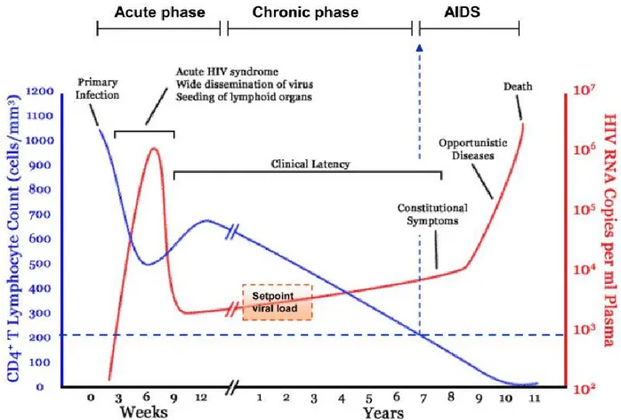
Although several decades have passed since the discovery of HIV, AIDS and HIV infection are still considered a strong threat to humans. The pathogenesis of HIV infection starts with a variable asymptomatic period before the onset of AIDS symptoms. This period is characterized by different pathogenic processes, such as severe depletion of CD4+ T cells, viral load increase, immune system activation, and intestinal barrier damage. As presented in Figure 1, the pathogenesis of HIV infection can by classified into three main phases: (1) acute primary infection, (2) chronic asymptomatic phase, and (3) AIDS.
1.1.2.1 Acute primary infection
The acute primary infection stage includes virus transmission and initial immune responses. HIV successfully infects CD4+ T cells and dendritic cells (DCs) present in mucosal membranes [7]. DCs are specialized antigen-presenting cells responsible for presenting the virus to naïve T cells leading to their activation. Infected DCs and macrophages play an important role in spread of infection by migrating to draining lymph nodes, especially intestinal lymph nodes [8]. Antigen-presenting cells (APC) also indirectly spread the infection by activating CD4+ cells and making them more susceptible to HIV infection. This APC response is preceded by substantial production of proinflammatory cytokines, such as interferon gamma (IFN-γ), interleukin (IL)-6 (IL-6), IL-10, IL-12, and IL-18 from DCs, macrophages, and natural killer (NK) cells [9, 10]. Furthermore, activated CD4+ cells become more susceptible to HIV infection. The production of this storm of cytokines enhances the ability of the virus to infect more CD4+ T cells. Since infected CD4+ T cells are the target of cytotoxic T lymphocytes (CTL) [11], acute primary infection is characterized by an intense decrease in CD4+ T cell count
in the gut mucosa [12]. When infected in vitro, CD4+ T cells mainly die by pyroptosis, a process that involves cellular swelling, cell membrane rupture, and release of intracellular contents in to the extracellular environment [13]. Unlike apoptosis, pyroptosis is a form of cell death associated with production of inflammatory cytokines, such as IL-1b and IL-18 [14]. During the infection process, incomplete reverse transcription products resulting from abortive HIV infection are sensed by the cytosolic DNA sensor, the interferon-γ-inducible protein 16 (IFI16), which leads to inflammasome assembly and activation of caspase-1 [15]. Caspase-1 activation is known to cause cell death via pyroptosis [16].
1.1.2.2 Chronic asymptomatic phase
Depletion of CD4+ T cells due to cytotoxic T lymphocytes and apoptosis continues during the chronic asymptomatic period of HIV infection. Meanwhile, production of new CD4+ T cells in the thymus is disrupted. HIV infection has been shown to strongly affect thymus function and decrease the production of new CD4+ T cells [17]. Furthermore, the immune system fails to clear the virus and viral proteins, which maintains the production of proinflammatory cytokines leading to chronic inflammation. This results into defective intestinal barriers and translocation of bacterial toxins into the circulation, which directly activate DCs and macrophages to produce more proinflammatory cytokines [18]. Continued viral production, together with high viral load and chronic inflammation, dramatically depletes CD4+ T cell count and accelerates AIDS symptoms.
Figure 1. A typical natural time course of HIV infection from an acute infection until the onset of AIDS
The Figure shows the typical time course of HIV infection comprising three phases: acute infection phase, chronic phase, and AIDS. The acute infection phase typically lasts for weeks and involves a sharp reduction in CD4+ T cell count and increase in viral load. The chronic phase is typically asymptomatic and spans several years. Continued viral production and reduced function of the immune system give rise to the symptoms observed during the final AIDS phase.
Reproduced with the permission from Elsevier Limited communications: An P, et al [19] author copyrights 2010.
1.1.3 Structure of HIV
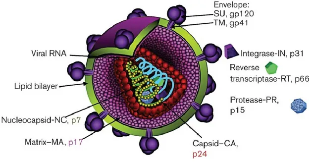
HIV is a lentivirus from the Retroviridae family [20]. HIV-1 and HIV-2 are the two types of viruses that cause AIDS. HIV-1 is distributed worldwide and it is the more virulent type, while HIV-2 is more localized to western and central Africa. As presented in Figure 2, HIV contains two identical copies of single-stranded RNA. Both types of virus encode nine open-reading frames responsible for protein production. The three main genes containing the viral structural proteins necessary to build a new virus are gag, pol, and env [21]. The env gene encodes glycoprotein (gp)-160, which is cleaved by the cellular enzyme furin to gp120 and gp41 proteins to form the viral envelope. The gag gene is cleaved into structural protein products (p24, p7, and p6) to form the viral core. The pol (DNA polymerase) gene is necessary to synthesize viral DNA and integrate it with host DNA to enhance viral reproduction. In addition to gag, pol, and env, HIV has two essential regulatory genes and four important accessory genes. The two essential regulatory genes regulate the expression of virion proteins (rev) and HIV trans-activator (tat) [22]. Rev protein is responsible for exporting intron-containing HIV-1 RNA from the cytosol to the nucleus during viral replication [23]. Tat protein plays an essential role in regulating the transcription of the full length of the RNA genome. In the absence of tat, RNA polymerase II fails to synthesize full-length viral transcripts [24]. The other four regulatory proteins are virus infectivity factor (Vif), viral protein u (Vpu), negative factor (Nef), and viral protein R (Vpr). HIV-1 vif encodes the highly conserved 23-kDa Vif protein, which is generated at a late stage of the virus life cycle and confers HIV infectivity [25]. Vif protein binds and degrades APOBEC-3G, an RNA editing enzyme, via proteasomal pathway. This way, Vif prevents the viral RNA from the hypermutating effects of ABOBEC-3G and enhance viral replication [26] Vpu is specific to HIV-1 and involved in CD4+ molecule degradation and deactivation of the nuclear factor kappa B (NF-κB) pathway [27]. Vpu is also capable of inducing pores in the cell membrane to ensure successful release of virus particles (virions) from the infected cells [28]. In the absence of Vpu, newly produced HIV particles remain attached to the cell surface and are not released from the interferon-inducible cellular restriction factor, BST2 or tetherin [29]. HIV-1 overcomes this BST-2-induced host protective mechanism by degrading tetherin via ubiquitination [30]. In contrast, Nef is involved in many functions during virus replication. It protects infected cells from CTL-induced lysis by interfering with the
expression of major histocompatibility complex (MHC) class I antigens [31]. It also causes the release of tumor necrosis factor alpha (TNF-α) from peripheral blood mononuclear cells (PBMCs) and provides microenvironment support for viral replication [32]. Nef activates T cells [33], and reduces the repertoire of antibodies produced by B cells by inhibiting the switch from immunoglobulin (Ig)-M (IgM) to IgA and IgG [34]. Vpr is a 14-kDa multifunctional protein that increases permissiveness and viral production [35]. It also plays an important role in promoting provirus entry into the nuclei of nondividing cells [36]. Moreover, this protein may participate in HIV-1 NK cell dysfunction by up-regulating NKG2D ligands and promote NK cell mediating killing of the virus-infected cells [37]. Also, Vpr has a substantial role in activation of long-terminal repeats (LTR) transcription [35]. The HIV transcription promoter is encoded by two LTRs: LTR3 and LTR5.
Figure 2: HIV-1 virion structure.
HIV virion main structures, envelope, capsid, matrix, nucleocapasid, lipid bilayer and viral RNA with detailing the localization of viral proteins. Abbreviations: SU, surface unit; gp120, glycoprotein 120; TM, transmembrane; gp41, glycoprotein 41.
Reproduced with the permission from copyright clearance center communications: Steckbeck JD, et al [38] author copyrights 2013.
1.2. Intestinal Permeability
1.2.1 Structure of the Epithelial Barrier
The intestinal tract comprises polarized monolayer epithelial cells, which are attached to each other by a complex structure known as the epithelial barrier. The intestinal epithelial barrier is a protective physiological barrier that regulates the transportation of water and nutritional particles. It also prevents bacteria, bacterial toxins, degraded food particles, and digestive enzymes from entering the circulation [39]. Different substances can be transported across intestinal barriers by paracellular or transcellular transport. Paracellular intestinal transport refers to small molecules that can pass between epithelial cells through the intercellular complex [40]. In contrast, transcellular intestinal permeability describes the process whereby substances pass through individual cells. As presented in Figure 3, the intercellular complex is composed of three structures: tight junctions (TJs), adherens junctions (AJs) and desmosomes, [41]. TJs and AJs are found on the apical side and play an essential role in the formation and maintenance of the integrity of the barrier. Disruption of these junctions induces intestinal leakage and increases antigenic and lipopolysaccharide (LPS) presentation to the immune system.
TJ structures are predominantly constructed from two components: transmembrane proteins, including occludin, junctional adhesion molecule A (JAM-A), and members of the claudin family. Cytoplasmic TJs proteins include zonula occludens types 1–3 (ZO-1, ZO-2, and ZO-3), cingulin, and afadin [42]. Both transmembrane proteins and cytoplasmic proteins are directly linked with underlying cytoskeletal actin filaments [43]. AJs, which are predominantly composed of β-catenin and p120 catenin, are in direct contact with E-cadherin and its actin cytoskeleton intracellular domain. Furthermore, the apical side of intestinal epithelial cells contains an abundance of actin filaments (F-actin) and nonmuscle myosin II that reorganize the actin cytoskeleton. F-actin is found as a circumferential belt at the level of the AJs and as a dense meshwork at the level of the TJs. Depolymerization of F-actin plays a principle role in epithelial barrier integration and junction function [44].
Figure 3: Structure of epithelial barrier
The Figure shows the distribution of Actin Filaments (F-Actin) and non-muscle myosin II (NM II) in intestinal cells. It demonstrates also the location of tight junctions (TJ) proteins such as: zonula occludens (ZO), claudin, occludin and junctional adhesion molecule (JAM) and adherens junctions (AJs) proteins such as catenins, E-cadherin, and nectins.
Reproduced with the permission of Frontiers in Bioscience communications: Ivanov AI [44] author copyrights 2008.
1.2.2 HIV Infection and Intestinal Permeability
Histological abnormalities of the intestinal mucosa were one of earliest observations made in 1984 in patients with AIDS [45]. Later on, the medical term HIV enteropathy was established to describe HIV-associated inflammation, diarrhea, malabsorption, and increased intestinal permeability. Interestingly, HIV-associated morphological changes in the GI epithelial layers, such as crypt hyperplasia and villous atrophy, occur without detection of further pathogens [46]. Intestinal permeability increases five-fold in HIV patients compared with healthy individuals [47]. This increase in permeability correlates with the significant increase in plasma LPS in patients with chronic HIV. In addition, progression of HIV toward AIDS correlates with circulating LPS levels. Interestingly, HAART decreases the level of plasma LPS in treated patients [18]. In support of this observation, LTNP and EC patients have less viral replication in the GI tract, and this reflects on LPS plasma levels [18, 48].
The mechanisms by which HIV leads to mucosal barrier dysfunction are yet to be established. Several researchers have demonstrated the direct effect of the virus itself, and of viral proteins, in breaching the intestinal barrier. Incubating the T84 intestinal cell line with different strains of HIV virus in vitro for 24 hours generated several interesting findings [49]. HIV downregulates production of occludin, ZO-1, and claudins 1–5, and disrupt TJs. Cells exposed to HIV exhibit decreased transepithelial resistance (TER). TER and transepithelial electrical resistance (TEER) are two related techniques to measure permeability but differ in methodology. Furthermore, bacterial and viral translocation across intestinal cells occurs after cells are treated with HIV [49]. Interestingly, HIV-related proteins play a key role in affecting epithelial barriers in different organs. HIV-1 Tat protein induces oxidative stress and causes alveolar epithelial dysfunction in animal models [50]. In the intestinal epithelial cell line Caco2, Tat protein induces caspase-3 activation and causes apoptotic cell death [51]. Tat protein inhibits cells proliferation and affects intestinal cells integrity by decreasing TEER [52]. Moreover, HIV-1 gp120 plays an important role in increasing intestinal permeability. gp120 decreases TER
and stimulates microtubule depolymerization in the intestinal cell line HT29 [53]. TER, which decreased in the T84 cell line after incubation with HIV, increased following treatment with a gp120 antibody [49]. On the other hand, HIV infection is associated with upregulation of many proinflammatory cytokines, such as IL-1β, TNF-α, and IL-6. These cytokines have a principal effect on intestinal permeability, which will be addressed in a later section of this thesis.
1.2.3 HIV Infection and the GI Immune System
It is widely believed that the intestinal immune system is central to the development of HIV pathogenesis. Most of the essential viral infection events, such as transmission, viral replication, and CD4+ T cell destruction are localized to intestinal tissue. Statistics indicate that 90% of worldwide infections are transmitted by genital mucosa and intestinal tissue [54]. This is because the intestine is the largest immunological organ in the human body, and contains almost 40% of lymphocytes, the majority of the T cell population, and lymphoid follicles forming Peyer’s patches [55, 56]. In addition, CD3+ T cells compose 90% of intraepithelial lymphocytes [57].
1.2.4 Cytokines and Regulation of Intestinal Permeability
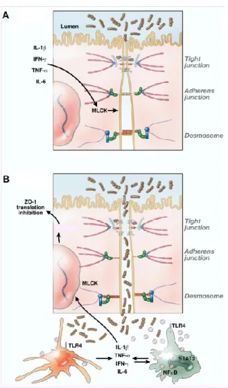
Different inflammatory conditions, such as Crohn disease, ulcerative colitis, and irritable bowel syndrome, or infections due to bacteria, such as Clostridium difficile and Vibrio cholerae disrupt the intestinal barrier and produce a “leaky” intestine. Increased intestinal permeability enhances antigenic penetration into the lamina propria. As shown in Figure 4, antigen-presenting cells, such as DCs, macrophages, and T helper cells, respond to this invasion of antigens and produce proinflammatory cytokines and results in disrupted intestinal barrier. The role of key proinflammatory cytokines in increasing intestinal permeability will be discussed in further detail.
Figure 4: Possible molecular mechanisms implicated in intestinal epithelial barrier disturbance
(A) Normal structure of intestinal epithelial barrier with normal junctional complexes including tight junctions, adherens junctions and desmosomes. The Figure also shows the role of proinflammatory cytokines such as IL-1β, TNF-α, INF-γ and IL-6 in initiating myosin light-chain kinase (MLCK) expression and activation. (B) Disrupted epithelial barrier results in activation of macrophages and DCs through TLRs, and increase in proinflammatory cytokine production by activating STAT3 and NFκB pathways. This leads to further dysfunction of the barrier and increased permeability through Zonula occludens-1 (ZO-1) translocation.
Reproduced with the permission of Elsevier communications: Lackner AA, et al [58], author copyrights 2009.
1.2.4.1 IFN-γ
The potential for cytokines to increase intestinal permeability was first reported in 1998.
Madara et al. showed a dose- and time-dependent increase in the TJ permeability of T89
intestinal cells in the presence of IFN-γ [59]. IFN-γ reduces TEER in T84 monolayer cells, which is prevented in the presence of anti–IFN-γ antibodies that block IFN-γ receptors [60].
Watson et al. proposed that TJs have two different types of pores; one responsible for large size
molecules and the other for smaller molecules. The authors also demonstrated the ability of IFN-γ to permit large-sized molecules, but not small-sized molecules, to cross TJs [61]. IFN-IFN-γ affects intestinal junctions proteins, occludin, claudin-1, and JAM-A by different mechanisms. IFN-γ increases endosomal uptake of occludin through macropinocytosis and disrupts TJs [62]. This property requires phosphorylation of myosin light chain (MLC). Furthermore, IFN-γ–induced reduction in TEER is reversed by inhibiting Rho kinase activation and by using phosphatidylinositol 3'-kinase (PI3-kinase) inhibitors [63, 64].
1.2.4.2 IL-1β
IL-1β is a proinflammatory cytokine belonging to the IL-1 family. IL-1β is produced by many immune cells, such as eosinophils, macrophages, T cells, and DCs [65-67]. Treatment of Caco2 cells with IL-1β induces the expression of other inflammatory cytokines, e.g., TNF-α and IL-8 [68]. A significant and dose-dependent decrease in TEER (80% reduction following three days of treatment) is reported with 1β on the basolateral but not on the apical side [68]. IL-1β causes a time-dependent disruption in TJs by decreasing occludin and redistributing claudin-1, and this effect is prevented in the presence of NF-κB inhibitors [69]. The increase in Caco2 permeability induced by IL-1β is associated with an increase in MLCK protein level [70]. However, IL-1β affects the distribution of TJ and AJ proteins in corneal pigment epithelial cells, and decreases TEER in simian virus 40-transformed human corneal epithelial (HCE) cells [71]. A similar effect was observed in pulmonary epithelial cells [72].
1.2.4.3 TNF-α
TNF-α is mainly produced by activated macrophages and T lymphocytes during inflammation. It also plays an essential role in inducing necrosis in tumor cells [73]. Interestingly, TNF-α is undetectable in healthy people, while it is detectable in tissues and serum
during inflammation or infection. The ability of TNF-α to increase epithelial permeability was reported in different cells types. TNF-α dose-dependently decreased TER in retinal epithelial cells [74]. Similarly, TNF-α is associated with increased intestinal permeability in Caco2 and T84 cells [75, 76]. However, TNF-α more aggressively disrupted epithelial TJs in the HT29 cell line. Treatment of HT29 with 100-ng/mL TNF-α induced an 81% decrease in TER after 24 hours. This response was reversed in the presence of tyrosine kinase and protein kinase A inhibitors [77]. Several researchers have demonstrated the principal role of MLCK protein to increase intestinal permeability by cytokines, such as IFN-γ and IL-1β. For example, Ma et al. demonstrated correlation between increased Caco2 TJ permeability in response to TNF-α and elevation in MLCK [78]. Similarly, blocking NF-κB inhibits elevation in MLCK gene expression and TJ disruption [78]. In contrast to IFN-γ and IL-1β, TNF-α induces apoptosis by activating the caspase cascade. Treating HT29 cells with TNF-α enhances apoptosis two fold compared with untreated cells. This could promote greater intestinal permeability by increasing the gaps between adjacent intestinal epithelial cells [79].
1.2.4.4 Transforming growth factor-β
Transforming growth factor β (TGF-β) is a multifunctional cytokine produced by many cells, including macrophages and intestinal epithelial cells. It controls cell proliferation and differentiation. Unlike many other cytokines, TGF-β enhances the function of the intestinal barrier by increasing expression of claudin-1 [80]. TGF-β augments basal resistance, as measured by TEER, of the T84 colonic epithelial cell line. It also shows strong ability to reverse the decrease in TEER induced by IFN-γ [81]. Furthermore, it inhibits the disturbance in TJs caused by Escherichia coli O157:H7 [80]. However, this protective effect toward epithelial barriers is selective for intestinal tissue. Woo et al. reported that TGF-β disrupts breast epithelial cells in an animal model [82]. In testicular Sertoli cells, TGF-β increases expression of occludin and other TJ proteins, and disrupts the epithelial barrier [83].
1.3. Platelets as Immune and Inflammatory Cells
1.3.1 Platelets as Immune Cells
Platelets are the second most numerous cells in the human body and are only surpassed in number by red blood cells. In humans, there are approximately 200,000 platelets per μL of blood [84]. They are very small anuclear circulating cells, with an approximate diameter of 1– 2 μm and lifespan between 8 and 10 days. They are shed in the blood stream from megakaryocytes, their progenitor cells. In contrast to platelets, megakaryocytes predominantly reside in bone marrow and are very large polyploid nuclei cells. Platelets contain approximately 60 different granules, which are classified under four types: (1) α-granules, (2) dense granules, (3) lysosomal granules, and (4) T granules [85]. The largest number and variety of platelet proteins are stored in α-granules, of which 50–60 are present per platelet. Proteomic analysis demonstrated that α-granules contain 284 proteins [86]. It is interesting to note that α-granules are the largest of platelets granules (200–400 nm in diameter) and secrete many important immunological proteins, such as like platelet factor 4, CXC chemokine receptor (CXCR) types 1–4, platelet-derived growth factor, TGF-β, and RANTES (Regulated upon activation, normal T-cell expressed, and presumably secreted) [87]. Dense granules are the second most common type of platelet granules. These are smaller (~150 nm in diameter) and less in number (3–8 per platelet) than α-granules. Dense granules contain molecules, such as adenosine triphosphate (ATP), adenosine diphosphate (ADP), serotonin, and glutamate [88, 89], and play a key role in inflammation by interacting with immune cells and amplifying inflammatory responses. Lysosomal granules are sparse; however, these granules contain degradative enzymes, such as aryl sulfatase, that play a role in wound healing and repair [90]. T granules were most recently discovered and play a role in TLR subtype 9 (TLR9) organization and function [91].
Platelets play an essential role as cellular mediator of thrombosis and hemostasis. In addition, platelets are considered as the most numerous immune cells. By producing serotonin, platelets participate in differentiation of monocytes to DCs and activated naïve T cells [92, 93]. Platelets are a major source of CD40 ligand (CD40L), which has been shown to increase T cell immunity to viral infection and induce DC maturation [94]. A mouse model showed that Tat
causes shedding of CD40L from platelets [95]. Platelets also release RANTES, which recruits leukocytes to inflammatory sites and plays a role in the differentiation of NK cells [96]. Furthermore, platelets express different members of the TLR family, such as TLR1, TLR4, TLR6, TLR8, and TLR9 [97]. LPS interacts with TLR4 to activate platelets that induce neutrophils to form neutrophil extracellular traps to restrict bacterial invasion [98]. Platelets are also considered as a source of several proinflammatory cytokines, such as IL-6, IL-8, and IL-1β [99].
1.3.2 Platelets and IL-1β Synthesis
It was thought for some time that platelets solely released premade proteins upon activation. The ability of activated (but not inactivated) platelets to express IL-1 was demonstrated in the late 1980s [100]. Surprisingly, Denis et al. demonstrated the presence of precursor mRNA (pre-mRNA) in platelets and proplatelet extensions of megakaryocytes. Activation of platelets by thrombin has been shown to splice IL-1β pre-mRNA to mature mRNA and synthesize IL-1β protein [101]. Interestingly, platelet synthesis of IL-1β is associated with clot formation and maturation. Use of monoclonal antibodies to block β3 integrins markedly decreases IL-1β synthesis and prevents fibrin mesh retraction [102].
1.3.3 Platelets and HIV Infection
Thrombocytopenia is one of the most common complications of HIV infection and occurs in more than 25% of AIDS patients [103]. It is also one of the first HIV symptoms exhibited in the acute phase of the infection [104]. The occurrence of thrombocytopenia following the viral infection is multifactorial in origin; it may be the result of the devastation caused by platelet-associated IgG, hypersplenism, inhibited production of thrombopoietin, and the harmful effect of the virus on platelets [105]. HIV infection initiates platelet activation and there appears to be a correlation between disease severity and degree of platelet activation [106]. Furthermore, changes in the morphology of platelets has been observed in HIV/AIDS patients [107]. Although HAART administration remarkably increases platelet count in patients [108], the role of platelets to enhance or clear the infection is still very controversial. It is not yet clear
whether platelets participate in spreading infection by internalizing HIV, or whether platelets aid immune cells to recognize and destroy the virus. Zucker-Franklin et al. reported that platelets and megakaryocytes internalize HIV [109]. It has been suggested that virus particles are endocytosed by platelets, enclosed within platelets, or come into contact with platelet α-granules. Two mechanisms for HIV entry into platelets have been proposed: (1) the virus fuses with CD4+ coreceptors, and (2) the virus is endocytosed by DC-specific intercellular adhesion molecule-3-grabbing non-integrin (DC-SIGN). Platelets do not express CD4, the primary HIV receptor; however, they do express CXCR1, -2, and -4 coreceptors [110]. HIV has been shown to bind to platelets via DC-SIGN [111, 112], which is expressed by DCs, DC-derived monocytes, and lymphoid tissue. DC-SIGN–expressing cells internalize HIV-1 and retain viral infectivity for days before infecting T cells [113]. Approximately 15% of platelets express DC-SIGN. This finding supports the notion that HIV endocytosis occurs via functional DC-SIGN [105]. In addition to DC-SIGN, HIV-1 can be captured by C-type lectin-like receptor 2 (CLEC-2) that is also expressed by platelets. CLEC-2 has been identified as a platelet activating receptor for the snake venom rhodocytin [114]. In contrast to DC-SIGN, CLEC-2 facilitates HIV-1 capture by platelets independent of viral envelop protein. CLEC-2 inhibitors clearly reduce HIV-1 binding to platelets [112]. Furthermore, knock-down of the CLEC-2 ligand, mucin-like membranous glycoprotein podoplanin, in human embryonic kidney 293 contains the SV40 T-antigen (HEK 293T) cells by short hairpin RNA results in a strong decrease in HIV-1 transmission to platelets after incubation with infected 293T cells [115]. More research into the role of platelets in HIV infection is necessary since it is unclear how platelets affect the pathogenesis of HIV.
1.4. Interleukin 18
IL-18 is a multifunctional pro-inflammatory cytokine, which belongs to the IL-1 famaily (IL-1F) of cytokines. The family family members are briefly discussed below.
1.4.1 Interleukin 1 Family
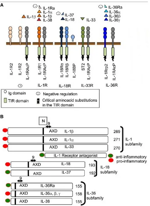
The IL-1 family comprises 11 members classified into three subfamilies: (1) the IL-1 subfamily consisting of IL-1α, IL-1β, IL-1RA, and IL-33, (2) the IL-18 subfamily consisting of IL-18 and IL-37, which have shorter propieces than IL-1 and IL-33, and (3) the IL-36 subfamily containing the shortest propieces, such as IL-36α, IL-36β, IL-36γ, IL-38, and IL-36Ra (reviewed in ref [116, 117]). A pro-piece is the N-terminal part that is removed from the precursor by proteolytic cleavage to generate mature and functional form of a cytokine. Most members of the IL-1 family are proinflammatory cytokines, except IL-37, which is an anti-inflammatory cytokine. This family has four signalling receptors: IL-1R1 and IL-1 receptor accessory protein (IL-1RAcP) for IL-1, ST2 and IL1RAcP for IL-33, IL-18Rα and IL-18Rβ for IL-18, and finally 1RAcP and 1Rrp2 for 36. Also, there are two decoy receptors in the 1 family, IL-R2 and IL-18BP [116]. The following paragraphs summarize the immunological functions of IL-1 family members, with particular emphasis on IL-18, which will be described in detail.
The IL-1α precursor is present in many organs, such as kidney, liver, lung, and the GI tract. Furthermore, it is also expressed by several cells, especially monocytes and B lymphocytes [118]. Precursor and processed mature forms of IL-1α are released by endothelial cells undergoing apoptosis [119]. Precursor IL-1α is cleaved by calpain, a calcium-activated cysteine protease, and processed into mature IL-1α [120]. Both forms of IL-1α are functional and considered alarming cytokines because these forms initiate many inflammatory reactions [121]. In contrast to IL-1β, IL-1α is localized in the nucleus and moves to the cytosol in cells undergoing necrosis [122].
Unlike IL-1α, precursor IL-1β is not functionally active, and needs to be cleaved by caspase-1 to produce its active form. Also, caspase-1 needs to be processed by newly assembled inflammasomes to become active [123]. However, processing of mature IL-1β is also induced
by non–caspase-1 mechanisms in caspase-1 knockout mice [124], and non–caspase-1 cleavage of precursor IL-1β is believed to be mediated by proteinase 3 (PR3) in neutrophils [125]. IL-1β is produced by monocytes, DCs, macrophages, NK cells, and B lymphocytes. It serves many immunological functions, especially in T cell activation. In the presence of 6 and TGF-β, IL-1β stimulates T helper type 17 (Th17) cell development [126]. IL-IL-1β in combination with IL-23 also initiates the development of Th17 [126]. IL-1β is necessary for NK cells to produce IL-22 and for NKT cells to produce IL-17 [127]. Furthermore, IL-1β plays an important role in chronic inflammatory diseases, such as type 2 diabetes, and cancer angiogenesis and metastasis [128, 129]. In animal models, IL-1β–deficient mice have lower inflammatory responses compared with wild-type mice [130], and develop fewer tumors compared with wild-type or IL-1α-deficient mice [131]. The effects of IL-1 in inducing fever and activating the hypothalamus– pituitary–adrenal axis are well described [117].
IL-33, which is also known also as 11th member of IL-1 family (IL1F11), is cleaved by caspase-1, as with IL-1β and IL-18. However, in contrast to IL-1β and IL-18, precursor IL-33 (30 kDa) is biologically active, whereas its processed forms are less active [132]. Like the precursor form of IL-1β, IL-33 is also processed by PR3 in neutrophils [133]. IL-33 binds to the ST2 receptor and IL-1RAcP coreceptor, and the soluble forms of ST2 and IL-1RAcP negatively regulate IL-33 [134]. IL-33 is predominantly produced by endothelial cells, epithelial cells, and fibroblasts, and can amplify both Th2 and Th1 responses to tumors and viral infection. In the presence of IL-12, IL-33 induces IFN-γ production by CD8+ and NK cells. On the other hand, IL-33–activated DCs support polarization of Th2 cells [135]. IL-33 plays an essential role in allergic lung inflammation since it enhances type 2 inflammation [136]. Interestingly, IL-33 shows impressive cardioprotection [137], and has been shown to reduce atherosclerotic plaque formation [138]. In HIV infection, low levels of IL-33 and high levels of its natural antagonist, ST2, were detected in the sera of HIV/AIDS patients compared with healthy uninfected individuals [139]. Unfortunately, no clear data about the role of IL-33 in HIV progression are available, and this field certainly warrants more attention.
IL-1RA is the natural antagonist of the cytokine IL-1. It binds to IL-1R, but it does not recruit IL-1RAcP,thereby preventing IL-1 biological responses [140]. Four isoforms of IL-1RA
exist. The sIL-1RA isoform, which is secreted by virtually every cell that produces IL-1, except endothelial cells, and three other intracellular (ic) isoforms icIL-1RA1, -2, -3, which are found in a variety of cells, such as activated fibroblast, fibroblast-like COS cell line, and the human liver carcinoma HepG2 cell line [140, 141]. IL-1RA knockout mice are smaller than wild-type mice and intraperitoneal LPS injection appears to be more frequently lethal in the knockout than wild-type mice [142]. In human, elevated IL-1RA is observed in patients with immune diseases, such as chronic rheumatic disease, whereas reductions are observed in pancreatic islets from patients with type 2 diabetes [143, 144].
Among the IL-1 family of cytokines, IL-37, which is also termed IL-1F7, is the cytokine that possesses anti-inflammatory characteristics. It is classified as belonging to the IL-18 subfamily and has five isoforms, with 37b being the most effective. Similar to 1α and IL-33, IL-37 is translocated to the nucleus after activation, which is induced by caspase-1 cleavage [145]. TGF-β is the most efficient stimulus for IL-37 production [146], and using siRNA to block production of IL-37 induces IL-1β production by two- to three-fold [146]. IL-37 transgenic mice have very low inflammatory responses and are protected against LPS challenge, dehydration, acidosis, and hyperkalemia. Also, these mice have low levels of inflammatory cytokines, such as IL-6 and TNF-α [146]. No data are available concerning IL-37 during HIV infection.
The IL-36 subfamily consists of five cytokines: IL-36α, IL-36β, IL-36γ, IL-38, and IL36Ra. The three IL-36 isoforms α, β, and γ, are also known as IL-F6, IL-F7, and IL-F8 and possess agonist characteristics [147]. IL-36 cytokines are expressed by various cells, such as bronchial epithelial cells, monocytes, macrophages, brain tissue, and keratinocytes [148]. LPS has been shown to induce IL-36γ, but not the α and β isoforms, from THP-1 [148]. However, T lymphocytes express both IL-36α and IL-36β [149]. Like IL-18 and IL-1β, IL-36 needs to be processed to become fully active, however, the enzyme responsible for IL-36 activation is still unknown [147]. Nonprocessed IL-36 is less active than its processed form [147]. IL-36α overexpressing transgenic mice suffer from acanothosis and hyperkeratosis skin lesions [150]. Although rhinovirus infection in human is associated with high expression of IL-36γ from
bronchial epithelial cells [151], no information is available about the role of IL-36 cytokines during viral infections like HIV, hepatitis C, or influenza.
IL-36 receptor antagonist (IL-36Ra) is the natural antagonist for several forms of IL-36 and can inhibit NF-κB activation induced by IL-36 in the jurkat cell line [152]. The main role of IL-36Ra is regulation of skin inflammation. Deficiency of IL-36Ra in IL-36α transgenic mice increases skin lesions [150]. Clinical data shows a positive effect of using anti-TNF in psoriasis patients and this improvement is associated with a decrease in IL-36 agonist and IL-36Ra [153]. A few reports have discussed the role of IL-36 and IL-36Ra in other organs, such as lung and joints [154, 155]. This area of research area remains to be explored.
IL-38, also known as IL-F10, is the 10th member of the IL-36 subfamily. It shares 40% homology with both IL-36Ra and IL-1RA [156]. Precursor IL-38 is not cleaved by caspase-1 as it does not have a caspase-1 cleavage site. IL-38 is expressed in skin epithelial cells and proliferating B cells of the tonsils [157]. Although very little is known about the biological function of IL-38, it was recently reported that IL-38 reduces Th17 responses to heat-inactivated
Candida albicans [158]. Different ligands and receptors for the IL-1 family are presented in
Figure 5: Ligands and Receptors in the IL-1 Family.
(A) A schematic demonstration of ligands and receptors of the IL-1 family. (B) Subfamilies among IL-1 ligands, classified by the length of the N-terminal prodomain. Numbers related to amino acids sequence.
Reproduced with the permission of Elsevier limited communications: Garlanda C, et al [116], author copyrights 2013.
1.4.2 IL-18 Production and Activation
IL-18 was discovered in 1987 in the serum of mice injected intraperitoneally with heat-killed Propionibacterium acnes and challenged with LPS [159]. It was cloned for the first time from the liver of these mice and was termed IFN-γ-inducing factor (IGIF) [160]. IGIF, which is now known as IL-18 and also termed IL-1F4, consists of 192 amino acids for the precursor form and 157 amino acids for the active form [161]. The gene encoding IL-18 gene is ocated on chromosome 11 in humans and chromosome 9 in mouse [162, 163]. IL-18 is a member of the IL-1 family and shares characteristics with some IL-1 family members. Comparable to IL-1β, IL-18 is synthesized in its inactive precursor form and remains intracellularly located. Many cells in the body can produce IL-18, and expression of IL-18 has been detected in macrophages, DCs, monocytes, and Kupffer cells [164, 165]. In addition, intestinal epithelial cells and osteoblasts can produce 18 [166, 167]. Activation and processing of the mature form of IL-18 can occur by canonical and noncanonical pathways.
The canonical IL-18 processing pathway involves cleavage by caspase-1. Caspase-1 itself is activated by assembling Nod-like receptor 3 (NLRP3) inflammasomes and other intracytoplasmic sensors like IFI16 and AIM2. The assembled inflammasome consists of pro– caspase-1, apoptosis-associated speck-like protein containing a caspase recruitment domain (ASC), and NLRP3. Inflammasomes involving NLRP have been shown to assemble after threat signals, such as bacterial toxins, environmental pollutants, or viral infections [168, 169]. Necrotic cells that release ATP can also induce inflammasome assembly [170]. After inflammasome assembly, active caspase-1 is released and cleaves the 24-kDa precursor of IL-18 to release IL-18 kDa mature IL-IL-18 that induces IFN-γ [171].
Unexpectedly, caspase-1–deficient mice reportedly produce IL-18 [172], and this finding was explained by showing different noncanonical production pathways for IL-18. Omoto et al. described the ability of granzyme B to cleave precursor IL-18 and produce biologically active IL-18 in caspase-1–deficient cells [173]. Similar to IL-1β, PR3 induces secretion of active IL-18 in the presence of a caspase-1 inhibitor from human oral epithelial cells
[174]. In addition, merpine-β, a tissue-specific metalloproteinase, can process pro–IL-18 to its active form. Merpine-β knockout mice appear to have low serum levels of IL-18 [175]. Fas/FasL interaction is another noncanonical production pathway for IL-18. Kupffer cells that expressed Fas were obtained from liver cells in mice treated with Propionibacterium acnes. Interestingly, these cells produce active IL-18 when cultured with recombinant FasL or cells expressing FasL [172]. A recent study by Bossaller et al. described that Fas signaling induces active caspase-8 in macrophages and DCs, which results in the expression of active IL-18 and IL-1β [176]. On the other hand, caspase-3 can cleave both precursor and mature IL-18 to two inactive products in the THP-1 cell line [177].
1.4.3 IL-18 Receptors and Signaling
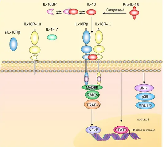
The IL-18 receptor (IL-18R) is a heterodimer containing IL-18Rα and IL-18Rβ. IL-18 signaling occurs when free active IL-18 binds to the IL-18Rα receptor. Although IL-18 binds to IL-18Rα with low affinity, this binding results in the recruitment of the coreceptor IL-18Rβ and forms a high affinity complex that initiates intracellular signaling [178]. After the formation of the IL-18/IL-18R complex, a signaling cascade is initiated through myeloid differentiation factor 88 (MyD88). Then, phosphorylation of IL-1R–associated kinases (IRAKs) takes place followed by IRAK dissociation from the complex and binding to TNF receptor-associated factor 6 (TRAF6). Finally, TRAF6 activates NF-κB and induces a signal [179]. See [Figure 6] for the signalling cascade initiated by IL-18 binding to its receptor. This signaling pathway is supported by observations in animal models. MyD88 knockout mice are not able to develop IL-18– mediated responses [180]. Furthermore, absence of IL-18Rβ prevents IL-18 signaling. For example, the human lung epithelial cell line A549 lacks IL-18Rβ. No IL-18 signal can be induced until IL-12 is present. IL-12 has been shown to induce expression of the missing receptor [181].
Figure 6: Interleukin-18 signal transduction.
The schema shows IL-18 signal transduction cascade. Precursor Pro-IL-18 is activated by caspase-1 to biological active IL-18. IL-18 binding protein (IL18-BP) binds to IL-18 and inhibits its function. Free IL-18 binds to IL-18Rα and IL-18Rβ, which transduce signals via the adaptor protein MyD88. This initiates IL-1 receptor activating kinase (IRAK) autophosphorylation and interaction with the TNFR-associated factor-6 (TRAF6), which induces nuclear translocation of the nuclear factor κB (NF-κB). Activation of STAT3 and the mitogen-activated protein kinase (MAPK) p38, JNK and ERK could also be initiated also by engagement of the IL-18R complex.
Reproduced with the permission of BioMed Central Ltd communications: Alboni S, et al [182], author copyrights 2010.
1.4.4 IL-18 Binding Protein
IL-18 binding protein (IL-18BP) is the natural antagonist of IL-18. IL-18BP binds with IL-18 and prevents its recruitment to the IL-18R. Mouse IL-18BP shares around 65% of the characteristics of human IL-18BP. IL-18BP is detected in many organs in human, such as PBMCs, small intestine, thymus, and ovarian tissue [183, 184]. In the mouse, it is found in lung, heart, and placenta [185]. In the serum of healthy individuals, IL-18BP levels are normally 20 fold higher than IL-18 [186]. Four isoforms (a, b, c, and d) exist in humans and two isoforms (c and d) exist in the mouse. Although IL-18BP b and d isoforms are not capable of neutralizing IL-18, isoforms a and c have high affinity to bind and deactivate IL-18 [187]. The detection of human IL-18BPb and IL-18BPd cDNA in a monocyte library suggests a role for these isoforms in IFN-γ regulation during inflammation [188].
IL-18BP is induced and upregulated by IFN-γ, thus establishing a negative feedback mechanism. It is detectable both in vitro and in vivo [189]. Paulukat et al. demonstrated that epithelial cell lines express IL-18BP after treatment with IFN-γ, and that indicates the self-limitation of IL-18 functions [190]. This concept is clinically supported with data obtained from a hepatitis patient treated with IFN-α who demonstrated high serum production levels of IL-18BP [191]. In this regard, IL-27, which has both pro and anti-inflammatory characteristics, can also induce IL-18BP by feedback mechanisms [192]. IL-18BP serves some protective functions against diseases, such as collagen-induced arthritis and contact hypersensitivity [193, 194]. Neutralizing IL-18 by injecting IL-18BP in BALB/c mice protects the animals against adriamycin nephropathy, and results in less frequent proteinuria and kidney dysfunction [195]. The same protective effect can be observed by neutralizing IL-18 by IL-18BP in cases of renal ischemia reperfusion injury and liver disease [196, 197].
1.4.5 IL-18 as an Immunoregulatory Cytokine
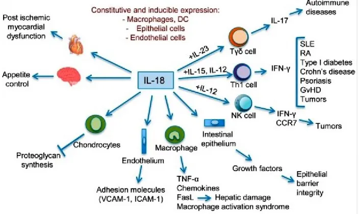
Although IL-18 does not induce fever or prostaglandin production, the cytokine promotes inflammation by various mechanisms [198, 199]. The importance of IL-18 as an immunoregulatory cytokine can be summarized by its ability to induce IFN-γ and FasL.
Furthermore, according to the presence of other cytokines, IL-18 can participate in Th1, Th2, and Th17 response depending upon the cells milieu [Figure 7]. In the presence of 12 or IL-15, IL-18 can induce IFN-γ from various types of cells that participate in Th1 responses. In the absence of IL-12 or IL-15, IL-18 loses its capacity to induce IFN-γ, but initiates its role in Th2 diseases [181]. IL-18 knockout mice weigh 2–3 times more than wild-type mice [200]. This finding reveals the role of 18 in energy homeostasis. Regardless, 18 is necessary for IL-23 signaling and functions synergistically with IL-IL-23 to initiate potent Th17 responses [126]. Blocking IL-18 in an experimental model of autoimmune encephalomyelitis has therapeutic benefits [201]. A similar effect is noted in animal models with other Th17 diseases like arthritis and heart disease [202, 203]. On the other hand, induction of FasL is a unique property of this multifunctional cytokine. IL-18 increases expression of FasL in cases of liver injury by inducing Fas-dependent hepatocyte apoptosis [204]. Furthermore, IL-18 mediates proapoptotic signals in renal tubular cells by increasing FasL expression. siRNA knockdown of FasL gene expression in these cells markedly decreases IL-18-induced apoptosis [205]. A previous report from our laboratory has shown that IL-18 induces FasL in the NK cell line NK92 as well as in primary human NK cells [206].
Figure 7: IL-18 in Innate and Adaptive Immunity.
Different biological effects of IL-18 and clinical conditions in which increased IL-18 activities have been implicated. SLE, systemic lupus erythematosus; RA, rheumatoid arthritis; GVHD, graft-versus-host disease; VCAM-1, vascular cell adhesion molecule 1; ICAM-1, intercellular
adhesion molecule 1.
Reproduced with the permission of Elsevier limited communications: Garlanda C, et al [116], author copyrights 2013.