Sleep sharpens sensory stimulus coding in human visual cortex after
fear conditioning
Virginie Sterpenich
a,b,c, Camille Piguet
a,c, Martin Desseilles
a,c,d, Leonardo Ceravolo
b, Markus Gschwind
a,e,
Dimitri Van De Ville
c,f,g, Patrik Vuilleumier
a,b,c, Sophie Schwartz
a,b,c,⁎
a
Department of Neuroscience, Faculty of Medicine, University of Geneva, Switzerland
b
Swiss Center for Affective Sciences, University of Geneva, Switzerland
c
Geneva Neuroscience Center, University of Geneva, Switzerland
d
Department of Psychology, University of Namur, Belgium
eNeurology Service, Department of Clinical Neurosciences, Centre Hospitalier Universitaire Vaudois (CHUV), Lausanne, Switzerland f
Department of Radiology and Medical Informatics, University of Geneva, Switzerland
g
Institute of Bioengineering, Ecole Polytechnique Fédérale de Lausanne, Switzerland
a b s t r a c t
a r t i c l e i n f o
Article history: Accepted 4 June 2014 Available online 14 June 2014 Keywords: Conditioning Emotion Perceptual learning Memory consolidation Sleep Functional MRI Amygdala Fusiform cortex
Efficient perceptual identification of emotionally-relevant stimuli requires optimized neural coding. Because sleep contributes to neural plasticity mechanisms, we asked whether the perceptual representation of emotionally-relevant stimuli within sensory cortices is modified after a period of sleep. We show combined ef-fects of sleep and aversive conditioning on subsequent discrimination of face identity information, with parallel plasticity in the amygdala and visual cortex. After one night of sleep (but neither immediately nor after an equal waking interval), a fear-conditioned face was better detected when morphed with another identity. This behav-ioral change was accompanied by increased selectivity of the amygdala and face-responsive fusiform regions. Overnight neural changes can thus sharpen the representation of threat-related stimuli in cortical sensory areas, in order to improve detection in impoverished or ambiguous situations. Thesefindings reveal an important role of sleep in shaping cortical selectivity to emotionally-relevant cues and thus promoting adaptive responses to new dangers.
© 2014 Elsevier Inc. All rights reserved.
Introduction
One essential function of perception is to achieve efficient detection and discrimination of relevant information in the environment even when sensory cues are variable and incomplete. It is well established that sensory processing of emotionally-relevant stimuli is enhanced to allow rapid attention orienting and adapted responses to potential threats (Vuilleumier and Driver, 2007). However, how emotional learn-ing induces long-lastlearn-ing changes in sensory cortices in humans remains to be clarified. Animal studies have shown that pairing a stimulus with an aversive experience (e.g. electrical shock) through associative Pav-lovian conditioning can subsequently shift the tuning curves of neurons in sensory cortices towards the characteristic sensory features of the conditioned stimulus and/or increase the number of neurons representing that stimulus (Gdalyahu et al., 2012; Resnik et al., 2011; Weinberger, 2004, 2007). Such remodeling of sensory representations may critically depend on modulatory signals from the amygdala
(Armony et al., 1997; Chavez et al., 2009; Duvarci et al., 2009; Shaban et al., 2006). Likewise, in humans,Li et al. (2008)showed that initially indistinguishable odors (mirror-image molecules) can be transformed into discriminable percepts after aversive conditioning, associated with distinctive activations in the olfactory cortex as demonstrated by functional Magnetic Resonance Imaging (fMRI). Other studies in humans suggest that emotional relevance enhances perceptual process-ing in sensory cortices via feedback from the amygdala (Rotshtein et al., 2010; Vuilleumier et al., 2001, 2004), and also promotes long-term re-tention of emotional memories through a modulation of hippocampal systems (LaBar and Cabeza, 2006; Phelps, 2006). However, a role for the amygdala in mediating the effects of emotion on sensory plasticity and cortical selectivity in humans has not been demonstrated.
Recently, evidence has accumulated to show that neural changes un-derlying the consolidation of perceptual or emotional memories benefit from sleep (Diekelmann and Born, 2010; Maquet, 2001; Stickgold and Walker, 2013). For example, long-lasting improvement in perceptual learning tasks requires (rapid eye movement (REM) and non-rapid eye movement (NREM)) sleep and involves plasticity in early sensory cortices (Aeschbach et al., 2008; Gais et al., 2000; Schwartz et al., 2002; Stickgold et al., 2000; Yotsumoto et al., 2009). Similarly, emotion-al memories, fear conditioning, and extinction of a conditioned response
⁎ Corresponding author at: Department of Neuroscience, Faculty of Medicine, University of Geneva, rue Michel-Servet 1, 1211 Geneva 4, Switzerland. Fax: +41 22 379 5402.
E-mail address:[email protected](S. Schwartz).
http://dx.doi.org/10.1016/j.neuroimage.2014.06.003
1053-8119/© 2014 Elsevier Inc. All rights reserved.
Contents lists available atScienceDirect
NeuroImage
are consolidated by a period of sleep following initial exposure (Baran et al., 2012; Payne and Kensinger, 2011; Sterpenich et al., 2007, 2009), particularly REM sleep (Menz et al., 2013; Nishida et al., 2009; Pace-Schott et al., 2009; Wagner et al., 2001). However, it is unknown whether sleep contributes to the remodeling of sensory representations of emotionally-relevant stimuli in sensory cortices, enhancing their per-ceptual discriminability subsequent to emotional learning.
Here, we hypothesized that fear learning modifies the perceptual sensitivity of sensory areas to discriminative stimulus features and is consolidated during sleep. This mechanism would allow the formation of a stronger,fine-tuned representation of fear-conditioned stimuli in cortical perceptual systems, possibly through offline reprocessing dur-ing sleep (Stickgold and Walker, 2013). Using fMRI, we tested this hy-pothesis by having human volunteers perform a discrimination task on morphed photographs of faces (Rotshtein et al., 2005), before and after one of the faces was associated with an aversive sound (see
Methods). To assess the conjoint effects of emotion and sleep, face discrimination was tested again after a 12-h delay containing either one normal day of wakefulness or one night of sleep in two different groups of participants. Our results show that aversive conditioning enhanced the detection of the conditioned face in morphed stimuli, i.e., based on reduced visual information. Critically, such perceptual im-provement only emerged after one night of sleep, and implicated selec-tive changes in face-responsive brain regions. These data demonstrate that sleep contributes to the sharpening of neural representations of emotionally-relevant stimuli in sensory cortices, improving their subse-quent perceptual discriminability.
Methods
General experimental design
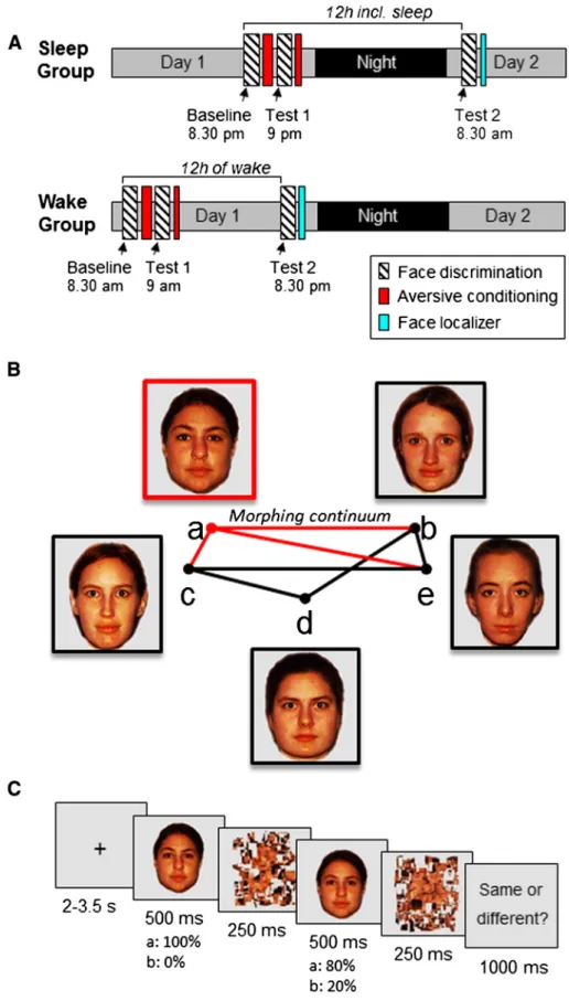
The study consisted of two experimental parts separated by a 12-h time interval and participants were randomly assigned to either a Sleep (n = 16) or a Wake group (n = 16,Fig. 1A). During a minimum of 4 days preceding the experiment, all participants followed a constant sleep schedule (23:00–07:00 or 24:00–08:00 ± 30 min). Compliance to the schedule was assessed using a sleep diary and wrist actigraphy (Actiwatch, Cambridge Neuroscience, Cambridge, UK). Because we aimed at minimizing any experimental stressors (seeResultssection), the participants from the Sleep group slept in familiar conditions at their home with actimetry (but no EEG) (Hu et al., 2006; Payne et al., 2008).
For thefirst experimental part, subjects from the Sleep group came to the lab at 20:30 or at 21:30 whereas those from the Wake group came at 08:30 or 09:30. After an assessment of their subjective vigilance state (Karolinska Sleepiness Scale, KSS: 10-point visual analogue scale from 1: very sleepy to 10: very alert) (Akerstedt and Gillberg, 1990) and a 5-min computerized psychomotor vigilance task (PVT;Cajochen et al., 2004; Dinges and Powell, 1985), participants were scanned while they performed the face discrimination task (Baseline; see details below), followed by a conditioning session, during which one face was aversively conditioned. After the conditioning, the participants complet-ed a second session of the face discrimination task (Test 1, similar to Baseline) to test for any immediate effect of the aversive conditioning on the face discrimination task. A short conditioning session was includ-ed at the end of thefirst experimental part in the MRI.
To test for the effect of sleep, the face discrimination task was admin-istered again after a 12-h delay, with or without an intervening night of sleep. Thus, after thefirst experimental part, participants from the Sleep group slept at their home; sleep was assessed via questionnaires (The St. Mary's Hospital Sleep Questionnaire) (Ellis et al., 1981) and actimetry. Participants from the Wake group were allowed to go about their normal daily activities during the waking interval; they were instructed to avoid intense cognitive or physical activity, and not to sleep during the day, which was confirmed by questionnaires
and actimetry. All participants came back 12 h later for the second experimental part, during which they again performed the discrimina-tion task (Test 2), using the same experimental protocol and condidiscrimina-tions as before. This second fMRI session aimed to assess any behavioral and neural effects of the aversive conditioning (by comparing trials with or without the conditioned face) and of sleep (by comparing the groups) on face discrimination thresholds, and any interaction of both factors. In total, the participants performed the face discrimination task three times: before (Baseline) and immediately after (Test 1) one face was conditioned, and after a 12-h consolidation period (either with or with-out sleep; Test 2). All sessions of the face discrimination task (20 min each), the conditioning task (12 min for the conditioning task and 2.5 min for the mini-conditioning task) and the face localizer (5 min) were performed in the MRI. During thefirst session (before the 12 h period of sleep or wake), participants stayed about 1 h in the MRI and during the second session (after the 12 h period) participants stayed about 45 min in the MRI.
Population
Thirty-two healthy participants (16 women and 16 men, mean age ± SD: 21.2 ± 3.6 years) participated in this study. A semi-structured interview established the absence of neurological, psychiatric, or sleep disorders. All participants were non-smokers, moderate caffeine consumers, and did not take any medication. They were not depressed as assessed by the Beck Depression Inventory (Steer et al., 1997), and had low to moderate anxiety levels as assessed by the Beck Anxiety Inventory and the STAI-T (Spielberger, 1983). Extreme morning and evening types were excluded (Horne and Ostberg, 1976). None of the participants complained of excessive daytime sleepiness as assessed by the Epworth Sleepiness Scale (Johns, 1991) or of sleep disturbances as determined by the Pittsburgh Sleep Quality Index Questionnaire (Buysse et al., 1989). All participants were right-handed as indicated by the Edinburgh Handedness Inventory (Oldfield, 1971). Partici-pants were randomly assigned to either a Sleep group (n = 16, 8 fe-males, age 22.4 ± 4.0 [mean ± SD]) or a Wake group (n = 16, 8 females, age 20.0 ± 2.6). The groups did not differ in age or self-assessment questionnaire responses, including depression, anxiety, sleepiness, sleep quality and circadian rhythms (t-tests, all pN 0.05). All participants gave their written informed consent to take part in this study and received afinancial compensation for their participation. The study was approved by the Ethics Committee of the Geneva Univer-sity Hospitals.
Stimuli and tasks Stimuli
We selectedfive photographs of female faces with a neutral expres-sion, which were clearly identifiable as five distinct individuals (Fig. 1B). We then applied a linear morphing procedure to generate 15 equidistant steps or linear‘morphs’ between each possible pair of original faces (Morpheus Photo Morpher,www.morpheussoftware.net). Scrambled, low-pass versions of these pictures were generated using dedicated Matlab (R2009b, MathWorks Inc., Sherbom, MA) scripts and served as vi-sual masks.
Face discrimination task
During the face discrimination task, two face stimuli were presented in a rapid succession, always including one original face and the same face morphed with another face, and participants judged whether the two faces depicted the same or different individuals (Fig. 1C). In total, 5 distinct original identities were used, all unfamiliar to the participants (Fig. 1B). We expected that participants would respond‘same’ on trials where both faces were close in morphing distance, thus sharing a high proportion of visual features; whereas participants should judge as ‘dif-ferent’ those trials where the morphed face contained more visual
Fig. 1. Task and design. A: Two groups of participants were scanned twice each, with the fMRI sessions separated by a 12-h interval with or without sleep (resp. Sleep and Wake groups). The discrimination task was repeated three times: before conditioning (Baseline), immediately after conditioning (Test 1), and after the 12-h interval (Test 2). Functional MRI was also acquired during conditioning (after the Baseline face discrimination task) and during a functional face localizer performed at the end of the protocol. B: Stimuli were composed of 5 face photographs from different individuals, and morphed faces generated between pairs of these 5 faces using a linear interpolation, resulting in 13 equidistantly morphed faces. One original face was randomly chosen to be conditioned with an aversive sound (the face a in this example; counterbalanced across participants). For each participant, 7 morphed series were randomly selected (out of the 10 possible ones) to contain 3 series with the CS+ face (red lines) and 4 series with CS− faces only (gray lines). C: Design of the discrimination task for one trial. Two faces of the same pair were presented successively. One face is an original face and the second is a morphed face, created by mixing this original face with another one to various degrees along a linear continuum.
information from another distinct face identity. Each of the 15 possible morphing distances between two faces was tested, from distance 1 (twice the same original face) to distance 15 (both original but different face identities in the pair). The morphing distance and the order of the presentation of the two faces within a trial (i.e., original face could either precede or follow the morphed face) were randomized. The task of the participants was to decide whether both faces were photographs of the same individual or of two different individuals. We told the participants that we included different shots of the same individuals taken under slightly variable viewing angle or lighting. Each trial started with a central fixation cross (mean duration = 2.75; ranging from 2 to 3.5 s). Then, a first face picture was presented (500 ms; degrees of visual angle: 5.4° × 5.4°), immediately followed by a visual mask (250 ms). A second face picture was then presented (500 ms) also followed by a second mask (250 ms). At the end of the trial, the participants gave their response on a keypad (‘same/different’; maximum response time: 1 s). Eight brief pauses (6-s grayfixation-cross), each after 30 trials, were also included.
To check that the 5 face identities could clearly be distinguished from each other, an independent group of 9 participants who did not participate in the main experimental protocol performed the face dis-crimination task on 290 stimuli (29 possible combinations of 2 face stimuli between each of the 10 possible pairs of original faces). Based on these results, we excluded one face pair (pair a–d;Fig. 1B) for which participants generated some incorrect‘same’ responses on trials composed of two distinct original faces.
For the fMRI experiment and for each participant, one of thefive original faces was randomly chosen to be conditioned (see below). Three series of morphed faces contained the to-be-conditioned face (in the example ofFig. 1B, the CS+ pairs are a–b, a–c and a–e), while four series only contained non-conditioned faces (onFig. 1B, the CS− pairs are b–e, c–e, c–d, b–d). Because one face pair was removed (a–d), the minimal number of morphing continuums that could be gen-erated from any face was 3 (Fig. 1B). Therefore, each CS+ was randomly chosen among the 5 faces and mixed with 3 of the 4 remaining faces. In order to avoid an imbalance between the number of CS+ containing pairs and CS− only pairs, and to make sure that each individual face was shown in at least two distinct morph continuums, we set the num-ber of CS− only pairs to 4. Thus, in total each participant was tested on 7 distinct series of morphed faces. Each face discrimination assessment (Baseline, Test 1, Test 2) was composed of 203 different pairs of faces, including all morphing distances (from 1 to 15) for the 7 selected morph continua, presented across two fMRI runs lasting 10 min each.
For the analysis of the face discrimination performance, discrimina-tion thresholds and slopes were obtained by logisticfit of the curves for each pair of faces in each session and each condition (Fig. 2A). Discrim-ination thresholds were defined here as 50% ‘different’ responses and expressed in morphing distance with equidistant steps from 1 (same faces) to 15 (two distinct original faces). For the analysis of the face discrimination task during Baseline, Test 1, and Test 2, we averaged individual performance for all possible CS+ pairs, which included the original CS+ (or to-be-CS+) face and a morph containing information from the CS+ (or to-be-CS+) face mixed with one of the CS− faces (i.e., pairs a–b, a–c, etc. inFig. 1). The same procedure was used for all CS− pairs containing one original CS− (or to-be-CS−) face together with a morph between the latter and another CS− (or to-be-CS−) face (i.e., pairs c–d, c–e, etc.). This yielded 6 threshold estimations and 6 slope estimations for each participant. The difference between test and baseline was used as dependent measure, i.e. test subtracted from baseline, to minimize the variance due to the randomization of the face pairs across conditions and participants. All pairs between individ-ual faces with more than one non-zero value (‘different’ responses) were included. Participants had a maximum of 1 s to respond on each trial and, whenever they did not respond on time, this was considered as a miss. To ensure validfitting of the discrimination curve, hence
robust threshold estimation, any face pair with more than 5 misses over the 15 tested steps was removed from further analysis. In total, 98.4% of the pairs were included in the analysis.
Aversive conditioning
For each participant, one face (randomly chosen among the 5 origi-nal faces) was aversively conditioned. During a partial conditioning fMRI protocol, half of the presentations of that face was followed by an aversive sound (auditory unconditioned stimulus, US), which was composed of 3 bursts of white noise (125 ms each, separated by 50 ms silent gaps). Presentations of 15 CS+/US and 15 CS+ were ran-domly intermixed with the 4 remaining original faces (CS−). Face stim-uli were presented one at a time (2.5 s each) followed by a varying interval (ISI: 4–5.5 s, mean = 4.75). Each face was presented 30 times; all the stimuli were presented in an intermixed, random order. Five pauses, each after 25 faces, were introduced (5-s grayfixation cross). Sound volume of the US was adjusted for each individual at a very unpleasant yet not harmful level. The conditioning was performed across two successive fMRI runs of 5 min each. At the end of Test 1, a short version of the conditioning task was administered to minimize any possible extinction effect due to the presentation of the CS+ during the discrimination task; the 5 original faces were presented 5 times each, in a random order, and the conditioned face was always associated with the presentation of the white noise. The effectiveness of condition-ing was verified by pupil dilatation and by fMRI data (see Supplemental data, Fig. S1).
Fig. 2. Behavioral results of the discrimination task. A: Sigmoid curve during the discrim-ination task from the Baseline session for all participants (n = 32). The mean discrimina-tion curve (computed independently of group and emodiscrimina-tional condidiscrimina-tions) was a logistic function with a slope of 2.77 ± 0.04 and a center of 7.13 ± 0.21 (mean ± sem). B: Differ-ence of discrimination thresholds between Test 1 or 2 and Baseline session for the CS+ and CS− in each group of participants; * indicates significance at p b 0.05 (mean + sem).
A semi-structured debriefing after the end of the experiment re-vealed that 27 subjects (out of 32) did not notice the morphing proce-dure, and 30 subjects were explicitly aware that conditioning occurred on one particular face.
Face localizer
To identify brain regions selectively involved in the visual processing of faces, a functional face localizer task was administered at the end of the second experimental part. Participants were shown photographs of faces, places (houses/landscapes), and scrambled versions of these stimuli, organized in 4 blocks per category (10.4 s each) presented in a pseudorandom order (i.e. no more than 2 blocks of the same category in a row). The set of faces stimuli was different than the one used in the face discrimination task. Each picture was displayed during 750 ms followed by a gray screen of 500 ms. To ensure that the par-ticipants paid attention to the stimuli, they were instructed to press a key whenever the same photograph was shown twice in an immedi-ate succession.
Functional MRI MRI data acquisition
MRI data were acquired on a 3 T whole body MR scanner (Tim Trio, Siemens, Erlangen, Germany) using a 12-channel head coil. Functional images were acquired with a gradient-echo EPI sequence (repetition time [TR]/echo time [TE]/flip angle: 2200 ms/30 ms/85°) and parallel imaging (GRAPPA; acceleration factor: 2). Each functional image comprised 36 axial slices (thickness: 3.4 mm, no gap; voxel-size: 2.8 × 1.8 mm; FOV: 235 × 235 mm; 128 × 84 voxels) oriented parallel to the inferior edge of the occipital and temporal lobes. Each session of the face discrimination task was acquired across two successive runs of 270 and 275 brain volumes respectively, the conditioning task across two runs of 175 volumes each, and the face localizer in one run of 130 volumes. For each fMRI run, thefirst three volumes were discarded to account for spin saturation effects. Structural images were acquired with a T1-weighted 3D sequence (MPRAGE, TR/inversion time [TI]/TE/ flip angle: 1900 ms/900 ms/2.32 ms/9°; FOV: 230 × 230 × 173 mm3;
256 × 246 × 192 voxels, resulting in voxel dimensions: 0.9 mm isotro-pic). Visual stimuli were presented on a back projection screen inside the scanner bore using an LCD projector (CP-SX1350, Hitachi, Japan), which the participant could comfortably see through a mirror mounted on the head coil. Responses were recorded via an MRI-compatible re-sponse button box (HH-1 × 4-CR, Current Designs Inc., USA).
MRI data analysis
Functional MRI data were analyzed using the Statistical Parametric Mapping software (SPM5;http://www.fil.ion.ucl.ac.uk) implemented in MATLAB (MathWorks Inc., Natick, MA). Functional scans were realigned, corrected for slice timing, normalized to the MNI EPI template (voxel size: 3 × 3 × 3 mm3), and spatially smoothed with a Gaussian
kernel of 8 mm. Data were then analyzed using a two-step procedure taking into account the intra-individual and inter-individual variances. For each participant, brain responses were modeled at each voxel using the general linear model and main contrasts of interest were com-puted. The resulting individual maps of t-statistics were then used in second-level random effect analyses. These second level analyses consisted in one-sample t-tests in each group separately or for testing common effects in both groups, and two-sample t-tests comparing effects between the groups (Sleep vs. Wake). Statistical inferences were corrected for multiple comparisons using Gaussian randomfield theory at the voxel level in small spherical volumes (radius 10 mm) around locations of interest, based on our functional face localizers (face vs. scramble for detection of brain regions involved in face percep-tion, and scramble vs. face for brain regions involved in the processing of non-face specific visual details).
For the analysis of each face discrimination session (Baseline, Test 1, Test 2), 4 main trial types were modeled: pairs containing the to-be-CS+ (Baseline) or to-be-CS+ face (original to-be-CS+ and to-be-CS+ morphed with a CS−) identified as ‘same’ (CS+ same) or ‘different’ by the participant (CS+ diff), and the CS− pairs (original CS− and same CS− morphed with another CS−) identified as ‘same’ (CS− same) or ‘different’ (CS− diff). Two additional regressors corresponded to hybrid trials composed of one original CS− and the same CS− morphed with a CS+, identified as ‘same’ or ‘different’. Note that we included hybrid tri-als to make sure that all possible morph continuums for the 7 selected face pairs were presented to each participant. However, because the hy-brid trials have an equivocal status regarding conditioning (only one stimulus with a variable amount of information about the CS+ in the trial), we did not consider them in the main contrasts reported in the present study. Thus, all possible combinations of faces between CS+ and CS− were included in the task and entered the design matrix as separate regressors convolved with a canonical hemodynamic response function (HRF). Parametric modulators for each regressor were also added to represent the actual morphing distance between face stimuli in each individual trial (i.e. morphing steps between the two stimuli in the pair, ranging from 1 to 15). In our analyses, we took advantage of repetition suppression effects to look at the sensitivity of brain re-gions to stimuli eliciting distinct perceptive judgments (‘same’ or ‘differ-ent’ responses) and as a function of the physical morphing distance. Repetition suppression of activity can trackfine variations in BOLD re-sponse and potentially allows inferences about the nature of represen-tations in different cortical regions (Grill-Spector et al., 2006; Henson et al., 2000; Rotshtein et al., 2005; Vuilleumier et al., 2002). Wefirst identified brain regions showing significant activation with our task (“same” + “diff”), and then those showing stronger repetition suppres-sion for pairs of faces that the participants judged as‘same’ vs. those judged as‘different’ across all trial types and for both groups together during the Baseline session. To directly test our main hypothesis that a period of sleep influences the effect of conditioning on the neural repre-sentation of faces, we compared the subjective discrimination for CS+ and CS−, separately (CS+ same vs. CS+ diff; CS− same vs. CS− diff). Then we tested the effect of objective discrimination, represented by the real distance of morphing between the two faces of a pair. We com-pared the effect of morphing distance (represented by the parametric modulator of the events) separately for the conditioned pairs and the neutral pairs. This contrast evaluated brain regions significantly less acti-vated for small distance of morphing (where the two faces are very similar). Finally, to assess the influence of a period of Sleep (or Wake), we identified brain regions whose activity changed after the 12 h period, comparing regions more activated during Test 2 than Test 1. This resulted in a triple interaction, Sleep vs. Wake group, Test 2N Test 1, large N small distances for the CS+ trials. For the conditioning task, onsets for 3 trial types were modeled: CS+/US, CS+, and CS−. We computed a main linear contrast to estimate brain responses to the CS+ compared to the CS−.
The face localizer task was analyzed with the 3 conditions (faces, places, and scrambled stimuli) modeled as blocks convolved with the HRF. Face-selective regions were then identified by comparing blocks of faces to blocks of places or scrambled stimuli (Tables S2 and S3).
Note that for all the tasks (aversive conditioning, face discrimina-tion, and localizer), movement parameters estimated during realign-ment were added as regressors of no interest in thefirst-level analyses and a high-passfilter was implemented using a cut off period of 128 s in order to remove the low frequency drifts from the time series.
Sleep and vigilance parameters
During the week preceding the experiment, sleep quantity (number of hours) and quality (10-point scale) were assessed by questionnaires;
data from the 4 days preceding thefirst experimental part were analyzed. Actigraphy data were analyzed for the night preceding the experiment. Actigraphy data were available for 7 participants of the Sleep group and 13 participants of the Wake group (this was due to technical problems that led to data loss). We performed Student's t-tests to compare mean values on each of these measurements for the Sleep and Wake groups.
Objective and subjective levels of vigilance were assessed once dur-ing each visit, just before the MRI session. We performed two repeated-measure ANOVAs on the mean reaction times (RTs) for the PVT (exclud-ing the 2% slowest RTs) as well as the KSS scores with the visits (first, second) as within-subject factors and the Group (Wake, Sleep) as between-subject factors.
Results
This discrimination task allowed us to test for any change in the per-ceptual representation of conditioned face resulting in a shift of recogni-tion ability along the morphing continuum (Fig. 2A). In addition, because each trial in the task was composed of a rapid succession of two faces with a variable degree of perceptual dissimilarity, we could exploit repetition suppression effects to probe the functional selectivity of neural populations within specialized brain regions (Grill-Spector et al., 2006; Rotshtein et al., 2005), both before and after conditioning. As repetition suppression indexes a facilitation in processing a repeated stimulus relative to a novel stimulus (Grill-Spector et al., 2006), we expected that greater repetition suppression would occur for pairs containing the conditioned face if its cortical representation was consolidated after learning and/or sleep, and that such changes should arise in sensory areas holding stimulus-specific traces along the visual hierarchy.
Performance on the face discrimination task
For each participant, psychometric curves were obtained for each session of the task (Baseline, Test 1, Test 2) by plotting the propor-tion of‘different’ responses against the morphing distance for each of the pairs containing the CS+ and those containing the CS−. Discrimination threshold (defined at 50% of ‘different’ responses) and slopes were computed by logisticfit (Fig. 2A) and confirmed that initial face discrimination performance did not differ between face identities prior to conditioning or between groups of partici-pants. Indeed, an ANOVA on the discrimination threshold values at Baseline yielded no significant effects of future conditioning status (CS+, CS−), Group (Sleep, Wake), or any interaction between both factors (all pN 0.20).
We then tested for any modification of the discrimination thresholds subsequent to conditioning and/or sleep. To do so, wefirst removed the baseline threshold for each individual face pair in each participant, to minimize the variance due to the randomization of the face pairs across conditions and participants. Then, threshold values were submitted to an ANOVA with Conditioning (CS+, CS−) and Test (1, 2) as within-subject factors, plus Group (Sleep, Wake) as between-within-subject factor, followed by planned post-hoc comparisons. While there was no main effect of Group (F(1,29) = 0.19, p = 0.66) or Conditioning (F(1,29) = 0.31, P = 0.58) nor triple interaction (F(1,29) = 2.26, p = 0.14), we observed a trend for an effect of Test session (F(1,29) = 3,77, p = 0.062) and, most importantly, a significant in-teraction between Group and Test (F(1,29) = 4,81, p = 0.036). As suggested onFig. 2B, the latter observation could reflect a shift in threshold for the CS+ stimuli from Test 1 to Test 2 that might be greater for the Sleep group compared to the Wake group. This interpretation was confirmed by posthoc tests showing that the interaction Group by Test was significant for the CS+ (F(1,29) = 4.55, p = 0.04), but not for the CS− (F(1,29) = 0.29, p = 0.60). Moreover, the change in dis-crimination threshold from Test 1 to Test 2 was significant in the
Sleep group selectively for the CS+ trials (mean ± SD: Test 1: −0.17 ± 0.80, Test 2: 0.47 ± 1.33, p = 0.02), not for the CS− trials (Test 1: 0.03 ± 1.13, Test 2: 0.20 ± 1.54, p = 0.27;Fig. 2B). No such effect was found in the Wake group, neither for the CS+ (Test 1: − 0.11 ± 1.24, Test 2: − 0.22 ± 1.44, p = 0.67), nor for the CS− (Test 1: 0.11 ± 0.99, Test 2: 0.16 ± 1.02, p = 0.70). Finally, because performance at baseline was subtracted from performance at Test 1, any threshold difference during Test 1 would suggest an immediate ef-fect of conditioning. When directly comparing the CS+ and CS− during Test 1 (Group × Conditioning posthoc ANOVA), we did not observe any significant main effect or interaction (all p N 0.3), thus confirming that the conditioning had no immediate effect on the face discrimination task, for neither group.
The same ANOVA performed on the slope of the discrimination curves revealed no significant main effect or interaction (Table S1). Finally, the ANOVA on the reaction times (RTs) revealed no main effect of Group (Sleep vs. Wake, F(1,29) = 0.68, p = 0.79), Conditioning (CS+ vs. CS−, F(1,29) = 0.22, p = 0.64), or Test (Test 1 vs. Test 2, F(1,29) = 1.05, p = 0.31), but a significant triple interaction between these factors (F(1,29) = 4.26, p = 0.048), due to shorter RTs during Test 2 for CS+ face pairs compared to CS− face pairs in the Sleep group (CS+:−83 ± 52, CS−: −63 ± 56, p = 0.027), which was ab-sent in the Wake group (CS+:−61 ± 54, CS−: −69 ± 59, p = 0.36, Fig. S2). As we observed for the threshold measurements, the Group × Conditioning ANOVA on the RT data from Test 1 was not significant (all pN 0.4).
In sum, these behavioral results show that, after a night of sleep (compared to a day of wakefulness), participants identified the CS+ face more readily in morph stimuli when they contained less informa-tion about the original CS+ face, and also responded faster to them than to CS− faces. Thus, after conditioning and after sleep, participants were able to correctly detect the CS+ in morphs at a farther distance from the original face picture. This effect is consistent with a perceptual generalization of the conditioned response to sensory cues sharing fea-tures with the original CS+ (Resnik et al., 2011). Importantly, because we used the exact same procedure to assess behavioral performance during baseline and during the testing sessions, it is unlikely that our re-sults can be explained by any preexisting difference between the groups. Functional MRI of the face discrimination task
To assess the neural underpinnings of the observed shift in the dis-crimination threshold for conditioned faces, we acquired fMRI data at 3 T (seeMethods) during each session of the face discrimination task (Baseline, Test 1, and Test 2, in both the Wake and Sleep groups), as well as during aversive conditioning and during a face-object localizer session (Fig. 1A). Using the general linear model and a standard two-level approach in SPM, our analysis of the Baseline sessionfirst con-firmed that face processing during the discrimination task robustly acti-vated a distributed network of face-responsive regions, including the bilateral fusiform face area (FFA), occipital face area (OFA), and lingual gyrus (results not detailed). These regions overlapped with those re-vealed by the functional face localizer (Fig. S3). We then determined whether face pairs perceived as‘same’ vs. ‘different’ during the Baseline discrimination session yielded reduced activity in visual cortices due to repetition-suppression effects (Grill-Spector et al., 2006; Rotshtein et al., 2005). As anticipated, we found decreased activation in the poste-rior fusiform gyrus, extending into the lingual gyrus (27x,−78y, −9z; Z-score: 3.24) in both Sleep and Wake groups. Critically, however, these regions did not overlap with face-selective areas in fusiform cortex (FFA) as identified by the independent face localizer (Tables S2–S3) in each participant (seeMethods). Note that the same contrast performed on the three sessions together revealed a significant activation in the same region of the posterior fusiform/lingual gyrus (27x,−75y, −9z; Z-score: 3.94), suggesting that this region may be generally involved in visual stimulus matching for both groups and irrespective of the
conditioning status of the face pairs. These data indicate that the repeti-tion effects, though driven by facial similarity, primarily reflected per-ceptual processes attuned to low-level visual features of faces, rather than more abstract face identity representations (Grill-Spector et al., 2006; Rotshtein et al., 2005). This interpretation is consistent with the difficult visual demands of the visual discrimination task, and further supported by the fact that the same posterior fusiform/lingual region was also activated when participants had to judge the repetition of scrambled pictures (during the face localizer,Fig. 3A in cyan), which can only be achieved by detecting changes in low-levels features of the pictures. No significant activations were found for the Baseline dis-crimination session when directly comparing both groups, and when comparing the to-be-CS+ and to-be-CS− faces.
Afirst critical question was whether aversive conditioning had an immediate influence on the processing of the conditioned face during the discrimination task. We therefore compared pairs containing the CS+ face with pairs containing only CS− in the session immediately fol-lowing conditioning (Test 1). We found increased activation in the amygdala (21x,−6y, −33z, Z-score: 3.28) and superior temporal sulcus (48x,−33y, −9z, Z-score: 3.72). These results indicate that the acquired aversive value of the CS+ persisted during the discrimination task and activated brain regions typically engaged by the social and emotional appraisal of faces. Importantly, during Test 1, prior to the 12-h delay, there was no significant difference between the groups when comparing‘same’ vs. ‘different’ responses for face pairs containing the CS+ or the CS−.
More critically, we next tested whether changes in brain activity between groups arose at Test 2, after the 12-h interval containing either sleep or wakefulness, paralleling the observed changes in behavior. Comparing‘same’ vs. ‘different’ responses to face pairs with the CS− (CS− same b CS− diff) activated several regions in both groups of
participants (Table 1,Fig. 3A), particularly the posterior fusiform region, which overlapped perfectly with the visual area activated by the same contrast in the Baseline session (also present in Test 1, not reported). Again, this region did not correspond to the FFA. Sleep and Wake groups did not differ for this contrast, as verified by direct group comparisons. The same comparison between‘same’ and ‘different’ responses but now performed on the CS+ trials (CS+ sameb CS+ diff) did not elicit any significant activation that was common in the Sleep and Wake groups, suggesting a divergence in processing CS+ cues (but not CS−) after sleep.
Because our main hypothesis was that sleep would specifically en-hance the consolidation of the acquired aversive value associated with the CS+, we directly compared both groups on the contrast above (CS+ sameb CS+ diff). This comparison (Sleep N Wake) revealed that the right amygdala was less activated when both faces in a CS+ trial were perceived as‘same’ (vs. ‘different’) in the Sleep group, but not in the Wake group (Table 2,Fig. 3B). This indicates a sensitivity to face repetition arising only for the fear-conditioned stimuli and after a sleep delay, consistent with a strengthened contribution of the amygda-la to the processing of emotionally-relevant material after sleep. The re-verse contrast (WakeN Sleep) did not show any significant activation.
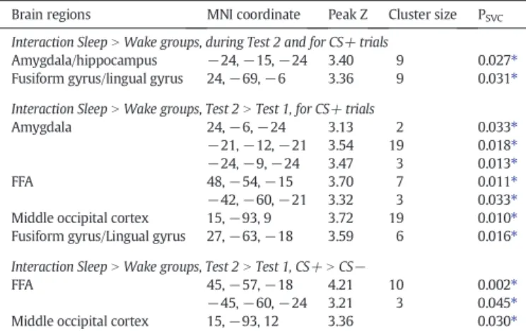
All the comparisons above contrasted trials based on the partici-pants' own subjective responses (face pairs perceived as same or differ-ent) during the face discrimination task. To reveal brain regions whose activity varied as a function of the objective visual distance between two faces in a pair (from 1 to 15), parametric modulators based on morphing distance were included in a second analysis. These parametric modula-tors identified regions whose activity showed linear decreases in activity with increasing visual similarity between the two faces in a trial (i.e. greater repetition suppression), independently of the subjec-tive response of the participant (which was not linear, seeFig. 2A). For CS− trials, we observed no significant parametric effect for either the Sleep or Wake groups separately, or for any comparison between groups. For CS+ trials, however, the parametric modulation by morphing distance revealed that participants who slept after the conditioning acti-vated significantly less the left amygdala and posterior fusiform/lingual gyrus as a function of reduced morphing distance, relative to the partici-pants who stayed awake (Table 3). To specifically demonstrate the effect of a period of Sleep (as compared to a period of Wake) on the consolida-tion of the neural representaconsolida-tion of the CS+ faces, we computed a triple interaction with the factors Group, Test, and Morphing Distance. This
Fig. 3. Effect of subjective discrimination (response‘same’ vs. ‘different’) for CS− and CS+ face pairs during the Test 2 session. A: Brain activation associated with subjective discrim-ination of morphed faces in CS− trials, for both groups of participants (Sleep group and Wake group), involving posterior fusiform/lingual cortex (yellow cluster). The parameter estimates are illustrated for CS− ‘same’ and ‘different’ responses. This region does not cor-respond to the fusiform face area (FFA) as defined during the face localizer task (seeFig. 4) but instead overlaps with the network involved in the detection of low-level visual fea-tures (scrambleN face, in cyan). B: Brain activation associated with subjective discrimina-tion of morphed faces on CS+ trials, greater for Sleep group than Wake group (SleepN Wake) in right amygdala. The parameter estimates are described for CS+‘same’ and ‘dif-ferent’ responses. Activations are displayed on the mean structural MR image, at p b 0.001 uncorrected.
Table 1
Regions showing increased repetition suppression (i.e. reduced activity) during Test 2 for CS− face-pairs perceived as ‘same’ (compared to ‘different’ responses) for both Sleep and Wake groups.
Brain regions MNI coordinate Peak Z Cluster size
PSVC
Fusiform gyrus/lingual gyrus/LOC 27,−78, −6 15,−81, −12 4.19 3.78 48 48 0.002⁎ 0.008⁎ Middle occipital gyrus 36,−75, 12 3.59 48 0.015⁎ Parieto-occipitalfissure −24, −72, 9 4.86 137 0.033⁎⁎ ⁎⁎ Significant at p b 0.05 FWE corrected for the whole brain volume.
⁎ Significant after small volume correction using the face localizer contrast scramble vs. face.
Table 2
Regions showing increased repetition suppression (i.e. reduced activity) during Test 2 for a CS+ trial perceived as‘same’ (compared to ‘different’ responses). Interaction SleepN Wake groups.
Brain regions MNI coordinate Peak Z Cluster size PSVC
Amygdala 27, 3,−27 3.69 8 0.011⁎ Middle cingulate gyrus −9, −3, −42 3.45 6 0.03⁎ ⁎ Significant after small volume correction using the face localizer contrast face vs. scramble for the amygdala, and scramble vs. face for the cingulate.
analysis confirmed that the amygdala and lingual gyrus were selectively modulated by objective visual similarity in the Sleep group but not in the Wake group, more so during Test 2 than Test 1 (seeFig. 4). Importantly, this analysis also revealed an additional significant activation in the ante-rior fusiform cortex, specific to the Sleep group. This region overlapped with the FFA obtained from the face localizer (Fig. 4, in cyan). Finally, tivation of the right FFA also survived in a last interaction taking into ac-count the Conditioning status of the face pairs, thus confirming increased FFA response selectively to the CS+ containing pairs and after a period of sleep.Table 3reports the results from this quadruple interaction with the factors Group (Sleep vs. Wake), Test (Test 2 vs. Test 1), Conditioning (CS+ vs. CS−), and Morphing distance. In other words, only the Sleep group showed discriminative responses in face-selective areas that were linearly sensitive to the visual similarity with the original CS+ face. To further investigate the pattern of activity in the anterior, face-responsive fusiform regions, we extracted the parameters estimates (beta values) for the FFA as functionally defined in each individual by the separate localizer run (seeMethods). A repeated-measure ANOVA was performed with Conditioning (CS+, CS−) and Response (different, same) as within-subject factors, and Group (Sleep, Wake) as between-subject factor on Test 2. We observed a marginal effect of Conditioning (F(1,30) = 3.19, p = 0.08) and a significant interaction between Re-sponse and Group (F(1,30) = 5.53, p = 0.025). Crucially, planned com-parisons demonstrated that the FFA was significantly less activated for face pairs in the CS+ same than CS+ diff condition (p = 0.04) for the Sleep group, whereas this difference was not significant for the Wake
group (p = 0.91) nor for the CS− condition (Sleep: p = 0.15, Wake: p = 0.61). These results converge to indicate that after one night of sleep (but not after a similar delay without sleep), both the amygdala and the face-responsive fusiform cortex exhibited a stronger sensitivity to identity-specific sensory attributes in the CS+, leading to differential repetition effects for the fear-conditioned and sleep-consolidated face stimuli. This provides thefirst demonstration that sleep may promote the reconfiguration of freshly encoded emotional memories in visual cortex, and thus enhance perceptual tuning to stimulus cues previously associated with an aversive experience. Moreover, such remodeling was observed for higher-level visual representations presumably mediating face identity recognition in our discrimination task (i.e. FFA), instead of more posterior regions of the fusiform cortex that extract more basic vi-sual features (Weiner and Grill-Spector, 2010) and show more general repetition effects irrespective of conditioning.
Finally, based on previous evidence that the amygdala can modulate FFA response to emotional faces (Vuilleumier et al., 2004), we tested whether FFA response to the CS+ (vs. CS−) correlated with amygdala response to the CS+ (vs. CS−) during the critical session on the second day (Test 2). Such a correlation was found significant in the Sleep group but not in the Wake group, suggesting that these two regions could be functionally more tightly connected after sleep (r2= 0.3, p = 0.02) than after wakefulness (r2= 0.009, p = 0.2) during the discrimination
of conditioned faces. Sleep parameters
Sleep was monitored over 4 nights prior to thefirst fMRI session (see
Methods), and showed no difference between the two groups on any sleep measure, including mean subjective sleep quality on a 10-points scale (see Table S4, t = 0.55, p = 0.59), number of sleep hours (t = 1.16, p = 0.25), and sleep efficiency estimated from the actimetry data for the night preceding the test (t =−1.17, p = 0.26). During the 12-h interval between experimental parts 1 and 2 (Fig. 1A), the Sleep participants reported a total sleep time of 7.77 ± 0.63 h, not differ-ent from their previous nights (p = 0.21), and a quality of sleep slightly lower (6.93 ± 1.58) than that of the preceding, non-experimental night (p = 0.001). Sleep efficiency of the experimental night (83.86 ± 3.73) was not significantly different from the night before the experiment (p = 0.15) for the Sleep group. Because thefirst and second experimen-tal sessions took place at different times of the day for each group, we also checked that vigilance state, assessed by the psychomotor vigilance task (PVT) and Karolinska Sleepiness Scale (KSS), did not differ between the groups. ANOVAs of the mean RTs in the PVT and KSS scores with Ex-perimental Part (1, 2) as within-subject factor and Group (Sleep, Wake) as between-subject factor revealed no significant main effects, nor any interaction between factors. Collectively, these data suggest that any be-havioral or neural difference observed between the groups during the
Table 3
Regions showing increased repetition suppression (i.e. reduced activity) during Test 2 modulated by the objective morphing distance between both faces (from 15 to 1) in each trial.
Brain regions MNI coordinate Peak Z Cluster size PSVC
Interaction SleepN Wake groups, during Test 2 and for CS+ trials
Amygdala/hippocampus −24, −15, −24 3.40 9 0.027⁎
Fusiform gyrus/lingual gyrus 24,−69, −6 3.36 9 0.031⁎
Interaction SleepN Wake groups, Test 2 N Test 1, for CS+ trials
Amygdala 24,−6, −24 3.13 2 0.033⁎
−21, −12, −21 3.54 19 0.018⁎
−24, −9, −24 3.47 3 0.013⁎
FFA 48,−54, −15 3.70 7 0.011⁎
−42, −60, −21 3.32 3 0.033⁎
Middle occipital cortex 15,−93, 9 3.72 19 0.010⁎
Fusiform gyrus/Lingual gyrus 27,−63, −18 3.59 6 0.016⁎
Interaction SleepN Wake groups, Test 2 N Test 1, CS+ N CS−
FFA 45,−57, −18 4.21 10 0.002⁎
−45, −60, −24 3.21 3 0.045⁎
Middle occipital cortex 15,−93, 12 3.36 0.030⁎
⁎ Significant after small volume correction using the face localizer contrast face vs. scramble for the amygdala and the FFA, and contrast scramble vs. face for the lingual gyrus.
Fig. 4. Effect of objective discrimination (real morphing distance between two faces, from 15 to 1) for CS+ morphing more for Test 2 than Test 1. The amygdala and the fusiform cortex (resp. left and right panel, in yellow) are less activated for face pairs with small than large distance along their morphing continuum, more for the Sleep group than the Wake group and more at Test 2 than at Test 1. Face-selective activations from a separate localizer task (face vs. scramble) are shown in cyan. Activations are displayed on the mean structural MR image of the whole group, at pb 0.004 uncorrected for display purposes.
discrimination task cannot be attributed to differences in vigilance state between the groups at the time of testing. Note also that, for the time period between fMRI sessions, participants from both the Wake and Sleep groups reported no physical or psychological stressor. Because the goal of our study was to study the impact of sleep on the perceptual discrimination of conditioned visual stimuli, we purposefully chose ex-perimental conditions minimizing any additional stress that would in-terfere with the consolidation of a newly acquired emotional association. For this reason, we also deliberately avoided sleep depriva-tion in the Wake group, which is known to elicit high levels of stress (van der Helm et al., 2011).
Discussion
Imagine you walk on a crosswalk; a car unexpectedly comes from a side road and hits you to the ground. Through the car's windshield, you clearly see the face of the driver who swiftly drives away without stopping. Unhesitatingly, a witness helps you stand up again. Some day later in a musical event, you suddenly recognize the face of the felo-nious driver popping out among the crowd, despite changes in outfit; however, during the same event you fail to recognize the witness among the musicians. Our study provides a neural mechanism account-ing for such a scenario that may happen in our daily lives, and suggests that the brain encodes threat stimuli in a way that modifies sensory pro-cesses to maintain better traces and allow more efficient detection, even in changing environments. While the prioritization of emotionally-relevant information for attention and subsequent declarative memory has been extensively studied (Phelps and LeDoux, 2005; Vuilleumier et al., 2001), comparatively little is known about the impact of stimulus emotional significance on long-term perceptual representations and perceptual decisions (Chavez et al., 2013; Li et al., 2008; Lim and Pessoa, 2008; Resnik et al., 2011; Weinberger, 2004). Moreover, recent brain imaging studies suggest that the consolidation of aversive associ-ations may benefit from post-encoding sleep (Menz et al., 2013; Pace-Schott et al., 2009; Sterpenich et al., 2007), but whether this also gives rise to enhanced perceptual sensitivity and discrimination ability for emotionally-relevant information has remained hitherto unknown. We designed the present experiment to demonstrate changes in the discrimination performance and the neural coding of face stimuli after conditioning followed by a period of sleep. Specifically, we predicted fa-cilitated access to the visual representation of the individual CS+ face and increased activity in face-responsive regions, beyond regions cod-ing for low-level visual features. Here we used a face discrimination task adapted from a study of face recognition (Rotshtein et al., 2005), in which the authors used morphed stimuli from pairs of famous faces and established the role of the right fusiform gyrus in extracting infor-mation about face identity, rather than lower-level physical attributes of the faces. Consistent with previous research, behavioral responses to morphed faces in our study followed a sigmoid psychophysical curve for face identity decisions (Beale and Keil, 1995; Rotshtein et al., 2005). This task offered ideal behavioral characteristics to monitor any shift in discrimination threshold (50%‘different’ responses) as a func-tion of condifunc-tioning and/or sleep. Critically, we did not observe any im-mediate effect of conditioning on the discrimination task, but significant changes emerged after a 12-h delay including a night of sleep (Test 2), with shorter reaction times for CS+ containing trials, and a systematic shift in the discrimination threshold away from the CS+ face. In other words, participants were able to detect the identity of the CS+ face when fewer details from this face were physically present in the image. How can this be explained?
On the one hand, such threat detection benefit is reminiscent of the ‘face in a crowd effect’ in which emotionally-relevant faces (e.g. with angry expression) are detected more rapidly and more accurately than neutral faces (Feldmann-Wustefeld et al., 2011; Hansen and Hansen, 1988; Ohman et al., 2001). Such privileged processing of emotional in-formation is presumably mediated through feedback signals from the
amygdala (Vuilleumier, 2005; Vuilleumier and Driver, 2007). While conditioning may involve enhanced arousal and attention, an effect shared by all conditioning paradigms, the modulation of sensory corti-ces by attention or emotion is produced by different sourcorti-ces. The specif-ic activation of the amygdala, in the absence of increased recruitment of fronto-parietal attentional networks, favors for a sensory modulation by emotion. Thus, one plausible interpretation is that the visual features of the CS+ face have become more salient through consolidation of the ac-quired aversive association. Increased amygdala recruitment due to the consolidation of fear learning during sleep, as reported here, may favor the processing of the CS+ during the face discrimination task. These re-sults add support to the notion that sleep may promote the consolida-tion of emoconsolida-tional memories (Baran et al., 2012; Hu et al., 2006; Menz et al., 2013; Payne and Kensinger, 2011; Sterpenich et al., 2007, 2009; Wagner et al., 2001). This hypothesis is also consistent with ourfinding of increased amygdala-FFA functional connectivity after sleep, selective-ly when CS+ face information is present.
On the other hand, the observed shift in discrimination threshold away from the original CS+ face might reflect changes in neuronal selectivity to facial information within visual cortex itself. Whether aversive conditioning increases the selectivity or the sensitivity of per-ceptual processes has been debated. In the auditory domain,Resnik et al. (2011)showed that aversive learning induces wider stimulus gen-eralization, with tones further away from a CS+ tone becoming less dis-tinguishable by human subjects. This mechanism may underlie fear generalization, whereby stimuli resembling those previously associated with negative consequences can also elicit fear responses (Ghirlanda and Enquist, 2003; Pavlov, 1927). Conversely, in the olfactory domain,
Li et al. (2008)showed that aversive conditioning enhances the discrim-ination of initially undistinguishable sensations and modulated patterns of neural activity in primary olfactory cortex in humans. Fast and efficient defensive behavior may require both high sensitivity to percep-tually similar stimuli, and high selectivity along relevant perceptual di-mensions distinguishing threat (CS+) from safety (CS−) signals in order to avoid overgeneralization of aversive associations (Lissek et al., 2010). The present results support the notion that adapted responses to threat-related stimuli involves both enhanced perceptual sensitivity to sensory features of CS+ cues, and enhanced cortical selectivity through a remodeling of neural representations in higher-level visual regions. Specifically, increased amygdala activity and functional cou-pling with sensory cortices may explain sensitivity to sensory attri-butes of fear-conditioned stimuli (even when the latter contain less information about the CS+; i.e. more distant morphs), while increased repetition-suppression effects in the face fusiform cortex is consistent with finer perceptual tuning of face-responsive visual regions to identity-specific attributes of the conditioned face.
Indeed, by using a repetition-suppression paradigm in which two faces were presented in a rapid succession (seeMethods), we assessed how brain responses were modulated by differences in sensory physical attributes between two stimuli (Grill-Spector et al., 2006). Specifically, we could determine how fear conditioning modified cortical activity as a function of the visual similarity between faces in each pair, while systematically varying their perceptual distance along the morphing continuum. Our fMRI results show that face stimuli used in our task re-cruited a distributed network of regions involved in faces processing (including FFA and OFA in visual cortices), but that the subjective dis-crimination of face pairs presented in rapid succession relied on a more posterior region in the lingual gyrus, outside face-responsive re-gions identified by the face localizer (when non conditioned and not consolidated by sleep), and observed across all sessions. The repetition effects in lingual gyrus for face pairs perceived as‘same’ may suggest that the face discrimination task primarily implicated a matching of basic visual features rather than the extraction of facial features from the two successive photographs (Ostwald et al., 2008). A similar pattern was found for the CS− face after conditioning. By contrast, visual recog-nition processes encoding face identity in the FFA proper were recruited
for the CS+ face and thisfine-tuning occurred only after a night of sleep (not after an equivalent period of wake). These data point to more elab-orate (perhaps holistic) processing of identity-specific face information for the CS+ stimuli, rather than just low-level visual features as observed for the CS− (Henson et al., 2000; Rotshtein et al., 2005). In keeping with this interpretation, at the end of the experiment during debriefing, 9 participants from the Sleep group (only 3 from the Wake group) reported to have‘personalized’ the faces, using nicknames and mentioning resemblance with specific people. This anecdotal observa-tion offers an eloquent illustraobserva-tion of the processing of the CS+ face as an individual identity, presumably mediated by greater recruitment of the FFA. Overall, our fMRI results therefore provide compelling novel ev-idence that, after a night of sleep, visual responses in extrastriate corti-ces become more selectively tuned to discriminative information from fear-conditioned stimuli.
Whereas previous studies demonstrated that performance in a visu-al texture discrimination task benefits from sleep after intense practice and involves plasticity in early visual cortices (Aeschbach et al., 2008; Gais et al., 2000; Mascetti et al., 2013; Schwartz et al., 2002; Stickgold et al., 2000; Yotsumoto et al., 2009), here we show that behavioral changes in perceptual discrimination abilities after sleep can be induced by incidental emotional experience and imply the emergence of selec-tive responses in higher-levels, category-selecselec-tive areas of the visual hi-erarchy. Furthermore, these effects occurred after a single, brief session of aversive conditioning, considerably shorter than the perceptual train-ing effects for visual texture cues (Schwartz et al., 2002; Stickgold et al., 2000), and unrelated to the discrimination task itself. This altogether attests the power of emotion signals in shaping cortical selectivity to behaviorally relevant stimuli. Collectively, thesefindings highlight how sleep may contribute to the remodeling of neural circuits involved in the processing of emotionally-relevant information to forge more effective and long-lasting representations within associative visual cortices, beyond low-level sensory regions. Because we did not record sleep using polysomnography in this study, we cannot attribute the ob-served effects to any specific sleep stage. However, based on previous work from our and other teams (Menz et al., 2013; Nishida et al., 2009; Sterpenich et al., 2014), we hypothesize that REM sleep may favor the reorganization of cortico-cortical representation of the faces in the occipital cortex and would thus be particularly beneficial for the consolidation of the perceptual representation of the conditioned face. Further research is needed to confirm this hypothesis.
In this study, we used an experimental protocol that allows testing for the effects of sleep, while minimizing unspecific stress (no sleep deprivation), which could possibly interfere with the consolidation of the freshly encoded emotional association. Although this type of proto-col has been extensively used in prior studies (e.g.Hu et al., 2006; Payne et al., 2008), we need to rule out circadian influences in our measure-ments. Firstly, there was no significant group difference in objective (PVT) and subjective (KSS) vigilance levels across experimental ses-sions. Secondly, we observed no behavioral or neural difference be-tween the two groups during the Baseline session or during Test 1. Importantly, we did notfind any behavioral difference for the CS+ and CS− between the groups during Test 1. Thirdly, in our main analy-ses, we tested for behavioral and neural changes from Test 1 to Test 2 in each individual, avoiding group comparisons of data from different circadian phases. We therefore believe that our mainfindings cannot easily be attributable to circadian factors, but more likely result from neural changes taking place during sleep. Yet, despite these precautions, whether some of our results could be driven by time of day influence cannot be excluded (e.g. impact of cortisol levels on the processing of emotional stimuli). These data thus constitute new evidence for a role of sleep in both quantitative and qualitative changes of memory repre-sentations (Diekelmann and Born, 2010; Maquet, 2001; Stickgold and Walker, 2013), here driven by emotional significance, arising in category-specific sensory cortices, and characterized by stimulus-specific increases in both sensitivity and tuning.
Conclusion
In sum, our study demonstrates that a night of sleep promotes re-organization in the cortical representation of faces. Recognition of affectively-salient face information becomes less dependent on low-level visual details but more reliant on identity-specific infor-mation, leading to distinctive shifts in discrimination performance and selective repetition suppression effects in visual cortex and amygdala. Thesefindings highlight the ecological benefit of a night of sleep in upgrading cortical representations for emotionally-relevant memories, and consolidating aversive associations through amygdala signaling. An optimal balance between these mechanisms respectively ensures selectivity and sensitivity in learning new threat-related cues in the environment, and can thus promote adaptive responses to dan-gers even when sensory inputs from potential threats are ambiguous or degraded.
Supplementary data to this article can be found online athttp://dx. doi.org/10.1016/j.neuroimage.2014.06.003.
Acknowledgments
This research was supported by grants from the Swiss National Sci-ence Foundation (No. 320030_135653), the Mercier Foundation, and the National Center of Competence in Research (NCCR) Affective Sci-ences financed by the Swiss National Science Foundation (No. 51NF40-104897) hosted by the University of Geneva.
References
Aeschbach, D., Cutler, A.J., Ronda, J.M., 2008.A role for non-rapid-eye-movement sleep homeostasis in perceptual learning. J. Neurosci. 28, 2766–2772.
Akerstedt, T., Gillberg, M., 1990.Subjective and objective sleepiness in the active individ-ual. Int. J. Neurosci. 52, 29–37.
Armony, J.L., Servan-Schreiber, D., Romanski, L.M., Cohen, J.D., LeDoux, J.E., 1997.Stimulus generalization of fear responses: effects of auditory cortex lesions in a computational model and in rats. Cereb. Cortex 7, 157–165.
Baran, B., Pace-Schott, E.F., Ericson, C., Spencer, R.M., 2012.Processing of emotional reac-tivity and emotional memory over sleep. J. Neurosci. 32, 1035–1042.
Beale, J.M., Keil, F.C., 1995.Categorical effects in the perception of faces. Cognition 57, 217–239.
Buysse, D.J., Reynolds 3rd, C.F., Monk, T.H., Berman, S.R., Kupfer, D.J., 1989.The Pittsburgh Sleep Quality Index: a new instrument for psychiatric practice and research. Psychi-atry Res. 28, 193–213.
Cajochen, C., Knoblauch, V., Wirz-Justice, A., Krauchi, K., Graw, P., Wallach, D., 2004. Circa-dian modulation of sequence learning under high and low sleep pressure conditions. Behav. Brain Res. 151, 167–176.
Chavez, C.M., McGaugh, J.L., Weinberger, N.M., 2009.The basolateral amygdala modulates specific sensory memory representations in the cerebral cortex. Neurobiol. Learn. Mem. 91, 382–392.
Chavez, C.M., McGaugh, J.L., Weinberger, N.M., 2013.Activation of the basolateral amyg-dala induces long-term enhancement of specific memory representations in the cere-bral cortex. Neurobiol. Learn. Mem. 101, 8–18.
Diekelmann, S., Born, J., 2010.The memory function of sleep. Nat. Rev. Neurosci. 11, 114–126.
Dinges, D., Powell, J., 1985.Microcomputer analyses of performance on a portable, simple visual RT task during sustained operations. Behav. Res. Methods 17, 652–655.
Duvarci, S., Bauer, E.P., Pare, D., 2009.The bed nucleus of the stria terminalis mediates inter-individual variations in anxiety and fear. J. Neurosci. 29, 10357–10361.
Ellis, B.W., Johns, M.W., Lancaster, R., Raptopoulos, P., Angelopoulos, N., Priest, R.G., 1981.
The St. Mary's Hospital sleep questionnaire: a study of reliability. Sleep 4, 93–97.
Feldmann-Wustefeld, T., Schmidt-Daffy, M., Schubo, A., 2011.Neural evidence for the threat detection advantage: differential attention allocation to angry and happy faces. Psychophysiology 48, 697–707.
Gais, S., Plihal, W., Wagner, U., Born, J., 2000.Early sleep triggers memory for early visual discrimination skills. Nat. Neurosci. 3, 1335–1339.
Gdalyahu, A., Tring, E., Polack, P.O., Gruver, R., Golshani, P., Fanselow, M.S., Silva, A.J., Trachtenberg, J.T., 2012.Associative fear learning enhances sparse network coding in primary sensory cortex. Neuron 75, 121–132.
Ghirlanda, S., Enquist, M., 2003.A century of generalization. Anim. Behav. 66, 15–36.
Grill-Spector, K., Henson, R., Martin, A., 2006.Repetition and the brain: neural models of stimulus-specific effects. Trends Cogn. Sci. 10, 14–23.
Hansen, C.H., Hansen, R.D., 1988.Finding the face in the crowd: an anger superiority ef-fect. J. Pers. Soc. Psychol. 54, 917–924.
Henson, R., Shallice, T., Dolan, R., 2000.Neuroimaging evidence for dissociable forms of repetition priming. Science 287, 1269–1272.
Horne, J.A., Ostberg, O., 1976.A self-assessment questionnaire to determine morningness– eveningness in human circadian rhythms. Int. J. Chronobiol. 4, 97–110.