Université de Montréal
The cardioprotective effect of a short-term aerobic exercise program and the mitochondrialpermeability transition pore ofthe Rat
par
Marc Ciminelli
Département de kinésiologie
Mémoire présenté à la faculté des études supérieures en vue de l’obtention du grade de Maître ès Sciences (MSc.) en sciences de l’activité physique
Décembre 2005
n
Université
(H’R
de Montréal
Direction des bibliothèques
AVIS
L’auteur a autorisé l’Université de Montréal à reproduire et diffuser, en totalité ou en partie, par quelque moyen que ce soit et sur quelque support que ce soit, et exclusivement à des fins non lucratives d’enseignement et de recherche, des copies de ce mémoire ou de cette thèse.
L’auteur et les coauteurs le cas échéant conservent la propriété du droit d’auteur et des droits moraux qui protègent ce document. Ni la thèse ou le mémoire, ni des extraits substantiels de ce document, ne doivent être imprimés ou autrement reproduits sans l’autorisation de l’auteur.
Afin de se conformer à la Loi canadienne sur la protection des
renseignements personnels, quelques formulaires secondaires, coordonnées ou signatures intégrées au texte ont pu être enlevés de ce document. Bien que cela ait pu affecter la pagination, il n’y a aucun contenu manquant.
NOTICE
The author of this thesis or dissertation has granted a nonexclusive license allowing Université de Montréal to reproduce and publish the document, in part or in whole, and in any format, solely for noncommercial educational and research purposes.
The author and co-authors if applicable retain copyright ownership and moral rights in this document. Neither the whole thesis or dissertation, nor substantial extracts from it, may be printed or otherwise reproduced without the author’s permission.
In compliance with the Canadian Privacy Act some supporting forms, contact information or signatures may have been removed from the document. While this may affect the document page count, it does flot represent any loss of content from the document.
Ce mémoire intitulé:
The cardioprotective effect of a short-term aerobic exercise program and the mitochondrial permeability transition pore of the Rat
présenté par Marc Cirninelli
a été évalué par un jury composé des personnes suivantes:
Luc Léger Président-rapporteur
Yan Burelle Directeur de recherche
François Peronnet Membre du jury
Résumé
En 2002, les maladies cardiovasculaires ont constitué la plus grande cause de décès à l’échelle mondiale, devançant le cancer et les maladies pulmonaires. L’infarctus du myocarde (1M) est généralement la première manifestation d’une maladie cardiovasculaire ischémique et il a été démontré que la pratique régulière d’exercices constitue un moyen efficace pour diminuer plusieurs facteurs de risque associés au développement de maladies cardiovasculaires tels l’hypertension, 1’ hypercholestérolémie, l’obésité et la résistance à l’insuline. De plus, il a été démontré que le taux de survie à l’issue d’un 1M est plus élevé chez les sujets actifs comparativement aux sujets sédentaires, indiquant que l’exercice protège le coeur contre ce genre de stress.
Plusieurs études, principalement chez le Rat, ont effectivement démontré qu’un programme d’entraînement en endurance d’une durée variant de trois à cinq jours jusqu’à quatre mois apportait une protection cardiaque contre l’ischémie-reperfusion (I-R). Cette protection se caractérise par une diminution du dommage tissulaire et par une meilleure récupération de la fonction contractile en reperfusion. Cependant, les mécanismes impliqués dans cet effet protecteur demeurent encore mal compris.
Par ailleurs, il est bien connu que la reperfusion d’un tissu ischémique peut engendrer des dommages au niveau des mitochondries. Des dysfonctions mitochondriales qui résultent en une diminution ou une abolition de la production d’ATP engendrent, affectent directement ta fonction contractile et mènent éventuellement à la mort cellulaire par nécrose. De plus, il est maintenant reconnu que la mitochondrie joue un rôle important
dans la signalisation de l’apoptose par sa capacité à relâcher plusieurs protéines pro apoptotiques qui sont normalement séquestrées dans la matrice mitochondriale ou l’espace inter-membranaire. Plusieurs études menées au cours des dernières années ont montré que l’ouverture du pore perméable de transition (PPT), un canal non spécifique à haute conductance de la double membrane mitochondriale, était directement impliqué dans les deux formes de mort cellulaire dans le coeur suite à une période d’I-R.
L’effet de l’entraînement sur la fonction mitochondriale et plus particulièrement sur la régulation du PPT n’a jamais été exploré dans l’optique d’expliquer l’effet cardio protecteur de l’exercice contre l’I-R. Les travaux effectués au cours de cette Maîtrise avaient donc deux objectifs. Le premier objectif était de mettre sur pied la technique de séquestration mitochondriale du [3H]deoxyglucose ([311]-DOG), une approche permettant de quantifier l’ouverture du PPT in situ dans le coeur isolé du rat perfusé en mode Langendorff. Le deuxième objectif était d’appliquer cette méthodologie pour déterminer
si l’entraînement à court terme (5 jours consécutifs de cours sur tapis roulant à une
intensité approximative de 75% du VO2max) diminue l’ouverture du PPT pendant la reperfiision (40minutes) suite à une épisode ischémique (30minutes).
Les résultats obtenus ont montré que l’entraînement permettait de diminuer le dommage mitochondrial tel que démontré par une moins grande perte du recouvrement de l’enzyme citrate synthase dans la fraction mitochondriale suite à l’I-R. De plus, l’incorporation de [3H]-DOG dans les mitochondries était de 30-40 % inférieure à celle retrouvée dans les mitochondries cardiaques provenant d’animaux contrôles, indiquant une inhibition du
l’ouverture du PPT. Par contre, dans les conditions de perfusion utilisées, l’entraînement n’a pas amélioré la récupération fonctionnelle et la relâche de LDH malgré une réduction significative du dommage mitochondrial induit par le PPT.
Dans l’ensemble, les résultats de ce travail mettent en évidence que l’entraînement à court terme peut atténuer le dommage mitochondrial et l’ouverture du PTP qui survient en reperflision suite à une période d’ischémie. Par contre, dans les conditions expérimentales utilisées, nous n’avons malheureusement pas été en mesure de déterminer la contribution du PPT comme effet protecteur de l’entraînement à court terme contre les dysfonctions contractiles et le dommage tissulaire, rapportés dans la littérature.
MOTS CLÉS: exercice, ischémie-reperfusion, cardioprotection, perméabilité transitionnelle, deoxyglucose
Summary
Ischemic heart disease claimed more lives worldwide in 2002 than any other single disease. Myocardial ïnfarction is commonly the initial manifestation of ischemic heart disease, and exercise training has been shown to decrease a number of the risk factors for myocardial infarction including hypertension, hyperlipidemia, obesity, and insulin resistance, and has also been reported to improve chance of survival in humans afier an ischemic event. Although exercise has proven beneficial in terms of cardioprotection, mechanisms for such an effect have yet to be elucidated.
Recent experimental studies have confirmed that both short-term (days) and long-term (weeks to months) endurance exercises provide myocardial protection against ischemia reperfusion (I-R) injury in rats. Specifically, short-term exercise (i.e. 1—5 bouts of endurance exercise) reduces cardiac injury and enhances myocardial contractile recovery from an I-R insuit as evidenced by an improved recovery of lefi ventricle developed pressures. While it is widely accepted that exercise improves tolerance against myocardial I-R, the mechanism(s) responsible for tins exercise-induced cardioprotection remains elusive.
Reperfiision of the ischemic tissue has been implicated with oxidative modifications and functional impairment of mitochondria. It is for this reason that many studies have focused on the mitochondria as a potential key player in ischemia-reperfusion injury. Indeed, failure to produce ATP as a resuit of mitochondrial dysfunction is believed to mark the transition toward tissue necrosis and contractile failure during early reperfusion.
In addition, mitochondriaare known to play a key role in signalling apoptotic ceil death through the release of several pro-apoptotic proteins that are normally confmed to the mitochondrial matrix or inter-membrane space. Opening of the permeability transition pore (PTP), a non-specific high conductance channel spanning the double membrane system ofthe mitochondria is known to trigger both necrosis and apoptosis.
The effect of exercise training on many aspects of mitochondrÏal function, and particularly on the regulation and behaviour of the PTP, has neyer been addressed in the context of explaining the cardio-protective effect against I-R. Therefore, the goals of the present Master’s thesis were to implement the mitochondrial [311J2-deoxyglucose entrapment method, a technique that allows to quantify PTP opening in situ in the isolated, Langendorff, perfused heart of rats and to apply this methodology to determine whether short-term (5 consecutive days of treadmiil nmning at approximately 75% VO2max) aerobic training reduces the occurrence of PTP opening during reperfusion (40 minutes) following an ischemic episode (30 minutes).
Resuits have shown that citrate synthase (CS) recovery was significantly greater in the exercise trained compared to control, suggesting a reduction in mitochondrial damage. furthermore, the incorporation of [3HJ-DOG within the mitochondria was shown to be reduced by 3 0-40% in trained animais, indicating a reduction of PTP opening. However, under the experimental conditions used, training did not resuit in a significant improvement in functional recovery and LDH release despite a significant reduction in PTP-induced mitochondrial damage, which is in contrast with several other reports.
Taken together the present resuits provide evidence that short-term training can attenuate mitochondrial damage and PTP opening which normally occurs in the heart following ischemia-reperfusion. However, under the experimental conditions used, the contribution of this mitochondrial protection to the protective effect of short-term training reported could flot be established.
KEY WORDS: exercise, ischemia-reperfusion, cardioprotection, permeability
Table of Contents
Résumé iii
English Summary vi
Table ofContetits ix
figtire Legends
Table Legends xii
Abbreviations xiii
Acknowledgements xiv
Chapter 1- Liter ature Review
1 INTRODUCTION 2
2 T11E MITOCHONDRIAL PER1tIEABIUTY TRANSITION PORE AND ITS ROLE
IN ISCIIEMIA-REPERFUSION INJURY IN THE HEART 5
2.1 IN VITRO STUDIES ON THE MITOCFIONDRIAL PERMEABILTY TRANSITION 5
2.1.] Consequences oJPTPopening011 niitochondria in vitro 5
2.1.2 Molecular identitv ofthe PTP 6
2.1.3 Regulatorvproperties oftÏie PTP 9
2.2 EFFECT 0F ISCHEMIA-REPERFUSION ON THE PTP 9
2.2.1 Overview ofthe metabolic anciionic consequences of ischemia— repeijitsion in the
heart 9
2.2.2 Cctrdio-protective effects ofpharmctcologiccd PTP mli ibitors 11 2.2.3 Assessment of PTPopening in situ in Hie ischeinic-repeifised heurt 17
2.2.3.1 Mitochondrial NAD release 17
2.2.3.2 Mitochondrial [3H] -deoxyglucose entrapment 22
2.2.3.3 Principle 23
2.2.4 ExperimentaÏ protocoL 27
2.2.4.1 Calculation ofthe ÏH]-DOG index 27
2.2.4.2 Role ofthe PTP inhibition in cardio-protectioiv 29
2.2.4.2.1 Cyclosporin A and Sanglifebrin A 29
2.2.4.2.2 Pyruvate and Propofol 31
3 Cardio-protection induced by short-term training .35
3.1 ANIMAL MODELS AND TRAINING PARADIGMS 36 3.2 EFFEcT 0F SHORT-TERM TRAINING ON MYOCARDIAL FUNCTION AFTER ISCHEMIA
AND ON MARKERS 0F TISSUE DAMAGE 3$
3.3 MEcHANIsMs UNDERLYING THE CARDIO-PROTECTIVE EFFECT 0F SHORT-TERM
EXERCISE TRAINING 43
3.3.] Hectt-shockproteins 43
3.3.1.1 Overview ofthe heat-shock family ofproteins 43
3.3.1.2 HSP-mediated cardio-protection 44
3.3.1.3 Mechanisms underlying HSP-mediated cardio-protectioir 46 3.3.1.4 Implication of HSP ‘sin training-induced cardioprotection 47 3.3.2 Oxidcttive stress cindantioxiclant responses 49
3.4 IS THEPTP INVOLVED IN THE PROTECTIVE EFFECT 0F SHORT-TERM TRAINING9 50
Bibliography for LiteratureReview 52
Chapter 2- Effect of short-term training on mitochondrial permeabïlity transition pore
opening following myocardial ischemia-reperfusion 5$
Abstract 59 Introduction 60 Methods 62 Resuits 66 Discussion 68 Article References 74 Figure Legends 77 Tables Legends 79 Figures $0 Tables $5
Figures in Chapter-1
Figure 1 Most popular and documented hypothesis regarding the pore composition. The adenylate translocator (ANT) of the inner membrane, the voltage dependent anion channel (VDAC) of the outer membrane, and the matrix enzyme cyclophulin-D (CypD). Other proteins associate with the complex as ïndïcated. Pg.8
Figure 2 Involvement ofthe PTP in ischemia-reperfusion —induced celi death. Lack of oxygen and circulating substrate rapidly leads to the abolition oxidative phosphoiylation and a rapid ATP content and accumulation ofP and W. Upon reperfusion, there is an excessive Ca2 uptake, which, coupled with oxidative stress and prevailing high P and low ATP can provoke PTP opening. Pg. 12
Figure 3 Resuits of lefi ventricular developed pressure (LVDP) and end diastolic pressure (EDP) following 15 minutes ofreperfusion after 30 or 40 minutes ofischemia with or without CsA. Pg. 14
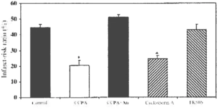
Figure 4 Reduction in the infarct size with the use of CsA. Pg. 15
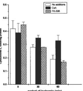
FigureS Mitochondrial cytochrome c content afier a penod ofischemia. CsA protects heart mitochondria from loss ofcytochrome e during ischemia but notFK-506.Pg. 16
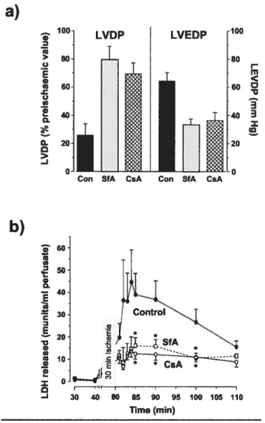
Figure 6 $fA and CsA protect the ischemic rat heart from reperfusion injury. In panel a, greater fiinctional recovery ofthe SfA- and CsA-treated hearts
afier a 30-min reperfusion is reflected in higher values for the LVDP and lower values for the LVEDP. Values ofthe LVDP after reperfision are presented as a percentage of the pre-ischemic value and were significantly greater (p< 0.00 1) with either SfA or CsA treatment, whereas values for
the LVEDP were significantly lower (p< 0.00 1). In panel b, the release of
lactate dehydrogenase (LDH) into the perfusate from the same hearts used in panel a was measured as an indicator of necrotic ce!! death
(,
p < 0.05 for CsA- or SfA-treated hearts versus controls). Pg. 18
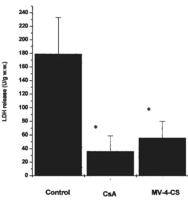
Figure 7 CsA and MV-4-CS reduces the release ofLDH in the coronary effluent induced by postischemic reperfusion. Pg. 19
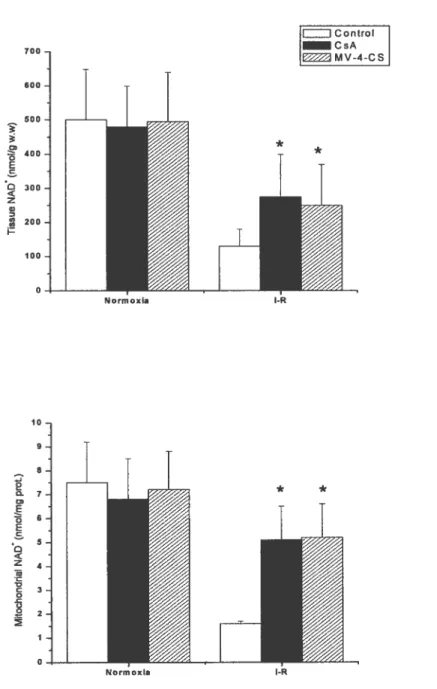
Figure $ CsA and MV-4-CS reduces the decrease in tissue and mitochondrial NAD+ contents associated with postischemic reperfusion. Pg. 20
Figure 9 [3HJ-DOG-6P degradation through non-specific dephosphorylation occurs slowly compared to its uptake, [3Hj-DOG-6P can accumulate in significant amounts in the cytosol. Pg. 9
Figure 10 Typical experimental protocol used for the assessment ofPTP opening using the [3HJ-DOG entrapment method. Pg. 26
Figure 11 Result ofDOG indexes either before 30 minutes of global ischemia or
after isohemia and 25 minutes ofreperfusion. Pg. 30
Figure 12 Effect of pywvate treatment on the mitochondrial permeability transition and lefi ventricular developed pressure (LVDP) durïng ischemia
reperfusion. Pg. 32
Figure 13 Effect ofpropofol treatment on the mitochondrial permeability transition and lefi ventricular developed pressure (LVDP) during ischemia
reperfusion. Pg. 34
figure 14 Effect of in vivo ischemia-reperfusion on arrhythmia scores. Pg. 39 Figure 15 Analysis ofthe effect of long-duration I-R (60 min ischemia-120 min
reperfusion) on the risk ofinfarction. Pg. 40
Figure 16 Postischemic recovery of lefi ventricular pressure and rates of contraction
(+dP/dt) and relaxation (-dPldt). Pg. 42
Figures in Chapter-2
figure 1 Analysis of heart rate and lefi ventricular pressure during perfusion in control and trained rat hearts. Pg. $1
Figure 2 Analysis of lactate dehydrogenase release throughout perfusion in control
and trained rat hearts. Pg. 82
Figure 3 Citrate synthase recovery in normoxia and ischemia-reperffision experiments. Pg. $3
Figure 4 PTP index and PTP index per unit citrate synthase recovered in normoxia and ischemia-reperfusion experiments. Pg. 84
Tables in Chapter-1
Table 1 Regulation ofPTP opening by an array ofdifferent physiological effectors. Pg. 10
Table 2 Summary of short-term training studies. Pg. 37
Table 3 Summary of different HSP groups and theirprimaiyfunctions. Pg. 45
Tables in Chapter-2
Table 1 Myocardial function before ischemia and at the end ofreperfùsion in hearts from control and trained rats. Pg.85
Abbreviations
ADP =adenosin di-phosphate
Mf apoptosis-inducing factor ANT = adenylate transiocator
ATP= adenine tri-phosphate
Ca2= calcium ion
CO cardiac output CS =citrate synthase CsA= cyclosporin A CVD =cardiovascular disease CypD cyclophilin-D Da dalton’s DOG deoxyglucose EndoG= endonuclease G HR= heart rate
HSE = heat shock element
11Sf= heat shock factor
HSP = heat shock proteins
11W hydraulic work
JPC= ischemic pre-conditioning
I-R=ischemia-reperfusion
LCA= left ascending coronaiy artery
LDH = lactate dehydrogenase
LVDP lefi ventricular developed pressure
MPTP mitochondrial permeability transition pore
P inorganic phosphate
PTP=permeability transition pore
RPP rate pressure product SfA= sanglifehrin A
Smac/DIABOLO=second mitochondria-derived activator of caspases-direct inhibitor of
apoptosis binding protein with low P
SP = systolic pressure
VDAC = voltage-dependent anion channel
Acknowledgements
f irst of ail I would like to thank Dr. Yan Burelle for his exceptional support as well as his encouragement throughout my studies at the Université de Montréal. I am extremely grateful for his dedication and his avallability in order to achieve my success and my time spent with him has rewarded me with an enriching experience. Mthough we have had many obstacles, his perseverance has given me the power to overcome anything within my path.
I would also like to dedicate this prolonged work to my parents, Gabriel and Solange Ciminelli, for ail their support throughout the years. Thanks to their guidance and constant motivation, not oniy to compiete my Master’ s but in every facet of my life, I have become what I am today.
I grateffihly acknowledge the rest of my family and ftiends for providing support in their own speciai ways, and of course my Melina, who has aiways been there for me, during the good times but especially during the not so good times. She has always iistened to me and has helped me in any possible way she could, which has given me the drive needed to accomplish this huge task.
1 Introduction
Cardiovascular diseases (heart disease and stroke) are the leading cause of deaffi in Canada (36 % of total mortality). Ischemic heart disease accounts for the greatest percentage of deaths at 20 %, of which half are attributable to complications of acute myocardial infarction (29). The total cost of cardiovascular disease, which is the leading cause of hospitalization for Canadian men and women, was estimated at 19.8 billions $ in 1993, of winch more than a third was atbibutable to coronary heart disease (4.8 billions $)(29). As a resuit, the socio-economical impact of ischemic heart disease is great, underscoring the importance of primary prevention to reduce both the mortality and costs associated with tins disease.
In tins regard, there exists a large body of epidemiological studies in humans to support the notion that regular exercise is associated with a reduction in the incidence of cardiovascular disease (CVD). Moreover, the survival rate of heart attack vicfims is greater in active individuals compared with sedentary ones (29) (71). These beneficial outcomes are at least partly due to systemic adaptations to regular exercise resulting in a reduction of several risk factors for the development of CVD. These adaptations include a reduction in blood pressure, an improvement of the plasma lipid profile, an amelioration of glucose tolerance and insulin sensitivity and improved weight management (29, 46, 90).
In addition to these beneficial systemic effects, a number of studies using animal models have shown that regular exercise is associated with an increased tolerance of the myocardium to ischemia-reperfusion (I-R) injury (7, 22, 40, 64, 80, 85) (10, 21, 41-43, 61, 63, 65-67, 82, 83, 91, 92) and exogenous oxidative stress (91). Indeed both short-term (22, 40-42, 63-65, 67, 80, 92)
and long-term (7, 10, 21, 43, 61, 66, $2, $3, 85, 91) aerobic training was shown to improve functional recovery and reduce tissue damage in isolated ischemic-reperfused hearts (7) (10, 43, 61, 63-67, $0, 91, 92) or in hearts submitted to lefi ascending coronary artery ligationin vivo (21, 22, 40-42, $3, $5).
The cellular mechanisms underlying this improvement in myocardial tolerance to I-R are flot yet well established. Studies performed over the recent years have focused on training-induced adaptations in the expression of heat shock proteins (HSP’s) (61, 64, $0) (22, 67), antioxidant defence systems (21, 41, 66, $2), sarcolemmal Ca2 handiing (7), myocardial energy metabolism (10) and endothelial function (95). However, there is currently no clear consensus on the respective role, mechanisms of action and relative importance of each of these components in protecting the trained heart against I-R injury.
On the other hand, it is well established that mitochondria play a key role in ceil death following I-R in several tissues including the heart. Indeed, failure to produce ATP as a resuit of mitochondrial dysfunction is believed to mark the transition toward tissue necrosis and contractile failure during early reperfusion (23, 26, 78). In addition, mitochondria are known to play a key roTe in signalling apoptotic cell death through the release of several pro-apoptotic proteins that are normally confined to the mitochondrial matrix or inter-membrane space (26, 28, 32, 39, 50). Although necrosis is the main form of celi death encountered following I-R, apoptosis has been observed in celis located in periphery of the necrotic zone and which have been less severely affected by I-R (15, 39).
A number of experimental evidence accumulated over the recent years indicate that the opening of the permeability transition pore (PTP), a high conductance non-specific channel spanning the double membrane system of mitochondria is involved in triggering both forms of celi death during reperfusion of the ischemic heart, particularly necrosis (14, 15, 39, 98). Direct pharmacological inhibition of the PTP using cyclosporin A (CsA) and its analogs were shown to improve functional recovery and reduce tissue damage following I-R. Moreover, cardio protective strategies such as ischemic preconditioning (55), administration of pyruvate (60) and the anti-oxidant anaesthetic propofol (56) were shown to mediate their beneficial effect at least partly through a reduction of PTP opening providing further support for the important role of this phenomenon.
Despite these evidences, the effect of exercise training on many aspects of mitochondrial function, and particularly on the regulation and behavior of the MPTP lias neyer been addressed in the context of explaining the cardio-protective effect against I-R. Therefore, the goals of the present Master’s thesis were to implement a new technique that allows to quantify PTP opening
in situ in the isolated perfused heart and to apply this methodology to determine whether short term aerobic training reduces the occurrence of PTP opening during reperfusion following an ischemic episode. The mitochondrial entrapment technique developed by Halestrap and colleagues was chosen based on the existing literature in ifie pcrfused heart. As for the training mode! used, it was justified by several studies reporting that short-term training induces a robust cardio-protection characterized by an increased functional recovery and a reduction in tissue damage.
The literature review included in this ffiesis will be divided in three sections. The first section provides an overview on the mitochondrial PTP and its involvement in ischemia-reperfusion injury in the heart. The second section focuses on the methodologies available to assess PTP opening in situ in isolated perfused hearts. finaily, in the third section, the existing literature on the cardio-protective effect of short-term training in rodent models and the possible underlying mechanisms are reviewed.
2 The mitochondrïal permeability transition pore and ïts
role in ïschemïa-reperfusïon injury in the heart
2.1 In vitro studies on the mitochondrïal permeability transition
2.1.1 Conseguences of PTP opening on mitochondria in vitro
The mitochondrial permeability transition was initially described in isolated mitochondria in order to explain a sudden increase of the inner membrane permeability to solutes in the presence of a high calcium concentration ([Ca2j) (97). Although initially thought to be due to unspecific membrane damage it is now widely accepted that this phenomenon is actually caused by the opening of ifie PTP, a non-specific high conductance channel spanning the double membrane system ofthe mitochondria.
Opening of the PTP causes an immediate collapse of the proton electro-chemicai gradient, massive ATP hydrolysis through the reversai of the FoFlATPase and equilibration of solutes with a molecular weight of less than 1500 Da (97). At least in vitro, this phenomenon induces high amplitude swelling of the mitochondrial matrix ultimateiy leading to the rupture of the
outer-membrane. PTP opening is also associated with the release of several pro-apoptotic proteins usually confined to the inner membrane space including the mobile electron carrier cytrochome e, apoptosis-inducing factor 1 (MF), Second mitochondria-derived activator of caspases — direct inhibitor of apoptosis binding protein with low P1 (SmacIDIABLO),
endonuclease G (EndoG), the serine-protease OMI/HtrA2, and possibly some pro-caspases (26, 31, 32).
These proteins act at different sites and through different mechanisms to initiate a regulated cascade of cellular dismantiement. Cytochrome c binds to APAF-1 and allows formation of the apoptosome, leading to cleavage and activation of pro-caspase-9 thus ffiggering the proteolytic caspase cascade (69). AIF (70) and endonuclease G (49) migrate to the nucleus where they cause chromatin condensation as well as large scale nucleosomal DNA fragmentation. Smac/DIABLO release in the cytosol potentiates some forms of apoptosis by neutralizing one or more members of the inhibitory apoptosis proteins (IAP) (1). Finally OMI/HfrA2, through its interactions with )UAP, is able to potentiate caspase activity.
2.1.2 Molecular identitv of the PTP
The actual molecular nature of the PTP is flot well established and is a subject of current debate. However it is generally agreed that the PTP is formed by ifie assembly of a supramolecular complex that spans the double membrane system of mitochondria (97, 98). In addition, the proteins involved in this process usually carry specific physiological roles within mitochondria
and their involvement in PTP formation requires the presence of adverse conditions (15, 39, 48, 62).
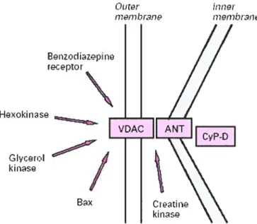
The most popular and documented hypothesis regarding the pore composition is that it is formed by three core components (figure 1) (15): the adenylate transiocator (ANT) of ifie inner membrane, the voltage-dependent anion channel (VDAC) of the outer membrane, and the matrix enzyme cyclophilin-D (CypD). Under normal conditions, these proteins assemble at contact sites between the two membranes where they allow ATP and ADP exchange between the matrix (15). Under adverse conditions such as Ca2 loading and excessive levels of oxidative stress, these proteins would transform into large conductance non-specific pores. This phenomenon would be exacerbated by the binding of CypD to ANT. It should be noted that several proteins are believed to interact with ANT and VDAC to promote or inbibit PTP opening including several kinases (hexokinase, gÏycerol kinase, mitochondrial creatine kinase), and members of the Bd-2 family of proteins (bid, bax and bd-2, bdlxL) (15).
However, an increasing amount of evidence suggests that ANT, VDAC and CypD are flot obligatory components of the PTP (48, 62). Indeed, altemate models suggest that the PTP could be formed by aggregates of misfolded proteins within the mitochondrial membranes that would accumulate as a resuit of damage (48, 62). According to this hypothesis, the PTP could thus be formed by a variety of mitochondrial proteins and flot exclusively of ANT, VDAC and CypD. One of the implications of this model is thus that the composition of the PTP could vary according to the tissue studied and the triggering conditions.
H€xokinis€
tnz’:’ciizI)IFii1
ptoi
Figure 1: Most popular and documented hypothesis regarding the pore composition. Taken from Crompton, M 1999. The adenylate transiocator (ANT) of the inner membrane, the voltage-dependent anion channel (VDAC) of the outer membrane, and the matrix enzyme cyclophilin-D (CypD) Other proteins associate with the complex as indicated.
iiut: I1?tfl)L!.fl Iv’inI kïns Ci iatin kins
2.1.3 Regulatorv properties of the PTP
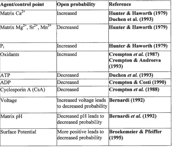
The regulation of PTP opening is mediated by an array of different physiological effectors. Table 1 gives a summary of the factors that play a role in either activation or inhibition of the PTP. Calcium accumulation in ifie matrix appears to be an absolute requirement for PTP opemng (14, 15, 98). In addition, a number of co-activators or antagonists further participate in pore regulation. Cations such as H, Mg2, Sr, and Mn2 can compete with Ca2 for binding on the PTP and thus act as inhibitors (3, 14). High levels of adenine nucleotides such as ATP and ADP limit PTP opening, possibly by binding to the ANT (3, 14). In contrast, high levels of inorganic phosphate significantly increase the susceptibility to Ca2tinduced PTP opening. A reduction in membrane potential will also favour perrneability transition since the PTP behaves as a voltage gated channel (14)). It is also well established that oxidation of the pyridine nucleotide pool (NADH and NADPII) favours PTP opening. finally PTP opening can be inhibited by the immuno-suppressant drug, cyclosporin-A (CsA) and its analogs (24, 35, 55) as well as by the new compound sanglifehrin A (13, 55) ifiat prevent binding ofthe cyclophilin-D to other putative PTP components.
2.2 Effect
of
ischemia-reperfusionon
the PTP2.2.1 Overview of the metabolic and ionîc consepuences of ischemia- reperfusion in the heart
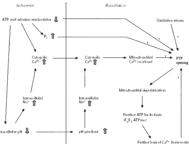
In the heart submitted to ischemia, lack of oxygen and circulating substrates rapidly leads to the abolition of oxidative phosphorylation, a rapid and important decrease in tissue ATP content and accumulation ofADP, P and
W
(37)Table 1: Regulation ofPTP opening by an array of different physiological effectors Agent/control point Open probability Reference
Matrix Ca2 Increased Hunter & Haworth (1979) Duchen et al. (1993) Matrix Mg2, Sr, Mn2 Decreased Hunter & Haworth (1979)
P1 Increased Hunter & Haworth (1979)
Oxidants Increased Crompton et al. (1987) Crompton & Andreeva (1993)
ATP Decreased Duchen et al. (1993)
ADP Decreased Crompton & Costi (1990)
Cyclosporin A (CsA) Decreased Crompton et aÏ. (1988) Voltage Increased voltage leads Bernardi (1992)
to decreased probability
Matrix pH Decreased pH leads to Bernardiet aL (1992) decreased probability
Surface Potential More positive leads to Broekemeier & Pfeiffer decreased probability (1995)
Accumulation of ADP will increase its conversion to IMP, adenosine, inosine and xanthine thus leading to a depletion of tissue adenylates. Lack of ATP will bring contraction to a hait ami cause a severe disturbance in ionic homeostasis, which is usually maintained by the action of the Nat’K ATPase pumps. During ischemia, Na and Ca2 will thus accumulate in the cytosol (37).
Re-introduction ofoxygen and substmtes upon reperfiision ofthe ischemic region aÏlows a partial or total recovery of electron flow through the respiratory chain and membrane potential (15). In presence of high cytosolic Ca2, this will rapidly lead to an important accumulation of Ca2 in the mitochondrial matrix (15). In addition, because ischemia causes damage to respiratory chain enzymes, electron leaks through these damaged complexes leads to an increase in the production ofreactive oxygen species (ROS) (15).
Therefore, figure 2 (15) demonstrates that most ofthe conditions required to ffigger PTP in vitro in isolated mitochondria prevail in cardiac celis following I-R. Based on tins analysis, Crompton (14, 15), Gnffiths (34, 35) and Halestrap (39) hypothesized that PTP opening occurred in the heart during reperfusion and was a significant contributor to tissue damage and contractile dysfimction.
2.2.2 Cardio-protective effects of pharmacoloqical PTP inhibitors
In support to tins hypothesis, several studies have shown that administration of the PTP inhibitor CsA in perfused hearts or isolated cardiomyocytes protected against reperfusion injury. In isolated perfused hearts, CsA administration was shown to resuits in an increased recovery of left
IiIir ATE h:IrIis
£FFIIIIii1 iii,I.:IiiIs !IIII t[’s
[•iii, Ii’iiIiiiI
L i 1 ‘VI I»ItI opening
7
Iii I Ii’: :III_i1i r F’Ja
Ï
VI t’1ii:I1:I1IiiII :1 1,111 iIî
t.I:It
I—I pI•iIII ilIil I;i I II. iIi p11
N
111i11].Ei I,js• f h:I 1i.StSiS
figure 2: Involvement ofifie PTP in ischemia-reperfusion —induced ce!! death. Taken from Crompton, M 1999. Lack of oxygen and circu!ating substrate rapid!y leads to the abolition oxidative phosphory!ation and a rapid ATP content and accumulation of P and H. Upon reperfiision, there is an excessive Ca2 uptake, which, coupled with oxidative stress and prevai!ing high P and low ATP can provoke PI? opening.
ventricular developed pressure (34, 35) (figure 3), a lower end diastolic pressure (34, 35) (figure 3), a reduction in infarct size (44) (figure 4), an improved recovery of adenylates homeostasis (35), a reduction in cytochrome e release and activation of apoptosis (6) (figure 5) and a better preservation of respiratory function in mitochondria isolated following I-R (6). Similarly, in isolated cardiomyocytes, CsA administration during simulated ischemia-reperfusion was shown to improve recovery of normal morphology and contractile activity (36) (25, 77).
While the effect of CsA in isolated mitochondria can unequivocally be attributed to PTP inhibition, its cardio-protective effect when administrated in intact hearts or cardiomyocytes is more difficult to interpret. Indeed, in addition to its effect on the PTP, CsA is a potent inhibitor of calcineurin, a Ca2/calmodulin-dependent protein phosphatase, which has been involved in celi death in the heart (86) (24).
However, studies performed with pharmacological agents that inhibit calcineurin but not ifie PTP, or solely inhibit the PTP without affecting calcineurin provided resuits that are compatible with a role of pore opening in I-R injury. Administration offK-506, a calcineurin inhibitor that does flot interact with the PTP (33) was shown to have limited effects on infarct size (44), mitochondrial cytochrome e release, caspase activation and DNA fragmentation in hearts submitted to I-R (6) (Figure 5).
In contrast, administration of the CsA analog N-methyl-valine-CsA (24) and the new compound sangÏifehrin A (13), which potently inhibit pore opening but have no effect on calcineurin, were shown to improve the recovery of contractile function (13) and reduce tissue damage as measured
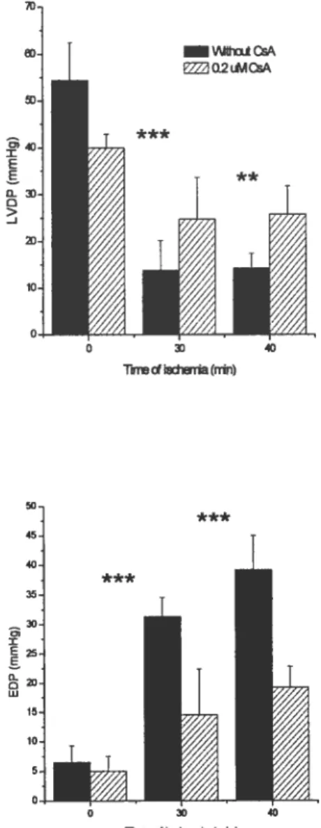
Figure 3: Resuits of left ventricular developed pressure (LVDP) and end diastolic pressure (EDP) following 15 minutes of reperfusion after 30 or 40 minutes of ischemia with or without CsA. Adapted from Griffiths, 1993 #53. ** P<0.02, P<0.01 between reperfused and control
hearts. —Woi cs E2 02Li.l cDsA ** *** 0 D > 1 o Twvecia (nin) —30. 35. 35. w 15-flrrw ofisd,enia f11m)
611 — Ç() 30 •- 11’ !LI -r
Figure 4: Reduction in the infarct size with the use of CsA. Taken from Hausenloy et al. 2002. Significantly different from control (*P<0.0001).
Iu
(j
s w e
ï
Figure5: Mitochondrial cytochrome econtent afier a period of ischemia. CsA protects heart mitochondria from loss ofcytochrome cduring ischemia but flot fK-506. Taken from Borutaite et al. 2003. , statistically significant effect of ischemia (P <0.01), if compared to control. #,
statistically significant effect of CsA (P <0.05), if compared to treatment wiffiout CsA in the same group.
No additions CsA FK-506
by LDH release in the coronary effluent (13, 24) (Figure 6 & 7). Taken together tins pharmacological evidence thus indicates that PTP opening occurs during reperfiision of the ischemic heart and that its inhibition resuits in a significant improvement in contractile recovery and a reduction in tissue damage.
2.2.3 Assessment of PTP openingin situin the ischemic-reperfused heart
One limitation of the pharmacological approaches used in the above mentioned studies is that the role of pore opening in ischemic injury is only inferred and PTP opening is not directly quantified. Moreover without a direct index of MTP opening, it is not possible to determine whether other cardio-protective strategies could mediate their effects through a reduction in the occurrence of PTP opening. In order to circumvent these limitations, two research groups have developed methods to estimate the extent of PTP opening that are applicable to studies in the intact heart: the measurement of mitochondrial NAD release and the mitochondrial [3H]-deoxygÏucose ([3HJ-DOG) entrapment technique.
2.2.3.1 Mitochondrial NAD release
NAD is a soluble electron carrier present at high concentration in the mitochondrial matrix. Given its molecular weight, NAD is readily released from the mitochondria upon opening of the PTP (24) (Figure 8). Once permeability transition has occurred, a significant amount ofNAD is
a)
— 100 w z > I-E ni w Q .c -u — 3 3 o. ID o.b)
60 50 G, o I I IFigure6: $fA and CsA protect the ischemic rat heart from reperftision injury. Taken from Clarke et al. 2002. In panel a, greater functional recovery ofthe SfA- and CsA-treated hearts after a 30-min reperfiision is reflected in higher values for the LVDP and lower values for the LVEDP. Values ofthe LVDP after reperfusion are presented as a percentage ofthe pre-ischemic value and were significantly greater (p <0.001) with either SfA or CsA treatment, whereas values for the LVEDP were significantly lower (p< 0.001). In panel b, the release oflactate
dehydrogenase (LDH) into the perfusate from the same hearts used in panel a was measured as an indicator ofnecrotic celi death (,p <0.05 for CsA- or $fA-treated hearts versus controls).
Con SfA CsA Con SfA CsA
30 40 80 85 90 95 100 105 110
240 220. 200 180 160 140 120 100 80 60 40 20 0. o) D o w o X Q -J * *
Ï
[
Control CsA MV-4-CSfigure 7: CsA and MV-4-CS reduces the release ofLDH in the coronary effluent induced by postischemic reperfusion. Adapted from Di Lisa et al. 2001. , P <0.01 statistical difference between freated and control hearts.
D, 400 E C 300 z w - 200 I— 100 LZI Control CsA MV-4-CS 700 600 500
I
0- —I
Normoxia 10 9 e 7 6 5 4 3 2 * *j
2 D D, E o E C n z w -C C o -cFigure 8: CsA and MV-4-C$ reduces the decrease in tissue and mitochondrial NAD+ contents associated with postischemic reperfusion. Taken from Di Lisa et al. 2001. *, P <0.01 statistical
difference between treated and control hearts.
released in the outer mitochondrial membrane (24). The remaining NAD that escapes degradation enters the cytosol, and if cellular integrity is altered, will be released in the coronary circulation (24). Only one study lias made use of this technique to further document the role of PTP opening in ischemia degraded by NAD glycohydrolase, an enzyme presumably located in the inter-membrane space -reperfusion damage (24). Di Lisa et al. (24) submitted isolated Langendorff-perfused hearts to a period of global ischemia of 30, 60 or 90 min followed by 30 minutes of reperfusion. NAD release in the coronary circulation was measured at regular intervals during reperfusion and at the end of the experiments hearts were homogenized and processed for isolation of mitochondria. NAD content was measured fluorimetrically in the whole homogenate and the mitochondrial fraction using an alcohol dehydrogenase assay. These values were compared to that measured in ftesffly isolated non-ischemic hearts. In hearts reperfused following 90 minutes of ischemia, mitochondrial and whole tissue JJ contents respectively decreased by 85 and 70 % compared to values obtained in non-ischemic hearts (Figure 8). This was accompanied by a significant leakage of NAD in the coronary effluent. In hearts perfused in the presence of the PTP inhibitors CsA and nmethyl-valine cyclosporin, these phenomena were aftenuated (figure 8). Moreover, a significant correlation between NAD release in the coronary effluent and the extent of tissue damage (as measured by the release of LDH) was observed. Based on these evidences, Di Eisa et al. (24) concluded that PTP opening was a causal event in the death of myocytes following I-R.
On the other hand, DiLisa et al (24) did flot report the results obtained in hearts submitted to shorter periods of ischemia (i.e. 30 mm) that are more frequently used in the literature. While the authors did not discuss this issue, preliminary work performed in our laboratory suggests that fuis
might be due to the fact that the method failed to detect significant levels of PTP opening under these conditions. Indeed, we observed that measurement of NAD using fluorescence methods yield variable resuits due to a low signal/noise ratio. Consequently, in hearts submitted to 30 min ischemia and 40 min of reperfusion, we consistently failed to detect significant and reproducible changes in whole tissue and mitochondrial NAD content. In addition, given the relatively low amount of NAD present in cardiac celis and because 30 min of ischemia is insufficient to cause large scale necrosis, the amount of NAD released in the coronary effluent was well below detection levels.
2.2.3.2 Mïtochondrial 13H1-deoxyglucose entrapment
Currently, one of the best strategies to directly assess PTP opening relies on the use of fluorescence microscopy to visualize diffusion within the mitochondrial matrix of exogenous fluorescent probes previously loaded in the cytosol. However, while this approach has provided valuable information about the role of the PTP in ce!! death, its use is limited to isolated ce!! models. for this reason, the group of Andrew Halestrap at the University of Bristol have made use of the same general principle to develop a method that can be applied to Langendorff perfused hearts and which relies on measurements of the incorporation in the mitochondrial compartment of an exogenous radioactive probe previously loaded in cardiomyocytes.
2.2.3.3 Princïple
In order to select the appropriate probe to accurately track PTP opening in this setting, several criteria had to be met.
Indeed, the probe:
1. Has to enter cardiomyocytes in relatively large amounts using existing sarcolemmal transporters,
2. has to display a relatively low level of toxicity to the celi, 3. has to exert a minimal effect on cellular metabolism,
4. has to remain relatively stable once within the cell i.e. its degradation by metabolic pathway has to be minimal within the time frame ofthe study, and has
5. to enter the mitochondrial compartment only when ffie PTP opens and to stay within mitochondria thereafler.
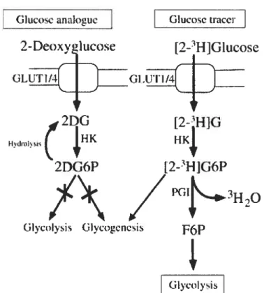
Based on these criteria, [3H1-DOG was selected as the probe of choice. Indeed, as a non metabolizable glucose analog, [3HJ-DOG rapidly enters cardiomyocytes through glucose transporters and is phosphorylated in [3HJ-DOG-6P in a step catalyzed by hexokinase. Because [3HJ-DOG-6P degradation through non-specific dephosphorylation occurs slowly compared to its uptake (54), [3HJ-DOG-6P can accumulate in significant amounts in the cytosol (Figure 9). This is a key factor in order to obtain a measure of PTP opening that offers a reasonable signal/noise ratio. On the other hand, because [3HJ-DOG phosphorylation results in Pi trapping, accumulation of great amounts of [311]-DOG-6P can resuit in a rapid depletion of myocardial ATP and PCr stores. However, this unwanted effect could be avoided if the [3H1-DOG concentration used is maintained below 2 mM (52, 54). The concentration that has typically been used for the
measurement of mitochondrial [3HJ-DOG entrapment is of 0.5 mM (34, 37, 54-56, 60), well below the concentration that will compromise energy homeostasis (52, 54).
Glucoseanalogue
R
Glycnlysis Glygenesis Gluco’e tracer[2-3HlGlucosc
F6Pk
[
GlycolysisFigure9: [3HJ-DOG-6P degradation through non-specific dephosphorylation occurs slowly compared to its uptake, [3Hj-DOG-6P can accumulate in significant amounts in the cytosol.
Takenfrom Hopkins et al. 2004.
2DG
[2-H]G [jJj,, r kh1K HKk2DGÔP
[2-I1IG6P
///
‘‘1’ø3H2O
Regarding the selectivity of [3H]-DOG-6P as probes for PTP opening, studies have shown that mitochondrial membranes have a low permeability for sugars in general. Indeed, when mitochondria are isolated in presence of ‘4C mannitol, only a small fraction of ‘4C becomes permanently associated with mitochondrial membranes. This amount represents 10-15 % of that expected for ‘4C-mannitol uptake and complete equilibration (94). On the other hand, PTP opening allows equilibration of solutes with a molecular weight of less than 1500 Da across mitochondrial membranes. Given this property, one ofthe assays commonly used to monitor PTP opening in vitro is to incubate mitochondria in a sucrose-based medium and to measure mitochondrial swelling secondary to the diffusion of sucrose and F120 in the mitochondrial matrix. Therefore, these properties insure that PTP opening in vivo will rapidly increase [3H]-DOG-6P entrapment in mitochondria and that [3HJ-[3H]-DOG-6P incorporation will be comparatively much less important in the absence ofPTP opening (figure 10).
Another key aspect regarding the accuracy of the method is that [311J-DOG-6P has to remain within mitochondria once PTP opening has occurred. In addition, accidentai PTP opening during the mitochondrial isolation procedure must be minimal. In order to limit the potential problems associated with these phenomena, Halestrap (34) proposed to homogenize heart tissues and isolate mitochondria using a rapid method and to supplement the buffers with a high concentration of EGTA (2 mM). Indeed, studies on isolated mitochondria have shown that once PTP opening has occurred, addition of EGTA to chelate Ca2 is able to close open pores and allow mitochondrial recovery (16).
26
[3H]..DOG (2deoxyglucose)
Load hean ceils vïth 2-deoxyglucose
DOG
‘—‘*DOG•6P
(DOG) ‘vhich is trapped in the cytosol
as DOG-6-P
DOG-8.P only enters mitochondna ïf pote opens
Pote closed
DOG.C•P
DOG-C•P
Pore open
Figure 10: Typical experimental protocol used for the assessment ofPTP opening using the [3HJ-DOG entrapment method. Taken from Halestrap et al. 2004.
Isolate mochonda in EGTA buffet. Open potes rapidly
close and trap DOG+ DOC-6-P in matrix.
2.2.4 Experimental protocol:
Figure 10 presents the typical experimental protocol used for the assessment of PTP opemng using the [3H]-DOG method. In practical terms, hearts are placed on the perfusion apparatus, instrumented for measurement of hemodynamic parameters and perfused in the non-recirculating mode with a normal Krebs-Henseleit (K-H) buffer. Following a period of stabilization, hearts are perfused for 20 min with the same buffer supplemented with 0.5 mM of [3H]-DOG (0.1 .tCi/mi). In order to maximize uptake of the tracer, the perfusion is performed in the re-circulating mode. Following this loading period, perfusion is switched back to the non-circulating mode with the [3H]-DOG-free K-H buffer in order to washout extracellular [3HJ-DOG. Ischemia is then initiated and following reperfiision, hearts are rapidly processed for the isolation of mitochondria and measurement of mitochondrial [3HJ-DOG entrapment.
2.2.4.1 Calculatîon of the (3H]-DOG index:
In order to achieve an accurate measurement of PTP opening, calculation of mitochondrial [3HJ-DOG entrapment has to take into account several confounding factors.
The first factor is that total tissue uptake of [3HJ-DOG through GLUT-mediated transport can
vary between experiments, thus resulting in various levels of cytosolic [3H]-DOG-6P accumulation. Similarly, depending on the severity of the damage induced by I-R, variable amounts of [3HJ-DOG-6P could leak out of cardiomyocytes that have loss membrane integrity.
failure to take this into account can lead to variations in the amount of mitochondrial [3HJ-DOG incorporation that are flot related do differences in PTP opening.
The second factor is that the amount of mitochondria recovered following isolation varies between days and according to the severity of I-R injury. Therefore, the total amount of [3HJ-DOG recovered will be influenced by the amount of mitochondria isolated. for these reasons, the PTP opening index (termed DOG index) is calculated as follows:
DOG index= iO x mito [3H1-DOG-6P per unit CS tissue [3HJ-DOG-6P per g wet weight
In this calculation, mitochondrial [3HJ-DOG-6P measured in d.p.m. is expressed per unit of citrate synthase. Ibis allows normalization for the amount of mitochondria recovered following isolation. In addition, in order to account for variations in the amount of cytosolic [3H]-DOG-6P present, mitochondrial [3H]-DOG-6P entrapment is expressed relative to tissue [3H]-DOG-6P per g wet weight. Therefore, by expressing the DOG index as a ratio, the measure is not influenced by loss ofcytosolic [3H]-DOG-6P.
On the other hand, one of the limits of the method is that PTP opening can only be measured in mitochondria that retained sufficient integrity to survive the isolation procedure (55) (37). Indeed, as discussed in our study, I-R induces a significant loss in the recovery of intact mitochondria following isolation and the DOG index fails to consider PTP opening that occurred in mitochondria that were lost. In order to take this phenomenon into account, recent studies have
thus normalized the DOG index by the total amount of citrate synthase recovered in the mitochondrial fraction by gram ofheart (55,37).
2.2.4.2 Role of the PTP inhibition in cardlo-protectïon:
This section will provide an overview of the resuits published in studies that have used the mitochondrial [3Hj-DOG method to investigate various cardio-protective strategies.
2.2.4.2.1 Cyclosporin A and Sanglifehrin A:
The effect of administering CsA and SfA, two direct inhibitors of the PTP, to perfiised hearts submitted to I-R has been investigated in two studies (34,55). Javadov et al. (55) have shown that administration of CsA during ischemia and early reperfiision was associated with a 20% reduction in the DOG index (figure 11 Panel A). As pointed out by the authors (37,34,55), this relatively small effect of CsA on PTP opening is somewhat surprising given the strong potency of CsA at inhibiting pore opening in isolated mitochondria.
However, as mentioned above (p 23), one limitation of the DOG index is that it fails to consider PTP opening in mitochondria that were lost during isolation due to excessive damage, thus underestimating true levels of pore opening. Because CsA allows to significantly affenuate the
160 12 80 10 8 60 D C/) o z X ‘ 40 X 0 o (3 0 20 g o
B
40 as 6) 30 25 t 20 • 1.8 t 0 o 1.0 D 05 00Non-Ischemic
I-R
Non-IschemicI-R
figure 11: Resuit of DOG indexes either before 30 minutes of global ischemia or after ischemia and 25 minutes ofreperfusion. Adapted from Javadov et al. 2003. TOP 2 graphs, statistical significance of SfA or CsA versus control hearts (*P<OE05;**P<0.01). BOTTOM 2 graphs, statistical sigriificance ofIPC versus control hearts (*P<0.05;**P<zO.01). IPC, ischemic pre conditioning; CsA, cyclosporin A; SfA, sanglifebrin A; CS, citrate synthase.
6
4
2
I-R
Non-scheniic Control SfA CA
I-R
A
I
loss of intact mitochondria observed in response to I-R, this underestimation is aftenuated. This phenomenon thus contributes to explain the apparently small effect of this drug on PTP opening. When this difference is taken into account by normalizing the DOG index values by the amount of citrate synthase recovered, the inhibitoiy effect of CsA on mitochondrial [3HJ-DOG entrapment is more compatible with the known potency of this drug at inhibiting pore opening (figure 11 panel B). Javadov et al. (55) have also shown that Sanglifehrin A (Sfa), a new agents that inhibits the PTP by binding to cyclophilin D, also reduced PTP opening in ischemic reperfused hearts (Figure 11).
2.2.4.2.2 Pyruvate and Propofol
Pyruvate supplementation was repeatedly shown to improve the functional recovery of the heart following ischemia-reperfusion (9, 11) (20). Kerr et al. (60) investigated wheffier the protective effect of pyruvate could be mediated by an attenuation of PTP opening. These authors reported that administration of 10 mM pyruvate during ischemia and reperfusion reduced mitochondrial [3HJ-DOG entrapment by 38% (Figure 12) and significantly retarded time to ischemic contracture, improved the recovery of lefi ventricular developed pressure (Figure 12), and lowered end diastolic pressure. However, in this study no aftempt was made to correct for differences in the recovery of intact mitochondria.
Three main hypotheses have been suggested to explain how pyruvate could inhibit PTP opening during reperfusion. The first hypothesis involves the fact that pyruvate allows maintenance of a more acidic intracellular pH during reperfusion. The second hypothesis is that pyruvate, by virtue
45
Figure 12: Effect of pyruvate treatment on the mitochondrial permeability transition and lefi ventricular developed pressure (LVDP) during ischemia reperfiision. Adapted from Kerr et al.
1999. Significant differences between control and pymvate-treated hearts (*P<O.05)
50 40’ 35 — 30 E 25 20’ > 15 10. 5.
C ontrol Pyruvate Controt P yravate
of its antioxidant properties (4, 20) could scavenge free radicals produced during reperfusion. finally, ifie third hypothesis is that pyruvate oxidation in the mitochondria could favour a better recovery of mitochondrial membrane potential (AP). Indeed, as described in section 2.1.3, an acidic pH, low levels of oxidative stress and an increase in A’P ail favour the maintenance of the PTP in the close conformation.
Similarly, the anaesthetic agent propofol (DIPRIVAN©, Zeneca Pharma), which is known for its capacity to scavenge free radicals (27, 73), was shown to reduce mitochondrial [311]-DOG entrapment by 26% and to improve functional recovery following I-R (figure 13) (56). This inhibition of PTP opening in situ was at least partly due to a direct effect of mitochondna rather than on infracellular modulators. Indeed, swelling assays performed on mitochondria isolated from non-ischemic hearts treated with propofol showed that significantly more Ca2 was required to open the PTP compared to that measured in mitochondria from control hearts.
Taken together, the studies on the cardio-protective effect of pyruvate and propofol thus suggest that several pharmacological agents that are flot directly targeting the PTP structure could exert their protective effects at least partly by affecting modulators of pore opening thus maldng the PTP a central target for cardio-protective strategies.
2.2.4.2.3 Ischemic pre-conditioning
Figure 13: Effect ofpropofol treatment on the mitochondrial permeability transition and lefi ventricular developed pressure (LVDP) during ischemia reperfusion. Adapted from Javadov et al. 2000. Significant differences between control and propofol-treated hearts (**P<0.025)
endogenous mechanisms that protect the heart against subsequent periods of prolonged ischemia. Typically, two episodes of brief ischemia (5 mm) intercalated with 5 min of reperfusion significantly attenuates tissue damage and improves the recovery of contractile function during reperfusion following periods of ischemia ranging between 20 to 30 min (44). Using ffie [3HJ-DOG technique, Javadov et al. (55) reported that IPC was associated with a significant reduction in the DOG index and an increase in the recoveiy of intact mitochondria following I-R (Figure
12). This inhibition of PTP opening by PC was also reported by other groups in perfiised hearts
(2), isolated cardiomyocytes (45) and mitochondria (2) using other methodologies.
The mechanisms by which IPC resuits in the inhibition of PTP opening are not yet defined and a detailed discussion on this issue is beyond the scope of this thesis. Inhibition of PTP opening could occur through an indirect mechanism by beneficially altering the intracellular milieu e.g. by attenuating cellular Ca2 overload (72, 87), and ROS production (75, 79). In addition PC could directly act at the level of mitochondria by beneficially altering factors that regulate PTP opening such as Ca2 loading, ROS production, membrane potential, matrix p11 and matrix adenylates (2).
finally, opening of mitochondrial KATp channels, which are involved in the IPC signalling
cascade, could underlie some ofthese effects (53, 72).
3 Cardio-protection induced by short-term training
A number of studies using rodent models have provided strong evidence indicating that hearts from trained animals arebetterprotected against contractile dysfunction and tissue injury induced by periods of ischemia ranging between 15 and40 min followed by 15-30 min ofreperfusion (22, 40-42, 63-65, 67, 80, 92). While this beneficial effect has been demonstrated using training
programs of various durations ranging from a few days to several weeks, the present review will focus on short-term training. In a first section, the effect ofthis type oftrainjng on the recovery of contractile function and tissue damage measured using various experimental models is presented. finally, in a second section, the main hypotheses concerning the mechanisms involved in the cardio-protective effect of short-term training are analyzed with an emphasis on how these could 5e linked to the regulation of the PTP.
3.1 AnimaI models and training paradigms
Table 2 provides an overview offfie type of animais, training paradigms and experimental models used in the eight studies that have investigated the effect of short-tenu training on myocardial protection against I-R (40-42, 63-65, 67, 92). In general, ail studies used treadmili nmning as the training modality. Most protocols involved 1 to 5 consecutive days of running for a duration of 60 min at a speed 30 m/min with a siope of 0%. The only two exceptions are the studies by Taylor et al. (92) and Lennon et al. (64) in which longer nmning periods (100 mm) and/or lower speeds were used (18-20 m!min). Experiments were generally performed 24 h following the last bout of exercise, except in the study by Lennon et al. (63) in which experiments were performed up to 1$ days aller training cessation. Both maies and females have been studied and the two genders appear to benefit from cardio-protection in response to this type of training. It should however be noted that one controversial study reported that a single bout of exercise was sufficient to induce cardio-protection against I-R in males but flot in females ($0), however this
Table 2: Summary of short-term training studies
Authors Animais Training model Exp. mode]
Hamilton Female Sprague-dawley 3-5d, 60 mm, 30 m./min. 0% In vivo occi. et al. 2001 (SD)-4 months lwk habituation at these sefting (5 24 h post
min+10-15 min increase daily) exercise
Hamilton female SD- 3 months 5d, 60 mm, 30 m./min, 0% In vivo occi. et al. 2003 lwk habituation at these sefting (10 24 h post
min +10-15 min increase daily) exercise Hamilton Male SD- 4 months, 300- 3d 60 mm, 30 m./min, 0% In vivo occi. et al. 2004 350g lwk habituation at these setting + 2 24 h post
d of rest exercise
Lennon et Male SD- 4 months, 300- 3d 60 mm, 30 m./min, 0% WH al. (2004) 350g lwk habituation at these sefting+ 2 24 h post
U of rest exercise
Lennon et Male SD- 6 months, 430- 3d 60 mm, 30 mimin or 18 mimin, WH
al. (2004a) 470g 0% 24 h post
lwk habituation at these seffing+ 2 exercise d ofrest
Lennon et Male SD- 4 months, 370- 3d 60 mi 30mlmin, 0% WH al. (2004) 400g 5 d habituation (begin lOmin at
30mlmin, with daily increases of 10 min until 50 minlday were achieved
Taylor et Female $D- 5-7 months 1 or 3d, 1 O0min, 20m./min. 6% WH
al. (1999) no habituation 24 h post
exercise Locke et al Male SD- 250-300g Male $D rats (250-300 g) Langendorff
(1995) 1 or 3 d 60 mm, 30 m.min-1, 0% 24 h post
study had received many comments and critiques (96). finally, the studies available can be distinguished according to the experimental model used to document cardio-protection. The two models that have been most frequently used are the ligation of the lefi ascending coronary artery (LCA) in vivo (40-42) and the isolated working heart preparation (63-65, 92). As for the
Langendorff perfusion, it has only used once by Locke et al. (67).
3.2 Effect of short-term training on myocardial function after ischemia and on markers of
tissue
damageStudies using in vivo LCA occlusion, a model of regional ischemia-reperfusion, have shown that short-term training resuits in a befter preservation of LVDP during ischemia and reperfusion, a reduction in the occurrence of arrhythmia (figure 14) during reperfusion and a reduction in the % ofrisk area infarcted (figure 15) (40) (41) (42).
A significant level of cardio-protection was also obtained with the isolated working heart preparation (63-65, 92). Tins mode! allows to study cardiac performance without the confounding effects of other organs systems, the systemic circulation, and a host of peripheral complications (88). Another advantage over other isolated heart models such as the Langendorff preparation is that it permits cardiac pump fimction to be measured while controlling cardiac filling pressure and aflerload. Under these conditions, short-term training was found to increase the recovery of cardiac output (CO) and hydraulic work (HW cardiac output x peak systolic pressure) by 24 to 57 % compared to that measured in hearts from sedentary control animaIs (Table 2) (63-65, 92). Lennon et al. (63, 64) also reported a 50-62 % reduction in the release of LDH in the coronary effluent, indicative of a better preservation of sarcolemmal integrity.
w o o U) (u E S-C (Sedentary Control) E-C (Exercise Control
E-AS (Exercîse, Antisense Olîgonucleotide) E-M (Mismatch Oligonucleotide)
figure 14: Effect of in vivo ischemia-reperfusion on arrhythmia scores. Taken from Hamilton et al. 2004. Significantly different from S-C (P <0.05).
C o o C ‘o (U r (U Il) r o
A
B
* *Figure 15: Analysis ofthe effect of long-duration I-R (60 min ischemia-120 min reperfusion) on
therisk ofinfarction. Taken from Hamilton et al. 2003. A) Risk area expressed as the percentage ofthe heart. B) Infarction area expressed as a percentage ofthe area at risk. * less than control diet/untrained (P<0.05)
Importantly, Lennon et al. (64) showed that an equal degree of cardio-protection could be obtained using high (75 % VO2max) and low (55 % VO2max) training intensities. Moreover, the same authors (63) have also showed that the cardio-protective effect induced by short-term training lasted up to fine days afier training cessation, indicating that this was flot due to acute changes induced by the last training session.
Interestingly, studies using the working heart mode! also reveal that the degree of cardio protection provided by short-term training depends on the type of hemodynamic parameter investigated. Indeed, while short-term training substantially increased the recovery of cardiac output and hydraulic work during reperfusion, it had either marginal or no effects on the recovery of heart rate (HR), systolic pressure (SP) and rate pressure product (RPP = HR x SP), which are parameters that are typically measured in Langendorif heart preparation (Table 2). However, the only study in which the Langendorif mode! was used reported that tbree consecutive days of training resulied in a significant improvement in the recovery of LVDP and rates of pressure development and relaxation (dp/dt max and mm) (67) (Figure 16). However, Locke et al. (67) fai!ed to find a cardio-protective effect following one day of training whi!e Taylor et al. (92), using the working heart mode!, were able to show a significant cardio-protection at this ear!y stage. Taylor et al. (92) suggested that parameters such as CO and HW, which depend on ventricular fihling, could be more sensitive to training adaptation compared to HR, SP and RPP.
Taken together, these studies thus indicate that training for short periods of time ranging between 1 and 5 days can confer a significant degree of cardio-protection against I-R, which can last up to fine days afier exercise cessation. This protection is characterized by an improved ability to
C z C C !Î100 ‘i-E u
r
A120!ioo
•- G) ø80 C) , . X— Q l2Q 100 60 40 80 30 min Ischemia 1chniIi 0 5 10 15 20 25 3045 minF*30 1ni(;•l Rcpcrfuslon (mi n) 60 40 B 20 o -S 0. E X 120 100 80 60 40 20 o
Figure 16: Postischemic recovery of left ventricular pressure and rates of contraction (+dP/dt) and relaxation (-dP/dt). Taken from Locke et al. 1995. Heat shock
(.) and
3 bouts of exercise (o). *p<o.05 compared with controls.recover contractile function, by a reduction in fue occurrence of arrhythmias and a reduction in tissue damage.
3.3 Mechanisms underlying the cardio-protective effect of short-term exercise training
Currently, two general mechanisms were proposed to explain the protective effect of short-term training against I-R induced contractile dysfunction and tissue injury. The first mechanism is related to an up-regulation in the expression of heat-shock proteins, particularly those of the 70-kDa family, while the second mechanism involves the upregulation of one or many antioxidant defence systems.
3.3.1 Heat-shock roteins
3.3.1.1 Ovewiew of the heat-shock famlly ofproteins
Stress proteins are synthesized in response to variety of stressfiil conditions including elevated temperature and oxidative stress ($4). Although there are two different classes of stress proteins, heat shock proteins and glucose-regulated proteins, experimental work related to exercise induced cardio-protection has only focused on the former.
Heat shock proteins (lISPs) are expressed in both prokaryotes and eukaryotes and can be separated in a number of different groups based on their molecular weight: 1) small lISPs (8-32 kfla); 2) 40- to 60 kDa HSPs; 3) 70 kDa lISPs; 4) 90 kDa HSPs; and 5)100- to 110 kDa HSPs ($4)(Table 3). The prîmary functions of these stress proteins are to control protein folding, to
prevent the denaturation and aggregation of intracellular proteins during stress, to accelerate the breakdown of damaged proteins and to function as molecular chaperones based upon their molecular weights.
Expression of HSPs is generally believed to be regulated at the level of transcription although transiational steps may also be important (84). The promoter regions of 1-ISP genes have been sequenced and shown to contain a higffly conserved cis-acting element, tenned the heat shock element (HSE). This HSE is required to transduce the effects of cellular stress to the transcriptional factors (HSFs).
There exist at least two distinct HSfs (11SF 1 and 11$F2) in mammals. 115F I is activated by heat, heavy metals, reactive oxygen species, and other factors that denature proteins. HSf2 is activated by hemin, used in heme-binding groups of myoglobin and catalase.
$tress-induced regulation of 115P transcription is mediated by HSf I binding to H$E. Activation of 11Sf is a multi-step process including oligomerization of inactive monomers to trimers, nuclear localization, HSf-DNA binding at the promoter region of the gene, final modulation of 11Sf leading to transcriptional competency.
3.3.1.2 HSP-mediated cardîo-protection:
Table3: Summary of different HSP groups and their primary functions
Name of stress Cellular Example(s) of cellular function Comments protein location
Ubiquitin (member Cytosol Damaged proteins are conjugated Ubiquitin levels in of small HSPs) to ubiquitin and targeted for ceils increase
degradation following cellular injury
HSP4O Cytosol Molecular chaperones and HSP4O family of regulators of HSP7O ATPase stress proteins
activity contains at Ieast 20
different proteins HSP72 Cytosol Molecular chaperone, prevention Postulated to play
and of protein aggregation, and an important role in nucleus refolding damaged proteins myocardial
protection against I Rinjury
HSP73 Cytosol Molecular chaperone, prevention Importance of and ofprotein aggregation, and 11SP73 in protection nucleus refolding damaged proteins against I-R injury is
unknown HSP9O Cytosol May function as a molecular At least two
chaperone during maturation of isoforms of HSP9O steroid receptor: assists in the exist
folding of newly synthesized peptides