1
Université de Montréal
The Effects of Stenting and Endothelial Denudation on
Experimental Aneurysm Healing and Gene Expression
Foltowing Endovascular Treatment
par:
TimE. Darsaut
Programme de Sciences Biomédicales
Faculté de Médecine
Mémoire présenté à la Faculté des Études Supérieures en vue de l’obtention du grade de Magister Scientiae (M.Sc.) en Sciences Biomédicales
Septembre 2006
.7-b)
‘7 r2
V .L’JJ
o
Direction des bibliothèques
AVIS
L’auteur a autorisé l’Université de Montréal à reproduire et diffuser, en totalité ou en partie, par quelque moyen que ce soit et sur quelque support que ce soit, et exclusivement à des fins non lucratives d’enseignement et de recherche, des copies de ce mémoire ou de cette thèse.
L’auteur et les coauteurs le cas échéant conservent la propriété du droit d’auteur et des droits moraux qui protègent ce document. Ni la thèse ou le mémoire, ni des extraits substantiels de ce document, ne doivent être imprimés ou autrement reproduits sans l’autorisation de l’auteur.
Afin de se conformer à la Loi canadienne sur la protection des renseignements personnels, quelques formulaires secondaires, coordonnées ou signatures intégrées au texte ont pu être enlevés de ce document. Bien que cela ait pu affecter la pagination, il n’y a aucun contenu manquant. NOTICE
The author of this thesis or dissertation has granted a nonexclusive license allowing Université de Montréal to reproduce and publish the document, in part or in whole, and in any format, solely for noncommercial educational and research purposes.
The author and co-authors if applicable retain copyright ownership and moral rights in this document. Neither the whote thesis or dissertation, nor substantial extracts from it, may be printed or otherwise reproduced without the author’s permission.
In compliance with the Canadian Privacy Act some supporting focms, contact information or signatures may have been temoved from the document. While this may affect the document page count, it does flot represent any Ioss of content from the document.
Faculté des études Supérieures
Ce mémoireintitulé:
The Effects of Stenting and EndotheLiat Denudation on
Experimentat Aneurysm Heahng and Gene Expression
Foltowing Endovascular Treatment
Présenté par:
TimE. Darsaut
A été évalué par unjury composé des personnes suivantes:
Président-rapporteur: Docteur Alain Rivard
Directeur de recherche: Docteur Jean Raymond
Co-directnce de recherche: Docteur Fatiha Bouzeghrane
Membre duJury: Docteur Tom Marotta
I dedicate this work to aïtpeopÏe affected bj neurosurgicaÏ iÏÏness
SOMMAJRE
Les récidives sont communes après le traitement endovasculaire des anévrismes intra crâniens. Une meilleure compréhension des mécanismes cellulaires et moléculaires de la trombose nitra-anévrismale après traitement pourrait conduire à de meilleures stratégies pour réduire les récidives.
La première étude traite des effets de l’utilisation d’endoproffièse (ou stent) et de la dénudation endothéliale sur l’évolution des anévrismes et l’occlusion des vaisseaux adjacents. Des anévrismes expérimentaux ont été créés et traités uniquement par cstenmig ou par ‘stenting’ avec ou sans dénudation endothéliale. Les résultats angiographiques et pathologiques des deux groupes ont été comparés. Le ‘stenting’ seul était insuffisant pour obtenir l’occlusion complète d’un anévrisme, mais les résultats se sont améliorés lorsque l’endoprothèse etait combinée avec la dénudadon endothéliale.
La deuxième étude porte sur les effets de l’endoproffièse sur l’expression génique des cellules de la paroi de l’anévrisme et s’efforce d’établir une corrélation entre les changements d’expression et les résultats angiographiques et pathologiques. Le profil d’expression de 14 gènes d’intérêt potentiel a été analysé par la RT-PCR sur les tissus du collet des anévrismes bilatéraux de la carotide, dont l’un d’eux a été traité par un endoprothèse.
L’utilisation de l’endoproffièse à amélioré les resultats angiographiques. Les profils d’expression mRNA étaient bien en rapport avec les effets attendus sur la formation néointirnale et l’organisation du thrombus et avec la recanalisation (ou formation d’un canal endoffiélialisé à l’intérieur du tbrombus); mais les différences entre les groupes soumis au ‘stenting’ et les groupes contrôles n’ont pas atteint le seuil de signification statistique.
SUMMARY
Recurrences are common afrer endovascular treatment of intracranial aneurysms. An
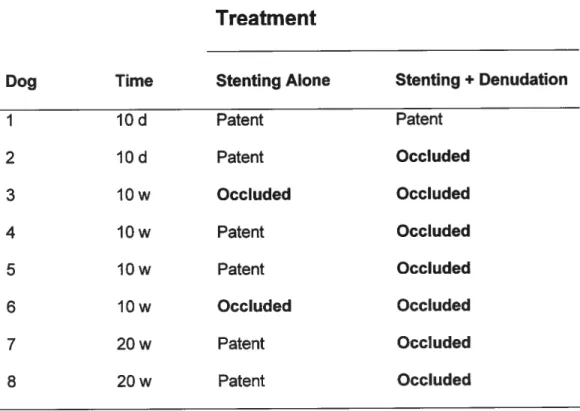
understanding of the cellular and molecular biology of intra-aneurysmal thrombus evolution followmg treatment promises to lead to better strategies to reduce recurrences. The first paper studies die effects of stenting and endothelial denudaflon on aneurysm and brandi vessel occlusion. Experimental aneurysms were created and treated with stenting alone or stenting and endothelial denudation. Angiographic and paffiologic findings were compared between die twogroups.
We found that stenting alonewas insufficient to lead to complete aneurysm occlusion, but resuits improved when stenting was combined with endoffielial denudation.
The second paper studies the effects of stenting on diegenetic expression of ceils ofdie aneurysm wafl and attempts to correlate changes in expression with angiographic and pathologic ffiidings. Bilateral carotid aneurysms were created; one was stented. Tissue from die aneurysm neck underwent RT-PCR at various times to create mRNA expression
proffles for 14 potentially interestinggenes.
Stenting led to angiographic improvement ofaneurysm appearance. mRNA expression proffles were in keeping widi expected effects on neointima fonnation and thrombus
organ2aton and with endothehali2ed channel formation within die thrombus, but differences between stented and control groups did not reach significance.
SOFAIAA1RE
IV
V
CONJENT
/I
LISP 0F JABLES
XI
LISTOFFIGURES
XII
LIST 0F ABBREVIATIONS
XIII
ACKNOWLEDGMENTS
XVII
f.
INrRODuCTION
1
7.1
INTRACRANIALANEURYSMS
J
1 .1 .1 Demographics
J
1.1.2 Etiopathogenesis
2
1.1.3 Ctinicat Manifestations
3
1.1.4 Comp[ications of aneurysm rupture
4
1.2
TREATMENTOFINTRACRANIALANEURYSMS
4
1.2.1 Open surgicat treatment
5
1.2.2 Endovascutar treatment
6
1.2.2.1 Coits
7
1.2.2.2 Stents
8
1.3
MECHANICAL AND HEMODYNAMIC FORCES ACTING ON
ANEURYSMS
13
1.3.1 Overview
13
1.3.2 Aneurysm Hemodynamics
14
1 .3.3 Mechanicat Forces Acting on Vascutar Ce[[s
15
1.3.3.1 Cyc[ic Wa[t Stress
16
1.3.3.2 Shear Stress
16
1.4
ENDOVASCULAR TREATMENT RESPONSE
17
1.4.1 Overview
17
1.4.1.1 Thrombosis and Inflammation
19
J .4.1 .2 Thrombus Organization
19
1.4.1.3 Neointima Formation
20
1.4.1.4 Re-endotheliatization
21
1.4.1.5 Recanatization
21
1.4.1.6 A more integrated view of aneurysm heating
22
1.4.2 Cetts invotved in the response to endovascutar treatment. .24
1.4.2.1 Endothetial. cetts
25
1.4.2.2 Neointimat cetls
27
1.4.2.2.1 Vascutar smooth muscle cetts
28
1.4.2.2.2 Myofibrobtasts
28
1 .4.2.3
Circutating cetts
29
1.4.2.3.1 Ptatetets
29
1.4.2.3.2 Leukocytes
30
1.4.2.3.3 Progenitorce[[s
31
1.4.2.3.3.2 Endothetiat progenitor cetts
.32
1 .4.3 Extracettutar matrix
33
1.4.3.1 Matrix metat[oproteinases and their inhibitors
33
1.5
REGULATION 0F THE VASCULAR RESPONSE TO INJURY
35
1.5.7 SoLubLe factors
35
1.5.7.1 Ptatetet derived gtowth factor-BB
36
1.5.1.2 Transforming growth factor-B1
37
1.5.1.3 Monocyte chemotactic protein-1
38
1.5.1.4 Tumour necrosis factor-o
39
1.5.2 Adhesion Motecutes
40
1.5.2.1 CeLt-cett interactions
40
1.5.2.1.7 Ptatetet endothetiat ce[t adhesion motecute- J
40
1.5.2.1.2 Vascutar cett adhesion mo[ecute-1
41
J .5.2.2 Cett-matrix interactions
42
1.5.2.2.1 Integrins
42
2.RESEARCHPRESENTATION
44
2.1
PROJECT FRAMEWORK AND CONCEPTUAL LINKS
44
2.2
HYPOTHESES
45
2.3
RESEJ&RCH GOALS
46
2.3.1 Generat Objectives
46
3.
MATERIALSANDMETHODS.47
3.1
IN VIVO METHODOLOGY
-SURGICAL MODELS
47
3.1.1 Laterat carotid aneurysm modet
47
3.1.2 Linguat aneurysm modet
48
3.1.3 Carotid bifurcation aneurysm modet
49
3.2
&NGIOGRAPH’(
51
3.2.1 Laterat carotid aneurysm model
51
3.2.2 Lingual. aneurysm modet
52
3.2.3 Carotid bifurcation model
53
3.2.4 Efficacy of endothetiat denudation
53
3.3
P8THOLOGY
53
3.3.1 Laterat carotid aneurysm modet
53
3.3.2 Linguat aneurysm modet
54
3.3.3 Carotid bifurcation modet
55
3.4
INVITROMETHODOLOGY
55
3.4.1 RNA extraction
55
3.4.2 Reverse transcription of RNA to DNA
56
3.4.3 PCR amptification
56
3.4.3 lmmunohistochemistry
56
4.
RESULTS
.59
4.1 The effects of stenting and endotheliat denudation on aneurysm
and branch occlusion in experimentaL aneurysm models
62
4.2 Effects of stenting the parent artery on aneurysm filling and gene
expression of varlous potentiat factors învoLved in healing of
experimentalaneurysms
85
5.
DISCUSSION
112
5.1
NEOINTIMA FORMATION I THROMBUS ORGANIZATION
113
5.2
REC&NLIZ4&1ION
118
5.3
STENTS AND BLOOD FLOW MODIFICATION
120
5.4
THEROLEOFTHEENDOTHELIUM
122
5.5
EXPERIMENTAL LIMITATIONS
125
5.5.1 Generat Limitations
125
5.5.2
Specific Limitations
126
5.5.2.1 Linguat Laterat Watt and Carotid Bifurcation Project
....126
5.5.2.2
mRNA expression Project
127
5.6
FUTURE DIRECTIONS
128
5.7
CONCLUSIONS
129
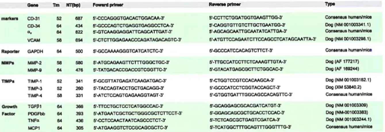
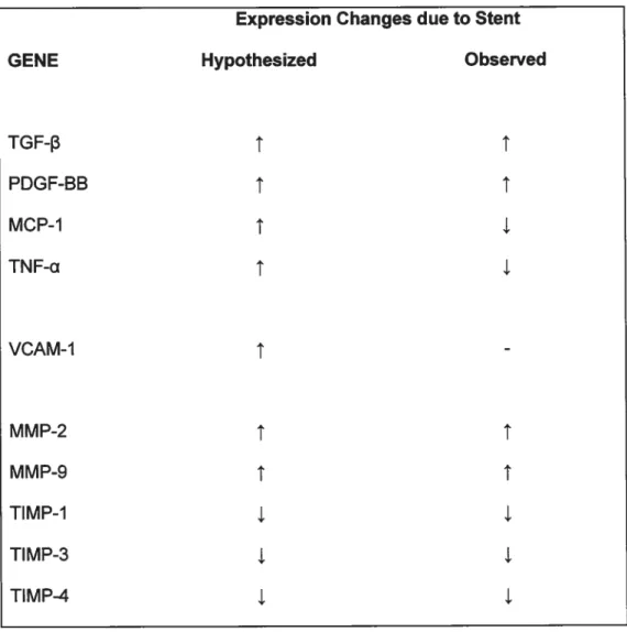
LIST 0F TABLES
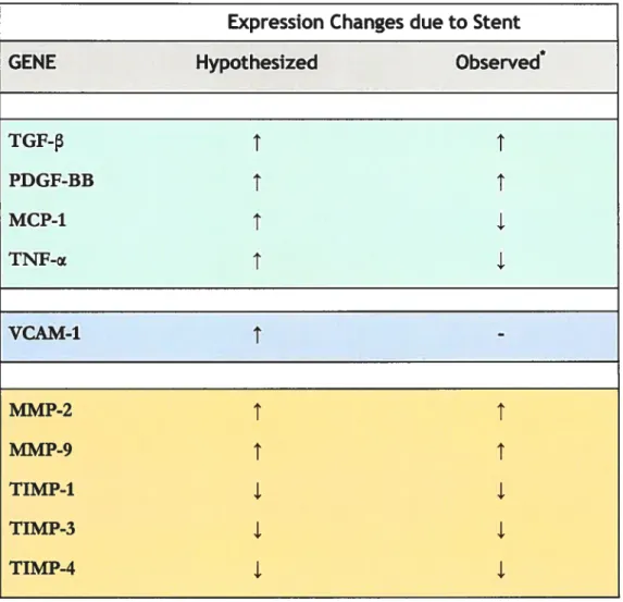
Table 1.
Genes potentia[[y invo[ved in the motecular mechanisms of aneurysm
heating, with the hypothesized expression changes obtained from a survey
of the titerature of other vascutar processes, and our experimentat
observations
43
Table 2.
LIST 0F FIGURES
FIGURE J .1
CI head showing diffuse subarachnoid
hemorrhage
3
FIGURE 1.2
Aneurysm clip across the neck of a sidewatt berry aneurysm,
removing it from the circutation
5
FIGURE 1 .3
Artist’s rendition of different endovascutar strategies to treat
aneurysms
7
FIGURE 1 .4
Artist’s rendition of endovascutar coiting in conjunction wïth
stenting
10
FIGURE 1.5
Schematic comparing ftow dynamics of non-stented versus
stented aneurysms
15
FIGURE 1 .6
Schematic showing process of recanatization fottowing
endovascutar treatment
23
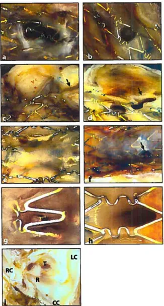
FIGURE 3.1
Construction of the wide neck aneurysm modet
50
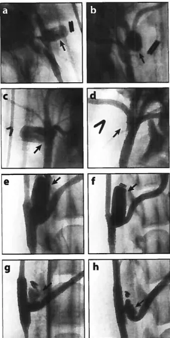
FIGURE 3.2
Angiogram
0ftypicat canine carotid taterat watt vein pouch
aneurysm at four weeks
51
FIGURE 3.3
Angiogram 0f typicat stented canine tinguat aneurysm
immediatety fottowing surgicat creation
52
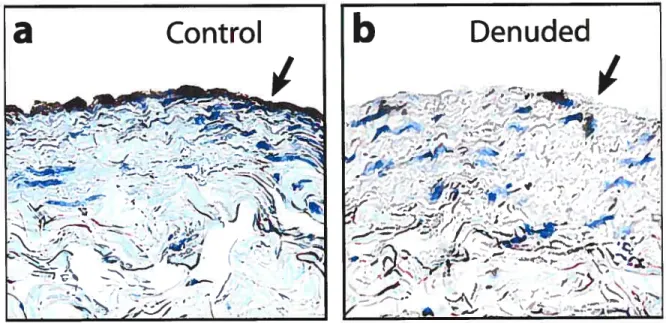
FIGURE 3.4
Photographs of a stented taterat watt aneurysm (on teft) and
an unstented control
54
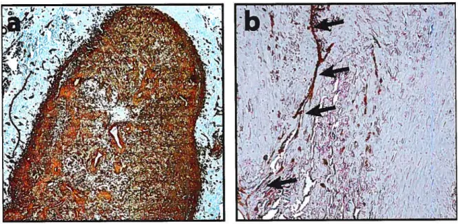
FIGURE 5.1
Photomicrographs of axiat sections through an
aneurysm
114
FIGURE 5.2
Photograph of coronal section through a taterat watt
aneurysm
116
FIGURE 5.3
Schematic showing the direction and attenuation of btood
ftow into a taterat watt aneurysm
119
FIGURE 5.4
Schematic ittustrating the targer aneurysm recurrence at the
distat tip compared to the proximat aneurysm tip
121
LIST 0F ABBREVIATIONS
ADP Adenosme diphosphate
ADPKD Autosomal dominant polycystic kidney disease
ASA Acetylsalicylic acid
BM-MNC Bone marrow-denved mononuclear ceils
C Celsius
CD Cluster of differentiation
CT Computenzed tomography
DNA Deoxyribonucleic acid
DVT Deep venous ffirombosis
EC Endothelial ceil
ECM Exfracellularmatnx
eNOS Endothelialnitricoxide synthase
EPC Endothelial progemtor ceil
E-selectin Endothelial-selectin
F French
G gravity
GAPDH Glyceraldehyde-3-phosphate dehydrogenase
GDC Guglielmi detachable coil
GSL Griffonia simplicifolia lectin
HC1 HydrocMoric acid
HPS Hematoxylin-phioxine-saffron
ICH Intracerebral hemorrhage
ISAT International subarachnoid aneurysm trial
ISUJA International study ofunrupturedintracranial aneurysms
MC/MPh Monocyte/macrophage
MCP-1 Monocyte chemotactic protein-1
IvflVfP Matrixmetalloproteinase
MPC Mesenchymal progemtor celi
MT-MMP Membrane typematrix metalloproteinase
NaC1 Sodium cifionde
NGS Normalgoat serum
NO Nitric oxide
PCR Polymerase cliain reaction
PDGF Platelet-derived growth factor
PDGFR Platelet-denved growffi factor receptor
PE Pulmonary embolus
PECAI\’I Plittelet-endoffiehal ceil adhesion molecule
RNA Ribonucleic acid
RT-PCR Reversetranscnptase polymerase cham reacfion
SAH Subarachnoid hemorrhage
oc-SIVJA Alpha-smooffi muscle actin
Taq Thermophilus aquaficus
TBT Tris-Buffered saiine-Tween
TEAM Trial of endovascularaneurysmmanagement
TGF Tissue growth factor
TIMP Tissue mbibitor of metalloprotemase
TNF Tumour necrosis factor
VCAM Vascular cd adhesion molecule
VE-cadhedn Vascul2r endoffielialcadhenn
VEGF Vascular endothelialgrowffifactor
VEGFR Vascular endoffielial growth factor receptor
VLA Very late antigen
VSMC Vascular smooth muscle ceil
The task ofscience is to stake out the Ïimits ofthe knowabÏe,
and to center consciousness within them.
ACKNOWLEDGMENTS
I would like to thank die following people:
Jean Rqymond, for bis rnentorship and guidance, attention to intellecmal detail, and exemplary leadership, but most of ail, for bis insistence on independent diought.
Igor Satakin, for ail die technical advices and surgical secrets. Thanks for die good times spent in the OR, angiography suite, lab, and cafetetia.
Gujilaine Gezy,the soul and glue of the NU, holding us together and malting countless contributions to bring about and shape this research year.
Audry Byteand the rest of the vivarium technicians, for help with the surgical and endovascular procedures.
Christelle Ogoudikpe, for help widi the biochemistry and molecular biology.
Fatiha Boueghrane, for helpfiil discussions as weil as the inimunohistochemistry.
Sophie Lerouge, Gittes SouÏe andJean Beigeron from CHUM, for helping to provide us widi stents.
The UniversiyofAïbertaneuivsulgeons, especiafly Vivek Mehia, for die opportunity to corne to Montreal.
Myparents, brothersandsisters, for moral and social support.
This work would not have been possible widiout die ffiiancial contribution from die
Canadian Institutes ofHeaÏth Research (CIHR; MOP-44062) and die Quebec Hearth and Stroke Foundation.
I would like to take this opportunity to thank die faculté des Études Supérieures, Programme de Sciences biomédicales of Université de MontréaÏand Dr Daniet Lajeunesse for dieir generosity in funding die Master prograrn scholarsbip. I was honored to be a recipient of this award.
This thesis comprises six chapters. The rst provides relevant background regarding intracranial aneurysms and how they can be treated, followed by an introduction to the cellular and molecular factors hkely involved in intracranial aneurysm healing. The second chapter explains the rationale, hypotheses, and goals of die experiments performed for this thesis, while the third chapter covers the materials and methodology used in completing and analyzing die experiments. The fourdi chapter is made up of the two manuscripts produced as a result of tins work. A general discussion of die material within die papers is covered in chapter five, with some concluding remarks. The sixth chapter provides references to die literature cited in this thesis.
J .1
INTRACRANIAL ANEURYSMS
1.1.1 Demographics
An aneurysm is an abnormal swelling along a hlood vessel[1J. The reported prevalence of unmptured aneurysms ranges from O.1-6%, but is likely considerably less than 5% [1]. Aneurysm rupture is die most common cause of spontaneous subarachnoid hemorrhage (SAH)[2], widi an incidence estimated at 10/100 000 population per year[3,
4J.
This devastating disease strikes women 1.6 times more dian men [1], usually between die age of 30 and 70 years, with a peak incidence during die 6 decade[5]. The consequences of SAH can be grave, widi approximately 45% overail mortality and moderate to severe disabffity in 30%[6-$]. In aduks, 85% of aneurysms are found in die antetior circulation, and most of diese aneurysms are saccular[2]. Postenor circulation aneurysms comprise l5%, a greater proportion ofwhich are fusiform.Aneurysms are much less common in children. Childhood aneurysms often bear atypical eflologic or morphologic features, and are suspected to represent a distinct padiological enflty[91.
1.1.2 Etiopathogenesis
The etiopathogenesis of most saccular aneurysms remains unknown. Cerebral blood vessels normally contain an outer adventifial layer, a middle layer of smooth muscle or media, an internai elastic lamina, and an innermost intimai layer. Cerebral vessels lack an extemal elastic lamina, which may predispose diem to aneurysmal formation. Microscopically, aneurysms are found to have structural changes in die wall of die blood vessel, including mural atrophy, loss of die internai elastic lamina and media, and thinning ofthe adventida[lO].
Aneurysm etiologies are commonly classified into acquired or congenital causes. Support for die hypodiesis that aneurysms are acquired lesions anses from die observation diat most cerebral aneurysms arise at brandi points or where arteties abmptly change curvature, implicating chronic hemodynamic stress as a etiologic agent
[9J.
In keeping widi this ffieory, die nsk factors most commonly linked to aneurysm formation and rupture are cigarette smoking and artetial hypertension[1, 11J. Furthermore, intracranial aneurysms are rareÏy found in children, and are found widi increasing incidence until die$ decade oflife[11j.
However, diere is supporting evidence for congenital predispositions to aneurysm development. Several hentable disorders are associated widi intracranial aneurysms, including Eliers-Daiilos type W, Marfan’s syndrome, neurofibromatosis type I, and autosomal dominant polycystic kidney disease (ADPKD)[l]. Aldiough a mutation at a pafficular gene locus has flot been discovered, intracranial aneurysms are known to run along familial lines[1 1]. Taken together, die evidence suggests diat die causes of aneurysm formation and rupture are multiple and multifactorial, most likely resulting from a
combination of genetic factors which predispose the arterial wall to form paffiological dilations in response to noxious environmental factors.
The remaining causes of aneurysm formation represent only a small minority of cases. Mycotic aneurysms are those that occur following infection, usually in association wiffi bacterial endocarditis[2], while post-traumanc aneurysms are an uncommon sequelae of trauma, usually following a penetrating injury to the arterial vessel wall[2j.
1.1.3 CLïnicat Manifestations
Unruptured aneurysms usually remain asymptomatic, alffiough they may present with signs and symptoms of mass effect, cortical or meningeal irritation, or occasionally with ischemic events from emboli originating from witbin the aneurysm. The majonty of aneurysms are diagnosed afrer rupture, which usuaily causes SAH, intracerebral hemorrhage (ICH) or subdural hematoma. The annual incidence of SAR in patients with unruptured aneurysms is stiil unknown, and is the topic of much ciment debate[12-14]. For mptured aneurysms, clinical symptomatology depends on aneurysm siae, location, bleeding sevetity, and the presence of associated hydrocephalus. Aneurysm rupture can resuit in sudden deaffi (as high as 65¾ of patients, as reported in die prospective arm of ISUJA)[12], while the remainder typically seek medical attention for a specirum of neurological signs and symptoms ranging from sudden headache to decreased level of consciousness[l, 2]. Typical clinical features include the sudden onset of severe headache, often with neurological deficits, associated nuchal pain and ngidity, a low-grade fever, and retmal hemorrhages. Figure 1.1 presents typical CT ndings ofaneurysmal SAH.
FIGURE 1.1
CT head showing diffuse subarachnoid hemorrhage.
Subsequent digital subtraction angiography revealeda lefr antenor choroidal artery aneurysm.
1.1.4
Complications of aneurysm rupture
3O5O% ofaneurysm ruptures are fatal prior to medical or surgical intervenfion[11. In ffie absence of prompt aneurysm obliteration, approxiruately haif of patients with aneurysm rupture will die within one month, usually due to re-hemorrhage[1J. For untreated patients who survive 6 mondis, ffiere is a 2°/o rupture rate per year for ten years, which fails to 1% after a decade[1]. Rebleeding has an associated mortality of 80%[l1. Mortality for patients who survive unifi treatment is approximately lO%, with 60% of survivors having a favorable outcome[5]. Overail mortality from SAH ïs approximately 60-65%[12, 15]. Offier potentially life-threatening early complications include acute hydrocephalus due to blockage of cerebrospinal flmd flow by thrombus formation within the ventricular system or basal cistems[2]. Vasospasm of major mtracranial vessels secondary to dot and dot breakdown products in the subarachnoid space can lead to cerebral ischemia or infarction. Offier potential complications of aneurysm rupture incÏude neurological deficits, treatment-related complications, systemic complications secondary to acute physiological stress, as well as complications secondary to irumobility. Chromc hydrocephalus is a common sequel of aneurysmal SAH, often requinng shunting of cerebrospinal fluid.
1.2
TREATMENT 0F INTRACRANIAL ANEURYSMS
Wbile the treatment of unruptured aneurysms, without more complete knowledge of the natural history, remains a malter of debate, there is litde controversy that mptured aneurysms should be treated to prevent further rebleeding. In general there are two options for the treatment of intracranial aneurysms; open suxgical or endovascular treatinent.
1.2.1 Open surgicaL treatment
Current surgical treatment 0f saccular aneurysms typically consists of a craniotomy to
place a meta]lic clip across ffie base of the aneurysm, effectively removing the aneurysm from the circulation and maintaining or recreating a normal arterial configuration (Figure 1.2). The aneurysm clip re-apposes the arterial mural structures between the clip blades, maintaining a continuous endothe]ial covering layer.
Figure 1.2
Aneurysm clip across the neck of a
sidewail berry aneurysm, removing it from the circulation. (From Boston Sdentiflc; www.bostonscieniific.com)
Surgical rates of morbidity and mortality for unruptured aneurysms are typically reported in surgical case senes to be <4% and <2%, respectively[16-1$], a likely underestirnate related to publication bias. However, the ISUJA investigators have repeatedly reported surgical morbidity and mortality rates for unniptured aneurysms ranging from 12.6-I 5.7%[l2, 19]. For rnptured aneurysms, in cases deemed salvageable, surgical mortality rates range from 7-29%[1 j. Morbidity due to surgery on mptured aneurysms is difficuit to separate from morbidity due to theaneurysm rupture itselE
Other surgical trealments for uncippable aneurysms depend on die nature, morphology, and location of the aneurysm, as well as die surgical exposure. Aneurysmorrhaphy with parent vessel reconstruction or surgical bypass following by parent vessel occlusion offer deffiiltive treatment, but are more technically difficuit and carry more nsk[20j. Other alternatives, such as aneurysm wrapping with muscle or mushn gauze to reinforce the wall, are of dubious efficacy, being used only when there are no offier options[20j. As the role of endovascular therapy is increasingly recognized, partial neck cipping followed by coilingis recognized as a viable alternative for difficult cases [20].
1.2.2 Endovascutar treatment
Endovascular treatment, an mcreasingly popular alternative to surgery, involves arterial puncture at a remote site, with navigation to and deposïtion of vatious matedals within the circulation. The tise in popularity was inifially due to the less invasive nature of endovascular treatment, which is now supported by the publication of the multicenter randomiaed ISAT trial of ruptured aneurysms, winch showed decreased morbidity and mortality at 1 year for endovascular treatrnent (23.7%) compared to surgery (3O.6%)[21].
In general, endovascular treatment strategies can be directed towards either the aneurysm or the parent ves sel (Figure 1.3). When the strategy targets the aneurysm, thrombogenic material (typically platinum coils) are deposited within the aneurysm fundus, in hopes of causing a locakzed reaction wiffi tbrombus formation, followed by dot organization and neointimal formation to obliterate the aneurysm and restore normal blood flow pattems. The strategy of coil deposiflon within aneurysms was shown wiffi ISAT to be of clinical benefit.
Treatment directed towards the parent artery can be eiffier ‘deconstructive’, occluding the parent vessel as well as the aneurysm, or ‘reconstructive’, using stents to alter aneurysm hemodynamics, provoke thrombosis and promote neointima formation at the junction of the parent vessel and aneurysm neck. Strategies to treat aneurysms using stents have neyer been proven beneficial but are increasing in populatity[22J. I will start by introducing the well-accepted embolization procedure and review the limitations of tins strategy. I will then discuss the potential benefits of stents as adjuncts to coil embolization, or as a prirnary mode of treadng intracranial aneurysms.
Artist’s rendition of different endovascular strategies to treat aneurysms
A) Coils deposited within the aneurysm. B) Endoluminal stent deposited in the parentartery to bridge the neck of the aneurysm. (from Boston Scientific; www.bostonscientific.com)
1.2.2.1 Colts
Although emboli2afion ofaneurysmswiffi vanous materials, including free coils, baRbons, and glue had been possible for decades for rare lesions, its clinical use remained exceptional unifi the introduction of Gugliehni detachable coils (GDCs) in 1991. Bnefly, endovascular treatment wiffi GDCs involves packing multiple thrombogenic platinum coils into an aneurysm to prevent blood from entering the fundus. The coils adjust to the contours of the aneurysm, and once sadsfactorily posirioned, the operator separates the coil from the stainless steel deliverywire through an electrolytic process. The key feature of GDCs was this detachment system, winch permitted the retrieval and reposifiornng of the coils when necessary, with detachment only when the coils were safely and stably positioned within the aneurysm. The improved safety proffleimparted by the detachment system led to the increased popularity of endovascular treatment, with littie concem for its long-term efflcacy. Afthough coiEng can lead to good outcomes, there are several
important drawbacks with tins technique. One occasionally encountered limitation of
Figure 1.3
endovascular therapy is difflcuk intravascular access due to atherosclerosis, which prevents proper negofiation of wires and caffieters to access the aneurysm. This difflculty is exacerbated when the aneurysm is located in tormous or distal vasculature. Complications at the arterial puncture site in the groin can also lead to chnically significant hemorrhages, occasionally requiring penpheral vessel stent-grafring. However, the most important drawback associated with endovascular coiling remains the angiographic recurrence rate, winch is reported to approach 2O4O%[23]. In a most recent case seties die retreatment rate following endovascular coiling reached 15% after two years, but the clinical significance of these recurrences in ternis of rehemorrhage was very modest [24]. Although the tisk of rupture of aneurysm recurrences is stiil not lmown, die fear of SIkH from an aneurysm recurrence continues to lead some neurosurgeons to treat aneurysms with surgical cipping. Attempts to decrease recurrence rates have focused mostly on coil modification; coatings to promote thrombosis [25], to increase aneurysm packing density[26j, emit radiation[27], or biologicailv active coatings[2$J have been introduced, but none have successfuily decreased recunence rates. Uquid embolic agents such as Onyx have also proven ineffecflve in tins regard[29]. Finaily, endovascular techniques are flot suitable for ail aneurysm morphologies; coils deposited witbin aneurysms with wide necks or a dome:neck ratio of <2 are at nsk of hermating into the parent vessel, leading to thrombo-embolic complications. Technical innovations, such as the balloon-remodeling technique have been developed to address tins, but tins adds complexity and procedural tisk[30]. The notion to use stents in die intracranial circulation originated from die need to prevent coil herniation in wide-necked aneurvsms.
1.2.2.2 Stents
Stents are made of tubular metaffic mesh, and were originaily designed to treat acute or subacute coronary artery occlusion following bailoon angioplasty, aiming to prevent recoil of die arterial wail after balloon dilatation. Balloon-expandable stents were introduced into atherosclerotic vessel segments and die balloon forcibly expanded to dilate die lesion, leaving die stent in situ in order to maintain vesse1 patency. Neuroendovascular therapists
have benefited from the cardiology experience regarding ischemic complications and vessel re-stenosis from neointimal hyperpiasia following stenting, but die cardiology literature is not aiways applicable to intracranial disease processes. Important pathological differences between diseased atherosclerotic blood vessels and vessels bearing aneurysms may alter the artetial response to stenting.
The use of stents for intracranial applications lias been hampered by die accessibility ta die tortuous intracranial circulation. Initial stenting attempts for intracranial pathologies used coronary stents as an ‘off-label’ indicaflon[31]. The balloon-expandable coronary stents are designed ta exert large radial forces ta force open atherosclerotic stenoses and then maintain vessel patency. The coronary stents available on die market were quite rigid, and neuroendovascular therapists found diem difficuit ta navigate through die carotid siphon at die skufl base[32]. Balloon-expanclable stents are limited by their poor flexibffity and ‘pushabffity’, as compared ta seif-expandable stents[33]. Nonedieless, in die less tortuous postetior circulation, balloon-expandable coronary stents have been successfully used ta treat aneurysms [34]. More recently, self-expandable stents designed specffically for intracranial applications have been developed. These devices and dieir deployment systems are more flexible, enabling intracranial navigation. In contrast ta dieir coronary counterparts, intracranial stents do flot need ta open stenotic segments, and self-expandable stents, winch exert smaller radial forces but adapt more closely ta die contours of die parent vessel, are more commonly used dian balloon-expandable stents. Self-expandable stents must nonedieless exert sufficient radial force ta praperly anchar die stent within die vessel, ta prevent stent migration, or ta prevent die hemiation of coils out of aneurysms[30].
In die treatment of intracranial aneurysms, stents are typically used in canjunction widi coils or odier embolic agents, aldiough diey can be used alone (Figure 1.4). As previously mentioned, die main utility of die stent in conjunction widi endovascular treatments is ta prevent die herniation of embolic material into die parent vessel, especially for wide neck or fusiform aneurysms [35, 36]. However, die use of stents for routine non-wide necked aneurysms has been suggested, due ta dieoretical effects of die stent on aneurysm healing. Stents are diought to act primarily by redirecting blood flow along die parent vessel,
alteringaneurysmalhemodynamics and promoting thrombosis [37, 38]. Redirecting blood flow forces may also promote more complete aneurysm healing and decrease recurrence rates by re-establishing a new endoluminal boundary that exciudes the aneurysm. The
stent may also bufferthe hemodynamic forces implicated in recunence, or alter die fluid
dynamics in die area of die stent, thereby promoting neointima formation [39]. The
effectiveness ofthestent atachievingthese goals depends primarilyon thestent design.
Figure 1.4
Artist’s rendiflon of endovascular coiling
in conjunction widi stenfing. Note how die stent prevents coil herniadon into
theparent vessel lumen.
The choice of stent should be based on die site and morphology of die aneurysm, the
stent design, as well as die delivery system. The relevant morphology of the
aneurysm/parent artery complex includes die caliber of die parent vessel, the desired length of“landingzone” for die stent, die widffi of die aneurysm neck, and die presence of branch or perforator vessels[30]. Stents are usually chosen to match die caliber ofthe
recipient vessel[401. Intendonal attempts to over-dilate cerebral vessels has led to extremely high morbidity andmortality rates[41]. Manufacturers recommend a “landing
zone” of 4-5 mm on eidier side of die aneurysm neck[32]. Longer stents present a larger thrombogenic surface area, and carry a greater theoretical tisk to branch or perforating vessels. M sites of tight vessel curvatures, a more flexible stent is required to conform to die parentartery.
The design of die stent includes die sire, composition, and structure, including the
number of struts per circumference. Clinicians can choose from a vatiety of lengths and diameters, but most stents in current clinical use are made of nitinol, a nickel-titanium
non-thrombogenic, and nitinol has been shown to be superior to odier metals with respect to neointimal hyperpiasia and in-stent stenosis[42]. for nitinoÏ stents, radio-opaque markers must be added to guide proper deployment, because nitinol is radiolucent.
A properly chosen stent structure is paramount to avoid complications[43]. Stent strut widdi and ceil size dictate die porosity of die stent, which is die ability for blood to continue to flow outside die confines of die stent mesh (ie: into die aneurysm or adjacent branch vessels) [30]. The ideal stent porosity alters blood flow sufflciently to provoke a localized tbxombosis within die aneurysm wbile sparing ail small vessels originating from under die stent[30]. These opposing goals may flot always be attainable with die stents currenily available, and compromise is often necessary. In cases devoid of critical perforators, where thrombosis is desired anywhere outside die confines of die stent, die ideal stent would be a stent covered with an impermeable membrane widi a non dirombogenic internai surface. Covered stents such as diese are usefiil to treat fusiform aneurysms widi no perforators arising from die parent vessel[441, and are also used to treat abdominal aortic aneurysms [45]. Stents widi a greater number of struts Qower porosity) are able to exert a greater amount of radial force. Radial force is an important consideration in adierosclerotic disease, where die main goal of die stent is to maintain vessel diameter, but for intracranial aneurysms, a minimum of radial force is required to prevent coil herniataon or stent migration. The number of struts correlates widi die amount of potentially thrombogenic surface area, and may pose a greater risk of parent vessel thrombosis. The number and type of hnkages between stent ceils furdier dictates die flexibiilty of die stent. These consideraflons have led stent manufacturers to create higliy flexible, high porosity stents widi few stent struts per cross-secflonal diameter.
The stent deployment system is also of crucial importance to a successftil endovascular procedure. The two most important features are navigabffity and mediod of delivery (self or bailoon-expandable). Stents designed for intracranial use are now sufflciendy navigable to access most aneurysms of die posterior and anterior circulation. Self-expandable stents offer several advantages compared to bailoon-expandable stents. These stents are deployed by puDing back die sheadi covering die spring-loaded stent, winch expands to fUI die contours of die parent vessel. The low radial force exerted by a seif-expandable
stent is sufficient to anchor the stent witbin tEe lumen, as well as to prevent coil herniation into tEe parent vessel in most cases if necessary. This meffiod of delivery does flot cause angioplasty-type injuries when deployed, wbich may reduce the incidence of neomtimal hyperpiasia and in-stent stenosis[46]. Bailoon-expandable stents are designed to expand to a frced volume, and are more prone to migration within tEe parent vessel if ffiey are undersized.
The tisks of stenting can be classified as acute or chromc. Thrombogenic nsks comprise tEe majoiity of acute and subacute tisks due to stenting[32]. The tbrombogenicity of intracranial stents is almost indisputable given the cardiology expetience with coronary stents[47], and as ail stenting procedures are now performed using vigorous anti-platelet regimens, it is ethically impossible to determine tEe nsk of tbrombosis due to stents by tEemselves. The best anti-platelet regimen bas flot been decided upon, but several have been suggested for intracranial stents under various circumstances [32, 35, 48]. Stent deployment also carties ischemic nsks to perforating vessels, by pardaily occluding vessel ostia originatingunder the stent, or causing embolization of fragments of atheroma [49]. It is important to note the difference in thrombotic tisks between die conventional coiling strategies that target aneurysms by depositing coils outside die normal circulation and stenting strategies that deploy devices within die parent vessel. The vessel trauma caused by die stent and the permanent deployment of a foreign body creates a risk of thrombosis to die exact vessel we aim to preserve. Thrombotic complications can also occur in a delayed fashion, requiring long-term anti-platelet ffierapy. The use of these agents afrer SAH may be a source of additional hemorrbagic complications, including aneurysm rebleeding, or foilowing odier procedures, such as extemal ventricular drainage. Odier risks of stenting include suboptimal placement, migration, fracture, or prolapse into the potential spaces outside the vessel lumen, such as a branch ostium or aneurysm [33, 50]. Catastrophic artetial perforation may also occur[33J. finaily, die artetial response to stenting can also lead to neointimal hyperpiasia and in-stent stenosis, winch is weil characteiized in the cardiology literature. Although the nature of die aneurysmal disease process differs from die atherosclerotic coronary situation, intracranial in-stent stenosis
due to exuberant neointima deposition with hemodynamically significant flow reduction has been reported[51].
1.2.3 Aneurysm recurrences foltowing treatment
Following complete surgical cipping as determined on angiography, aneurysm recurrence
rates range from 1.5 to 3%[52]. for surgically cipped lesions with known residua, die recurrence rate is 7-25%[52, 53]. Endovascular treatment is eidier incomplete or is associated withaneurysm recurrence in 2O4O% of cases [23]. Engineering and biomedical strategies to decrease recurrence rates through novel device designs and biological coil modifications have not solved die problem of recunences. Most developments were proposed by manufacturers and developed by engineers, with littie concems for the biological mechanisms involved in healing or recunences following endovascular freatment. There is a need for better characterization of die molecukr mechanisms occurring followmg aneurysm treatment to help engender and guide further attempts to decrease recurrence rates.
1.3
MECHANICAL AND HEMODYNAMIC FORCES ACTING ON
ANEURYSMS
1.3.1 Overview
Blood flow is a resuli of repetitive coordinated myocardial contractions which propel blood through the vascular tree. In vivo hemodynamic forces are complex; they vary with cardiac cycle, heart rate, blood pressure, blood viscosity, vessel architecture, and viscoelastic properties of die artery[54]. Forces generated by pulsaffle blood flow are attenuated pdmarily by two processes: elastic stretching of die arterial media, and friction between die elements of blood and die vessel wall. Chonic padiological elevation of diese forces has been implicated in die formation and rupture of intracranial aneurysms [1]. The nature and magnitude of hemodynamic force acting on an aneurysm depend on die location and morphology of die aneurysm. Following endovascular treatment, die
orgamzing thrombus is subjected to continued pulsatile blood flow, which potenlially impacts many of the ongoing biological processes.
Mechanotransducflon was initially thought to be due to activation of specific mechanoreceptors, such as a mechano-sensifive ion channels or ceil surface adhesion molecules, but there is also evidence for die entire endodielial ceil cytoskeleton to act as a mechanoreceptor [55, 56]. Eukaryotic ceils are able to maintain cytoskeletal rigidity through cytosolic ifiaments diat generate and resist mechanical loads [55, 57]. These
intracellul2r scaffolds aliow the ceil to respond to mechamcal signais from the
environment, by transducing cellular deformations through cellular signaling padiways, resulting in physiologic or paffiologic responses[58]. Forces of lesser magnitude that would flot normally deform die ceil soma are able to trigger mechanotransduction
signalingpaffiways through the deformation of ceil surface receptors, such as integrins[59, 60]. The establishment of appropriate ceil-ceil and ceil-matrix contacts is cnflcal for transduction of mechanical forces in smooth musde flssue[61]. Here, I will discuss die hemodynamics of lateral wall aneurysms, followed by a description of die two types of mechanical forces acting on vascular ceils.
1.3.2 Aneurysm hemodynamics
Aneurysm hemodynamics studies reveal diat flow within an aneurysm depends primarily
on the morphology of the aneurysm and the relationship to the parent artery[62]. For lateral wall aneurysms, three distinct zones have been descnbed: f) an inflow zone entenng die aneurysm at die distal portion of die ostium, 2) an outflow zone exiting die aneurysm at die proximal end of die ostium, and 3) a centrai slow flow vortex [63]. Modification of die inflow zone may provoke aneurysm thrombosis while maintaining
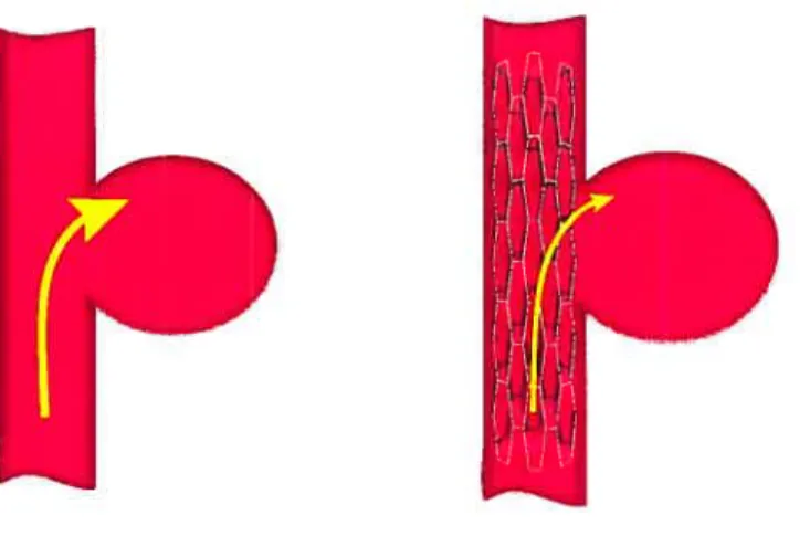
parent vessel patency [63]. The abffity of stents to modify die infiow zones of an aneurysm are well documented[62], and this effect of stents hkely contributes to die obliteration of literaiwallaneurysms (Figure 1.5) [42].
FIGURE 1.5
Schematic comparing flow dynamics of non-stented versus stentedaneurysms
Hemodynamic forces have long been implicated in ffie initiation of atherosclerosis, wiffi greater levels oflaminar flow conferring an atheroprotective phenotype[64]. There is an increased incidence of atheroma formation on the proximal portion of diseased branch vessels, purportedly due to decreased laminar shear stress or increased [641. Shear stress is lmown to modulate gene expression (see section 1.3.3.2) , and the loss of a protective
genetic expression proffle may trigger cellular and molecular events that account for affieroma formation localized to vessel bifurcations [65].
Blood flow into a lateralwall aneurysm concepmally resembles that into a brandi vessel; the distal portion ofthe ostium acts as a flow divider, and is exposed to higher levels of shear stress compared to the proximal aneurysm ostium [66]. The observed increased neointima formation at the proximal ostium [67] hkely reffects this difference in laminar shear stress between proximal anddistalostia.
1.3.3 Mechanical forces acting
on
vascutar cetts
Stretch and shear stresses are important physiologic stimuli knownto ttigger ceil signaling
ceils (VSMCs) are exposed to both types of mecharncal force, die sheax stress from blood flow is borne primarily by ECs, while VSMCs are primarily subjected to cycic stretch from pulsatile pressure[69]. The response to mechanical forces may be important to the occurrence of aneurysm recurrences following endovascular treamient.
1.3.3.1 Cyctic WaLt Stress
Cydic wall stress is the tensile stress vector acting perpendicular to the blood vessel lumen diat occurs as die blood vessel lumen is expanded widi systole[70]. This force, which primarily affects vascular smooth muscle ceils, resuks in 5-6% wall excursion at peak systole under normal physiological conditions, and can be as high as lO% under hypertensive condiflons[71]. Mechanical stretching of cultured VSMCs lias several effects, including reonentation of ceils, changing protein and DNA syndiesis, and increased production of extracellular matrix components [61]. Mechanical stretch also promotes stem ceils to differentiate into VSMCs, and may also lead to transdifferentiation of ECs into VSMCs[70j. Chronically elevated cydic wall stress is a feature of hypertension, which induces VSMC hypertrophy and hyperplasia [72]. Meclianical strain also leads to increased production of proteoglycans[73], and promotes extracellular matrix remodeling[70]. This cellular response aims to counterbalance die applied force by forming more extracellular matrix to buttress die vessel wall. Stent placement likely decreases die net cycic strain sensed by die artetial wall by absorbing some of die radially directed force of pulsaffle blood.
1.3.3.2 Shear Stress
Shear stress is die frictional force exerted at die endodielial apices caused by die flowing elements of die blood. The velocïty of flowing blood is maximal in die center of die vessel lumen, and decreases as it approaches die wall[74j. The fluid can be viewed as divided into a seties of adjacent layers moving at different velocities wiffi respect to each other. The velocity differential creates die shearing flow. Blood flow is disturbed at
branch points and curved areas of the vascular tree, compared to die simple laminar flow in straight parts of die aorta[69j. The endodielium acts as a sensing surface to transduce shear stress forces, using surface integrins, intercellular adhesion molecules, \ŒGFR-2, ion channels, G-protein coupled receptors, and ftimeric G proteins[69]. Endothelial ceils change dieir genetic expression profiles with the different types of shear forces, i.e.: laminar, disturbed, or oscfflatory flow[69]. Laminar flow resuits in die upregulation of anti proliferative, anti-in&mmatory, and differentiative properfies, widi down-regulation of genes associated widi ceil cycle progression and proliferation[69], which is of particular importance to adierosclerosis and localized neointima formation.
Abrupt reductions in fluid shear stress has been shown to induce VSMC proliferation in expedmental prosdietic grafts[75J. Stenting likely dismpts the laminar shear of blood entering die aneurysm, leading to turbulent blood flow. We hypothesize diat die stent related disruption in blood flow will resuit in a change in genetic expression at die level of ceils of die aneurysm wall, wbich we hope to correlate wiffi angiographic and paffiologic indicators of aneurysm healing.
1.4
ENDOVASCULAR TREATMENT RESPONSE
1.4.1
Overview
The primary goal of therapy is to protect patients from rupture or re-rupture; because of historical evidence wiffi surgical cipping and balloon embolizaflon, it is generally diought that stable, long-term angiographic exclusion of die aneurysm is die sunogate endpoint to aim for. To conflrm tins hypothesis, observational study of ffiousands of patients for many years would be necessary to determine dlinical efficacy, something winch is incompatible wiffi die steady technical and clinical advances in tins field. An alternative hypodiesis, from paffiological studies in animal models, suggests diat a thick, complete neointima is die signature of a stable occlusion. Furdier refinements suggest that a thick neointima completely closing die neck of die aneurysm is die resuk of normalized flow pattems, widi die mtra-aneurysmal thrombus fully organi2ed, widiout recanalization.
Endovascular therapy relies on the vascular biological response to lead to a successfiil outcome. Short terni goals of dierapy include occlusion of the aneurysm. While some mediods can achieve this by purely mechamcal means (surgical dipping, onyx glue or balloon embolization), endovascular treatment usually relies on thrombus to achieve tins goal. Subsequent evolution of die formed intravascular thrombus is crucial to the long term outcome of aneurysm treamient, wbich is dependent on the dominance of one of two phenomena: a) organization of the thrombus and neoindrna formation at the neck and b) recanalization [76]. The most important long-term outcome is protection from aneurysm rupture, but it is unknown what deg-ree of protection, if any, is conferred by partial aneurysm occlusion. Given die paucity of natural history data regarding angiographic recurrences, an ideal resuit would also produce stable, non-threatening angiographic resuits. The anticipated pathological correlates of this angiog-raphic endpoint would require die formation of a new, stable mural structure completely occluding die aneurysm, and covered by a continuous sheet of endothelium.
The molecular response to endovascular dierapy is a complex and incompletely understood process diat may be conceived of as a reaction to a fomi of vascular injury. The universal response to vascular injury involves neointirna formation, as docurnented after a wide array of physical or chemical injuries to die timer (endothelial) or outer (adventitial) layers of die vessel wall. The molecular response to vascular injury depends on die nature and severity of die injury, but generally involves every type of ceil in die vicinity (vascular smoodi muscle ceils, myofibroblasts, endodielial ceils, platelets and leukocytes) as well as circulating ceils, elements in die blood, and die extracellular matdx[56, 77, 78]. Because die response to embolic treatment is a relatively new area of investigation, many observed phenomena are interpreted in light of mechanisms and concepts previously evoked in odier more mature research areas, such as re-stenosis following artetial balloon injury widi or widiout stenting, angiogenesis, affierosclerosis, thrombosis, organization, inflammation, recanalizanon, and endodielialization. Most pertinent to this diesis are thrombosis and thrombus organization, and die processes of neointima formation, re-endodielialization, and recanalization.
1.4.1.7 Thrombosis and Inflammation
Clotting is a requisite component for circulatory liomeostasis, and is involved in the recovery of vascular integrity following injury[79]. For inapproptiate intravascular clotting, such as ffiat triggered by paffiological mechanisms, blood flow may be restored early, by thrombolysis, or late, through the process of recanali2ation[79]. Endovascular aneurysm treatment aims to form a thrombus localized witbin the aneurysm; the thrombotic reacflon to the introduction of an intravascular foreign body, as well as the tissue reaction to the dot, involves mediators classically considered as part of inflammation. There is a link between an excessively vigorous inflammatory reaction and neointima formation, at least in terms of clinical models of in-stent restenosis [801. A more detailed molecular explanation of die vast subjects of dirombosis and inflammation are considered beyond die scope of this thesis.
7.4.1.2 Thrombus Organization
Thrombus organi2ation is the process that converts a dirombus into tissue. In a successflilly organized thrombus, pathologists distinguish two sets of vessels: a) vasa vasorum of die outer and middle coats of the organized thrombus, and b) vessels diat form channels within die thrombus[79, $1]. The organi2ation of intravascular dots relies on several cellular events: i) die portion of the dot exposed to flowing blood is coated by monocytes, which subsequently penetrate the dot;ii) die flow-exposed surface of die dot is re-endodiehahzed, in) myoflbroblasts and neo-cap•es appear or develop within the tissues [79]. Occasionally, the neo-capfflaries destined to provide blood supply to the newly formed tissue flhling die aneurysm can re-join die parent vessel, contributing to thrombus recanalization (also see section 1.4.1.5).
In treated experimental aneurysm specimens, the tissue found witbin the aneurysmal sac continues as a ‘neointima’ at die neck, closing die entrance to die sac, and diere is no clear demarcadon between die so-called neointimal layer and die organized thrombus.
1.4.1.3 Neointîma Formation
An integral aspect of aneurysm healing is neointima formation at the neck, representing a universal response to vascular injury, characteri2ed by the formation of myxoid tissue with stellate-shaped smooth muscle ceils in a loose extracellular matrix[82j. Tradiflonaily, neointima formation was thought to occur in three phases: i) an acute phase, characten2ed by interactions of platelets, thrombin, and leukocytes which release biologicaily active mediators and activate medial smooth muscle ceils; ti) an intermediate phase, where smooth muscle ceils divide and migrate to die area of vessel injury; and iii) a chromc phase, where large amounts of extracellular matrix are produced and subsequendy undergo a process of remodehng[77]. It is important to note that die classic ‘neointima’ was descnbed in affierosclerosis models devoid of intima, or in endothelial injury models, leaving ail other panetal layers to participate in the pathophysiological process; die classical theory emphasized VSMCs migrating from die media to die sub-endothelium[77J, but others, using large animal models, descnbed die importance of thrombosis, followed by endothelia]ization, with VSMC inifitrating die thrombus as a key feature in neointima formation[83-85]. In aneurysms, afrer endovascular treatment, we wish to see ‘neointima’ replacing and closing die space where blood was flowing. Hence, a provisional manix (dot) is a sine qua non pre-requisite, ami die neointima formed in fact represents replacement of die dot by a new parietal layer composed of ‘organiaed dot’. The new parietal layer delimits die space wiffi residual blood flow (as dened by die endoffielium) from die contents of die occluded aneurysm, which subsequendy lls with connective tissue. In this perspective, diere is a continuum between dot orgai±ation (within die occluded sac) and neointima formation (closing die neck of a completely treated aneurysm).
The classical paradigm of neoinfima formation has been chailenged by die discovery and implication of circulating blood elements in vascu]ar repair. Circulating ceils, in particular endothehal progemtor ceils (EPCs), as well as dendiatic ceils, form part of a ever expanding list of ceils involved in this complex biology[86, 87]. The cellular milieu in wbich vascular heahng occurs consists of many different ceil types, producing and
responding to various chemical and mechanical stimuli. Some of ffie regulatory factors likely involved in tItis process will be discussed in section 1.5.
1.4.1.4 Re-endothetialization
The endothelial Iming regenerates to cover new mural structure fiffing the aneurysm orifice. Regenerated endothelial ceils on the neoindmal surface may migrate from adjacent healthy vascular wall, from the adventitial vasa vasoffim, or arrive from the bloodstream[86, $8]. Following stent implantation in animal models, 20% of endothelialization occurs by 4 days, <40% at 7 days, while endotheliali2afion is typically complete by 2$ days[89]. However, the importance of the process and timing of re endothelialization is a controversial topic[90]. One strategy in the cardiologists’ baffle against restenosis aims to reduce neointimal hyperpiasia by promotmg early re endothelialazation using growth factors such as VEGF [89]. Efforts within the neurovascular arena to diminish ffirombo-embolic complications originating from the thrombus around the coil mass have also targeted early re-endothehalization as a therapeutic goal[2$, 91]. However, our laboratory lias shown an association between the integrity of the endothelial hrting and recunences; early re-endotheliali2ation may counter balance thrornbus organization and promote the formation of endoffielialized channels witbin the thrornbus[92, 93].
1.4.1.5 Recanalization
Recanalization is a process that potentially compromises the long-terrn occlusion of endovascularly treated aneurysms. In general, circulatory horneostasis lias two limes of defense against intra-vascular thrombosis, winch in most cases is a paffiological occurrence. Acutely, thrornbolysis occasionally succeeds in opening up occluded vessels. However, when thrombolysis fails, the more chronic process of recanalization attempts to create new channels through or beside the dot to rejoin the patent portions of the vasculature. Much of the evidence for recanalizaflon cornes frorn work done on deep
venous thrombosis, akhough certain aspects of this process, such as ffie growth of prog-ressively maturing channels through a matrix share conceptual similaiities with angiogenesis and artenogenesis. The theoreiical importance of mechanical signaling is detailed in section 1.3.3.
Bnefly, die restoraflon of blood flow across occluded blood vessels seems to depend primarily on die activity of two ceil population, monocyte/macrophages (MC/MPh), and circuladng progenitor cells[79J. MC/MPhs, under the influence of the chemoldne MCP-1, act to penertate the extracellular matnx formed by die mass of platelets and fibrin, and create tubular spaces, or ‘tunnels’[94j. Matrix metalloproteinases are further known to be important to this process[941. These tunnels are subsequently seeded with circulating progemtor ceils. In turn, diese (at least in some hypoffiesized paradigms) will differentiate into different types of ceils depending on die depth of penetration. On die surface of die thrombus, they would differentiate into endothelial ceils, wbile ceils adopt a myoflbroblast or smooth muscle character in die deeper layers[79, 95]. Uhuînately, die capifiaries formed in die deeper layers may form communications with the endodielium hned clefrs at the surface of the thrombus. For pathological thrombi with a blood vessel, this can serve to re-establish blood flow across occluded vessels; in endovascularly rteated aneurysms, this potentially leads to recurrences.
1.4.1.6 A more ïntegrated view of aneurysm healing
Because research domains tend to develop along existing concepts, attempts to understand the mechanisms of aneurysm healing usually follow one of two paradigms. The rst paradigm relies on similarides between the formation of neointima in models of artetial restenosis and at die neck of rteated aneurysms, while die second emphasizes die similarities between angiogenesis and die formation of endoffieliaEzed clefrs in recunences. We have a tendency to conceive of die biological reaction to die presence of die endoluminaldotas consisting of two opposing phenomena; one assuring a permanent occlusion (organi2atton/neoindma formation) and die other promoting re-establishment of die lumen (recanalization). Boffi mechanisms, however, are in fact synchronous and
colkborative in restoling lumen patency and blood flow. It would be less efficient to attempt to remove die entire volume of die dot initially. Recanalization and re endodielialization of channels permits die rapid re-establishment of some blood fiow. Inifitration of die dot with myofibroblasts will allow die formation of an interconnected network of Œ-actin positive ceils and extracellular matrix diat can retract die organizing dot (Figure 1.6). As obstructing tissues are being ‘contracted’, die endodielialized spaces correspondingly expand, leading to a vessel lumen of increasing diameter and more efficient blood fiow. Seen in tins manner, bodi phenomena coninbute to recovery of die integrity of die vessel. In this integrating perspective, recana]ization leads to blood flow, and blood fiow promotes recanalization.
Ibrombus invaded bycxSMAceIlsto replace provisional matrix
Schematic showing process of recanalization following endovascular treatment, widi thrombus invasion by Œ-SMA ceils and formation of small endodielialized channels between thrombus and aneurysm wall. Subsequent dot contraction, widi continued hemodynamic forces, leads to channel expansion and aneurysm recurrence. (Illustration by G. Gevry)
One could also note diat endodielializafion, neointima formation, organization, and recana]izaflon are die names or labels used to focus on one or another aspect of die vascular healing phenomenon, winch in fact is a more complex and dynamic reaction dian diese concepts would suggest. These ternis permit us to orient research into aneurysm
FIGURE 1.6
healing, but ffiey have also been the origin of much confusion and misconception. Better characterization of the ceils and molecular mechanisms involved in thrombus organizanon
/
neointima formation and recanali2ation promises to provide dues winch can be used to decrease the rate of aneurysm recurrence. One of the goals of this work was to gain insight into the establishment of endotheliali%ed channels that we have found to form within some treated anearysms. For example, by examining the expression proffle of the endothelial marker CD34 over time, we hoped to gain insight into the ldnetics of endothelializanon of these channels, winch may represent the precursors to recurrences.1.4.2 CeIts involved in the response to endovascutar treatment
Traditionally, ceils were identified and cktssffied on the basis of microscopic appearance, relying on differential stain uptake or typical topograpincal loca]izations to distinguish ceil types. Advances in molecular diagnostics have added a level of complexity to ceil typologies, winch now rely on antibody recogrntion of cd surface markers with various degrees of specificity. The difflculty in exact determination of ceil nature and origin cornes from the fact that typical, charactenstic, or so-calied ‘specific’ markers can be descnbed only when ceils occupy their typical location and acquired their destined function. When anatomy and physiology are disturbed by some event, die transient, migrating, and proliferating ceil types harbour intermediate phenotypes appropriate to their momentary functions and can no longer be identified with certainty using these ‘specific’ markers.
Iniiially, die ceils diat form die neointirna were thought to be of smooth muscle ceil origin because of c-smooth muscle actin staining, but subsequent investigation has led to uncertainty regarding die nature and otigin of Œ-SMA+ ceils. This has led to the adoption of multiple paradigms, depending on the paffiobiologic process under snady. For example, -SMA+ ceils found witbin the neointima have been assigned a medial VSMC origin in die cardiology restenosis and affierosclerosis literature, while the vascular wound healing and dot organizaflon literature implicates die advenfltial myofibroblast as the progenitor of Œ-SMA+ ceils. More recendy, circulating precursor ceils have been
described, as well as a thesis involving the transformation of monocytes into myofibroblasts
[1•
Simihrly, classical theory taught that endoffiehal ceils covering an intravascular ffirombus migrated from adjacent healthy endothelial wall. Wiffi the recent discovery of circulating progenitor ceils, both the origin of the ceils that repopulate to coat the surface of the thrombus, as well as lime the channels within the thrombus ïs now under considerable scrutiny. Both vessel-wall denved as well as circulating endothelial ceils express endothelial-specffic markers, including CD34, VEGFR2, Tie-1, Tie-2, VE-cadhedn, E selectin[96], and currendy it is not possible to distinguish panetal from circulating endoffielial progenitor ceils.
In this thesis, I duly acknowledge the controversy surrounding the origins of -SMA+ and endothelial ceils, and categotize the ceils mvolved in the endovascular treatment response into endothelial, neointimal, and circulating ceils.
1.4.2.1 Endothelial ceits
Endoffielial ceils are specialized epithelial ceils that form a monolayer bamer between flowing blood and the blood vessel wall. The endothelium is supported by a basal lamina, winch rests on a thin layer of fibro-collagenous support tissue separating the intima from the vascular smooth muscle ceils. The endoffielium plays a key role in vascular homeostasis, modulating vascular tone, caliber, and blood flow in response to humoral, neural, and mechanical signais.
In general, the function of the endoffielium varies, depending on the state of the endothelial ceils. In the basal resting state, the endothelium forms a non-thrombogenic layer, and secretes various vasoactive substances, including minc oxide (NO) [97]. Niinc oxide, in addition to its vasodilatory effects, also modulates inflammation, platelet activation, and thrombosis[97]. Furffiermore, NO counteracts leukocyte-endothelium adhesions, VSMC proliferation, and platelet aggregation[97]. In response to appropriate
chemical or mechamcal signais (usually signaling dismption of endothelial integrity), endothelial ceils adopt an activated state by altering their genetic expression. There are several consequences of this phenotype switch: endothelial ceils secrete growth factors and trigger signaling cascades affecting adjacent endothelial ceils and VSMCs, resuHng in ceil migration and proliferation. These signais ftirther lead to extracellular mattix degradation to facffitate ceil migration. Finally, endothelial ceils alter their surface
molecular expression proffle. Upregulated surface adhesion molecules senTe to arrest
circulating ceils, including platelets and leukocytes, to participate in the process.
The intravascular thrombus formed witbin an aneurysm (usually following endovascular treatment, although spontaneous aneurysm thrombosis can occur) unclenches a seties of
chemical signais that resuks in, among offier things, endoffielial ceil activation. The presence of endothelial ceils witbin the ensuing chemical milieu of the maturing dot is associated with incomplete aneurysm healing and recunences [93, 98]. The expression of non-thrombogenic and thrombolytic surface molecules by endoffielial ceils within the dot bas been suggested to coninbute to tEe process of recana]ization and aneurysm
recunence[93J. Following endovascular therapy, tEe non-endothelialized portion of tEe maturingdotexposed to blood flow presents a potenfially thrombogenic surface. Efforts to promote early endothelial coverage of tEe dot have been suggested as a strategy to decrease thromboembolic complications[28, 91]. However, strategies tEat promote early re-endothelializaflon may also increase recurrence rates by prematurely populating the dot with endoffieliaEzed channels.
In our publication, we used CD34 as a marker of ceils of endothelial lineage, hoping to follow tEe process of formation of endotheliakzed channels within tEe tErombus. We hypothesized that CD34 expression would increase steadily wiffi time as channels became lined, and then p’ateau. We further hypothesized that the effects of stenting on mechanotransduction would decrease tEe ability of these channels to form, a difference tEat we hoped would be reflected in CD34 expression.
1.4.2.2 Neointimat cetLs
11e origin of die oc-smooffi muscle actin (Œ-SMA) positive ceils found within die neointima remains unclear (see section 1.4.2). Vascular smoodi muscle ceils and myoflbroblasts are the most widely accepted candidates, alffiough there is evidence that monocytes may also transdifferentiate into oc-SMA+ cells[79]. One explanaflon is that following vessel injury, different types of local ceils as weil as arrested circulaling ceils respond to local signals and revert to a ‘defau]t’ ceil type best suited to die repair process[99]. Under tins concept, it is possible for ail tbree ceil types to contribute to die body of &-SMA+ ceils observed wiffiin die neointima.
In this work, we assayed for die expression of Œ-SMA mRNA. In an arteri2i balloon mjury model, smooth muscle ceils are known to begin to proliferate in the media after approximately 24 hours [100]. Afrer 4 days, diey niigrate into the intima, where diey continue to proliferate and form extraceilular matrix, a process which continues until steady-state is reached at 3 months, at wbich point die intima is estimated to comprise 20% ceils and $O% matrix [101]. Recent work suggests diat other ceil types may also play a significant role in this process [102, 103]. In a porcine vascular thermal injury model diat quantifled macrophage and myofibroblast ceils at the site of injury, die appearance of these other ceils types was found at 14 days, and continued to increase until 2$ days [7$].
In our work, we hypoffiesized diat stent deployment would lead to an increase in -SMA+ ceils within die neointima that would peak at 4 days and continue to steadily increase over time, diereby resembling an arterial balloon-injury response.