UNIVERSITÉ DE MONTRÉAL
DÉVELOPPEMENT D’UNE MÉTHODE D’ÉLASTOGRAPHIE PAR
RÉSONANCE MAGNÉTIQUE POUR L’ÉVALUATION DU DISQUE
INTERVERTÉBRAL
PIERRE-FRANÇOIS BEAUCHEMIN DÉPARTEMENT DE GÉNIE MÉCANIQUE ÉCOLE POLYTECHNIQUE DE MONTRÉAL
MÉMOIRE PRÉSENTÉ EN VUE DE L’OBTENTION DU DIPLÔME DE MAÎTRISE ÈS SCIENCES APPLIQUÉES
(GÉNIE MÉCANIQUE) MARS 2014
UNIVERSITÉ DE MONTRÉAL
ÉCOLE POLYTECHNIQUE DE MONTRÉAL
Ce mémoire intitulé:
DÉVELOPPEMENT D’UNE MÉTHODE D’ÉLASTOGRAPHIE PAR RÉSONANCE MAGNÉTIQUE POUR L’ÉVALUATION DU DISQUE INTERVERTÉBRAL
présenté par : BEAUCHEMIN Pierre-François
en vue de l’obtention du diplôme de : Maîtrise ès sciences appliquées a été dûment accepté par le jury d’examen constitué de :
M. COHEN-ADAD Julien, Ph.D., président
Mme PÉRIÉ-CURNIER Delphine, Doctorat, membre et directrice de recherche Mme CHERIET Farida, Ph.D., membre et codirectrice de recherche
REMERCIEMENTS
Mes remerciements vont tout d’abord à mes directrices de recherche, Delphine Périé-Curnier et Farida Cheriet, pour m’avoir permis de réaliser ce projet qui me tenait à cœur, pour leur support, pour leurs conseils, pour m’avoir fait confiance et donné de la latitude dans mes travaux.
Merci à tous mes collègues pour m’avoir permis de remettre les choses en perspective tout au long de ma maîtrise. Merci encore à Delphine Périé-Curnier et à Florent Salako pour les opportunités d’enseignement qui m’ont été offertes.
Merci au Pr. Philip Bayly, au Pr. Joel Garbow et à John Schmidt de la Washington University à Saint Louis pour m’avoir accueilli dans leur laboratoire et m’avoir permis de réaliser des expériences essentielles à mon projet.
Merci à toute ma famille pour m’avoir supporté tout au long de mes études et à tous mes amis pour les bons moments.
Ce projet a été financé en partie par le Groupe de Recherche en Science et Technologies Biomédicales (GRSTB), la fondation du CHU Sainte-Justine, le Conseil de Recherche en Sciences Naturelles et Génie du Canada (CRSNG) et le fond de recherche du Québec en Nature et Technologie (FRQNT).
RÉSUMÉ
La dégénérescence discale est un problème ayant des répercussions sociales et économiques importantes. Malheureusement, les outils de diagnostic sont encore limités en ce qui a trait au diagnostic précoce et non-invasif de la pathologie. Comme des changements de comportements mécaniques sont présents dès les premiers stades de dégénération, des efforts ont été réalisés pour caractériser mécaniquement le disque intervertébral (DIV) par imagerie médicale. L’élastographie par résonance magnétique (ÉRM) permet de mesurer quantitativement les propriétés mécaniques de tissus en mesurant la propagation d’ondes de cisaillement. Cette technique a donc été identifiée comme possédant un fort potentiel pour la détection précoce des changements de propriétés mécaniques du DIV.
L’hypothèse sous-jacente à ce projet est qu’il est possible de mesurer les propriétés mécaniques du disque intervertébral en analysant la propagation d’ondes de cisaillement par ÉRM.
Pour réaliser des acquisitions ÉRM sur le DIV, un montage expérimental adapté doit être conçu. De nombreux types de montages emploient une variété d’orientations d’excitation, de types d’actuateurs et de matériaux d’enrobages. Dans le but d’étudier différentes orientations d’excitation, des montages non-magnétiques permettant de faire varier ce paramètre ont été conçus. Le DIV étant un tissu rigide, des fréquences élevées sont nécessaires pour son évaluation. Un actuateur piézoélectrique a été choisi pour permettre son orientation libre dans l’appareil d’imagerie par résonance magnétique (IRM) et générer des vibrations de bonne amplitude dans la plage de fréquence étudiée (1000Hz-1800Hz). Le silicone wirosil® a été choisi comme matériau d’enrobage puisqu’il est possible d’obtenir une rigidité comparable au tissu étudié et d’éviter les échanges avec le tissu. De plus, il est simple à préparer et produit une bonne intensité de signal sur les images IRM.
Un algorithme de simulation d’ondes de cisaillement a été implémenté. Les forces et les faiblesses de l’algorithme d’inversion algébrique de l’équation différentielle (AIDE), de la méthode des champs virtuels (VFM) et de la formulation faible présentée par Cortès et al. (FF) ont été évaluées. Trois paramètres ont été étudiés, soit le niveau de bruit, la résolution (ou la taille des voxels) et la rigidité (ou le rapport longueur d’onde-champ de vision). Les résultats de ces simulations ont montré que, parmi les trois algorithmes testés, l’algorithme VFM présente les
meilleures performances en ce qui a trait à une application à des données expérimentales. Un algorithme d’estimation locale de fréquence (LFE) partagé par le Bayly Research Group a également été évalué et a démontré des performances adéquates.
Des expériences préliminaires ont été réalisées dans le but de définir les meilleurs paramètres d’acquisition ÉRM sur un appareil IRM préclinique 4.7T. Pour ce qui est de la séquence, les meilleurs résultats ont été obtenus avec une séquence spin echo couplée à des gradients de sensibilisation au mouvement de 15 G/cm, un temps d’echo (TE) de 25 ms et une taille de voxels isotropique de 1mm. Parmi les différentes orientations d’excitation testées, l’excitation par aiguille concentrique a produit le plus de déplacements dans le DIV. De plus, les mouvements étaient significativement limités à un seul composant de mouvement, ce qui a permis de négliger les autres et d’accélérer le processus d’acquisition.
Des acquisitions ÉRM ont été réalisées sur trois DIVs bovins (n=3) isolés à des fréquences allant de 1000Hz à 1800Hz par intervalles de 200Hz. Les différents algorithmes évalués dans l’étude de simulation ont été appliqués en plus d’un algorithme d’estimation de fréquence locale (LFE) ayant été partagé par le Bayly Research Group de la Washington University à Saint Louis. Les modules de cisaillement ont été calculés dans deux régions d’intérêt du DIV, soit la région centrale définissant le nucleus pulposus et la région périphérique définissant l’annulus fibrosus.
Étant donné la faible taille de l’échantillon (n=3), il n’a pas été possible de réaliser un test de normalité et d’établir des résultats statistiquement significatifs. Toutefois, les modules de cisaillements calculés dans la région de l’annulus fibrosus se sont révélés plus élevés que ceux obtenus dans le nucleus pulposus. L’ordre de grandeur des modules de cisaillement obtenus correspond bien à la littérature en tenant compte des différences de conditions expérimentales. Des tests mécaniques réalisés sur le matériau d’enrobage permettent également de valider l’ordre de grandeur des résultats obtenus.
Cette étude a permis le développement d’une méthode ÉRM conçue pour le DIV. Nous avons également démontré la possibilité d’effectuer des mesures de module de cisaillement de manière locale sur le DIV. Les résultats obtenus sont encourageants en ce qui a trait à l’application de l’ÉRM au DIV. Cette étude pourra servir de base aux futures applications à des DIVs dégénérés, in situ et in vivo.
ABSTRACT
The degenerative disc diseases have important social and economic repercussions. Unfortunately, diagnostic tools are still limited regarding early and non-invasive diagnostic of the pathology. As mechanical behavior changes can be observed in the early stages of disc degeneration, efforts have been made to mechanically characterize the intervertebral disc (IVD) using medical imaging. Magnetic resonance elastography (MRE) allows quantitative measurements of tissues mechanical properties by measuring shear waves propagation. Thus, this technique has been identified as having strong potential for detecting the early mechanical properties changes in the IVD.
The hypothesis is that it is possible to measure the mechanical properties of the IVD by analysing shear wave propagation using MRE.
To realize IVD MRE, an experimental setup must be designed. Many types of setups use different excitation orientations, actuator types and embedding materials. With the objective to study different excitation orientations, non-magnetic setups allowing variation of that parameter have been designed. As the IVD is a stiff tissue, high frequencies would be needed for its evaluation. A piezoelectric actuator was selected in order to allow variable orientations in the magnetic resonance imaging (MRI) system and generation of sufficient amplitude vibrations in the studied frequency range (1000Hz-1800Hz). Wirosil® silicone was chosen as embedding material as it is possible to produce a stiffness comparable with the studied tissue, to eliminate fluid exchanges with the tissue. Moreover, it is easy to prepare and produces high intensity MRI images.
A shear wave simulation algorithm was implemented. Advantages and disadvantages of the algebraic inversion of the differential equation (AIDE) algorithm, the virtual field method (VFM) and weak formulation presented by Cortès et al. (FF) were evaluated. Three parameters were studied, the level of noise, the resolution (or voxel size) and the stiffness (or wavelength to field of view ratio). The results showed that, between the three algorithms tested, VFM presents the best performance regarding the application to experimental data. A local frequency estimation
(LFE) algorithm shared by the Bayly Research Group was also evaluated and presented adequate performance.
Preliminary experiments were realized in order to define the best MRE acquisition parameters on a 4.7T preclinical MRI scanner. For the MRI gradient sequence, the best results were obtained with a spin echo sequence using 15 G/cm motion sensitization gradients, a 25ms echo time (TE) and 1mm isotropic voxels. Between the different excitation orientations tested, concentric needle excitation produced the maximum displacement in the IVD. Moreover, motion was restricted to a single component, allowing us to neglect the two other components and accelerating the acquisition process.
MRE acquisitions were realized on three bovine isolated IVDs (n=3) at frequencies from 1000Hz to 1800Hz with 200Hz intervals. The different algorithms evaluated during the simulation study were applied in addition to a local frequency estimation (LFE) algorithm shared by the Bayly Research Group from the Washington University in Saint Louis. Shear moduli were calculated in two regions of interest in the IVD, the central region corresponding to the nucleus pulposus and the peripheral region corresponding to the annulus fibrosus.
Considering the small sample size, it wasn’t possible to realize a normality test and establish statistically significant results. However, shear moduli computed in the annulus fibrosus region were higher than those obtained in the nucleus pulposus. The magnitude of the shear moduli obtained corresponded well to the literature considering the differences in experimental conditions. Mechanical tests realized on the embedding material also allowed validation of the shear moduli magnitude.
This study allowed the development of MRE method applied to the IVD. It also showed the possibility of making local shear moduli measurements in the IVD. The results are encouraging regarding MRE applications to the IVD. This study could be used as a base for MRE studies on the degenerated, in situ and in vivo IVD.
TABLE DES MATIÈRES
REMERCIEMENTS ... III RÉSUMÉ ... IV ABSTRACT ... VI TABLE DES MATIÈRES ... VIII LISTE DES TABLEAUX ...XII LISTE DES FIGURES ... XIII LISTE DES ANNEXES ... XVIII
INTRODUCTION ... 1
CHAPITRE 1 REVUE CRITIQUE DE LA LITTÉRATURE ... 3
1.1 Article 1 : Mechanical characterisation of intervertebral disc degeneration: a review of the promising magnetic resonance elastography techniques ... 3
1.1.1 Introduction ... 4
1.1.2 Multiparametric MRI ... 6
1.1.3 Magnetic resonance elastography ... 7
1.1.4 Intervertebral disc ... 9
1.1.5 High frequency magnetic resonance elastography ... 11
1.1.6 Future challenges ... 14
1.1.7 Acknowledgements ... 15
1.1.8 References ... 16
1.1.9 Figures and tables ... 23
1.2 Complément sur les techniques de mesure de l’élasticité ... 27
1.2.1 Tests mécaniques ... 29
1.3 Complément sur les techniques d’inversion... 32
1.3.1 Équations du mouvement ... 33
1.3.2 Ajustement de l’équation différentielle ... 35
1.3.3 Inversion algébrique de l’équation différentielle ... 36
1.3.4 Estimation locale de la fréquence ... 37
1.3.5 Formulation faible ... 38
1.3.6 Modèles éléments finis ... 41
CHAPITRE 2 OBJECTIFS-HYPOTHÈSES ... 42 2.1 Introduction ... 42 2.2 Questions de recherche ... 42 2.3 Objectif général ... 42 2.4 Objectifs spécifiques ... 42 2.5 Hypothèse scientifique ... 43
2.6 Originalité et critères de succès ... 43
CHAPITRE 3 CONCEPTION DU SYSTÈME D’ÉLASTOGRAPHIE ... 44
3.1 Montages expérimentaux ... 44
3.1.1 Montage pour système clinique ... 45
3.1.2 Montages pour systèmes précliniques ... 48
3.1.3 Système d’actuation ... 50
3.1.4 Synchronisation avec le système IRM ... 50
3.1.5 Validation du système ... 51
3.2 Méthodes de reconstruction ... 53
3.2.1 Techniques de traitement de l’image ... 54
CHAPITRE 4 COMPARAISON DES ALGORITHMES SUR DONNÉES SIMULÉES... 64
4.1 Simulation du champ de déplacement par l’équation directe ... 65
4.2 Paramètres comparés ... 67
4.2.1 Bruit gaussien ... 68
4.2.2 Échantillonnage spatial ... 69
4.2.3 Rigidité ou rapport longueur d’onde-champ de vision ... 69
4.3 Méthode de comparaison ... 71
4.4 Résultats ... 71
4.4.1 Effet du bruit ... 72
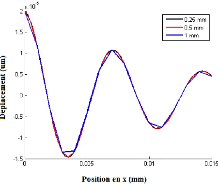
4.4.2 Effet de la taille des voxels ... 74
4.4.3 Effet de la rigidité ... 75
4.4.4 Interdépendance des paramètres ... 76
4.4.5 Évaluation du module G’’ ... 79
4.5 Discussion ... 79
4.6 Complément pour la méthode LFE ... 80
4.6.1 Résultats ... 81
4.6.2 Discussion ... 82
CHAPITRE 5 APPLICATION DE LA MÉTHODE AU DISQUE INTERVERTÉBRAL ... 84
5.1 Méthode expérimentale ... 84
5.1.1 Préparation des échantillons ... 84
5.1.2 Acquisition de l’image anatomique ... 87
5.1.3 Acquisition des données ÉRM ... 87
5.1.4 Traitement des données ... 90
5.2.1 Résultats préliminaires ... 92
5.2.2 Résultats de l’étude de fréquence ... 94
5.2.3 Validation ... 102 5.3 Discussion ... 105 5.3.1 LFE ... 105 5.3.2 VFM ... 106 5.3.3 AIDE ... 106 5.3.4 FF ... 107
5.3.5 Évaluation du module de perte ... 107
5.3.6 Limites ... 107
CHAPITRE 6 DISCUSSION GÉNÉRALE ... 110
CONCLUSION ... 114
RÉFÉRENCES ... 116
LISTE DES TABLEAUX
Tableau 1-1 : Table 1 - MRE experiments in the kHz range ... 23
Tableau 1-2 : Hypothèses d’inversion directe de l’équation algébrique ... 36
Tableau 4-1 : Plages de variation des paramètres ... 71
Tableau 4-2 : Estimations des modules de cisaillement pour des conditions idéales ... 72
Tableau 4-3 : Résumé des performances des algorithmes ... 79
Tableau 4-4 : Erreur absolue ... 81
LISTE DES FIGURES
Figure 1-1-1 : Figure 1 - displacement field components of the brain in the x, y and z directions (a, b and c) in micrometres at an arbitrary time point for an in vivo dataset. The total amplitude image (d) indicates the strong attenuation occurring within the brain as the shear waves move towards its centre. (reproduced with permission [42]) ... 24 Figure 1-2 : Figure 2 - An example of an in vivo reconstruction. (a) T2-weighted MR image; .... 24 Figure 1-3 : Figure 3 - Shear waves, or transverse wave, produce motion perpendicular to the
direction of propagation. In comparison, longitudinal waves, or compression waves, generate motion along the same axis as the direction of propagation. ... 25 Figure 1-4 : Figure 4 - Calibrated phantom used to verify the MRE technique and reconstruction. ... 25 Figure 1-5 : Figure 5 - Components of motion at 1250Hz for (A) normal and (B) degenerated
IVD (reproduced with permission [54]) ... 26 Figure 1-6 : Figure 6 - Shear waves propagating through the psoas (reproduced with permission) ... 26 Figure 1-7 : Figure 7- Experimental setup for generating shear waves in an ex vivo IVD ... 27 Figure 1-8 : Différentes méthodes d’élastographie (Adapté de Mariappan[3]) ... 28 Figure 3-1 : Schéma du système ÉRM. La séquence de gradients est contrôlée par la console et
est synchronisée avec le générateur de fonction par un pulse TTL. Le signal du générateur de fonction est amplifié puis filtré avant de stimuler l’actuateur piézoélectrique (Piezo) dans le montage. ... 44 Figure 3-2 : (1) Actuateur piézoélectrique et (2) une unité fonctionnelle vertèbre-DVI-vertèbre.
Une vertèbre est fixée au montage et une vertèbre liée à l’actuateur par une transmission flexible. ... 46 Figure 3-3 : Montage clinique enrobé in situ, le DVI (D) étant situé entre les vertèbres (V), avec
excitation mécanique par la vertèbre (a) ou par aiguille (b). ... 47 Figure 3-4 : Montage clinique planaire ... 47
Figure 3-5 : Montage clinique avec aiguille ... 48
Figure 3-6 : Montage préclinique planaire ... 49
Figure 3-7 : Montage préclinique concentrique ... 49
Figure 3-8: Actuateur avec chargement ... 51
Figure 3-9 : Position du vibromètre laser ... 51
Figure 3-10 : Déplacements mesurés par vibrométrie laser ... 52
Figure 3-11 : Déphasage 2D ... 53
Figure 3-12 : Déphasage 1D ... 53
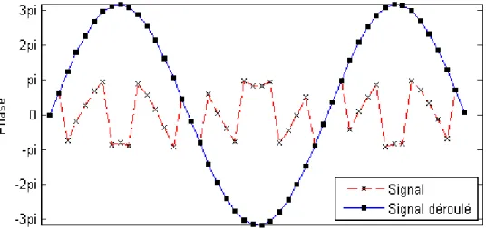
Figure 3-13 : Déroulage de la phase ... 54
Figure 3-14 : FFT temporelle ... 55
Figure 3-15 : Filtre gaussien 1D ... 56
Figure 3-16 : Filtre directionnel ... 57
Figure 3-17 : Indice de sensibilité des champs virtuels en fonction du nombre d’onde et de la phase. ... 62
Figure 3-18 : Résultats de l’algorithme VFM en ... 62
Figure 4-1 : Processus d’évaluation des algorithmes ... 64
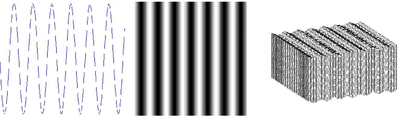
Figure 4-2 : Simulation élastique sur un vecteur en 1D, sur une tranche en 2D et sur un volume en 3D ... 66
Figure 4-3 : Simulation viscoélastique sur un vecteur en 1D, sur une tranche en 2D et sur un volume en 3D ... 66
Figure 4-4 : Effet du bruit ... 68
Figure 4-5 : Effet de l’échantillonnage spatial ... 69
Figure 4-6 : Effet de la rigidité sur la longueur d’onde ... 70
Figure 4-7 : Justesse des algorithmes testés pour G’=50kPa et une taille de voxel de 0.25mm. ... 73
Figure 4-9 : Erreur absolue des algorithmes testés pour G’=50kPa et un niveau de bruit nul ... 75
Figure 4-10 : Justesse de la méthode AIDE pour G’=50kPa ... 76
Figure 4-11 : Justesse de la méthode AIDE pour une taille de voxel de 1 mm ... 77
Figure 4-12 : G’ calculé vs G’ simulé pour une taille de voxel de 0.25 mm et un niveau de bruit nul ... 78
Figure 4-13 : G’ calculé vs G’ simulé pour une taille de voxel de 1 mm et un niveau de bruit nul ... 78
Figure 4-14 : Justesse de l’algorithme LFE pour G’=50kPa et une taille de voxel de 1 mm ... 81
Figure 4-15 : G’ calculé vs G’ simulé pour une taille de voxel de 1 mm et un niveau de bruit nul ... 82
Figure 5-1 : Disque vertébral isolé ... 85
Figure 5-2 : Position de l’actuateur ... 86
Figure 5-3 : Position du montage dans l’antenne ... 86
Figure 5-4 : Excitation latérale ... 89
Figure 5-5 : Excitation axiale ... 89
Figure 5-6 : Excitation concentrique ... 89
Figure 5-7 : Algorithme de traitement des données ... 90
Figure 5-8 : Définition des régions d’intérêt ... 92
Figure 5-9 : Vue anatomique haute résolution du DIV ... 92
Figure 5-10 : Cartes de déphasage (unités en rad) ... 95
Figure 5-11 : Cartes de module de cisaillement (unité en Pa) ... 95
Figure 5-12 : Résultats des algorithmes d’inversion dans le NP ... 96
Figure 5-13 : Résultat des algorithmes d’inversion dans l’AF ... 97
Figure 5-14 : Module de cisaillement de l’AF et du NP par LFE ... 98
Figure 5-16 : Module de cisaillement de l’AF et du NP par AIDE ... 100 Figure 5-17 : Module de cisaillement de l’AF et du NP par FF... 101 Figure 5-18 : Module de perte de l’AF et du NP par AIDE ... 102 Figure 5-19 : Résultats de validation du matériau d’enrobage par analyse de cisaillement
LISTE DES SIGLES ET ABRÉVIATIONS
ÉRM Élastographie par résonance magnétique DIV Disque intervertébral
ÉUS Élastographie par ultrasons
SE Spin echo
GE Gradient echo
LFE Local frequency estimation (estimation locale de fréquence) VFM Virtual field method (méthode des champs virtuels)
AIDE Algrebraic inversion of the direct equation (inversion alg. de l’équation directe) FF Formulation faible
NP Nucleus pulposus
AF Annulus fibrosus
G’ Module de cisaillement de conservation G’’ Module de cisaillement de perte
LISTE DES ANNEXES
ANNEXE 1 – Résultats de simulations ... 126 ANNEXE 2 – Résultats obtenus par méthode LFE ... 132
INTRODUCTION
La dégénérescence discale est une pathologie douloureuse entraînant de nombreuses consultations médicales chaque année. Les coûts importants et la perte de qualité de vie qu’elle engendre sont un fardeau tant au niveau personnel que social. Malgré les avancées dans le domaine, le diagnostic précoce et le traitement de cette maladie restent encore un défi. Les propriétés du disque intervertébral (DIV) changent de manière importante lors de la dégénérescence, mais leur évaluation est difficile à cause de la position anatomique du tissu. L’approximation indirecte des propriétés mécaniques par imagerie à résonance magnétique (IRM) multi-paramétrique a démontré un potentiel certain pour l’évaluation non-invasive du DIV. Toutefois, il n’existe actuellement pas de technique permettant d’évaluer les changements intrinsèques des propriétés mécaniques de manière non-invasive. L’évaluation mécanique du disque permettrait de détecter la pathologie dès les premiers stades de dégénération et d’effectuer un suivi quantitatif global de sa progression. La connaissance de l’environnement mécanique du DIV qui entraîne une réponse anabolique ou catabolique des cellules permettrait de proposer de nouveaux traitements à la dégénérescence du DIV qui sont aujourd’hui limités à la discectomie et la fusion.
L’élastographie par résonnance magnétique (ÉRM) consiste à visualiser la propagation des ondes mécaniques dynamiques dans un tissu biologique grâce à une séquence d’imagerie par résonance magnétique sensibilisée au mouvement et d’en extraire le module d’élasticité associé. Cette technologie est apparue comme une technique sensible et non-invasive pour l’évaluation des propriétés des tissus volumineux et mous favorisant la propagation des ondes. Comme le DIV est un tissu petit, rigide, viscoélastique et entouré d’un environnement osseux, une technique adaptée doit être développée.
L’objectif de ce projet est de développer une méthode d’ÉRM capable d’identifier les propriétés mécaniques dynamiques d’un DIV bovin in vitro de manière quantitative. Cela nécessitera la conception et la fabrication d’un système d’actuation non-magnétique synchronisé à une séquence IRM sensibilisée au mouvement pour acquérir le champ de déplacement et l’évaluation d’algorithmes d’inversion pour définir le mieux adapté au DIV. Les résultats devraient permettre d’évaluer différents paramètres d’acquisition et de définir les mieux adaptés au DIV.
Un article de revue de littérature mettra en évidence le potentiel de l’élastographie par résonance magnétique en ce qui a trait à l’évaluation du DIV et des compléments viendront l’enrichir en décrivant les techniques de mesure d’élasticité et d’inversion applicables. Les objectifs et hypothèses seront définis et la méthode sera présentée tant du point de vue de la conception du système d’actuation que des algorithmes d’inversion testés. Enfin, les résultats de l’application de la méthode à des données simulées et au DIV seront présentés et discutés.
CHAPITRE 1
REVUE CRITIQUE DE LA LITTÉRATURE
1.1 Article 1 : Mechanical characterisation of intervertebral disc
degeneration: a review of the promising magnetic resonance
elastography techniques
Pierre-François Beauchemin 1,2, Delphine Périé 1,2, Farida Cheriet 1,2, Sabine Bensamoun 3
1 École Polytechnique, Montréal, QC, Canada
2 Centre de recherche, CHU Sainte-Justine, Montréal, QC, Canada
3 Biomechanics and Bioengineering Laboratory, Université de Technologie de Compiègne, Compiègne, France
Cet article a été soumis le 18 septembre 2013 à la revue scientifique Journal of Biomechanical
Engineering
Abstract
Intervertebral disc (IVD) degeneration is a painful condition that can be caused by different factors including mechanical loadings, trauma, genetics or aging. Current clinical evaluations are limited to anatomic changes and pain response. However, during degeneration the IVD undergoes composition changes affecting its material properties which are not currently assessed clinically. Also, treatments have been disappointing because of poor understanding of the tissue biomechanics. New methods must be found in order to non-invasively assess the material properties and the mechanical environment of the IVD.
Multi-parametric MRI allows evaluation of tissue properties based on correlations with MRI parameters and was applied to the IVD. However, the extrapolation of the mechanical environment within the intervertebral disc from this technique remains difficult.
Magnetic resonance elastography (MRE) can allow non-invasive mechanical characterisation of the IVD by using vibrations to probe the tissue and phase-contrast MRI methods to image the propagation of acoustic shear waves via motion-sensitization gradients that
are phase-locked to the cyclic shear deformations. Inversion algorithms are then used to calculate the internal distribution of the shear stiffness within the tissue from the displacement field obtained through acquisitions. This could result in new diagnostic and treatment options.
This review overview multi-parametric MRI, the physical principles involved in MRE and the characteristics of the IVD with regards to MRE application. The last section discusses the adaptation of high frequency MRE to assess the mechanical environment of the IVD.
1.1.1 Introduction
Disc degenerationDegenerative disc disease provokes shape, structure and composition changes within the intervertebral disc (IVD). In clinical practice, this degeneration is investigated by physical examination, patient history, radiography/CT scan, magnetic resonance imaging (MRI) and/or discography. During degeneration the IVD undergoes many pathological changes. The loss of proteoglycan is accompanied by dehydration and height diminution. With matrix disorganization, these changes have a significant effect on the mechanical properties of the IVD and result in problematic stress concentration associated with discogenic pain [1].
Diagnosis
Radiography/CT scan is very limited as it is mainly used to exclude other diagnosis of other confounding conditions such as scoliosis, instability, spondylolisthesis or fracture [2].
MRI has the advantage of being able to assess directly neural and disc structures. Loss of intensity on T2-weighted images is associated with dehydration of the disc and can be related to disc degeneration [3]. Although, loss of signal can be caused by other conditions, such as disc protrusion, herniation or aging, and don’t necessarily indicate a symptomatic disc. This technique is an important tool but isn’t enough to diagnose disc degeneration in asymptomatic discs [2] and remains limited in the detection of early degenerative changes [4]. MRI remains the safest option in IVD evaluation as it is completely non-invasive and doesn’t require radiation exposure.
Discography is an invasive procedure which consists of injecting a contrast agent to image the disc structure. It is the only test giving both anatomical and functional information
about the IVD. The stage of disc degeneration is determined according to the shape and density of the dye’s shadow. For example, leakage of the dye outside of a globular pattern at the center of the disc is an abnormal pattern. During this test adjacent discs are used as control and should be pain free to confirm the diagnosis. This test is viewed as the most important diagnostic tool for degenerative disc disease, but should not be used for screening purposes because of its invasive nature [2, 5, 6]. This technique should be used carefully as it was determined that modern discography result in accelerated progression of degenerative changes [7]. Indeed, needle punctures provoke altered strain behavior in the annulus fibrosus which may initiate disc degeneration [8].
Treatments
The current major clinical procedures for treating disc degeneration have been disappointing because of subsequent degeneration at adjacent disc levels [9]. Although core trunk muscle strengthening and physical therapy are believed to help relieve back pain, physical activities could potentially lead to further IVD repair [10]. However, the thresholds beyond which physical activity causes injury or disease have to be defined. The knowledge of the mechanical environment within the IVD would be a critical step towards understanding its evolution and the guidance of the therapeutic treatment. Non-invasive imaging techniques allowing definition of the mechanical behavior of the IVD are required.
Imaging of mechanical properties
In order to increase MRI utility in the assessment of degenerative disc disease and to overcome its limitations, multi-parametric MRI was presented as a sensitive way to evaluate degenerative changes in the IVD [11]. However, the extrapolation of the mechanical environment within the IVD from this technique remains difficult.
Magnetic resonance elastography (MRE) can allow non-invasive mechanical characterisation of the IVD by using vibrations to probe the tissue and phase-contrast MRI methods to image the propagation of acoustic shear waves via motion-sensitization gradients that are phase-locked to the cyclic shear deformations. Inversion algorithms are then used to calculate
the internal distribution of the shear stiffness within the tissue from the displacement field obtained through acquisitions [12].
The next sections of this review will overview multi-parametric MRI, the physical principles involved in MRE and the characteristics of the IVD with regards to MRE application. The last section will discuss adaptation of high frequency MRE to assess the mechanical environment of the IVD.
1.1.2 Multiparametric MRI
Multi-parametric MRI is a technique that correlates quantitative measurements of MRI parameters, such as relaxation times, with tissue properties. It offers potential to assess changes in water content and biochemical composition of soft tissues that occur with aging and degeneration. Studies have correlated IVD water, proteoglycan, and collagen content to the following MRI parameters: longitudinal magnetization recovery (T1), the transverse magnetization decay (T2), and the magnetization transfer ratio (MTR) [13-15]. The apparent diffusion coefficient (ADC) was found to be directly related to the proteoglycans content and inversely related to the collagen denaturation [16]. Thus, multi-parametric MRI has been investigated as a sensitive and non-invasive technique for describing the alterations in mechanical properties of IVDs.
The mechanical properties of IVDs of bovine tails and their changes with the alteration of the tissue composition by enzyme treatment were characterized, and multiple linear regressions were investigated between MRI parameters and biomechanical properties. In the nucleus pulposus, correlations were obtained between the hydraulic permeability and T2 or ADC, and a trend was found between the compressive modulus and ADC [17]. Correlations were also found between the permeability or the compressive modulus and the spin lock relaxation time T1ρ in both nucleus pulposus and annulus fibrosus [18]. Moreover, the multiple linear regressions between the mechanical properties and MR parameters showed that the permeability of the nucleus pulposus and the swelling pressure of the annulus fibrosus can be expressed as a linear combination of T1, T2, T1ρ, MTR and ADC with a coefficient of determination of 0.8 and 0.7 respectively [18]. Weak linear correlations were found between the osmotic pressure and T1ρ of the human nucleus pulposus [19]. The compressive moduli and the permeabilities of isolated
IVDs can be assessed mostly by MTR and diffusion sequences with a coefficient of determination of 0.5-0.8 [20]. Moreover, this last study showed that multi-parametric MRI associated to principal component analysis and agglomerative hierarchical clustering are promising tools to classify the degenerated IVDs and further find biomarkers and predictive factors of the evolution of the pathologies. The immediate static compression of the isolated bovine IVD did not lead to any significant change of the MRI parameters, except for the diffusion that decreased in the direction of the compressive stress [21].
This technique has the advantage of being performed using standard MRI equipment without the need of special hardware. However, multi-parametric MRI doesn’t offer a direct evaluation of the disc integrity. In comparison, MRE allows computation of the intrinsic shear modulus instead of estimating material properties through correlations. Both techniques provide complementary information that can be used to assess composition and structural changes during degeneration.
1.1.3 Magnetic resonance elastography
MRE is a non-invasive imaging technique allowing quantification and mapping of the mechanical properties of biological tissues by imaging propagating shear waves using MRI. It was first introduced by Muthupillai in 1995 [22] and there are now a great number of MRE methods that can be differentiated by tissue of interest, mechanical excitation, data acquisition protocol and data processing algorithms.
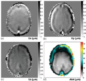
To characterize the mechanical properties of a tissue, its response to a mechanical stimulus must be measured and the property computed. In MRE, mechanical excitation is introduced in the tissue or sample using a vibrating device synchronized to the acquisition sequence. Data acquisition is performed using a MRI phase-contrast imaging technique allowing the mapping of the displacement field and thus visualization of propagating shear waves in the tissue (Figure 1). Once processed, this displacement map is used to compute shear modulus estimations (Figure 2). The main applications of MRE focus on relatively large and soft tissues or organs like the liver, the kidney, breast, skeletal muscle and brain [23-26]. These applications have been reviewed [27-29] and the technique is being adapted at high frequencies to tissues as small and stiff as trabecular bone [30], hyaline cartilage [31], mouse brain [32] and IVD [33, 34].
Elasticity and shear modulus
Under assumptions such as linear elasticity and isotropy, propagating harmonic waves can be used to determine the Lamé constants, λ and μ, respectively characterizing longitudinal and shear deformation. When subjected to vibrations, longitudinal and shear waves (Figure 3) propagate through the tissue. Longitudinal waves can have wavelengths of tens of meters in soft tissues, making accurate estimation of λ very difficult [35]. Assumptions neglecting their contribution can be made and partial filtering is possible because of their very low spatial frequency [36]. MRE focuses on the characterization of the shear modulus (μ), because shear waves have much shorter wavelengths.
Displacement measurement
Measurement of the propagating shear waves produced by a vibrating device is performed by using motion-sensitized MRI sequences. Motion sensitization is achieved through motion encoding gradients, which are oscillatory gradients synchronized to the mechanical excitation. These gradients introduce a phase difference proportional to the displacement of the material as shown in Equation 1. Thus, it is possible to obtain a displacement map by plotting the phase differences of the received signal.
( ⃗ ) ( ⃗ ⃗⃗ ) ( ⃗⃗ ⃗ ) Equation 1
Where: γ is the gyromagnetic ratio, N is the number of oscillations, T is the period of the oscillation, ⃗ is the gradient strength, ξ0 is the displacement vector, ⃗⃗ is the wave vector, ⃗ is the
spin position vector and α is the relative phase of the mechanical and magnetic oscillations [28]. Equation 1 shows that the phase offset is not only proportional to the displacement, but also to the gradient strength and to the number of bipolar waves. Thus, sensitization to very small displacement can be achieved by increasing the motion encoding gradient strength and the number of cycles. However, experiments have shown that even with careful synchronization, the phase variance increases with the number of bipolar waves [37].
During a single acquisition only one direction of motion can be captured in function of the orientation of the motion encoding gradient. However, by repeating the experiment with three
orthogonal directions of motion sensitization, it is possible to acquire a 3D displacement field [38].
Data processing
Following certain acquisitions, steps such as phase unwrapping [39] and filtering [38] may be necessary. Once the raw data has been clean up and artifacts removed, inversion algorithms are applied in order to compute the stiffness map from the displacement fields. These algorithms aim to solve the equations of motion by using various assumptions. These techniques can be either direct or iterative and have been reviewed in multiple articles [28, 36, 40].
As most biologic tissues exhibit viscoelastic properties, the complex shear modulus (G*) composed of the storage (G’) and loss (G”) modulus can be considered [41, 42]. These components are respectively related to the elastic and viscous portion of the complex shear modulus [43]. Both provide useful information regarding material properties of assessed as shown in figures 2 and 4. The use of biphasic model is also possible and makes it possible to estimate the time-harmonic pressure field resulting from excitation in fluid-saturated soft tissues [44, 45].
1.1.4 Intervertebral disc
The IVD’s main physiological functions are to support the loads applied on the spine while allowing flexibility of the upper body in all directions. In addition, it dissipates energy from impacts and acts as a shock absorber. In addition to factors such genetics and aging [46], mechanical loadings [47] and injuries [48] can also initiate degenerative disc disease which is a very painful condition.
Morphology & composition
The IVD has a small and irregular geometry as height ranges from 4 to 12 mm [49-51] and consists of two principal structures, the annulus fibrosus and nucleus pulposus, located between two cartilaginous endplates. The annulus fibrosus is located in periphery of the disc and is composed of lamellae of fibrous cartilage and collagen fibers wrapping the nucleus pulposus. The nucleus pulposus is a gelatinous tissue composed mainly of water, proteoglycan and a
network of collagen. As their composition and organisation differs, both the annulus fibrosus and nucleus pulposus have significantly different mechanical properties.
Mechanical properties
Viscoelastic properties and changes with degeneration of both principal structures of the IVD have been estimated through mechanical tests. As it is a cartilaginous tissue, the annulus fibrosus is significantly stiffer than soft tissues traditionally assessed by MRE [29]. In the annulus fibrosus, the dynamic shear modulus |G*| ranges from 100kPa to 400kPa depending on loading conditions and degeneration state and exhibit predominant elastic behavior as the tangent of the phase offset tan(σ) range from 0.1 to 0.7 [52]. The nucleus pulposus is significantly softer than the annulus fibrosus as |G*| ranges from 5kPa to 60kPa, with a higher energy dissipation as tan(σ) ranges from 0.43 to 0.33 [53]. With IVD degeneration, |G*| and tan(σ) increased in the annulus fibrosus while in the nucleus pulposus, |G*| increased and tan(σ) decreased [1]. This indicates that the viscoelastic shear modulus could be used as a biomarker for degenerative disc disease.
Differences have also been reported in the permeability of normal and degenerated IVDs, with increases in hydraulic permeability between 6% and 40% being measured. Intradiscal pressure was also showed to decrease with degeneration. The results from some researchers have been reviewed [1]. Advances in magnetic resonance poroelastography could allow further investigation of these pathological changes.
MRE applications
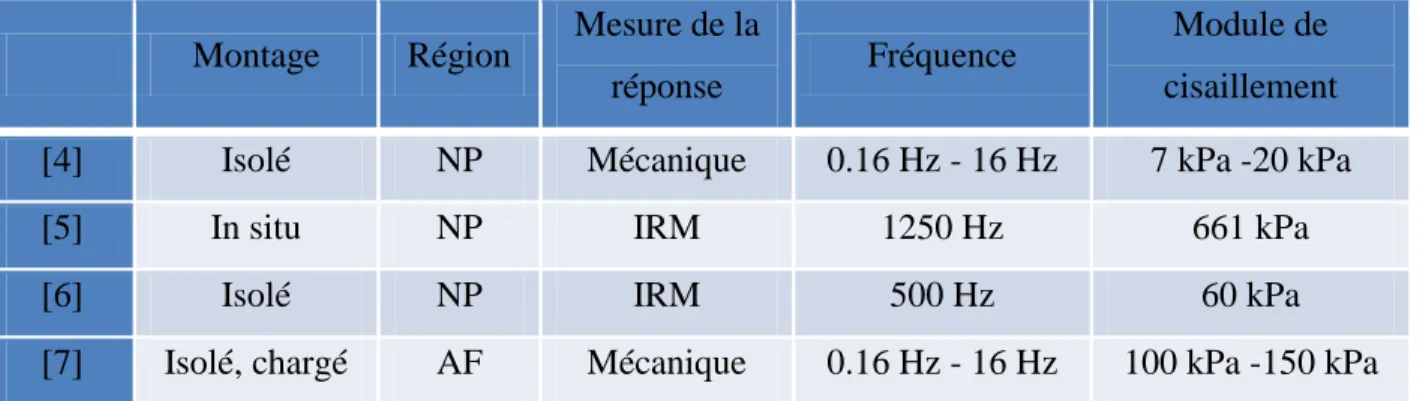
Few studies examined the adaptability of MRE to the IVD. Acquisitions have been performed on ex vivo bovine nucleus pulposus in the 500-1000 Hz range [34], with 500Hz offering the best operational compromise, and on ex vivo human nucleus pulposus [54] at 1250 Hz. In comparison, the annulus fibrosus presents a greater shear modulus and its best operational frequency can be estimated in the kHz range.
Cortes [54] also pointed the importance of acquiring dimensional displacement on 3 directions as a component showed much lower amplitudes than the other two (Figure 5). Moreover, acquisition in the annulus fibrosus can be complicated because of low signal and lower relaxation times limiting the number of motion encoding gradients [34].
Local frequency estimation and direct inversion algorithms were tested by Dunn [34], while Cortes [54] developed a robust method based on the first derivatives of the displacements because of noisy wave data. In both cases only the elastic component could be determined even though the IVD exhibits viscoelastic and poroelastic properties.
MRE can also be applied to surrounding tissues in order to assess the environment of the pathological IVD that will induce behavioral changes of the back muscles and ligaments [46]. In a recent study Bensamoun et al. [55] has demonstrated during a liver clinical test the potential of the MRE technique to provide the mechanical properties of the environmental liver tissue such as the psoas. A better knowledge of the back muscle behavior could enhance the development of rehabilitation program for IVD disease. Thus, in the present study, MRE tests were performed with a pneumatic driver placed on the low part of the back and the acquisitions of the MRE phase images (Figure 6) were recorded according to Bensamoun’s protocol which was previously described for muscle tissue. The clear propagation of the shear wave within the psoas muscle revealed the strong potential of the MRE technique to quantify the stiffness of the psoas and therefore other back muscles.
1.1.5 High frequency magnetic resonance elastography
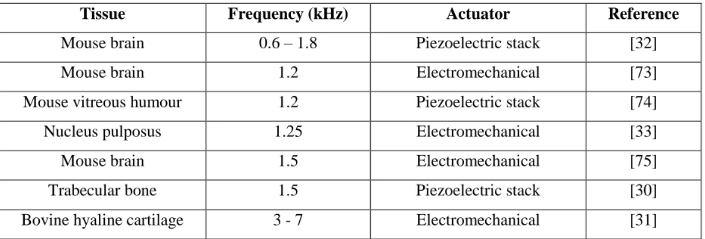
Considering that wavelength increases with stiffness and the small dimensions of the IVD, high frequency experiments in the kHz range are needed to obtain adequate results. Indeed, reducing the wavelength of the shear deformation by increasing the frequency allows the assessment of smaller and stiffer tissues as it reduces the residual fitting error [56]. However, high motion amplitude has to be achieved as mechanical vibrations of high frequency attenuate faster [29]. The best operational trade-off must be found that maximizes the resolution while allowing sufficient penetration in the tissue. High power actuation systems are needed to provide sufficient motion throughout the tissue at high frequencies. There are still few MRE experiments being reported in the kHz range. Applications at frequencies above 1 kHz are presented in Table 1.
Mechanical excitation
Many types of actuation systems can be used to generate the mechanical wave in MRE [57-59], but only a few can be used effectively in the kHz range. High frequency experiments typically use electromechanical or piezoelectric actuators as pneumatic actuators are limited to low frequency applications because of synchronization difficulties with the imaging sequence [60].
Electromechanical actuators are cheap, simple to design and provide oscillations of good amplitude in the kHz range. However, they have to be oriented perpendicularly to the MRI main magnetic field as they use it to generate movement and can create artifacts in their surroundings. They can overheat if used for long periods of time and the coil is sensitive to the switching gradients of the imaging sequence leading to imprecise motion amplitude and phase [61]. Cortes[33] used this type of actuator at 1250 Hz on the IVD and Lopez successfully used this type of actuator up to 10 kHz to characterize hyaline cartilage. In the second case, the use of a local gradient coil allowed the actuator to be located outside the range of the motion encoding gradients [31].
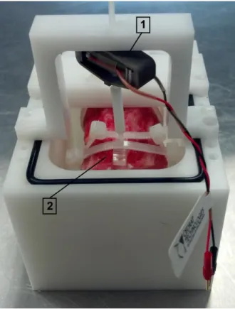
In comparison, piezoelectric actuators can be oriented freely in the MRI system and provide precise and stable excitation but can be costly because of the need for high-voltage amplifiers. Piezoelectric stacks [32, 59] are generally used in order to obtain sufficient motion. Indeed, Clayton used an amplified piezoelectric actuator up to 1.8 kHz. The actuator was in constant motion throughout the experiment in order to obtain a true steady-state response and didn’t suffer from overheating [32]. Piezoelectric stacks can be used to produce shear waves in an ex vivo IVD by using adjacent vertebrae as fixation elements (Figure 7).
Data acquisition
When using high frequency motion to excite the tissue, the specifications of the MRI must be analysed to make sure sufficiently power motion encoding gradients can be obtained. Fast switching gradients require hardware with high slew rates in order to achieve sufficient magnetic strength while keeping synchronization with the mechanical motion. The motion encoding gradient maximum strength is limited by hardware, but above a critical frequency, the maximum strength can be said to be directly proportional to the slew rate and inversely proportional to the oscillation frequency.
This phenomenon was reported [62] and experimental measurements of gradient strength in function of frequency were obtained. To increase the motion encoding gradient amplitude, Lopez built a custom-made z-axis gradient-coil that could achieve gradient strength 5 times greater than a clinical MRI at 5000Hz. This coil was designed for small samples of hyaline cartilage and used with a customized gradient-recalled-echo sequence [62]. This type of equipment is very helpful to increase the phase contrast but is still limited to applications on tissue samples.
Adaptation of the technique to mouse brain was possible through the use of a small-animal MR scanner at 4.7T. The sequence used was a modified spin echo with sinusoidal motion encoding gradients [32].
Validation methods
Most MRE setups are first tested on soft gels phantoms with known mechanical properties in order to evaluate the method’s reliability. Studies evaluating the viscoelastic shear modulus used dynamic shear testing or oscillatory rheometry [63] as a validation tools. These techniques allow the characterization of both components of the complex shear modulus but are limited to frequencies below 400 Hz [56, 64]. Also, comparison to mechanical testing can be difficult because of the difference in loading conditions.
Validation methods for MRE experiments focusing on the elastic shear modulus also include ultrasound elastography [24, 65]. Cross validation was performed and a significant correlation was found with no systematic divergence between the two methods [66]. Although no comparison was made with MRE, ultrasound elastography has been applied to intervertebral discs under loading and a significant correlation was established between the shear wave speed and the stiffness from mechanical testing [67].
In order to characterize stiff tissues like the IVD, new validation methods have to be considered. The hyper-frequency viscoelastic spectroscopy of biomaterials is a new technique allowing measurements of the complex shear modulus on a wide frequency range. This technique was tested on a variety of soft gels [68] with frequencies ranging from 10 Hz to 1000 Hz, is non-destructive and performed on small samples in controlled conditions [69].
1.1.6 Future challenges
MRE represent a potentially power evaluation tool but challenges remain to adapt MRE to the IVD. Until now, noise problems have been encountered during MRE acquisitions of the nucleus pulposus and have prevented analysis of the annulus fibrosus. This problem could be overcome by increasing motion encoding gradients strength or the amplitude of motion in the tissue through the use of specialized equipment, such as local gradient coils and optimized motion actuators.
Work was done to use the elastic shear modulus of the nucleus pulposus as a biomarker for disc degeneration, but both elastic and viscous components have this potential. The modelling of the wave propagation in the IVD should be improved. Until now only the elastic portion of the shear modulus in the nucleus pulposus has been characterized. Measurement of the viscoelastic behavior could prove itself very useful as it undergoes changes in both the annulus fibrosus and the nucleus pulposus during degeneration. The viscous portion can be determined using specific inversion algorithms or by acquiring data at multiple frequencies [28]. If acquisitions in the annulus fibrosus can be achieved, its non-linear and anisotropic behavior should be considered as they also vary with degeneration [70].
Validation through dynamic mechanical testing is still limited to low frequencies but recent techniques such as hyper-frequency viscoelastic spectroscopy seem promising. Although similar testing frequencies could be achieved, differences in loading conditions need to be addressed as they can influence mechanical properties. Establishment of a gold standard allowing direct comparison of the values obtain through MRE still remains.
The effect of many conditions on MRE of the IVD should also be studied in order to assess its response to various patterns. It was already shown that MRE of the skeletal muscle is sensitive to variation in loading conditions [71], age and voluntary contraction [72].
MRE could also benefit in the definition of multi-parametric MRI correlations. MRE would allow correlation between mechanical and MRI parameters both obtained almost simultaneously. Effective clinical tests based on these two methods could then be developed with non-invasive procedure or exposition to radiations.
MRE has already demonstrated its potential to be used as viable clinical tool for evaluation of liver fibrosis [29] and could be applied to the assessment of degenerative disc disease. It is also time efficient as it can be added to standard MRI acquisitions. Many of its characteristics make it an ideal tool to broaden our understanding of the IVD mechanics and evaluation of degenerative disc disease progression. It could allow the definition of new biomarkers based on shear modulus that could prove very useful as ex vivo mechanical testing demonstrated significant changes between the different severity stages of degenerative disc disease [52, 53]. Also, mapping of the tissue elasticity could possibly allow early localisation of specific areas of the disc being affected by degeneration.
1.1.7 Acknowledgements
Funded by the Natural Sciences and Engineering Research Council (NSERC) of Canada (discovery grant), the Groupe de Recherche en Sciences et Technologies Biomédicales (GRSTB), the Foundation of Stars and the Foundation CHU Sainte-Justine.
1.1.8 References
[1] Wang, Y., Chen, H. B., Zhang, L., Zhang, L. Y., Liu, J. C., and Wang, Z. G., 2012, "Influence of Degenerative Changes of Intervertebral Disc on Its Material Properties and Pathology," Chin J Traumatol, 15(2), pp. 67-76.
[2] Hasz, M. W., 2012, "Diagnostic Testing for Degenerative Disc Disease," Advances in orthopedics, 2012(pp. 413913.
[3] Rahme, R., and Moussa, R., 2008, "The Modic Vertebral Endplate and Marrow Changes: Pathologic Significance and Relation to Low Back Pain and Segmental Instability of the Lumbar Spine," AJNR. American journal of neuroradiology, 29(5), pp. 838-42.
[4] Luoma, K., Vehmas, T., Riihimaki, H., and Raininko, R., 2001, "Disc Height and Signal Intensity of the Nucleus Pulposus on Magnetic Resonance Imaging as Indicators of Lumbar Disc Degeneration," Spine J, 26(6), pp. 680-6.
[5] Modic, M. T., Masaryk, T. J., Ross, J. S., and Carter, J. R., 1988, "Imaging of Degenerative Disk Disease," Radiology, 168(1), pp. 177-86.
[6] Peh, W., 2005, "Provocative Discography: Current Status," Biomedical imaging and intervention journal, 1(1), pp. e2.
[7] Carragee, E. J., Don, A. S., Hurwitz, E. L., Cuellar, J. M., Carrino, J. A., and Herzog, R., 2009, "2009 Issls Prize Winner: Does Discography Cause Accelerated Progression of Degeneration Changes in the Lumbar Disc: A Ten-Year Matched Cohort Study," Spine J, 34(21), pp. 2338-45.
[8] Michalek, A. J., Buckley, M. R., Bonassar, L. J., Cohen, I., and Iatridis, J. C., 2010, "The Effects of Needle Puncture Injury on Microscale Shear Strain in the Intervertebral Disc Annulus Fibrosus," The spine journal : official journal of the North American Spine Society, 10(12), pp. 1098-105.
[9] Helgeson, M. D., Bevevino, A. J., and Hilibrand, A. S., 2013, "Update on the Evidence for Adjacent Segment Degeneration and Disease," The spine journal : official journal of the North American Spine Society, 13(3), pp. 342-51.
[10] Brisby, H., Wei, A. Q., Molloy, T., Chung, S. A., Murrell, G. A., and Diwan, A. D., 2010, "The Effect of Running Exercise on Intervertebral Disc Extracellular Matrix Production in a Rat Model," Spine J, 35(15), pp. 1429-36.
[11] Welsch, G. H., Trattnig, S., Paternostro-Sluga, T., Bohndorf, K., Goed, S., Stelzeneder, D., and Mamisch, T. C., 2011, "Parametric T2 and T2* Mapping Techniques to Visualize Intervertebral Disc Degeneration in Patients with Low Back Pain: Initial Results on the Clinical Use of 3.0 Tesla Mri," Skeletal radiology, 40(5), pp. 543-51.
[12] Muthupillai, R., and Ehman, R. L., 1996, "Magnetic Resonance Elastography," Nature medicine, 2(5), pp. 601-3.
[13] Antoniou, J., Pike, G. B., Steffen, T., Baramki, H., Poole, A. R., Aebi, M., and Alini, M., 1998, "Quantitative Magnetic Resonance Imaging in the Assessment of Degenerative Disc Disease," Magnetic resonance in medicine : official journal of the Society of Magnetic Resonance in Medicine / Society of Magnetic Resonance in Medicine, 40(6), pp. 900-7.
[14] Chatani, K., Kusaka, Y., Mifune, T., and Nishikawa, H., 1993, "Topographic Differences of 1h-Nmr Relaxation Times (T1, T2) in the Normal Intervertebral Disc and Its Relationship to Water Content," Spine J, 18(15), pp. 2271-5.
[15] Weidenbaum, M., Foster, R. J., Best, B. A., Saed-Nejad, F., Nickoloff, E., Newhouse, J., Ratcliffe, A., and Mow, V. C., 1992, "Correlating Magnetic Resonance Imaging with the Biochemical Content of the Normal Human Intervertebral Disc," Journal of orthopaedic research : official publication of the Orthopaedic Research Society, 10(4), pp. 552-61.
[16] Antoniou, J., Demers, C. N., Beaudoin, G., Goswami, T., Mwale, F., Aebi, M., and Alini, M., 2004, "Apparent Diffusion Coefficient of Intervertebral Discs Related to Matrix Composition and Integrity," Magnetic resonance imaging, 22(7), pp. 963-72.
[17] Perie, D., Iatridis, J. C., Demers, C. N., Goswami, T., Beaudoin, G., Mwale, F., and Antoniou, J., 2006, "Assessment of Compressive Modulus, Hydraulic Permeability and Matrix Content of Trypsin-Treated Nucleus Pulposus Using Quantitative Mri," Journal of biomechanics, 39(8), pp. 1392-400.
[18] Mwale, F., Demers, C. N., Michalek, A. J., Beaudoin, G., Goswami, T., Beckman, L., Iatridis, J. C., and Antoniou, J., 2008, "Evaluation of Quantitative Magnetic Resonance Imaging, Biochemical and Mechanical Properties of Trypsin-Treated Intervertebral Discs under Physiological Compression Loading," J Magn Reson Imaging, 27(3), pp. 563-73.
[19] Nguyen, M., Johannessen, W., Yoder, J. H., Wheaton, A. J., Vresilovic, E. J., Borthakur, A., and Elliott, D. M., 2008, "Noninvasive Quantification of Human Nucleus Pulposus Pressure with Use of T1ρ-Weighted Magnetic Resonance Imaging," Journal of bone and Joint Surgery, 90(pp. 796-802.
[20] Recuerda, M., Perie, D., Gilbert, G., and Beaudoin, G., 2012, "Assessment of Mechanical Properties of Isolated Bovine Intervertebral Discs from Multi-Parametric Magnetic Resonance Imaging," BMC musculoskeletal disorders, 13(1), pp. 195.
[21] Manac'h, Y. G., Perie, D., Gilbert, G., and Beaudoin, G., 2012, "Sensitivity of Multi-Parametric Mri to the Compressive State of the Isolated Intervertebral Discs," Magnetic resonance imaging, pp.
[22] Muthupillai, R., Lomas, D. J., Rossman, P. J., Greenleaf, J. F., Manduca, A., and Ehman, R. L., 1995, "Magnetic Resonance Elastography by Direct Visualization of Propagating Acoustic Strain Waves," Science, 269(5232), pp. 1854-7.
[23] Bensamoun, S. F., Glaser, K. J., Ringleb, S. I., Chen, Q., Ehman, R. L., and An, K. N., 2008, "Rapid Magnetic Resonance Elastography of Muscle Using One-Dimensional Projection," Journal of magnetic resonance imaging : JMRI, 27(5), pp. 1083-8.
[24] Bensamoun, S. F., Wang, L., Robert, L., Charleux, F., Latrive, J. P., and Ho Ba Tho, M. C., 2008, "Measurement of Liver Stiffness with Two Imaging Techniques: Magnetic Resonance Elastography and Ultrasound Elastometry," Journal of magnetic resonance imaging : JMRI, 28(5), pp. 1287-92.
[25] Mariappan, Y. K., Glaser, K. J., Hubmayr, R. D., Manduca, A., Ehman, R. L., and Mcgee, K. P., 2011, "Mr Elastography of Human Lung Parenchyma: Technical Development, Theoretical Modeling and in Vivo Validation," Journal of magnetic resonance imaging : JMRI, 33(6), pp. 1351-61.
[26] Sack, I., Beierbach, B., Hamhaber, U., Klatt, D., and Braun, J., 2008, "Non-Invasive Measurement of Brain Viscoelasticity Using Magnetic Resonance Elastography," NMR in biomedicine, 21(3), pp. 265-71. [27] Glaser, K. J., Manduca, A., and Ehman, R. L., 2012, "Review of Mr Elastography Applications and Recent Developments," Journal of magnetic resonance imaging : JMRI, 36(4), pp. 757-74.
[28] Manduca, A., Oliphant, T. E., Dresner, M. A., Mahowald, J. L., Kruse, S. A., Amromin, E., Felmlee, J. P., Greenleaf, J. F., and Ehman, R. L., 2001, "Magnetic Resonance Elastography: Non-Invasive Mapping of Tissue Elasticity," Medical image analysis, 5(4), pp. 237-54.
[29] Mariappan, Y. K., Glaser, K. J., and Ehman, R. L., 2010, "Magnetic Resonance Elastography: A Review," Clinical anatomy, 23(5), pp. 497-511.
[30] Chen, J., Mcgregor, H., Glaser, K., Mariappan, Y., Kolipaka, A., and Ehman, R., 2009, "Magnetic Resonance Elastography in Trabecular Bone: Preliminary Results," eds., Honolulu, pp.
[31] Lopez, O., Amrami, K. K., Manduca, A., and Ehman, R. L., 2008, "Characterization of the Dynamic Shear Properties of Hyaline Cartilage Using High-Frequency Dynamic Mr Elastography," Magn Reson Med, 59(2), pp. 356-64.
[32] Clayton, E. H., Garbow, J. R., and Bayly, P. V., 2011, "Frequency-Dependent Viscoelastic Parameters of Mouse Brain Tissue Estimated by Mr Elastography," Physics in medicine and biology, 56(8), pp. 2391-406.
[33] Cortes, D. H., Magland, J. F., Wright, A. C., Barocas, V. H., and Elliott, D. M., 2012, "Magnetic Resonance Elastography of Intervertebral Disc - a New Biomarker for Disc Degeneration," eds., pp. 83-84. [34] Dunn, T., 2005, "Magnetic Resonance Elastography at 3 Tesla: Implementation, Validation and Application to a Degenerative Disc Model," Ph.D. thesis, http://search.proquest.com/docview/305415884?accountid=40695 University of california, San Francisco.
[35] Manduca, A., 2005, Advanced Image Processing in Magnetic Resonance Imaging, CRC Press, Analysis of Dynamic Magnetic Resonance Elastography Data.
[36] Manduca, A., Oliphant, T. E., Dresner, M. A., Lake, D. S., Greenleaf, J. F., and Ehman, R. L., 2002, "Comparative Evaluation of Inversion Algorithms for Magnetic Resonance Elastography," eds., Washington, D.C., pp. 997-1000.
[37] Othman, S. F., Zhou, X. J., Xu, H., Royston, T. J., and Magin, R. L., 2007, "Error Propagation Model for Microscopic Magnetic Resonance Elastography Shear-Wave Images," Magnetic resonance imaging, 25(1), pp. 94-100.
[38] Manduca, A., Lake, D. S., Kruse, S. A., and Ehman, R. L., 2003, "Spatio-Temporal Directional Filtering for Improved Inversion of Mr Elastography Images," Medical image analysis, 7(4), pp. 465-73.
[39] Ghiglia, D. C., and Pritt, M. D., 1998, Two-Dimensional Phase Unwrapping: Theory, Algorithms, and Software, Path-Following Methods.
[40] Doyley, M. M., 2012, "Model-Based Elastography: A Survey of Approaches to the Inverse Elasticity Problem," Physics in medicine and biology, 57(3), pp. R35-73.
[41] Cheng, S., Gandevia, S. C., Green, M., Sinkus, R., and Bilston, L. E., 2011, "Viscoelastic Properties of the Tongue and Soft Palate Using Mr Elastography," Journal of biomechanics, 44(3), pp. 450-4.
[42] Green, M. A., Bilston, L. E., and Sinkus, R., 2008, "In Vivo Brain Viscoelastic Properties Measured by Magnetic Resonance Elastography," NMR in biomedicine, 21(7), pp. 755-64.
[43] Meyers, M. A., and Chawla, K. K., 2008, Mechanical Behavior of Materials, Cambridge University Press,
[44] Perrinez, P. R., Pattison, A. J., Kennedy, F. E., Weaver, J. B., and Paulsen, K. D., 2010, "Contrast Detection in Fluid-Saturated Media with Magnetic Resonance Poroelastography," Medical physics, 37(7), pp. 3518-26.
[45] Perrinez, P. R., Kennedy, F. E., Van Houten, E. E., Weaver, J. B., and Paulsen, K. D., 2010, "Magnetic Resonance Poroelastography: An Algorithm for Estimating the Mechanical Properties of Fluid-Saturated Soft Tissues," IEEE transactions on medical imaging, 29(3), pp. 746-55.
[46] Urban, J. P., and Roberts, S., 2003, "Degeneration of the Intervertebral Disc," Arthritis research & therapy, 5(3), pp. 120-30.
[47] Lotz, J. C., Colliou, O. K., Chin, J. R., Duncan, N. A., and Liebenberg, E., 1998, "Compression-Induced Degeneration of the Intervertebral Disc: An in Vivo Mouse Model and Finite-Element Study," Spine J, 23(23), pp. 2493-506.
[48] Osti, O. L., Vernon-Roberts, B., and Fraser, R. D., 1990, "1990 Volvo Award in Experimental Studies. Anulus Tears and Intervertebral Disc Degeneration. An Experimental Study Using an Animal Model," Spine J, 15(8), pp. 762-7.
[49] 1978, "Mechanical Response of the Lumbar Intervertebral Joint under Physiological (Complex) Loading," The Journal of Bone & Joint Surgery, 60(1), pp. 41-55.
[50] Lin, H. S., King Lui, Y., Ray, G., and Nikravesh, P., 1978, "Systems Identification for Material Properties of the Intervertebral Joint," Journal of biomechanics, 11(1–2), pp. 1-14.
[51] Taylor, J. R., 1975, "Growth of Human Intervertebral Discs and Vertebral Bodies," Journal of anatomy, 120(Pt 1), pp. 49-68.
[52] Iatridis, J. C., Kumar, S., Foster, R. J., Weidenbaum, M., and Mow, V. C., 1999, "Shear Mechanical Properties of Human Lumbar Annulus Fibrosus," Journal of orthopaedic research : official publication of the Orthopaedic Research Society, 17(5), pp. 732-7.
[53] Iatridis, J. C., Setton, L. A., Weidenbaum, M., and Mow, V. C., 1997, "Alterations in the Mechanical Behavior of the Human Lumbar Nucleus Pulposus with Degeneration and Aging," Journal of orthopaedic research : official publication of the Orthopaedic Research Society, 15(2), pp. 318-22.
[54] Cortes, D. H., Magland, J. F., Wright, A. C., and Elliott, D. M., 2013, "The Shear Modulus of the Nucleus Pulposus Measured Using Magnetic Resonance Elastography: A Potential Biomarker for Intervertebral Disc Degeneration," Magnetic resonance in medicine : official journal of the Society of Magnetic Resonance in Medicine / Society of Magnetic Resonance in Medicine, pp.
[55] Bensamoun, S. F., Robert, L., Leclerc, G. E., Debernard, L., and Charleux, F., 2011, "Stiffness Imaging of the Kidney and Adjacent Abdominal Tissues Measured Simultaneously Using Magnetic Resonance Elastography," Clinical imaging, 35(4), pp. 284-7.
[56] Okamoto, R. J., Clayton, E. H., and Bayly, P. V., 2011, "Viscoelastic Properties of Soft Gels: Comparison of Magnetic Resonance Elastography and Dynamic Shear Testing in the Shear Wave Regime," Physics in medicine and biology, 56(19), pp. 6379-400.
[57] Rossman, P. J., Muthupillai, R., and Ehman, R. L., 1999,
[58] Tse, Z. T., Janssen, H., Hamed, A., Ristic, M., Young, I., and Lamperth, M., 2009, "Magnetic Resonance Elastography Hardware Design: A Survey," Proc Inst Mech Eng H J Eng Med, 223(4), pp. 497-514.
[59] Uffmann, K., Abicht, C., Grote, W., Quick, H. H., and Ladd, M. E., 2002, "Design of an Mr-Compatible Piezoelectric Actuator for Mr Elastography," Concepts in Magnetic Resonance, 15(4), pp. 239-254. [60] Uffmann, K., and Ladd, M. E., 2008, "Actuation Systems for Mr Elastography: Design and Applications," IEEE engineering in medicine and biology magazine : the quarterly magazine of the Engineering in Medicine & Biology Society, 27(3), pp. 28-34.
[61] Uffmann, K., 2001, "Characterization of an Electromagnetic Actuator for Mr Elastography," eds., Glasgow, pp.
[62] Lopez, O., Amrami, K. K., Manduca, A., Rossman, P. J., and Ehman, R. L., 2007, "Developments in Dynamic Mr Elastography for in Vitro Biomechanical Assessment of Hyaline Cartilage under High-Frequency Cyclical Shear," Journal of magnetic resonance imaging : JMRI, 25(2), pp. 310-20.