Université de Montréal
GENETIC-EPIDEMIOLOGIC ANALYSIS 0F X4NACTWATION SKEWING IN
HUMAN FEMALES: SUGGESTIVE EVIDENCE FOR TWO DISTINCT TRAITS
par
Robert Mio
Département de médecine
Faculté de médecine
Thèse présentée à la Faculté des études supérieures
en vue de l’obtention du grade de
Philosophiae Doctor (Ph.D.)
en sciences biomédicales
novembre 2003
u)
L
u36
V.
c3—
Université
de Montréal
Direction des bibliothèques
AVIS
L’auteur a autorisé l’Université de Montréal à reproduite et diffuser, en totalité ou en partie, par quelque moyen que ce soit et sur quelque support que ce soit, et exclusivement à des fins non lucratives d’enseignement et de recherche, des copies de ce mémoire ou de cette thèse.
L’auteur et les coauteurs le cas échéant conservent la propriété du droit d’auteur et des droits moraux qui protègent ce document. Ni la thèse ou le mémoire, ni des extraits substantiels de ce document, ne doivent être imprimés ou autrement reproduits sans l’autorisation de l’auteur.
Afin de se conformer à la Loi canadienne sur la protection des renseignements personnels, quelques formulaires secondaires, coordonnées ou signatures intégrées au texte ont pu être enlevés de ce document. Bien que cela ait pu affecter la pagination,
il
n’y a aucun contenu manquant.NOTICE
The author of this thesis or dissertation has granted a nonexclusive license allowing Université de Montréal to reproduce and publish the document, in part or in whole, and in any format, solely for noncommercial educational and research purposes.
The author and co-authors if applicable retain copyright ownership and moral rights in this document. Neither the whole thesis or dissertation, nor substantial extracts from it, may be printed or otherwise reproduced without the author’s permission.
In compliance with the Canadien Privacy Act some supporting forms, contact information or signatures may have been removed from the document. While this may affect the document page count, it does flot represent any loss of content from the document
faculté des études supérieures
Cette thèse intitulée:
GENETIC-EPIDEMIOLOGIC ANALYSIS 0f X-INACTIVATION SKEWING IN
HUMAN FEMALES: SUGGESTiVE EV1DENCE FOR TWO DISTINCT TRAITS
présenté par:
Robert Mio
A été évalué par un jury composé des personnes suivantes:
Présidente-rapporteur
Manjunath Puftaswamy, PhD
Directeur de recherche
Lambert Busque, MD (fRCPC)
Membre du jury
Eric Milot, PhD
Examinateur externe
Anna Naumova, PhD (McGill University)
Gary Gilliland, MD PhD (Harvard University)
Représentante du doyen
Dennis Claude Roy, MD (fRCPC)
III
SUMMARY
C
_
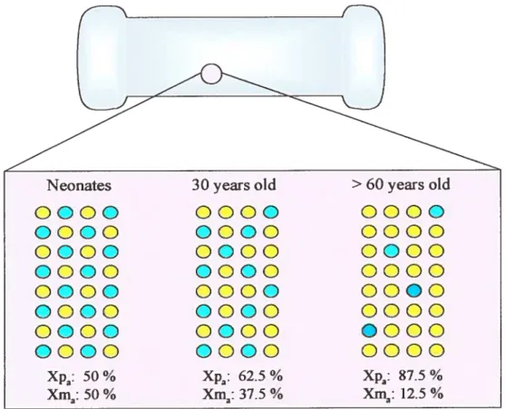
Introduction: X mactivation (Xi) randomly mactivates a single X chromosome (maternai or
patemai) in female somatic ceils. In a significant proportion of females however, Xi is flot random, a phenomenon termed skewed Xi [arbitrarily defmed as preferentiai inactivation (75%) of the maternai (Xm) or patemal X (Xp)]. The proportion ofXp inactivated relative to Xm is termed the X inactivation ratio (XIR). The study of Xi in humans is hampered by two unrelated phenotypes. i) TheprimaryXi trait. Initiated during early embryogenesis, in a developmental context, it induces a similar Xi pattern, which can vary ftom random to skewed, among varions tissues. A resulting skewed Xi paffem, tenned primary skewing (PS), may resuit from a small number of stem ceils present when Xi is initiated However, other possibilities include genetic influences, such as heterozygosity for the X-linked Xce locus, as observed in certain mice hybrids. That 9% of human female neonates demonstrate a skewed Xi pattem in cord blood supports a PS trait in humans. ii) the secondwy skewing trait, usualIy associated with a skewed Xi pattem in a tissue-specific mariner, occurs after the initiation of Xi. Secondary skewing often resuits from a growth competition between X-linked alleles, such as in female carriers of various X-linked immunodeflciency disease alleles. Flowever, X-linked disease alleles are rare and do not explain the high prevalence of skewing (38%) as observed in peripheral blood (PB) of ‘healthy’ females 60 years of age and older. The latter trait, termed acquired skewing (AS), lias been assigned varlous etiologies. Recent data support an X-linked genetic component influencing hematopoietic stem ceil (HSC) growtWsurvival kinetics. Clinically, skewed Xi lias been associated with varlous biomarkers (breast cancer and recurrent spontaneous abortion for example). In light of this data, a study was undertaken to resolve the etiologies and biological / ciinical associations of Xi traits in liuman femaies. Methods: French Canadian nuclear families (females oniy) were recruited for study analysis. Two biological tissues were obtained: PB for analysis of the AS traitand buccal cetis (BC) for analysis of the primary Xi trait. PB was fractionated and cell-sorted to obtain pure-ceil populations. The XIR was determined by the HUMARA clonaiity assay. Xi phenotypes included. i) The )UR derived from BC for analysis of the primary Xi trait. ii) The XW of PB for analysis of the AS trait and iii) the relative value of AS obtained by quantitating deviation from the primary )UR (i.e., difference between the BC and
P13 )UR). Qualitative analyses included a skewed Xi pattem (25% deviation from random Xi for
the )UR and an AS value 0,25 for the relative AS trait). Subject data included a medical questionnaire and blood counts, which were analyzed as a function of Xi phenotypes. Genetic effects were determined by heritability studies. Resuits and Conclusion: 1144 females derived from
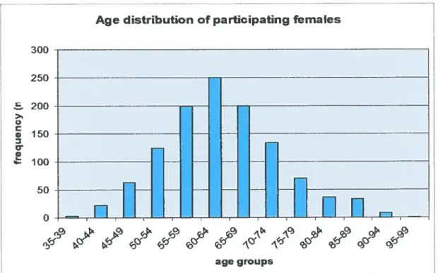
193 nuclear families were recru ited. Age ranged from 3$-96 years, with a mean of 63,3 years.
90,8% of females were informative for the HIJMARA assay. findings related to the BC XIR were
neonatal cord blood, i.e. 8,6%); ii) the incidence of Xi skewing was relatively stablewith advancing age (p=O,21); iii) the BC )UR was modestly correlated with that of PB lineages (O,46<r<O,56) and iv) heritabiÎity analysis revealed a genetic (piausibly X-linked) component (p<O,0001; h2O,30). These findings are similar to the Xce-influenced primary Xi trait in mice, supporting evidence for an XCE-Iike primary Xi trait in humans. further, this fmding suggests that the primary Xi trait in humans is flot strictly a stochastic process as previously suggested. Clinically, the BC and blood
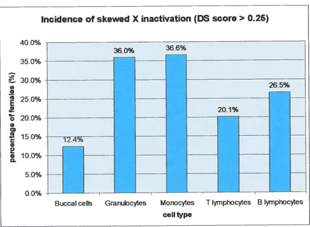
)UR was associated with asthma. Among hematopoietïc lineages, the incidence of skewed Xi was
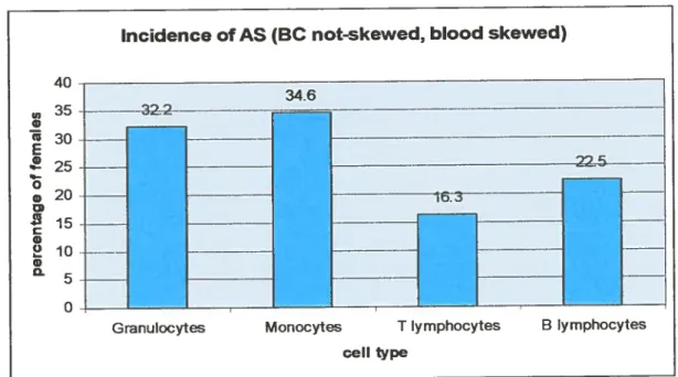
higherversus BC: granulocytes (PMN) 36%; monocytes 36,6%; T cells 20,1% and B cells 26,5%. The incidence of relative AS was 22,7; 27,2; 11,4 and 16,3%, respectively. Withthe exception of T ceils, signilhcant correlation of AS values among B ceils and myeloid lineages (0,73<r<0,$5) was
consistent with a FISC origin of AS. The incidence of AS (relative and absolute) increased
significantly with age, particularly for myeloid lineages. Lack of T celis contribution may be attributed to long-lived memory T cells. Heritability estimates attributed 20-39% of the variance of AS values to genetic effects (plausibly X-Iinked), supporting linkage studies to map the trait(s).
Clinically, increasing AS values were associated with a decreased eosinophil count. Because
eosinophïl count is a predictor of ail-cause mortaiity, the finding suggests AS may 5e associated with increased longevity. In effect, that the AS trait demonstrated a different biological profile versus the PS trait (i.e., late versus early-onset, increased incidence of skewing with age versus stable, different incidence of skewing, and a different clinical profile) is convincing evidence for
twodistinct traits.
Key words: X chromosome, acquired skewing, primary skewing, HUMARA, heritability,
hematopoietic stem ceNs, buccal ceils, human, females, Québec, hematopoietic lineages, association studies, clinical data, complete blood counts, age
V
RÉSUMÉ
C
.Introduction: Dans les cellules somatiques de femmes, I mactivation du chromosome X (iX) inactive, de façon aléatoire, l’un des deux chromosome X (maternel ou paternel). Cependant, dans une certaine proportion de femmes, l’inactivation n’est pas aléatoire, un phénomène appelé IX biaisé [arbitrairement défmi comme étant une inactivation préférentielle (?75%) du X maternel (Xm) ou paternel (Xp)J. La proportion de Xp inactivé relatif au Xm est appelé le ratio d’ inactivation du X (RIX). Chez l’humain, l’étude de iX est entravée par deux phénotypes non reliés: i) Le LX
primaire. Amorcé tôt durant l’embryogenèse, dans un contexte de développement donné, induit un RIX similaire dans divers tissus, soit aléatoire ou biaisé, ce dernier est appelé le biaisé primaire (BP). Le BP peut provenir d’un petit nombre de cellules souches présentent lors de l’initiation du
IX. D’autres causes incluant les influences génétiques, comme l’hétérozygocité du locus Xce lié au
X, observée dans certaines hybrides de souris. Le fait que chez 9% des nouveau-nés de sexe féminin on observe l’IX biaisé dans le sang du cordon suggère un phénotype BP chez l’humain, ii) Le phénotype biaisé secondaire, habituellement associé avec l’iX biaisé dans un tissu spécifique, a lieu après l’initiation de IX. Le biais est le résultat d’un désavantage de croissance conféré par des allèles mutant liés au X. Les exemples incluent des porteuses de différents allè]es de maladies immunodéficientes liées au X. Malgré cela, ces allèles sont rares et n’expliquent pas la haute fréquence (3 8%) de ce biais observé dans le sang périphérique ($P) chez des femmes âgées de 60 (au plus) et en santé. Ce phénotype, appelé biais acquis (BA), a été associé à diverses causes. De récentes données indiquent une composante génétique liée au X qui influence la cinétique des cellules souches hématopofétiques (CSH). Cliniquement, le phénotype iX biaisé a été associé à différents marqueurs biologiques comme le cancer du sein et l’avortement spontané répétitif
À
la lumière de ces données, une étude a été entreprise pour élucider les causes et les associations biologiques des deux phénotypes iX chez les femmes. Méthodes: Des familles Canadiennes Françaises (femmes seulement) ont été recrutées pour participer à l’étude. Deux tissus biologiques ont été recueillis pour analyse: le sang périphérique (SP) pour l’analyse du phénotype BA et des cellules buccales (CB) pour l’analyse du phénotype iX primaire. Le SP a été fractionné, et ses cellules classées pour obtenir des populations de cellules pures. Le RIX a été déterminé par la méthode de HUMARA. Les phénotypes de iX inclus sont: i) Le RIX obtenu des CB, représentatifdu phénotype IX primaire. ii) La valeur absolue du RIX pour ]‘analyze du phénotype BA et iii) la
valeur BA relatif calculé selon la différence entre les RIX primaires des CB et du SP. Pour les analyses qualitatives, le RIX biaisé (25%) et la valeur BA relatif (0,25) ont été utilisé. Les données cliniques incluant un questionnaire médicale et les analyse sanguines, ont été analysées en relation avec le lUX. Une composante génétiquelfamiliale des phénotypes a été évaluée par une
étude familiale. Résultats et conclusion: Parmi 193 familles, 1144 femmes ont été recrutées. Ces
(E
dernières étaientâgées entre 3$ et 96 ans, pour une moyenne de 63,3 ans. Les résultats obtenus pour le phénotype DC primaire sont de quatre ordres : i) la fréquence du DC biaisé dans les CB était faible (12,4%-similaire au résultat obtenu dans les cordons de nouveau-nés (8,6%)), ii) le RIX des CBétait relativement stable avec l’âge (p=O.2l), iii) le RIX des CB corrélait signilitivement avec celui
des types cellulaires du SP (O,46<r<O,56) et iv) l’héritabilité des RIX des CB suggère une composante génétique (p<O,000I; h2=O,30). Ces propriétés sont en lien direct avec un phénotype iX primaire, possiblement en relation avec un locus Xce lié au X. Ceci suggère que le IX primaire chez l’humain n’est pas un processus strictement stochastique comme suggéré précédemment. Cliniquement, les RIX ont été associé à une augmentation des cas d’asthme. Parmi les types hématopoïétiques, l’incidence du DC biaisé était plus élevée par rapport au CB: granulocytes (PMN) 36%, monocytes 36,6%; lymphocytes T 20,1% et lymphocytes B 26,5%. En utilisant la valeur BA
relatil l’incidence du BA (valeur 0,25) était 22,7; 27,2; 11,4 et 16%, respectivement.
À
l’exception des cellules T, chez un individu les RIX entre les différentes cellules hématopoïétiques corrèlent d’un façon significative (O,73<r<O,85), ce qui supporte une d’origine CSH. L’incidence de BA augmentent significativement avec l’âge, particulièrement pour les types myéloïdes. Une incidence du BA moins fréquente chez les lymphocytes T pourrait être attribuée à une plus grande
(E
longévité. Les études d’héritabilité ont attribué 20 à 39% de la variance des valeurs de BA auxeffets génétiques, ce qui supportent les études de liaisons génétiques pour localiser les phénotypes. Cliniquement, l’augmentation des valeurs de BA était associée à une diminution du nombre d’éosinophiles. Puisque le compte d’éosinophiles est associé à la mortalité, une association négative suggère que le BA est associé à la longévité. En conséquence, le fait que le phénotype BA ait démontré un profil biologique différent de celui du phénotype BP (i.e. : l’apparition acquise versus primaire, l’incidence de iX biaisé differentes et les profils cliniques differentes) suggère fortement deux phénotypes distincts.
Mots clés: chromosome X, biaisé acquise, biaisé primaire, HUMARA, héritabilité, cellules souches hématopoïétiques, cellules buccales, humain, femme, Québec, lignées hématopolétiques, études d’association, donnée clinique, analys&sanguines, l’âge
vi’
TABLE 0F CONTENTS
SUMMARY iii
RÉSUMÉ
yTABLE 0F CONTENTS vii
LIST 0F FIGURES xv
LIST 0f TABLES xvi
LIST 0F ABBREVIATIONS xviii
ACKNOWLEDGEMENTS xxi
Chapter 1
INTRODUCTION AND LITERATURE REVIEW 1
1 THE X CHROMOSOME 2
1.1 Basic principles 2
1.2 Lyon’s Hypothesis 2
1.3 Anatomical human X-mosaicism 3
1.4 Timing of X inactivation: concurrent with tissue dfferentiation 4
2 MECHANISM 0F X INACTWATION 5
G
Abbreviations: Xtm, maternai X; X”, paternal X; a, active; ï, inactive2.1 The X inactivation center (Xic/XIC) 562.2 Genetic elements ofthe Xic: Xist gene 9
2.2.1 Deveiopmental regulation ofXist 10
2.3 Characteristics acquired with Xist upregulation 10
Methylation ofCpG islands of Xi-linked genes 10
Asynchronous replication ofthe inactive X 10
Covalent modifications of X chromosome associated histones 12
Enrichment of histone variants 13
Enrichment ofmRNA species 13
2.4 Stability of X inactivation 13
3 REGULATORY ELEMENTS 0F THE )UC 13
3.1 further characterization of the Tsix gene 14
4 IMPRINTED X NACTIVATION 15
4.1 Preferential inactivation ofthe paternal X in extraembryonic tissues 15
4.2 X-iinprinting in humans 16
5 ESCAPE FROM X INACTIVATION 16
6 CLINICAL APPLICATION 0f THE LYON’S HYPOTHESIS 17
6.2 Principles of X-inactivation based clonality assays.17
6.2.1 DistinguishXpftomXm 1$
6.2.2 Distinguish active X (aX) from inactive X (iX) 1$
6.3 Utility of Xi based clonality assays 1$
6.3.IHUMARAassay 18
6.4 Hematological application of clonality assays 27
6.4.1 ClonaI origin ofhematologic malignancies 27
6.4.2 Stem celi origin of hematologic malignancies 27
6.4.3 Carrier detection ofX-linked disorders 27
6.5 Limitation of X-inactivation based clonalhy assay 2$
7 NONRAN1JOM X-INACTWATION 2$
7.1 Binomial distribution of X-inactivation ratios 2$
7.2 Skewed X-inactivation as a discrete trait 2$
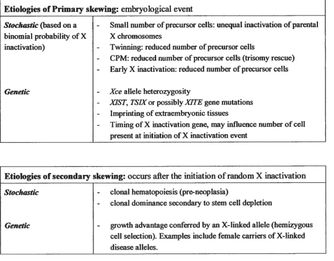
7.3 Etiologies of skewed X inactivation 29
7.3.1 Etiology ofprimary skewing: Stochastic event 31
7.3.2 Etiology of primary skewing: a heritable trait 31
7.3.2.1 Xce allele heterozygosity: a skewed Xi pattern in multiple tissues 32
7.3.2.2 )USTiTSIX aberrations as a cause ofprimaryskewing 33
7.3.3 Etiology of Secondary Skewing: selection against X-linked disease allele(s) 33
7.3.3.1 Suggestive evidence that secondary skewing may be incompletely penetrant 34
7.3.3.2 Nonhematopoietic tissues are generally robust to secondary skewing 35
7.4 Familial clustering of skewed Xi: Primary or secondary etiology9 35
7.5 Randomly ascertained families to identify the etiology of skewed Xi 37
$ iNCIDENCE 0F SKEWED Xi IN TFW GENERAL FEMALE POPULATION: variable 37
$.1 Higher incidence of skewed Xi in peripheral blood versus N}lTs 3$
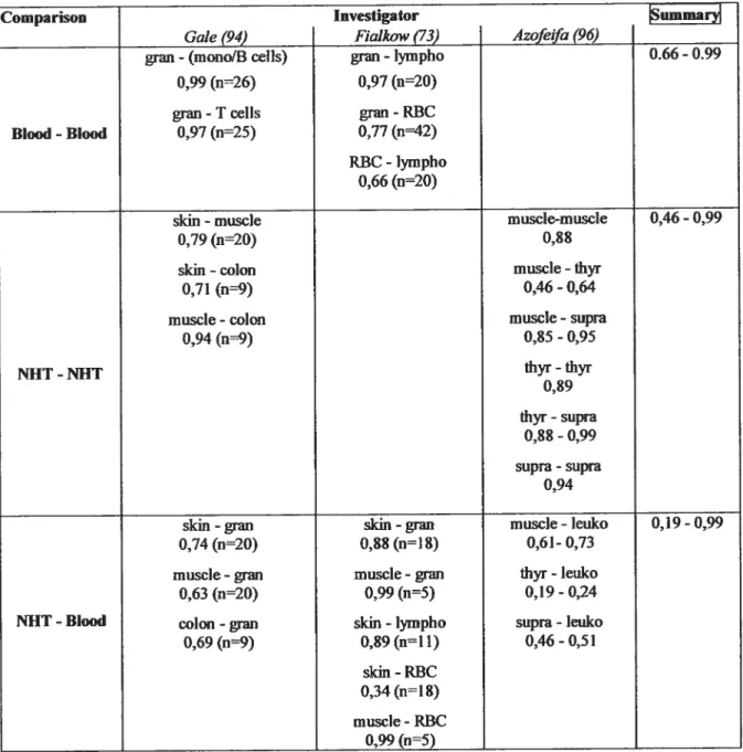
8.2 Correlation ofXIRs among varlous tissues 3$
8.2.1 Correlation ofXWs among blood ceil lineages: strong 38
$.2.2 Correlation ofXIRs among NIITs: moderate, evidencefor a body-wide Xli? 39
8.2.3 Correlation ofXIRs between blood celis and MITs: low 39
Concordance ofXipatterns between bÏoodandNHTs: evidence ofo body-wide Xi pattern....41 8.3 Suggestive evidence for anXCE-like X chromosome skewing trait 41
9 ANALYSIS 0f BLOOD Xi PATTERNS AS A FUNCTION 0F AGE 41
9.1 AS: confirmation studies 42
9.2 Clarifications in light of the AS phenomenon 43
10 ETIOLOGY 0F SKEWED Xi IN CORD BLOOD:primary skewing trait 43
Ix
11.1 Stochastic event: clonai dominance secondary to stem ce!! depletion 44
11.2 Stochastic event: clona! ‘pre-neop!asia” hematopoiesis 45
11.3 Continuation of the PS trait 46
11.4 Genetic: X-linked hemizygous cet! selection 46
12 X-LINKED HEMIZYGOUS CELL SELECTION ETIOLOGY 0F AS 46
12.1 AS in felines 46
12.1.1 Hematopoietic stem celi origin of AS 47
12.2 X-linked genetic basis of the AS trait extended to humans 4$
12.3 Twin studies support an X-Iïnked genetic basis to AS in humans 4$
12.3.1 Cone1ation ofXIRs in elderly MZ twin pairs 4$
12.3.2 Genetic contribution to the AS trait 50
12.4 Infrmnsic versus extrinsic factors in the etiology of AS 50
12.5 Molecular characteristics ofthe AS trait: Genetic model 51
13 CLII’IICAL ASSOCIATIONS 0f SKEWED Xi 52
13.1 Skewing and expression of X-linked disease allele(s) 52
13.2 SkewingandRSA 53
13.3 Skewing and susceptibility to ovariaWbreast cancer 53
13.4 Skewing and predisposition to autoimmunity 54
13.5 Skewingandlongevity 55
13.6 Therapeutic applications 55
14 HYPOTHESIS and GOALS 55
15 STRATEGIES, RATIONALE AND EXPERIMENTAL DESIGN 56
15.1 Recruitment ofelderly females 57
15.2 Genetic analyses: sib-pair approach to dissect a complex genetic trait 57
15.3 Collection offamilies 57
15.3.1 Benefit ofa re)atively isolated population 57
15.4 Sample size (7. estimation): power stiidies 58
15.5 Defmition of the phenotype 59
Chapter II 60
MATERIALS AND METHODS 60
2.1 Siibjects 61
2.2 Medical Questionnaire 61
2.2.1 Age 61
2.2.2 Smoking habits: environmental stimulus 61
2.2.3 family data: skewing and X-linked mutant alleJe(s) 62
2.3 Biological samples .63
C
2.3.1 Primary Xi pattern / PS trait 632.3.2 AS trait: peripheral blood 63
2.4 Complete blood counts 64
2.5 Cellular fractionation 64
2.6 Immunophenotyping and ceil sorting 64
2.7 DNA isolation from blood ceils 65
2.8 DNA isolation from buccal ceils 65
2.9 HUMARA clonality assay 66
2.9.1 HUMARA allelic ladder 67
2.9.2 Quantitation ofHUMARA alleles 67
2.93 ReÏiability of quantification: accuracy 6$
2.10 Calculation ofthe XIR 6$
2.10.1 DS score (degrec ofskewing) 6$
2.10.2 PAmat score (Proportion Active of the Maternally inherited X chromosome) 68
2.10.3 scoreS 69
2.IO.4A$Ds score 69
2.11 Discrete criteria to delineate a skewed Xi paffem 69
2.11.1 $kewing as a discrete trait 69
2.11.2 Acquired skewing as a discrete trait 70
2.12 familial resemblance of Xi paffem traits 70
2.12.1 familial aggregation of skewed Xi: recurrence risk ratio (RRR) 70
2.12.2 $ibling correlation ofXIRs by ANOVA 71
2.12.3 Heritability analyses 71
2.13 Intra-individual correlation ofX[Rs: 72
Body-wide Xi pattem: correlationoJXîRsbetween BC and hematopoietic lineages 72 HSC origin of AS: correlation ofXIRsamong hematopoietic lineages 72
2.14 Statistical analyses 72 Chapter III 73 RESULTS (PARTI) 73 3.1 Population data 74 3.2 Specimen data 74 3.3 Incidence of skewed Xi 74
3.3.1 Skewed X inactivation: DS score 0,25 74
BC skewing: primary skewed Xi pattem (PS) / body-wide skewing trait 74
x
3.3.2 Incidence ofthe AS trait: ASQL and ASQT analyses .75
Incidence of acquired skewing- qualitative analyses: ASQE 75
Incidence ofacqufred skewing- quantitative analyses: ASQI 75
3.4 Distribution and mean DS and ASDS scores 76
DSscore 76
A$usscore 76
3.5 XIRs as a function of age 77
3.6 Evidence supporting a primary Xi (PS) trait: intraindividual correlation ofXIRs between BC
and hematopoietic lineages 7$
3.6.1 Correlation ofPAmat scores between BC and leukocytes 79
3.6.2 Concordance for a skewed Xi pattern among various tissues 79
3.7 Evidence for a FISC origin of AS: intraindividual correlation ofXIRs among hematopoietic
lineages $0
3.7.1 Correlation ofPAmat scores among blood lineages: FISC origin $0
3.7.2 Correlation ofASp scores among blood lineages: FISC origin of AS $0
3.8 Suggestive evidence for distinct Xi-skewing traits: PS versus AS $1
DISCUSSION FOR PART I $2
Q
Popu]ation and specimen dataSuggestive evidence ofa primary Xi trait: Primary skewing trait $2$2Further characterization ofthe AS trait $4
Hematopoietic lineages implicated in the AS trait $5
AS represents departure fromtheprimaly XIR $6
HSC origin of AS $6
Suggestive evidence that primaiy skewing and AS are distinct traits $7
Clinical implications $7
Interpretation ofXipatterns for myeloprolferative disorders $7
Control tissue for X-linked disorders: $8
Chapter W 10$
RESULTS (PART II) 10$
4.1 Characteristics ofthe sample population 110
4.1.1 Hematopoietic indices 110
4.1.2 Clinical characteristics 111
4.1.2.1 Prevalence of clinicat traits versus reference popu]ations 111
4.1.3 Smoking characteristics 112
4.1.4 Family I parity data 112
4.2.1 Age as a confounding variable 112
Age-hematopoietic indices 113
Age-dlinical data 113
Age-smoking habits 113
Age-parity 114
4.2.2 Clinical data as a confounding variable 114
4.2.2.1 Clinical data-blood counts 114
4.2.2.2 Clinical data—parity data analysis 116
4.2.3 Smoke as a confounding variable 117
4.2.3.1 Smoke-clinical data 117
4.2.3.2 Smoke-blood counts 119
4.2.3.3 Smoke-parity 119
4.2.4 Parity data as a confounding variable 120
Parity and blood counts 120
4.2.5 Covariates associated with blood counts: multivariate analyses 120
4.2.6 Maximum tikelihood heritability estimates ofconfounding variables 120
Bloodcounts 120
fD
Parity data 121Smoking habits 121
4.3 Familial aggregation of skewed Xi pattems 121
4.3.1 Primary skewing trait (DS score 0,25) 121
4.3.2 AS trait (AS score 0,25) 122
4.4 Physiological relevance of skewed Xi pattems: association with biological variables 122
4.4.1 ilematopoietic indices and Xi skewing 123
4.4.1.1 DSscore 123
4.4.1.2 ASDS score 124
4.4.2 Clinical data and Xi pattems 125
4.4.2.1 DSscore 125
4.4.2.2 ASDS score 126
4.4.3 Parity data and Xi patterns 127
4.4.3.1 DSscore 127
4.4.3.2 ASDS score 12$
4.5 Etiologies ofskewed Xi pallems 12$
4.5.1 Envfronmental etiology: cigarette smoke 128
XIII
4.5.1.2 ASDS scores.129
4.5.2 Familiallgenetic etiology: sibling correlation ofXlRs and heritability coefficients 129
4.5.2.1 Xi Skewing trait: deviation from random Xi 129
4.5.2.2 AS trait: Deviation from the BC XIR 130
4.5.3 Segregation analysis of skewed Xi patteras (preliminary findings) 130
4.5.3.1 Segregation analysis ofskewed Xi patteras (DS score 0,25) 130
4.5.3.2 Segregation analysis ofthe AS trait (ASDS score 0,25) 131
DISCUSSION FOR PART fi 132
Descriptive statistics and confounding variables 132
Genetic component to variability ofhematopoietic celi numbers 133
Primary Xi pattem: BC XIR 133
Evidenceof an X-linked genetic component: familial aggregation / correlation analyses 133
Candidate gene: 134
Groundsfora geneticalÏy complex trait 134
Number ofprogenitor cetÏs present when Xi is initiated 134
Linkage analysis of a genetically complex trait 135
Parametric analysis 135
Nonparwnetric wzalysis 136
Attelle association 136
AS trait: hematopoietic )URs 137
Evidence ofan X-tinked genetic component: familial aggregation / correlation analyses 137
T lymphocyte XIRs: strong evidence ofafamilial /X-linkedgenetic component 13$
AS trait: groundsfora genetically complex trait 13$
Candidate gene(s) for AS: modulation of fiSC kinetics 13$
Role of enviromnental factor(s) in variance ofthe AS trait: cigarette smoke 139
Hematologic associations / consequence of AS 139
BloodXi-skewing (DS score) anti hemoglobin concentration 140
A$ trait (D$ and ASDS scores) and eosinophil counts 140
BloodXi-skewing (DS score) andptatelet count 141
Ciinical consequence oftheprimary Xi pattern 141
Primary Xi pattern and asthma 141
XCE-like tocus may influence expression ofX-tinked genes 142
Preferential inactivation0fXm versus Xp 143
Clinical associations ofthe AS trait 144
AS and immune-modulation. 144
Chapter V. 184
CONCLUSION, PERSPECTIVES AND FUTURE DIRECTIONS 184
5.1 Conclusion 185
Primary Xi trait 185
Genetic (X-linked) component to variability ofBC )URs 1$5
Clinical findings associated with the BC XLR 186
AS trait 186
Hematopoietic stem ceil origin of AS 186
Genetic (X-linked) component to variability of AS values 1 $6
Hematologic associations ofthe AS trait 186
Clinical associations of AS 187
5.2 Perspectives and f uture Directions 188
Reference List 191
APPENDIXES
Annex 1 ii
HERJTABILITY 0F NONMRANDOM X-INACTIVATION IN HEALTHY FEMALES ii
Annex2 xviii
SKEWED Xi AND LYMPHOCYTE CLONALITY xviii
Annex 3 xxii
EVII)ENCE 0F A HSC ORIGIN 0F SKEWING IN HUMANS xxii
Annex4 xxiv
PILOT STUDY: EVIDENCE FOR TWO Xi TRAITS IN ELDERLY HUMAN FEMALES xxiv
Annex5 xxxi
FORMULAIRE DE CONSENTEMENT xxxi
Annex6 xxxv
xv
LIST 0F FIGURES
Chapter 1
Figure 1 Timing of X inactivation in the mousemode! 6
figure 2 The Xic anditselements 7
Figure 3 Xist, Tsix and X inactivation 11
figure 4 ITUMARA clonai ity assay 22-23
Figure 5 X-Iinked hemizygous theory of AS 49
Chapter 3
Figure 6 Distribution offamily size (female siblings) $9
Figure 7 Age distribution of female participants $9
Figure 8 Incidence of skewed X inactivation(DS score 0.25) 90
Figure 9 Incidence of AS in hematopoietic lineages 91
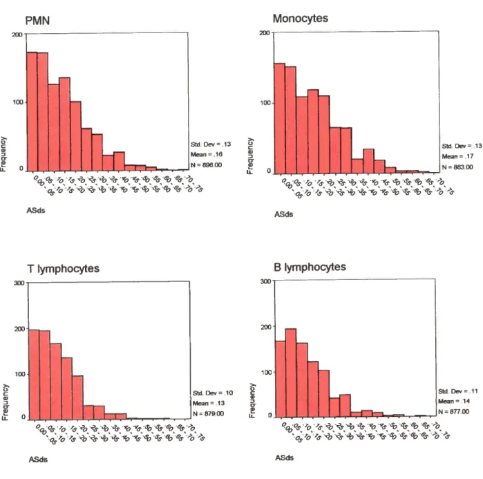
Figure 10 frequency distribution ofD$ scores 92
Figure 11 frequencydistribution of A$0scores 93
Figure 12 Age-PAmat scatter plots 94
(J
figure 13 Age-D$ scorescaffer plots 95Figure 14 Age-ASp, score scaffer plots 96
Figure 15 Age-ASDSscore scafterplots 97
figure 16 Body-wide )UR: intra-individua! correlation ofPAmat scores 98
Figure 17 Biood skewing: intra-individual correlation ofPAmat scores 99
Figure 1$ Acquired skewing: intra-individual correlation of A$pt score 100
Figure 19 Direction of AS relative to body-wide )UR 101
Chapter 4
Figure 20 Frequency distribution of blood counts 146-147
LIST 0F TABLES
Chapter 1
Table I The Xic and its elements 7
Table H Characteristics acquired with Xist upregulation 12
Table HI X inactivation based clonality assays 19
Table W Utility of the HUMARA assay 24-25
Table V Primary versus secondary etiologies ofskewing 30
Table W Intra-individual correlation of)UR among various tissues 40
Chapter2
Table VII CeIl-sorted populations and purity 65
Chapter 3
Table VII Number of informative females (DNA amplified 102
Table IX Incidence of skewing and AS (n3) 102
Table X Mean X inactivation ratios 103
Table XI Skewingas a function of age 104
Table XII Intra-individual correlation of )URs 105
Table Xffi Evidence supporting a body-wide skewing trait 106
Table XW Direction of AS relative to the body-wide XIR 107
Chapter 4
Table XV Descriptive statistics for hematopoietic indices 150
Table XVI Outiiers removed ftom blond counts 151
Table XVII Descriptive statistics of clinical data 152
Table XVIII Descriptive statistics of smoking habits 153
Table XIX Descriptive statistics of family / parity data 154
Table XX Association between age and hematopoietic indices 155
Table XXI Association between age and clinical data 156
Table XXII Association between age and smoking characteristics 157
Table XXHI Association between age and parity 158
Table XXW Association between clinical data and hematopoietic 159-160
Table XXV Association between clinical data and parity 161
Table XXVI Association between smoking habits and clinical data 162
Table XXVII Association between smoking habits and blood counts 163
Table XXVIII Association between smoking habits and parity 164
xvii
G
Table XXX Covariates associated with hematopoietic indices .166
Table XXXI Heritabilityestimatesof confoundingvariab]es 167
Table XXXII familial aggregation of skewing and AS 168
Table XXXIII Association between skewing and blood counts 169-170
Table XXXIV Associationbetween skewing and clinicaldata 17 1-172
Table XXXV Parity dataand skewing 173
Table XXXVI Association between smoking habits and skewing 174-175
Table XXXVII familial resemblance ofXIRs 176
LIST 0F ABBREVIATIONS
C
X recurrence risk ratio
ar allele ratio
APOE-4 apolipoprotein E -4
AS acquired skewing
BM bone-marrow
BMT bone marrow transplant
bp base-pair
BRCA 1 breast cancer I gene
CBC complete blood count
CPM conflned placental mosaicism
COPD chronic obstructive pulmonary disease
CysLT, cysteinyl leukotriene receptor
DMD Dtichenne muscular dystrophy
DNA deoxyribonucleic acid
dpc days post-coitum
DS degree of skewing
DZ dizygous
ES embryonic stem
ET essential thrombocythemia
FITC fluorescent isothiocyanate
G6PD glucose-6-phosphate dehydrogenase
GFP green fluorescent protein
GLM general linear model
UPRT hypoxanthine phosphoribosyltransferase
IISC hematopoietic stem celi
FIUMARA human androgen receptor
ICF immunodeficiency, centromeric instability, facial dysmorphism
Kb kilo base
LA linkage analysis
LI Jong interspersed repeat elements-I
LD Jinkage disequilibrium
LOH Ioss ofheterozygosity
xix
MDS myelodysplastic syndrome
(E
MLL mononuclear layerMPD myeloproliferatïve disorder
mRNA messenger RNA
MS multiple scierosis
MZ monozygous
ng nanogram
NHT nonhematopoietic tissue
OA osteoarthritis
PALA proportion active of the larger HUMARA allele
PAmat proportion active of the maternaI aJ]e]e
PAR pseudoautosomal region
PB peripherat blood
PCR polymerase chain reaction
PCV polycythemia PGK phosphoglycerate kinase PE phycoerythrin PMN polymorphonuclear celis P5 primary skewing RA rheumatoid arthritis
RBC red blood ceits
RE restriction enzyme
RFLP restriction fragment polymorphism
RNA rïbonucleic acid
RR relative risk
RSA recurrent spontaneous abortion
SA spontaneous abortion
SLE systemic lupus erythematosus
SNP single nucleotide polymorphism
SOLAR Sequential Oligogenic Linkage Analysis Routines
SRS simple repeat sequence
TE Tris-EDTA
TSG tumor suppressor gene
TDT transmission disequilibrium test
TRD transmission ratio distortion
VNTR variab]e number tandem repeat
WAS Wiskott-Aldrich syndrome
X-SCID X-linked severely combined immunodeflciency
Xa active X chromosome
Xce X-chromosome controtiing element
XCI X chromosome inactivation
Xi inactive X chromosome
XicDUC X inactivation center
)UP X inactivation pattem
)UR X inactivation ratio
Xist/XIST X-inactivation specific transcript
XLA X-Iinked agammaglobulinemia
Xm maternai X chromosome
xd
ACKNOWLEDGEMENTS
o
I am very grateful to my director and mentor Lambert Busque for timely discussions, intellectual stimulation, and for allowing me the opportunity and time allofted to advance the chromosome X project. Ris insight and encouragement were equally appreciated.
I thank the team members I’ve been collaborating with over the duration of the project, without
whom the project would flot have been feasible: in particular Sylvie Provost, Marianne Gingras and Sylvie fafard for technical experience, encouragement and timely discussions; Linda Lizotte and Nathalie Rathé for specimen and data collection; Marie-Pierre Dubé, Andrew Paterson and Dushanthi Prnnaduwage for statistical insight and analyses; and to Marc Lussier and Pierre Chagnon for project management, genetic expertise and encouragement. I am also grateful to Nathalie Labreque and Martin Guimond for technical assistance andlor ïmmunological insight. Technical assistance and discussion of provoking genetical issues with Patrick Scoif is gratefully acknowledged. My indebtedness goes to Alain Bonnardeaux, Denis Coumoyer, Iran Hoang and Claude Perreault for being members of my Masters thesis review board andlor pre-doctoral committee.
Q
xxiii
o
Through error you corne to the truth! I am a man because I err!
Fyodor Dostoevsky
o
Chapter I
2
I
THE
X CHROMOSOME
(
1.1 Basic principles
‘flic term “X chromosome”, used to denote the sex chromosome thatdetermines the development of
the homogametic female sex, lias been adopted in honor of Henking’s observation of a densely stained body that appeared in haif of secondary spermatocytes of the heteropteran insect,
Pyrrhocoris apterus. 11e was uncertain of the clear nature of die body so lie labeled it “X” for unknown, years later identified as the sex chromosome. The X is an exceptional chromosome as it
can undergo inactivation or reactivation - dependant on chromosome company, developmental
pathway (soma versus germiine) and stage of development. Among eutherian mammals, with few
exceptions, it is conserved in size and compromises roiighly 5% of the haploid genome (01mo,
1967). Moreover, as the X chromosome Iacks a pairing partner in males (aside from the PAR
regions that recombine with the Y), 01mo hypothesized that h is relatively protected from
rearrangements thus conserving gene linkage to the X chromosome across various eutherian
species.
Among mammalian species, X-Jinked gene dosage equivalence between XX females and XY males
is accomplished by a fundamental mechanism of gene regulation: X chromosome inactivation. By
the same mechanism, aneuploidy of the X chromosome (extra copies or monosomy) is well
tolerated. X inactivation (Xi) is initiated during eariy female development and is a fundamental
requirement for normal development. Failure to do so has been associated with severe
developmental defccts and embryonic lethality (Migeon et al., 1993). In partïcular, ectodermal ceil
death and absence of mesodermal formation lias been observed in mouse embryos bearing two active Xs (Takagi and Abe, 1990). To achieve gene dosage equivalence with the autosomes, gene
expression from the single active X is upregulated approximately 2-fold, as demonstrated in
different mice strains where a particular bous was X-linked in one but autosomal in another (Adier et al., 1997), consistent with Ohno’s hypothesis for an evolutionary requirement for high level gene expression from the single active X chromosome (01mo, 1967).
1.2 Lyon’s Hypothesis
The X chromosome inactivation hypothesis, first proposed by Mary Lyon (Lyon, 1961), states that: (ï) one of the two X chromosomes in mammalian female ceils îs genetically inactivated; (ii) the inactive X could be maternai or patemal in origin; (iii) inactivation occurs early in embryonic development and remains flxed in progeny celis. The latter is consistent with clonai inheritance of
X inactivation patterns (Davidson et al., 1963). In other words, “X chromosome inactivation is the transcriptional silenchig of a randomly selected X chromosome initiated in early female development”. As a resuit, females are functional mosaics for X-linked polymorphisms, with two distinct ceil lines/populations. Lyon based lier hypothesis on summation of the following observations: i) the Barr body (then referred to as the X-chromatin body) was formed by condensation of a single X chromosome (01mo et al., 1959); ii) the asynchronous labeling of the X chromosomes (Gilbert et al., 1962); iii) mice with an X0 genotype were normal fertile femafes, suggesting a single X is required for development (Welshons and Russell, 1959); and iv) mice heterozygous for an X-linked coat color gene (moftled, brindled, tortoise-sheli, or tabby - most of
which are lethal in the hemizygous state) present a variegated coat phenotype (Welshons and Russefi, 1959) (Lyon, 1960). In fact, in the heterozygous state, these genes give rise to a random patchy, somewhat linear, distribution of abnormal and wild type coat colors (Lyon, 1962). Dosage compensation by X inactivation lias been tested and extended to humans (Gartier et al., 1992). Indeed, very similar patterns can be seen in female carriers ofvarious X-lïnked skin disease alleles.
1.3 Anatomical human X-m osaicism
In humans, the unes of Blashko, a nonrandom developmental pattem of the skin, manifest in the heterozygous state of various X-linked gene defects, with mutant gene expression covering affected areas and wiid-type gene expression constituting normal skin. Examples of X-linked skin disorders
with clinical manifestations foflowing the Blashko unes are focal dermal hypoplasia,
chondrodyspiasia punctata, and hypohidrotic ectodermal dyspiasia (Happle, 1985). The nature and origins of the unes of Blashko may be best explained by the visible consequences of Lyonization (Flappie, 1985), possibly reflecting the stream or trend of embryonic tissue growth. They describe a V-shape over the spine, on the abdomen they frequently form whorls and on the limbs they mn in a more or less perpendicular linear direction. When compared to mosaic defects of human skin, the banding observed in mice is usually much more coarse, vaguely resembling the distribution of Biashko’s unes.
Although the linear skin lesions are likely to reflect cional prolïferation of two functionaily different ccli populations, analysis of smaH skin specimens (3-6mm) from normal individuals lias found similar X inactivation paffems UP) in different regions of the same individual, suggesting skin growth is characterized by considerable ccii mixing (Fiaikow, 1973). Similar findings were reported for uterine tissue (Linder and Gartler, 1965) and hair roots (Gartier et aI., 1969). Moreover, in human skin sampies composed of approximately 35 basai keratinocytes, a fme mosaic of tiles was observed, with the maternai or paternal X chromosome inactivated in each tue (Asplund et al.,
4
2001). It appears therefore that the distribution paftern of skin ceils may be influenced by X-liiilced genetic factors: in healthy females, a fme mosaic of tues; in carrier’s of certain X-Iinked skin disease alleles on the other hand, a linear pattem may be observed.
1.4 Timing of X inactivation:
concurrent with tissue d!fferentialion
Aftbough the process and mechanism of Xi bas been investigated in several species, a prime model
is the mouse as it is experimentafly amenable and a plethora of information is available. The
following is an outiine.
Pre-fertitizution
During early female development, oogenesis is characterized by reactivation of the inactive X, occurring prior to the leptotene stage (Kratzer and Chapman, 1981), (Gartler et al., I 9$0), ensuing in two active X chromosomes (Epstein, 1972). During mate gametogenesis on the other hand, the active X chromosome is progressively inactivated around fffst meiotic prophase (Lifschytz and Lindsley, 1972), ensuing in formation of the XY body. Akin to the female soma, the inactive X is relatively condensedlheterochromatic. In contrast however, althougb transcriptionally inactive (Richier et aI., 1992), flic CpG islands ofhouse keeping genes are relatively unmethylated (Driscoli and Migeon, 1990).
Post-fertilization
Key observations of Xi occurring post-fertilization have been documented in vivo using harvested mouse preimplantation embryos and in embryonic stem (ES) ceils. In both systems, initiation of Xi has been tightly regulated to tissue differentiation - sec figure 1 (page 6) . Upon fertilization, the
zygote and early blastocyst (prior to implantation) embodies two active X chromosomes (Gartier et al., 1972). Reactivation of the sperm-derived inactive X chromosome however has not been well characterized. Upon dïfferentiation, one X is inevitably selected for inactivation (Epstein et al., 197$), (Kratzer and Gartier, 197$), (Monk and Kathuria, 1977), (Monk and Harper, 1979), (Penny et al., 1996). In ceits destined to form extraembiyonic lineages (trophectoderm and primitive endoderm), the patemal X bears a putative imprÏnt marking the chromosome for early inactivation, wherein Xi is initiated 4,0-5,0 days post-coitum (dpc), shortly around or after the time of implantation (Takagi and Sasaki, 1975), (West et al., 1977), (Takagi et aL, 197$), (Monk and
Harper, 1979), (Costanzi et al., 2000). After implantation (5,5 — 6,5 dpc), a genome-wide
demethylation event is believed responsible for erasure of the parental imprint(s) (Rastan, I 9$2a) (Monk et al., 1987), ensuing in random inactivation of parental X chromosomes in cells ofthe inner ceti mass (Gardner and Lyon, 1971).
furthennore, aithougli preliminaiy data found that Xi occurs concurrently with tissue differentiation (Monk aiid Harper, 1979), unequivocal evidence was derived from a mouse une transgenic for an X-Iinked Lac Z transgene. The transgene, which was subject to inactivation, underwent inactivation in a developmental context, gradually proceeding in sub-populations and Jineages, and was vfrtually completed by 11,5 dpc (Tan et al., 1993; Tan et al., 2000). Thereafier, the inactive state of the chromosome was clonally inherited in daughter celis. lii humans, Xi is speculated to occur approximatety 16 dpc (Park, 1957).
2
MECHÀMSM 0f X INACTWATION
Xi is an intensely studied subject that involves several molecular mechanisms: counting and choosing, initiation/propagation and maintenance. An introduction to the underlying principles, properties and mechanisms of Xi wilJ be provided (for a comprehensive review see (Willard, 1996a; Avner and Heard, 2001)). The X-inactivation center (Xic), spanning approximately 1MB, is a key cis-regulator for the initiation of Xi. Several elements involved in the Xi process reside within the Xic (Figure 2, page 7) (Table I, page 7).
2.1 The X inactivation center (XicIXIC)
The fïrst dues for an “initiator of X chromosome inactivation” came from genetic studies wherein mutant X chromosomes lacking a particular region were unable to undergo inactivation. This deleted region was thereafier called the X chromosome inactivation center (Xic). Evidence that the Xic functions in cis lias been progressively defmed by the partial spreading of inactivation onto autosomal segments in X-autosome transiocations containiiig the XicDUC (Russell, 1963), (Rastan, 1983), (Cattanach et al., 1991), (Jeppesen and Turner, 1993; Jeppesen and Turner, 1993). Localization ofthe human )UC has been assigned to band Xq13 (Brown et al., 1991a), (Leppig et al., 1993), (Lafreniere et al., 1993). There is no convincing evidence for more then a single XIC (Wïllard, 1 996b). The corresponding syntenic region in mouse is somewhat smaller and the overali organization ofthe XicDUC is poorly conserved between human and mouse (Debrand et al., 1998).
Three general processes have been attributed to the Xic/XIC: counting, choice and
6
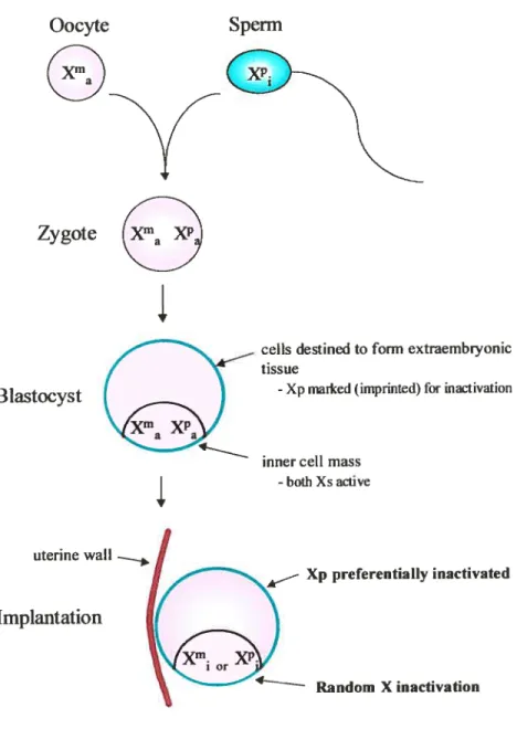
Figure 1. Timing and appearance of X inactivation during female mouse development. After
formation of the zygote, both Xs are relatively active. An imprinting pathway, where Xp (paternal
X chromosome) is preferentially inactivated, is operative in tissues destined to form extraembiyonic
structures. At around the time of implantation, random X inactivation begins in celis destined to form epiblast-derived tissues.
Oocyte
Sperm
Blastocyst
Implantation
celis destined to form extraembiyonic tissue
-Xpmarked(impnnted)forinactivation
inner celi mass
-both Xs active
Xp preferentially inactïvated
Random X inactivation Zygote
uterine walI
7 figure 2. The Xic region and its defmed elements. The Xic, located at Xq13, bas been implicated in three defined processes of X inactivation: ï) counting, ii) choice, and iii) initiationlpropagation. Genetic elements identifïed within the Xic that play a rote in the X inactivation process are Xist, Tsix and the Xce element. Distances between elements and the size of the genetic elements are approximate.
Xic (Xq13)
Xist gene expression
Xce element
4
Tsix gene expression
Table L The Xic and its elements.
Xic: A master control region required for the initiation of X inactivation. As the Xic functions in
cis, X chromosomes void of an Xic fait to undergo inactivation. A counting, choosing and spreading
role bas beenattributed to this locus. Effector elements residing within the Xic includeXist,Xceand
Tsix.
Xist: Localized to the Xic region, the X inactive specific transcript (Xist) is a non-protein coding
mRNA essential for the initiation of X inactivation in cis and appears to be the primary signal for propagation of inactivation along the chromosome.
Tsix: Tsix is a mRNA species transcribed antisense to and approximatety 15 Kb downstream of the Xist gene. In mice, the transcript spans the whole of the Xist gene while in humans a shorter transcript has been identified. Targeted deletions in the 5’ region of Tsix Ieads to skewed X inactivation and disrupts imprinted X inactivation in extraembryonic tissues, suggesting Tsix influences X chromosome choice. A possibte role of Tsix may be regulating Xist activity in cis.
Xe: The apparent function of the cis-acting X-chromosome controlling element (Xce) is to modulate the probability upon which a particular X chromosome is inactivated. In the mouse, a gradient of alleles bas been demonstrated. In the heterozygous state, the X chromosome bearing a stronger allele is preferentiatly setected to remain active versus the X with the weaker alfele, resulting in primary nonrandom X inactivation. Xce activity resides 3’ to Xist.
$
i) Counting: This step senses the number of X chromosomes in the celi and ensures that only a
single X chromosome remains active per diploid celi -ai] other X(s) are inactivated (Therman and
Patau, 1974) (Rastan, 1982a). The absolute requirement ofat Ieast two Xics is required for initiation of X inactivation (Rastan and Robertson, 1 9$5), (Rastan, 1983). How a single X is selccted is flot presently clear. It is speculated that a blocking factor produced in limited amounts binds to a single Xic (reviewed in (Migeon, 1994). Alternatively, allelic methylatïon differences within a proposed differential methylation region may be another (Chao et aI., 2002). Since the number of active X chromosomes is dependant on the number of chromosomal sets (ploidy number of autosomes), as there are one to two active X’s in XXX triploids (69 chromosomes) and two active X’s in X)OOC tetraploids (92 chromosomes), an autosomal origin for the blocking factor lias been supported (Carr, 1971), (Jacobs and Migeon, 1989). In fact, recent findings identified the CTCf insulator / transcription factor as ‘the’ or ‘one of’ several putative trans-acting “blocking facto?’. As several CTCF binding sites were identified in the mouse TsixIDXPas34 region, the authors postulate that Tsix and CTCF work together to designate the future active X by inhibiting Xist activity in cis (Chao et al., 2002). for a review of plausible mechanisms, see (Percec and Bartolomei, 2002). Ihe role for CTCF as the human “blocking factor” is presently unclear as the number of CTCf binding sites in the syntenic human region has signiflcantly less CTCf binding sites.
ii) Choice. During the choice process, one X is seiected to remain active and the other to be
inactivated. Although believed to be a random process, with maternai and paternal X boasting equal inactivation probabïlity, deviation from random inactivation has been noted. for example, in extra embiyonic lineages of mouse, the patemal X is marked for preferential inactivation. In the embryo proper ahernatively, the imprint is believed erased soon after implantation, ensuing in random X inactivation. Nonetheless, in mice, allelic variants ofthe Xce locus (the nature of which is presently unclear) can compromise choice, ensuing in primaiy nonrandom X inactivation pattems. A gradient of Xce alleles lias been identified, each influencing the probability of inducing cis-ïnactivation (Cattanach and Isaacson, 1967), (Cattanach et al., 1969), (Cattanacli and Perez, 1970). it is specuiated that the Xce affects affinity for the trans-blocking factor. Additional elements recently implicated in the choice step are Tsix (Lee and Lu, 1999), Xist (Marahrens et al., 1997), (Boumil and Lee, 2001), (Newali et aI., 2001) andXite (Ogawa and Lee, 2003). Further, as CTCF may act in concert with Tsix in designating the future active X, it may aiso play a rote in the choice step.
iii) Initiation I propagation: This step involves initiation and spreading of the inactivation signal onto neighboring regions in a cis-mediated manrier (Rastan, 1983), (Lee et ai., 1996), (Willard, I 996a). Given the cis-Iimited function of Xist, binding sites or nucleation centers (booster elements) have been specutated (Gartier and Riggs, 1983), (Riggs, 1990). As a candidate booster element,
Lyon (Lyon, 199$) proposed long interspersed repeat elements- 1 (Li), based on two observations. i) The X chromosome is enriched for sucli elements, approximately two-fold versus the autosomes (Bailey et al., 2000). ii) In the event of X-autosome transiocations, the extent of inactivation spreading onto autosomal regions correlated with levels of LI elements. Moreover, X-linked regions that contain genes that escape inactivation are sïgnïficantly reduced in Li content versus chromosomal segments containing genes subject to inactivation (Bailey et aI., 2000).
Although Xist RNA coating of the inactive X chromosome may have a chromatin-remodeling role, (Clemson et al., 199$), Xist expression per se may flot be required for ‘maintenance’ of inactivation (Csankovszki et al., 1999) (Wutz and Jaenisch, 2000) as Ioss of the )UC in humans bas flot been associated wïth instability ofthe inactive state (Brown and Willard, 1994), thus suggesting that once silencing bas been achieved, it is frreversible and thereof independent of Xist. Moreover, when Xist expression was reactivated from the active X or when ectopic Xist expression was induced, inactivation could flot be initiated (Wutz and Jaenisch, 2000) (Clemson et aI., 1998), consistent with a developmental context ofXist expression.
2.2
Genetic elements of the Xic: Xist gene
The Xist gene, for X inactivation specific transcript, is believed to play a primary role in the X inactivation process: I) it is located within the Xic (Xq 13.2), ii) is transcribed specifically from the inactive X chromosome in both human (Brown et al., 1991b) and mouse (Borsani et aI., 1991), (Brockdorff et aI., 1991), iii) is transcribed prior to Xi (Kay et al., 1993), and iv) bas been shown to be essential for cis-mediated Xi by loss-of-function experiments (Penny et al., 1996), (Marahreiis et al., 1997), (Lee et al., 1996). Although necessary for Xi, the Xist gene is flot required for male development since male mice bearing a Xist deletion are fertile and physiologically normal (Marahrens et al., 1997).
The Xist/XIST gene encodes a large untranslated RNA (Brown et al., 1992; Lee et al., 1996), (Brockdorff et al., 1992) localized to the nucleus (Brown et al., 1992; Clemson et al., 1996; Lee et
aL, 1996). Although both the mouse and human gene contain at least $ exons, the )U$T cDNA
spans a maximal 19.3 KB (Hong et al., 2000) whule Xist cDNA spans at least 17.4 KB, however smaller isoforms have been produced (Hong et al., 1999). The mouse and human Xist/XI$T gene exhibit moderate but significant conservation of sequence and gene structure (Nesterova et al., 2001).
10
2.2.1 Developmental regulation ofXist
Dynamicchanges in Xist expression markone of the earliest events of the Xi process (Figure I .3a).
In undifferentiated murine embryonic stem (ES) celis (both male and female), the X-chromosomes
are relatively active and marked by basal Xist expression (Hong et al., 1999; Tai et al., 1994), (Beard et al., 1995), (Daniels et al., 1997a), (Panning and Jaenisch, 1996) (Panning et al., 1997), (Panning et al., 1997), (Johnston et al., 199$). This RNA however accumulates at the site of transcription and does flot localize across the chromosome. Upon differentiation however, Xist RNA expression is upregulated (>1 5-fold) from the X destined for iriactivation and is downregulated on the future active X (Panning and Jaenisch, 1996) (Panning and Jaenisch, 1996). Xist upregulation is thought to resuit from an increased RNA haif-life (Panning et al., 1997), (Sheardown et al., 1997), possibly owing to a switch in Xist promoter usage (Johnston et al., 199$). However, recent data suggcst an altemate mechanism: Tsix gene interference (vide infra) (Warshawsky et al., 1999). Nonetheless, Xist accumulation is rapid, coating the inactive X within one celi cycle (Wutz and Jaenisch, 2000), (Panning et al., 1997). $everal thousand X-linked genes are transcriptionally
silencedshortly afler the accumulation ofXist RNA (Keohane et al., 1996).
2.3 Characteristics acquired with
Xist
upregulation
Acquisition of Xi involves several progressively acquired changes in structure and function,
includingrecruitmentof regulatory
/
chromatin proteins, many ofwhich have been identified. Both genetic and epigenetic modifications have been characterized, see Table II (page 12) for an outiine. Cytologically, the inactive X assumes a condensed, compact appearance coïned the Barr body (Barr and Bertram, 1949).Methylation of CpG islands of
Xi-linked genesDuring the course of Xi, the CpG islands of Xi-linked genes are generally hypermethylated (Wolf and Migeon, 1982; Wolf et aI., 1984) (Keith et aI., 1986) (Bird, 1986), erisuing in transcriptional
silencing.
Asynchronous replication of the inactive X
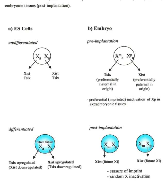
Figure 3. Xist, Tsix and X inactivation. a) Xis! and Tsix expression in undifferentiated versus differentiated ES ceils. b) Xist and Tsix expression in extraembryonic (pre-implantation) versus embiyonic tissues (post-implantation).
a) ES Ceils
undifferentiated Tsix upregutated (Xist downregulated) Xist upregulated (Tsix downregulated)b) Embryo
pre-imptantation-preferential (imprinted) inactivation of Xp in
extraembiyonic tissues
Xist Xist
Tsix Isix (preferentiallyTsix (preferentiallyXist maternai in paternal in
origin) origin)
differentiated post-implantation
futurefuture
XaX
Xist (future Xi) Xist (fiture Xi)
-erasure of imprint - random X inactivation
12 Table U. Genetic/epigenetic and morphological characteristics acquired with inactivation of the X chromosome.
Genetic nwdjficatians
- Xistgene upregulation
- Tsixgene downregulation
- General shutdown of most X-Iinked genes
Epigenetic nwd,ficatwns
- Xist mRNA coating
- hypermethylation of CpG islands
- asynchronous replication (usually late-repticating)
- hypoacetylation ofhistones 113 and 114
- hypermethylation ofhistone H3
- high concentration of core histone macroll2A 1.2
- enrichment for BRCA1 mRNA
- condensed chromatin structure, coined the Barr body
C
asynchronously (reviewed in (fleard et al., 1997)), namely in late S phase, (Boggs and Chinault, 1994), (Schmidt and Migeon, 1990). Genes that eseape X inactivation on the other hand, replïcate in synchronicity with its’ active X homologue (Boggs and Chinault, 1994).
Covalent modifications of X chromosome associated histones Histone acetylation
The transcriptionally active X is charactenzed by hyper-acetylation ofNTI2-terminal lysine residues of histone H3 and 114 (Jeppesen and Tumer, 1993), (Gilbert and Sharp, 1999), (Boggs et aI., 1996), while those associated with promoters of transcriptionally inactive genes of the inactive X chromosome are essentially hypoacetylated.
Historie methylation
During X chromosome inactivation, methylation of lysine 9 of histone 113 (H3-K9 methylation) occurs within or shortly afier Xist accumulation (fleard et al., 2001), (Mermoud et aI., 2002).
Enrich
ment of
histone variantsThe inactive X is further characterized by a high concentration of the core histone macrofl2Al .2 (Costanzi and Pehrson, 1998). Localization of macrofl2Al .2 to the inactive X may be mediated by interaction with Xist mRNA, possibly by formation ofa ribonucleoprotein compiex (Csankovszki et al., 1999).
Ennchment of mRNA species
Recent studies have demonstrated that BRCAI mRNA associates (possibly essential) with )UST mRNA in female somatic celis, mediating chromatin stability of the inactive X (Ganesan et al., 2002). Thereof in absence of functional BRCA1 (either by germiine mutations, LOil or RNA interference), i) the chromatin state of the inactive X was destabilized, ii) enrichment for macrofl2Al .2 was suppressed and iii) particular X inactivated genes (a GfP transgene for example) were partially upregulated, thus suggesting BRCAI plays a pivotai roie in mediating Xi. Thus, the gender-speciflcity upon which BRCAI mutations transcend to ovarian and breast cancer may be expÏained by genomic instability of the inactive X chromosome. Supportive evidence was derived from various BRCAI4 tumors, of which demonstrated defects in chromatin structure and over expression of several genes linked to the inactive X (Jazaeri et al., 2002). One hypothesis may be
that derepression of Xi leads to a dosage imbalance of genes required for normal
function!development of breast and ovaries.
2.4 Stability of
X
inactivation
Once Xi has been achieved, the chromatin state is clonally inherited (Davidson et ai., 1963) and the inactive state believed stably maintained for duration ofthe cell lifespan (Migeon, 1972). However, exceptions have been noted. i) The inactive X is reactivated as a normal part of oogenesis. ii) Albeit at a low ftequency (1 0 to I 0), in vitro cuitivation has been associated with reactivation of varions Xi-linked genes (Dyer et al., 1989) (Mohandas et al., 1981), (Graves, 1982). In addition, there ïs convincing evidence in mice that particular Xi-linked genes are partially reactivated with advancing age (Wareham et al., 1987) (Brown and Rastan, 198$).
3
REGULATORY ELEMENTS 0F THE XIC
To identify regulatoiy regions within the Xic, deletion and transgenetic analyses in and adjacent to the Xis! gene and their consequential effects on cis-mediated Xi were investigated.
14 Initiation etement
Targeted deletion of the 5’ region ofXist(7-Kb, including 30 bp of the promoter region) abolished initiationlpropagation (Penny et aI., 1996), delïmiting initiation of Xi to thïs region (Lee et al.,
1 999a).
Counting elements
The counting process was delImited to a 37-Kb region lying 3’ to Xist, but genetically separable from Tsix promoter and
Xite.
Moreover, as the aberrant X was also inactivated in differentiating XY ceils, sex-specific factors in the Initiation of Xi are putatively exclu ded.Choice elements
The choice process appears influenced by various elements within the Xic. i) The elusiveXce tocus, reputably associated with nonrandom Xi patterns (Caftanach and Papworth, 19$1), has been delimited to a rather large region 3’ to the Tsix locus (reviewed in section 7.3.2.1). ii) Targeted deletion of the Xist antisense transcript, i.e., Tsix, induces constitutive Xist expression and nonrandom Xi of the aberrant X (Lee and Lu, 1999). iii) The recently identified Xite gene (downstream from Tsix) also mediates choice as targeted deletion of the gene downregulated Isix activity in cis, resulting in Xist upregulation and preferential inactivation of the aberrant X (Ogawa and Lee, 2003). iv) In humans, a rare base-pair mutation in theXIST promoter region lias been associated with preferential inactivation of the mutant X (Plenge et al., 1997), suggesting mutation/polymorphisms in theXistgene may influence Xi pattems. A plausible mechanism may be increased Xist transcription, thus increased probability of undergoing Xi. y) lnduced chemical
mutagenesis of the mouse genome identified two autosomal bd (yet to be clearly identified) which when mutated altered X chromosome choice (Percec et aÏ., 2002).
3.1 Further characterization of the Tsix gene
The Tsix gene, so dubbed as it is franscribed antisense to
Xist,
encodes a 40-Kb transcriptoriginating some 2$ Kb downstream ofXist in both liuman and mouse (Lee et al., 1999b), (Sado et
al., 2001), (Migeon et al., 2001). Although originally deflned as having no conserved ORF, recent studies suggest it is partially processed, giving rise to a 2,7 and 4,3 Kb franscript (Sado et al., 2001). It is found exclusivety in the nucleus, but unlike Xist, remains bocalized to the Xic (Lee et aL,
1999b).
Because Tsix is i) transcribed antisense to Xist, ii) is concurrent with early Xist expression and iii) Tsix upregulation from
the
future Xa is associated with Xist downregulation (Panning and Jaeniscli, 1996), (Lee et al., 1999b), a role in regulating Xist activity bas been speculated. Avallable evidence suggests high Jevel Tsix expression represses Xist Jikely thougli a franscriptional mechanism (Lee and Lu, 1999), however other mechanisms are plausible (sec (Avner and Heard, 2001) for review). Once Xi is estabuished, Tsix expression from Xa ïs downregulated to undetectable levels.Similar to the munne homologue, human T$IX is expressed exclusively in epiblast derived ceils. However, it is truncatcd at its 3’ end, ending approximately at exon 5 of the XÎST gene, deriving a transcript of approximately 35 Kb (Migeon et al., 2001). Moreover, human T$IX lach the 5’ CpG is]and putatively required for murine Tsix imprinting fimction (Sado et al., 2001), suggesting TSJX may flot function like its murine counterpart (Migeon et al., 2001).
4
IMPRINTED X INACTWATION
Genomic imprinting is a process whereby gene function is affected in a parental origin-specific manner and is manifested as a difference in expression of parental alleles. The effects of genomic imprinting arise from differential epÏgenetïc modification of parental alleles during gametogenesis, followed by additional epigenetic processes that may occur afier fertilization (Latham, 1999). Genomic imprinting may limit the effects of growth factors in the embryo andlor in extra embiyonic tissues, in which there is differentiai parental investment (flaig, 1993). Strong epigenetic effect of parental iniprinting on the X inactivation proeess has been observed in mouse and weaker evidence has been found in human.
4.1 Preferential inactivation of the paternal X in extraembryonic tissues
li mice, unlike embryonic tissues that exhibit random Xi, the patemal X is preferentially inactivated in the first ceils to differentiate, the physiologicai consequence of which is presently not clear,
eventually giving rise to tissues destined for extra-embiyonic development (Takagi and Sasaki, 1975), (West et aÏ., 1977), (Harper et al., 1982), with $7-$$% oftrophoblasts possibly showing Xp inactivation (Takagi and Sasaki, 1975), (West et al., 1977). Resistance of Xm from undergoing inactivation lias been ascribed to acquisition of an imprint during oogenesis (Lyon and Rastan, 1984), (Tada et ai., 2000). flic Xist gene is thought to play a vital role in the imprinted pathway since female mice inheriting a patemai Xist deletion bear 2 active Xs and die early in embryogenesis. Mice inheriting a maternai Xist deletion on the other hand, are normal and exhibit
16
exclusive paternai Xi (Marahrens et al., 1997). In humans however, there is no evidence for
parental-specific preference of Xist expression (Daniels et al., 1997b) (Ray et al., 1997). Interestïngly however, preferential inactivation of Xp in extra-embryonic tissues may flot be related to imprinting at ail. Alternatively, relative to the active maternai X, the inactive X derived from
sperni may be more receptive to the inactivation signal (Monk and McLaren, 1981). Supporting
evidence was derived from elegant cloning experiments where the inactive X (maternai or paternal in origin) of the donor-derived nucleus was preferentially inactivated in extra-embtyonic tissues of the cioned embiyo (Eggan et aI., 2000), thus suggesting that the donor-derived inactive X, like in sperm, is poised for early inactivation.
4.2 X-imprinting in humans
Unlike the mouse, evidence for preferential inactivation of Xp in extraembryonic tissues in humans remains controversial. Although random Xi was observed in chorionic villi of eariy gestation (Migeon and Do, 1979), (Migeon et al., 1985), (Mohandas et aL, 1989), (Bamforth et al., 1996), preferentia! inactivation of Xp was observed by others (Harrison and Warburton, 1986), (ilarrison, 1989), (Goto et al., 1997) and partially supported by (Uehara et al., 2000). f urthermore, whereas trophoblasts from fuil-term placentas showed Jack of preferentiai inactivation of Xp (Looijenga et al., 1999), Xp was preferentialiy inactivated in another study (Ropers et al., 197$). Discrepancies may be explained by methodofogy used, tissuetypeandlor maternai ccli contamination.
5
ESCAPE
FROM X INACTIVATION
An intriguing aspect of Xi is that certain genes escape inactivation, resulting in expression from both X chromosomes. Approximately 30% of genes on the short arm of X and less than 5% of genes on the long arm escape Xi, averaging over 10% of X-linked genes (Carrel et aI., 1999). Relative to mice, humans are believed to have a larger number of genes that escape Xi (Disteche,
1999). These genes may be independent or clustered together, the latter found in or near the
pseudoautosomal region (PAR) (Lyon, 1962), (Miller and Willard, 199$), suggesting a
chromosomai domain model of regulation. It is unclear whether genes are initiaily inactivated and then re-activated, or simply remain active. it was hypothesized that genes escape Xi to compensate for functionai Y homologues in males. However, many genes that escape Xi do flot have an Y homologue, suggesting dosage imbalance between the sexes. Interestingly, several genes display
variable escape from Xi, being expressed from the inactive X in some females but subject
from Xi may be a significant factor for physiological and disease susceptibility differences
tE
between the sexes (see (BrownandRobinson, 2000) for review).6
CLINICAL APPLICATION 0F THE LYON’S HYPOTHESIS
Several methods have been developed to determine the clonai nature of a pathologicai tissue.
Originally these methods relied on specific gene defeets associated with particular cancers (reviewed in (Gilliland et al., 199 la)). For example (I) immunoglobulin and T ceil receptor gene rearrangements in iymphomas, (ii) cytogenetic and fluorescence in situ hybridization analysis of genetic aberrations (transiocations, deletions, duplications and inversions) in acute myeloid leukemia, chronic myeloid leukemia and myeloproliferative disorders, (iii) point mutations in criticai genes such as proto-oncogenes and tumor suppressor genes in hematological and gastrointestinal cancers, and (iv) viral integration in vfral-associated lymphomas. However, not ail tumors exhibit particular tumor-specific markers or the specific abnormalities are flot known.
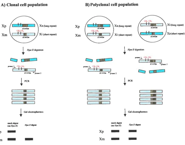
6.1 X inactivation-based clonality assays
Xi based clonaiity assays were developed on precepts ofthe Xi hypothesis (Linder and Gartier, 1965). These assays, winch do not rely on the presence of tumor-specific markers, have the advantage of detecting clonai derivation of ceils in any informative female. The finding that a pathological tissue is monoclonal is consistent with a neopiastic process (i.e., that a tumor bas arisen
from acquired somatic mutation(s) in a single progenitor celi (Nowell and Hungerford, 1960))
whereas normal and reactive celi populations (inflammatory tissues) are typically polyclonal in origin. Application of these assays is central to many aspects of our understanding ofthe normal biology of hematopoiesis as weil as the pathogenesis of hematologic malignancies ((Busque and Gilliland, 199$) for review). For example, the stem ceil origin of differentiated hematopoietic lineages (Gartler et al., 1969), the clonai origin of numerous hematological and non-hematological maiignancies (Linder and Gartler, 1965), and the stem celi origin of various hematologic malignancies. In addition, these assays can be utilized for detection status of female carriers of various X-linked disease alleles (reviewed in (Puck and Willard, 199$)).
6.2 Principles ofX-inactivation based clonatity assays
Xi-based ctonality assays, reÏy on two basic prerequisites. The first is to differentiate parental origin ofthe chromosomes: Xp from Xm. The second is to distinguish the active X (aX) from the inactive