Page de titre
Université de Montréal
“La pathophysiologie de la maladie de Ménière au niveau du sac
endolymphatique : une étude immunohistochimique de
l’aquaporine-2, le récepteur de Vasopressine V2R, NKCC2 et
TRPV4”
Par Marc-Henri Asmar
Département des Sciences Biomédicales
Faculté de Médecine
Thèse présentée en vue de l’obtention du grade de maîtrise en Sciences
Biomédicales, 2
èmecycle, option Générale
Langue de rédaction du manuscrit : Anglais
Dépôt initial le 31 Août 2016
Abstract
Objectives Endolymphatic sac (ELS) pathophysiology in Ménière’s Disease (MD) remains
poorly understood. We identified from the literature a group of proteins expressed on the ELS and involved in endolymph volume regulation: Aquaporin-2 (AQP2), vasopressin receptor V2R, Sodium Potassium Chloride Cotransporter type 2 (NKCC2) and TRP channel type V4 (TRPV4). Our objective was to determine whether their ELS expression was altered in MD, to better understand the pathophysiology of endolymphatic hydrops.
Methods Patients with definite MD undergoing endolymphatic duct blockage surgery were
recruited, as well as controls undergoing surgery for vestibular schwannomas (VS). ELS biopsies and blood samples for plasma Arginine Vasopressin (AVP) were obtained. Immunohistochemistry for AQP2, V2R, NKCC2 and TRPV4 was performed. Slides were scanned digitally for highly sensitive pixel density analysis by specialized software (VIS by Visiopharm®).
Results 27 definite MD patients and 23 VS controls were included. Global scores generated by
the software represent total and relative protein expression density of 3 staining intensity levels, exclusively on ELS epithelium. AQP2 expression density was significantly elevated in MD compared to VS (p = 0.018). There was no significant difference in plasma AVP, V2R, NKCC2 and TRPV4 expression.
Conclusion This original study evaluates simultaneous in-situ expression of AQP2, V2R,
NKCC2 and TRPV4 on the human ELS in MD, with a VS control group. Our results show only AQP2 up regulation on the ELS of MD patients. We suggest a constitutively increased expression of AQP2 in MD, independent of its regulatory axis (AVP-V2R). Acquired regulator sequence mutations could support this model.
Key words:
AQP2, V2 receptor, NKCC2, TRPV4, Vasopressin, Ménière’s disease, Vestibular schwannoma, Endolymphatic sacRésumé en Français
Objectifs La pathophysiologie de la maladie de Ménière (MM) demeure mal comprise. Nous
avons identifié dans la littérature un groupe de protéines exprimées sur le sac
endolymphatique (SEL) et impliquées dans la régulation du volume endolymphatique : l’Aquaporine-2 (AQP2), le récepteur V2R de vasopressine (AVP), le Co-transporteur de Sodium Potassium et Chlorure type 2 (NKCC2) et le canal TRP type V4 (TRPV4). Notre objectif est de déterminer si leur expression sur le SEL est altérée dans la MM, pour améliorer notre compréhension de la physiologie de l’hydrops endolymphatique.
Méthodes Recrutement des cas de MM et schwannomes vestibulaires (SV) comme contrôles,
le jour de leurs chirurgies respectives. Prélèvement de biopsies de SEL et sang pour AVP. L’immunohistochimie pour AQP2, V2R, NKCC2 et TRPV4 fut effectuée, et les lames scannées pour analyse digitale de densité d’expression par un logiciel spécialisé (VIS par Visiopharm®).
Résultats Total de 27 cas MM et 23 contrôles. Les scores générés par le logiciel représentent
la densité d’expression totale et relative des protéines, exclusivement sur l’épithélium du SEL. Les scores d’AQP2 sont élevés de façon significative dans la MM comparée aux contrôles (p = 0.018). Nous ne rapportons aucune variation significative pour AVP, V2R, NKCC2 et TRPV4.
Conclusion Cette étude originale évalue l’expression simultanée de AQP2, V2R, NKCC2 et
TRPV4 sur le SEL dans la MM, avec un groupe contrôle (SV). Nos résultats démontrent une augmentation isolée de l’AQP2 dans la MM. Nous proposons une surexpression constitutive de cette dernière, indépendante de son axe de régulation (AVP-V2R). Une mutation somatique au niveau des séquences régulatrices pourrait justifier nos observations.
Mots-clés :
AQP2, récepteur V2, NKCC2, TRPV4, Vasopressine, Maladie de Ménière, Schwannome vestibulaire, sac endolymphatiqueList of abbreviations
AQP2
Aquaporin-2
EDB
Endolymphatic Duct Blockage surgery
EL
Endolymph
ELH
Endolymphatic Hydrops
ELS
Endolymphatic sac
GS
Global Scores
HPS
Hematoxyline, Phloxine and Safran
IHC
Immunohistochemistry
MD
Ménière’s Disease
MRC
Mitochondria-rich cells of the endolymphatic sac epithelium
NKCC2 Sodium-Potassium-Chloride Cotransporter 2
PL
Perilymph
pAVP
Plasma Arginine-Vasopressin
RRC
Ribosome-rich cells of endolymphatic sac epithelium
TRPV4 Transient Receptor Potential Cation channel type V4
VS
Vestibular Schwannoma
VM
Vestibular Migraine
List of Tables
Table 1. Mean total area of marked regions of interest (ELS epithelium), expressed in squared
micrometers (page 40)
List of Figures
Figure 1. Ménière’s Disease Endolymphatic sac. (page 41) Figure 2. Vestibular Schwannoma Endolymphatic sac. (page 42)
Figure 3. Mean global scores for quantitative immunostaining analysis. (page 43)
Figure 4. Mean global scores for quantitative immunostaining analysis, excluding 3 patients
diagnosed with vestibular migraines. (page 44)
Figure 5. Hypothetical model for Aquaporin-2 and Endolymphatic hydrops in Ménière’s
Disease. (page 45)
Acknowledgements
I would like to thank the outstanding team at the histology lab of IRIC (Institute for Research in Immunology and Cancer, University of Montreal) for their technical expertise, help and guidance in conducting our experiments. Special consideration goes to Julie Hinsinger, Micheline Fortin and Mélanie Beland. I would also like to thank my exceptional mentor Dr. Issam Saliba for his guidance and collaboration, as well as Dr. Louis Gaboury for sharing his expertise in histopathology which was crucial for my experiments.
Table of Contents
I. Introduction
1. Inner ear fluid dynamics and endolymphatic hydrops
2. Inner ear Aquaporin expression, Aquaporin-2 and AVP receptor type 2 3. NKCC2
4. TRPV4
5. Objectives and experimental hypotheses II. Materials and methods
1. Study design and patient population
2. Endolymphatic sac tissue biopsy and blood sampling 3. Histology
4. Antibodies and Immunohistochemistry 5. Highly sensitive pixel density analysis 6. Statistics
III. Results
1. Patient demographics and plasma AVP
2. Immunohistochemistry: Global Scores for staining intensity 3. Total surface area of stained endolymphatic sac epithelium 4. Exclusion of patients diagnosed with vestibular migraines IV. Discussion
1. Plasma AVP, V2R and significance in Ménière’s Disease
2. Endolymphatic sac AQP2: AVP-V2R-AQP2 axis in Ménière’s Disease 3. NKCC2 and TRPV4 in Ménière’s Disease
4. Ménière’s Disease and genetics
5. Hypothetical model for endolymphatic sac pathology 6. Study limitations and future research
V. Conclusion VI. References
I. Introduction
Ménière’s Disease (MD) is a cochleo-vestibular disorder first described by Prosper Ménière in 1861 1, characterized by episodic vertigo, fluctuating sensorineural hearing loss, aural fullness and tinnitus. The incidence of MD is variable between populations, ranging from 4.3 to 15.3 cases per 100000 2,3. The pathologic marker of this disease, endolymphatic hydrops (ELH), remains poorly understood, despite decades of research. A number of
etiologic factors have been proposed to explain the origin of ELH: allergies 4, viral infections such as CMV 5, altered glycoprotein metabolism 6, autoimmune processes 7,8, hormonal dysfunction, genetic mutations or inner ear malformations. None was able to link the disease to a particular process with satisfactory evidence; researchers would present promising
findings such as increased circulating immune complexes (CIC) in MD 9 only to be countered by better-designed studies that could not replicate their findings 10.
Treatment for MD includes an initial trial of medical therapy and lifestyle changes such as restriction of caffeine, alcohol, theophylline and salt (CATS), that achieve satisfactory control in about two thirds of patients 11. Medical therapy often aims to reduce water retention with diuretics and vasodilators (betahistine) or provide symptomatic treatment for the severe nausea and vomiting that complicate vertigo spells. Upon failure of medical therapy, MD is
considered intractable. Several interventions are available to provide the best possible relief to patients, ranging from minimally invasive intratympanic corticosteroid injections 12 to more aggressive procedures such as endolymphatic sac decompression or shunting, and destructive procedures such as intratympanic Gentamycin or labyrinthectomy. The success of all
interventions described to date is questionable, and treatment ultimately depends upon the surgeon’s experience and patient’s preference. A recent Cochrane review of the literature for all surgical interventions could only find 2 studies that satisfied all inclusion criteria. In these 2 papers, only endolymphatic shunt surgery was properly evaluated, with a total of 52 patients, and no solid evidence for its efficacy was found 13. We have described a novel technique that controls MD symptoms with considerable success, supported by 5 years of experience: endolymphatic duct blockage surgery. The intervention consists of a modified endolymphatic
sac decompression with endolymphatic sac and duct dissection, followed by a crucial therapeutic step: blocking the endolymphatic duct with 2 titanium clips 14.
I-1. Inner ear fluid dynamics and endolymphatic hydrops
The inner ear is for the most part enclosed in bone and contains two non-compressible fluid compartments: the endolymph (EL), a K+ rich fluid enclosed in the membranous labyrinth, and perilymph (PL), a Na+ rich fluid enclosed in the bony labyrinth. The EL compartment extends into the endolymphatic sac (ELS), an extra osseous structure that is in direct contact with the neighboring dura and sigmoid sinus, making it an important point of contact and potential fluid exchange. Although its exact function is debated, it is widely assumed to be a site of EL resorption. Early experiments involving the ELS have shown that a dye injected at the
cochlear apex eventually migrated to the ELS 15. This direction of fluid migration was the basis for longitudinal EL flows, but was challenged by observations that the cochlea was also a self-regulating system under physiological conditions. This was the basis for the theory of local adjustments to EL volume, or radial EL flows 16,17. The cochleo-vestibular PL is sensitive to serum osmolarity fluctuations due to the relative permeability of the blood-perilymph barrier; Salt et.al demonstrated that osmolarity changes in the PL induced
longitudinal EL movements to correct the osmotically induced volume disturbances 18. Under physiologic conditions in vivo, such movements were not observed but rather in manipulated experimental cochleae, and would require large hydrostatic or osmotic disturbances to be induced. The cochlea compensates most disturbances with radial flow between the scala media (EL) and scalae vestibula/tympani (PL), facilitated by water channels (Aquaporin 4 and 5) between the apex and the cochlear duct. Eckhard et.al described it as the PL-EL water shunt 19.
Experimental animals have been of paramount importance in the study of inner ear fluid dynamics and most of our current knowledge stems from such experiments. In vivo studies in humans are virtually impossible due to restricted access to the bony labyrinth and imaging technology has only recently become sensitive enough for evaluation of inner ear fluids. Endolymphatic hydrops (ELH) being the pathologic hallmark of Ménière’s Disease, efforts were made to reproduce it experimentally. Pioneer studies demonstrated that surgical
obliteration of the endolymphatic duct in experimental animals could induce hydrops, very consistently in guinea pigs and rabbits, but less so in rats and mice 20-22. Rats were particularly resistant to the ablative procedure, developing ELH in only half the cases 22. The intervention also had repercussions in the cochlea, with one group reporting a disruption of enzymatic activity in the spiral ligament that preceded the development of ELH 23. It is likely that this model’s questionable replicability between species is due to inherent differences: radial and longitudinal flows may have variable contributions to the delicate fluid balance required for function in different species 24. Guinea pigs are the most routinely studied animals in these experiments, but they represent by no means a perfect model: they are slow and cumbersome to produce vestibular symptoms and hydrops is generated by ELS under-resorption, which may not reflect human ELH 25.
Endolymphatic hydrops in humans has been associated with Ménière’s disease, but its contribution to symptoms remains unclear. Early temporal bone studies on post-mortem specimens revealed that all MD subject exhibited ELH, but a significant number of hydropic bones had no history of MD 26. Merchant et.al did a similar review of temporal bones and
while all MD exhibited ELH, many cases had idiopathic ELH and a history of stable
sensorineural hearing loss with no vertigo, which do not qualify for a diagnosis of Ménière’s Disease 27. These findings suggest ELH is present but not exclusive to MD and this further complicates the treatment of this disease, as most therapies attempt to reduce ELH. The most recent study of this type evaluated ELH in MD live subjects by MR Imaging technique with intratympanic Gadolinium contrast injection: a significant correlation was found between the extent of Vestibular ELH and disease duration (over 2 years) as well as auditory function and vertigo. Interestingly, 17% of cases had contralateral mild ELH that was undiagnosed and asymptomatic 28. This suggests ELH may develop in patients well before symptoms of MD manifest.
I-2. Inner ear Aquaporin expression, Aquaporin-2 and AVP receptor
type 2
Water and ion transport in the inner ear are vital to maintain normal auditory function. As in all mammalian cells, Aquaporin (AQP) channels are the main gateways for water transport. They are integral trans membrane proteins that allow transport of water and small molecules such as urea and glycerol. AQP1, 2, 4 and 5 are selective water channels, and are of special interest in MD 29. The importance of these channels for auditory function is highlighted by genetically modified mouse models: AQP4 knockout mice exhibit profound deafness, while AQP1, 3 or 5 knockouts have normal hearing thresholds 30,31. AQP2 knockouts cannot be studied, as it is a non-viable mutation that severely impedes renal function.
In the cochlea, Eckhard et.al observed that EL flowed from the base to the apex when PL osmolarity increased or EL volume decreased, and from the apex to the base when PL
osmolarity decreased or EL volume increased. At the cochlear apex, outer sulcus cells (OSCs) express AQP4 on their basolateral membranes and AQP5 (low expression density) on their luminal membranes, as well as abundant AQP5 inside endosomal vesicles. They also detected cholinergic receptors (M3) in OSCs and supporting cells of the spiral ligament, which induced a translocation of AQP5 from vesicles to the apical membrane when stimulated, thus
increasing luminal density of AQP5 and allowing EL to be absorbed. Their findings suggest that the cochlea is regulated by autonomic stimuli in addition to osmotic changes. They speculated that autonomic hyperactivity or osmotic abnormalities could induce ELH by interfering with the AQP4-5 shunt of the cochlea 31. Correspondingly, some authors have
reported plasmatic hyperosmolarity in MD 32,33.
The endolymphatic sac (ELS), which is believed to be an important site of EL resorption, is often compared to the kidney because of its epithelial structure and expression patterns. Two main cell types populate its epithelium, the Ribosome Rich cells (RRC) which are similar to intercalated cells of the renal collecting duct, and Mitochondria Rich cells (MRC), which are similar to principal cells of the collecting duct. The renal collecting duct reabsorption of water is largely mediated by generated osmotic gradients, yet 20% of that process is regulated by the hormone vasopressin (AVP). This has been studied extensively and the mechanism involves
translocation of AQP2 from vesicles to the luminal membrane under AVP stimulation, via cAMP and Protein Kinase-A signaling pathways 34,35. In the ELS, the luminal epithelial membrane (apical) is in contact with endolymph while the basolateral membrane is in contact with loose connective tissue, forming a complex network of tubules in its intermediate and distal portions. AQP2 expression on ELS epithelial cells has been confirmed in several studies, both on ELS samples and cell cultures 36,37. It was also confirmed that, similarly to kidney cells, the majority of AQP2 cellular stores was confined to endosomal vesicles 36. Furthermore, cAMP agonists (cholera toxin) were shown to induce hydrops 36, and cAMP inhibitors (lithium) were shown to reduce EL volume 37, suggesting intracellular signaling
pathways similar to the renal AQP2 model.
AQP2 is a hormone sensitive water channel subject to AVP regulation and is only
expressed in certain key sites of water exchange. Several notable experiments have confirmed the involvement of AVP-V2R-AQP2 in the ELS. Kumagami et.al observed that chronic systemic AVP administration in guinea pigs decreased luminal membrane turnover, decreased fluid reabsorption and facilitated EL hydrops 38. Such findings suggest a paradoxical action of
AVP on ELS epithelium when compared to renal cells, while retaining the same molecular signaling pathway. A physiological purpose for this effect would be the maintenance of EL volume in the face of systemic hypovolemia, which is expected to increase plasma AVP (pAVP). Takeda et.al complemented these results by adding a V2R inhibitor to the experiment (OPC-31260). They concluded that AVP-mediated activation of V2R-AQP2 resulted in EL influx, while OPC-mediated inhibition resulted in EL efflux, providing substantial evidence for a potential hyperactivity of this pathway in MD 39,40. Indeed, Maekawa et.al reported elevated AQP2 and V2R mRNA in the ELS of MD patients when compared to vestibular schwannoma controls, confirmed by elevated proteins on western blot. Furthermore, they conducted an experiment on human ELS cultured cells that confirmed translocation of endosomal AQP2 to the basolateral membrane following AVP stimulation, in addition to endosomal trapping of luminal AQP2 and decreased luminal membrane turnover 41. It is important to note that the mechanism demonstrated here is the polar opposite of a renal cell response, where AVP increases luminal membrane AQP2 density 34. In the ELS, luminal membrane density of AQP2 is decreased with AVP stimulation. These results validate
observations that AVP may cause hydrops, but no other study reproduced the experiment on human ELS.
I-3. NKCC2
NKCC2 is a sodium potassium and chloride co-transporter or symporter because it
transports ions in the same direction. It is an electro-neutral channel with a stoichiometry of 1 Na+: 1 K+: 2 Cl-. Its expression in the renal thick ascending limb (TAL) of the loop of Henle is well documented, and is responsible for reabsorption of nearly 20% of filtered NaCl 42. The
function of this channel is always to transport ions from the extracellular compartment into the cell. In the ELS, the expression of NKCC2 was first suspected when NaCl movements out of the EL fluid were reported. The EL composition of the cochlea is different from that of the ELS; cochlear EL is rich in K+ and poor in Na+, while endolymphatic sac EL is rich in Na+ and poor in K+43. This equilibrium can only be achieved by active transport of Na+ and K+ into the ELS epithelium with concomitant secretion of Na+ by the ELS into the EL space. NKCC2 expression has been confirmed by immunohistochemistry in the ELS in several studies on both human and animal ELS samples 44-47. Localization of the channel has been compared to other secretory epithelia, namely salivary glands 44. Genetic studies also confirmed cDNA expression in the ELS 46 and detailed localization was described by Akiyama et.al: the luminal membrane of the ELS epithelium, with a more prominent expression on the proximal portion of the sac and less on the distal parts 47. These observations confirm active ion absorption by the ELS and suggest an important role in EL fluid homeostasis. Interestingly, a study also reported that AVP analogs increased the expression of NKCC2 in the thick ascending loop of Henle 48, suggesting potential interactions with the AVP-V2R-AQP2 axis.
I-4. TRPV4
TRPV4 is a Ca2+ dependent calcium entry channel, also permeable to Na+ and Mg2+ that is activated by cellular hypotonicity. Its function is often described as osmo-mechanosensation and activates several membrane and intracellular pathways. It is expressed in a variety of cell types including renal tubular epithelial cells, sweat glands, the stria vascularis and vascular endothelia, adipose, lung and nervous tissues, in addition to osmotic centers of the brain and
heart. Cell swelling and heat are its most potent activators 49,50. In rat nervous tissue, hypotonic stimuli and subsequent activation of TRPV4 induced AQP1 translocation in a
Ca2+/Calmodulin dependent manner. The same study found that AQP2 was not mobilized following hypotonic stimuli in the absence of AVP-V2R activation 51, suggesting an
independent but complementary response to hypotonic stress. TRPV4 may even have a more complex central function, as TRPV4 knockout mice exhibit an altered response to hypertonic saline loading suggestive of SIADH, i.e. a lower osmotic trigger level for secretion of ADH. Hyponatremia was not documented in this study and these observations were not made in physiologic conditions 52. The role of TRPV4 in AVP secretion or regulation is poorly
understood, but is of interest in MD. Expression of TRPV4 was confirmed in guinea pig 53 and human ELS 54, suggesting it may play a role in EL osmoregulation. A localization study was conducted on TRPV4 in the ELS, revealing its expression on the luminal membrane of
Mitochondria Rich Cells (MRC) of the ELS, with a much lower expression on Ribosome Rich Cells (RRC) 55. The authors also evaluated the regulatory volume decrease that is expected to occur after artificial cell swelling, a function believed to be linked to TRPV4. This
phenomenon did not occur in Ca2+ free or Gd3+ rich hypotonic solutions, both known to be
inhibitors of TRPV4 55. This confirms the osmoregulatory function of TRPV4 in the ELS.
I-5. Objectives and Experimental hypotheses
The objective of our immunohistochemical analysis of AQP2, V2R, NKCC2 and TRPV4 proteins in the endolymphatic sac was to determine if their expression was altered in
Ménière’s Disease. ELS samples from patients with vestibular schwannomas with no evidence of sac invasion or damage would constitute controls. Confirming elevated levels of plasma AVP in MD was a secondary objective.
The experimental hypotheses for this study were the following:
1. The V2R-AQP2 axis is hyperactive in MD; we expect to find elevated levels of V2R or AQP2 in affected endolymphatic sacs
2. NKCC2 expression is elevated in MD; we expect such findings because of reports of increased expression with AVP stimulation
3. TRPV4 expression is lower in MD; we expect that a down regulation would decrease the ELS response to hypotonic endolymph.
4. Plasma AVP is increased in MD; excessive stimulation of the V2R-AQP2 axis is expected to favor endolymphatic hydrops
II. Materials and methods
II-1. Study design and patient population
This is a cross-sectional, single center, single physician and case-control study conducted from 2014 to 2015 at our tertiary care center. Patients were eligible for enrolment if they had received a clinical diagnosis of MD according to the 1995 AAO-HNS criteria 56. These criteria are the following: (i) repeated attacks of vertigo lasting at least 20 minutes (ii) fluctuating cochlear symptoms often in the form of hearing loss in low to middle frequencies and (iii) exclusion of other causes. Intractable MD patients, after failing at least 6 months of medical therapy that includes diuretics, betahistine and CATS restrictions, opted for a surgical intervention: endolymphatic duct blockage surgery (EDB). They were offered the choice of participating in the present study on the day of surgery. Informed consent forms were obtained for all cases. Inclusion criteria consisted of (i) a clinical diagnosis of definite Ménière’s
Disease; (ii) failure of medical therapy and CATS restriction for at least 6 months; (iii) consent to undergo EDB surgery; (iv) at least 2 episodes of vertigo consistent with MD in the 60 days preceding the intervention. Exclusion criteria consisted of: (i) patient under 18 years of age; (ii) patient unable to provide informed consent; (iii) neurologic or psychiatric
conditions; (iv) history of previous ear surgeries; (v) documented otologic (different from MD), cardiac or renal conditions; (vi) pregnancy; (vii) documented syndrome or inner ear malformation.
Controls were eligible for enrolment if they had received a diagnosis of Vestibular
Schwannoma (VS) confirmed by magnetic resonance imaging, presenting for total excision by translabyrinthine approach, and had never been diagnosed with Ménière’s Disease. They were offered the choice of participating in the present study on the day of surgery and informed consent was obtained for all controls. Inclusion criteria consisted of: (i) consent to undergo surgery and (ii) intact endolymphatic sac confirmed by the surgeon in the operating room. Exclusion criteria were similar to the MD group in addition to having no history or suspicion of Ménière’s Disease. A total of 27 MD patients and 23 VS were enrolled.
Endolymphatic sac tissue biopsy and blood sampling
During EDB surgery for MD cases, the endolymphatic sac was exposed. ELS tissue biopsies were obtained from the lateral portion of the endolymphatic sac main body after clipping of the EL duct. Care was taken to meticulously dissect the adjacent dura matter to avoid sample contamination by dural cells. During VS total excision surgery by
translabyrinthine approach for controls, the endolymphatic sac was dissected and its lateral part was totally obtained. The adjacent dura was also carefully dissected to avoid sample contamination. Tissue samples were immediately immersed in a neutral buffered histological fixative (Tissufix® T-20, Chaptec Inc., Montreal QC, Canada) and transported to the histology laboratory. Blood samples from all participants were obtained preoperatively for plasma Vasopressin measurement.
II-2. Histology
ELS biopsies were allowed to fix in buffered fixative for 24 hours at room temperature. Fixed tissues were paraffin embedded and cut with a microtome (thickness of 4 micrometers). 4 samples were reserved for Immunohistochemistry and 1 was stained with Hematoxylin, Phloxine and Safran (HPS coloration). This protocol allowed us to stain collagen in yellow, red blood cells in bright red, cytoplasm and muscle fibers in pink, nuclei in blue. To
differentiate stromal tissue from epithelial and vascular structures and identify our regions of interest during analysis, we mounted slides with HPS coloration using sections adjacent to the Immunohistochemistry (IHC) sections. Each IHC mounted slide has a corresponding
and structurally similar HPS mounted slide, for a total of 5 slides per sample.
II-3. Antibodies and Immunohistochemistry
Primary rabbit polyclonal Aquaporin-2 antibodies were purchased from abcam® (ab85876; Cambridge, MA, USA), dilution 1:1000 and incubation for 1 hour at room temperature (RT). Primary rabbit polyclonal V2R (AVPR2) antibodies were purchased from Atlas Antibodies (HPA046678; Stockholm, Sweden), dilution 1:200 and incubation for 3 hours at RT. Primary rabbit polyclonal NKCC2 (SLC12A1) antibodies were purchased from Atlas Antibodies
(HPA018107; Stockholm, Sweden), dilution of 1:100, incubation for 1 hour at RT. Primary rabbit polyclonal TRPV4 antibodies were obtained from abcam® (ab39260; Cambridge, MA, USA) dilution 1:50 and incubation for 2 hours at RT.
Immunohistochemistry on paraffin-embedded samples was carried out using the automated Discovery XT staining platform from Ventana Medical Systems (Roche group, Tucson, AZ, USA). Samples were deparaffinized inside the immunostainer. Antigen recovery was
conducted using heat retrieval (Heat-Induced Epitope Retrieval) with either standard CC2 (Ventana Medical Systems, Inc., Oro Valley, AZ, USA) using a low pH citrate
buffer or standard CC1 (Ventana Medical Systems, Inc., Oro Valley, AZ, USA) using a high pH TRIS EDTA buffer.
Primary antibody anti-TRPV4 was detected using the ChromoMap DAB detection kit
(Ventana Medical Systems) and OmniMap anti-Rb HRP (Ventana Medical Systems, Inc., Oro Valley, AZ, USA). The anti-Rb HRP secondary antibody was applied for 32 minutes at room temperature. Primary antibodies anti-AQP2, anti-NKCC2 and anti-V2R were detected using the DABmap detection kit (Ventana Medical Systems, Inc., Oro Valley, AZ, USA) and a Biotin-SP-conjugated Affinipure Donkey Anti-Rabbit IgG (Jackson ImmunoResearch
Laboratories, Inc., West Grove, PA, USA). Slides were counterstained with Hematoxylin and cover-slipped.
II-4. Highly sensitive pixel density analysis
Immunostained slides were digitized at 40x magnification using the NanoZoomer 2.0-HT slide scanner (Hamamatsu Photonics, Boston, MA, USA) and visualized using the software NPDview2 (Hamamatsu Photonics, Boston, MA, USA). The epithelial structures of the endolymphatic sac were defined manually as specific regions of interest (ROI) on each slide. HPS stained slides of the corresponding sample were visualized simultaneously (NDPview2) to confirm the presence of epithelium in highlighted areas of the immunostained slide. Close proximity of the HPS section to the immunostained section enabled us to perform this operation with accuracy. Examples of MD slides for AQP2 are shown in Figure 1 and VS slides for AQP2 are shown in Figure 2.
Quantitative pixel density analysis was performed with the Visiomorph DP®
software (Visiopharm, Broomfield, CO, USA). Detection of DAB signal was done using HDAB-DAB color deconvolution and intensity parameters adjusted to generate proportional scores (Negate +255 for 8-bit values). Within the region of interest (ROI), an unsupervised k-means cluster analysis was performed to separate pixels into 4 classes corresponding to negative, low, moderate and strong IHC staining. On the software user interface, these 4 intensities are respectively represented by blue, yellow, orange and brown colorations. Examples of MD slides are shown in Figure 1 and VS slides are shown in Figure 2. Mean Intensity (MI) and Area were defined for each class and the global score (GS) for a given ROI was calculated as follows:
GS = [(MI x Area) LOW + (MI x Area) MOD + (MI x Area) STRONG] / Total Area of ROI
Global scores were generated by combining all ROIs defined on one image, which corresponds to all immunostained epithelial cells with one specific antibody on one specific ELS sample.
II-5. Statistics
Statistical analysis was completed using SPSS version 24 (IBM, Chicago, IL). For
continuous normally distributed variables, Student t-tests were used to compare results for our two groups. For proportions, the Chi-square test was used. For non-normally distributed data, the Mann-Whitney non-parametric test was performed. The Shapiro-Wilk test was used to assess normality of each data set. P < 0.05 was considered statistically significant.
III. Results
III-1. Patient demographics and plasma AVP
The mean age for our MD group (n = 27) was 54.3 ± 12.19 years. 7 (26%) of those patients were Male. The mean age for our VS group (n = 23) was 51.6 ± 10.7 years. 11 (47.8%)
patients were Male. There was no statistical significance to the observed differences (p = 0.92 and 0.11 for age and sex, respectively). Our groups were therefore homogenous. Mean plasma AVP was 16.66 ± 33.8 pg/mL for MD patients and 17.84 ± 23.57 pg/mL for VS patients and did not differ between our groups (p = 0.55). Both values were well above the upper normal limit of 3.5 pg/mL. Blood samples were obtained from patients the morning of surgery, implying they had been on NPO orders for 12 hours. We expected a significant elevation in pAVP levels in both groups, but suspected MD patients would exhibit a more exaggerated response, which was not observed. The rationale behind such expectations was that all MD patients were in the acute phase (active disease, as per inclusion criteria) and some groups have reported that pAVP is significantly higher during the acute phase when compared to the remission phase of MD, as well as compared to other vestibular disorders 57-59.
III-2. Immunohistochemistry: Global scores for staining intensity
Quantitative image analysis was performed on immunostained ELS samples. Each primary antibody was evaluated separately and the software generated a single Global Score (GS) for each sample. We report the mean GS calculated from all cases (MD) or controls (VS), with standard deviations. Mean global scores with standard deviations are depicted in Figure 3. For AQP2 endolymphatic sac immunostaining density, the MD group had a mean GS of 88.31 ± 19.85 and the VS group had a mean score of 75.83 ± 11.7. This difference was statistically significant (p = 0.018).For V2R endolymphatic sac immunostaining density, MD group had a mean GS of 22.36 ± 9.45 and VS group scored a mean 23.8 ± 13.91 (p = 0.793). For NKCC2, MD group scored 23.27 ± 8.62 and VS scored 23.11 ± 8.81 (p = 0.755). For TRPV4, MD group scored 87.05 ±
20.49 and VS group scored 89.85 ± 16.78 (p = 0.1). None of the differences observed for V2R, NKCC2 and TRPV4 were statistically significant.
These results describe an overexpression of AQP2 in the ELS epithelium of patients affected with Ménière’s Disease when compared to vestibular schwannomas. V2R is not overexpressed nor down regulated in the ELS by exposure to documented AVP elevation, NKCC2 is not overexpressed as we initially suspected and TRPV4 is not down regulated. Significance of these observations will be covered in depth in the discussion.
III-3. Total Surface area of stained endolymphatic sac epithelium
Total area of the ELS epithelium analyzed in each sample was reported. This measurement represents manually selected areas of interest, which we confirmed to be epithelial structures by juxtaposition with HPS stained slides of the same sample (Figure 2 and Figure 3). The total area was incorporated in the GS formula as well as partial surface area for every level of staining intensity. Total surface areas of ROIs are summarized in Table 1.AQP2 and TRPV4 total marked surface areas were not significantly different in MD and VS ELS samples. For ELS immunostaining of NKCC2 however, samples obtained from VS controls had more marked surface area than samples obtained from MD cases, and the difference was statistically significant: 61.29 x103 µm2 for MD ELS epithelium and 97.94 x103µm2 for VS ELS epithelium (p = 0.036). Immuostaining of V2R showed a similar trend but was not statistically significant: MD group had a total marked area of 69.4 x103 µm2 while VS group had total marked area of 113.74 x103 µm2 (p = 0.066).
The variable results we observed in surface areas of marked epithelia in MD and VS were mainly due to the limited sample obtained from patients undergoing Endolymphatic Duct Blockage surgery. In the control group, we were easily able to collect lateral ELS specimens.
III-4. Exclusion of patients diagnosed with vestibular migraines
In the year that followed the enrollment of Ménière’s Disease subjects, 3 patients continued to suffer from severe episodic spells of rotatory vertigo, accompanied by migraine type
were less prominent features of the spells, and the baseline levels of tinnitus and aural fullness these patients experienced everyday made it difficult for them to detect subtle changes during a vertigo spell. EDB has been a very successful intervention in our 5 years of experience, and rare cases that did not respond well were assessed carefully.
Interestingly, a detailed review of the patient’s histories revealed a frequent association between supposed Ménière’s attacks and symptoms consistent with migraine headaches. Furthermore, they had peculiar auras that preceded some of those attacks (tingling sensations on the tongue, metallic taste, or photopsias). Patients also reported feeling the auras quite often without experiencing a vertigo spell; the typical complaint would be bracing and preparing for vertigo to occur momentarily, and feeling relieved within minutes without experiencing the full vertiginous spell. These patients were placed on a trial of Amitriptyline with self-reported monitoring for vertigo spells; all 3 were successfully controlled within 2 weeks and did not experience another spell in the following year on amitriptyline treatment. We concluded that these patients were suffering from vestibular migraines and had been misdiagnosed as definite MD, despite satisfying all AAO criteria. Vestibular migraine is described as one of the main differential diagnoses of MD with many close similarities in terms of presentation and symptoms. In light of these observations, we repeated the analysis for quantitative IHC with exclusion of the aforementioned 3 patients from the MD group. Mean global scores (GS) are depicted in Figure 4.
For AQP2 endolymphatic sac immunostaining density, the MD group had a mean GS of 90.94 ± 20.08 and the VS group had a mean score of 75.83 ± 11.7. This difference was statistically significant (p = 0.003). For V2R endolymphatic sac immunostaining density, MD group had a mean GS of 22.93 ± 9.98 and VS group scored a mean 23.8 ± 13.91 (p = 0.983). For NKCC2, MD group scored 23.87 ± 9.11 and VS scored 23.11 ± 8.81 (p = 0.997). For TRPV4, MD group scored 88.4 ± 21.67 and VS group scored 88.42 ± 16.78 (p = 0.217). None of the differences observed for V2R, NKCC2 and TRPV4 were statistically significant. The result of our secondary analysis demonstrated that excluding vestibular migraine cases from the MD group not only amplified the statistical significance of the difference in AQP2 immunostaining density (p = 0.018 reduced to p = 0.003), but also greatly increased the p values for all the other variables.
IV. Discussion
IV-1. Plasma AVP, V2R and significance in Ménière’s Disease
Experimental insights into the V2R-AQP2 pathway pose important questions for the understanding of MD: is plasma AVP the main triggering offence in MD or is it merely a perpetuating insult? Reports of plasma AVP in MD are widely variable in the literature. Some authors report no elevation of pAVP in MD when compared to healthy controls 57, while others report elevated levels when compared to other vestibular disorders 60. Nausea andemesis are well known triggers of AVP release; Aoki et.al found that MD with nausea and emesis had higher pAVP than other vestibular disorders also with nausea and emesis,
suggesting minimal impact of nausea/emesis on AVP elevation in MD 58. Many also reported higher pAVP during the acute phase when compared to the remission phase within the same group of MD patients 57-59. However, there was no homogeneity between these studies and MD was not always defined by AAO criteria. Abnormal levels (> 3.5 pg/mL) were not always assessed and some studies did not have control groups 59,61. An interesting study found that all
MD patients who had normal pAVP levels would respond well to conservative therapy, suggesting a possible prognostic utility to AVP 62. Our results do not confirm the exclusive or exaggerated AVP elevations described by some authors in MD; we found MD patients and controls to respond in a similar fashion to 12 hours of no fluid or food intake.
Kitahara et.al went further in the investigation of AVP-V2R and reported from cell culture experiments that V2R was elevated in MD, both in mRNA and protein expression. They also found pAVP to be elevated in MD, and an increased sensitivity to AVP for cAMP activation
63. These results are conflicting because AVP is known to down regulate its receptor by
negative feedback even with acute AVP elevations 64, a process that should be amplified with increased sensitivity or increased serum AVP. V2R regulation may be more complex and involve other cellular or hormonal elements. Our results however, do not confirm the increased V2R described by Kitahara’s experiment. No difference was observed in MD endolymphatic sac expression of V2R when compared to controls, despite severely elevated levels of pAVP in both groups. Our findings suggest that V2R regulation may be
observed in the face of different AQP2/AVP profiles i.e. elevated AVP and elevated AQP2 in MD cases versus elevated AVP and lower AQP2 in VS controls.
More studies are needed to fully establish AVP variations in Ménière’s Disease; nonetheless, a central role for AVP in the pathology of MD remains a poor hypothesis, as systemic levels should affect both ears equally in all cases. Our results are on par with reports that did not find an elevation of pAVP in MD 57, at least when compared to VS. Given the NPO status of patients in preparation for their respective surgeries, it is expected for pAVP to be elevated in both groups, as we reported, but we found no particular elevation that could be attributed to MD. Also, none of our patients had any episodes of vertigo on the day of surgery nor during postoperative follow up, suggesting that acute changes in pAVP, large as they may be, are probably not predictive of MD exacerbations.
IV-2. Endolymphatic sac AQP2: AVP-V2R-AQP2 axis in Ménière’s
Disease
Experimental evidence has been critical for our understanding of endolymphatic hydrops. Takeda et.al’s notable experiments with V2 antagonist in the inner ear have shown that activation of the AVP-V2R-AQP2 axis resulted in endolymph influx while inhibition resulted in endolymph efflux from the EL compartment 39,40. Given such clinical evidence, there should be a high suspicion for hyperactivity of this pathway in Ménière’s Disease, with etiologic implications. However, it is not clear where the dysfunction of this pathway lies in the inner ear: EL hydrops in vivo is induced by obliteration of the endolymphatic duct in guinea pigs 20,21, less consistently in rats 22 and blockage of the duct in MD patients controls
the disease better than both endolymphatic sac decompression 14 and intratympanic
corticosteroid injections (unpublished data). V2R antagonists in guinea pigs also inhibit the experimentally induced hydrops 39, suggesting a V2R regulation of the EL volume
independent of the endolymphatic sac. It is tempting to extrapolate and conclude that the human endolymphatic sac may also not be involved in EL volume regulation, but it would contradict the staggering success of endolymphatic duct blockage 14. More time is needed to establish the long-term efficacy (beyond 5 years) of that intervention, but it would be careless to disregard the possibility that Ménière’s Disease pathology originates in the ELS. Our
understanding for EDB’s success with disease control is that blocking the EL duct prevents diseased ELS from communicating a volume influx to the cochlea. It is more likely that the guinea pig ELS, although an excellent source of experimental data, is not an accurate model of the human ELS. We can however make an important deduction from these experiments: the role of V2R-AQP2 is not limited to the endolymphatic sac, but is also relevant in the cochlea. Eckhard et.al performed an outstanding review of aquaporin channels in the inner ear and their link to hearing impairment 65. AQP2 is expressed in the human ELS as well as the stria vascularis, inner sulcus cells of the cochlea and Reissner’s membrane, indicating many sites of EL-PL exchange could be targets for V2R-AQP2 regulation. Because of confinement to incompressible compartments, only small hydrostatic pressure gradients exist between the EL and PL, and water movements across the PL-EL barrier are governed by osmotic gradients, with water permeation rates depending on aquaporin channel density of each cell membrane. It is of physiologic relevance to auditory function that the osmotic gradient across the PL-EL barrier remain tightly regulated in the cochlea, as EL volume must remain stable for normal hair cell function. The perilymph reflects capillary osmolarity at 289 mOsm/Kg H2O at steady
state; endolymph is maintained at 322 mOsm/Kg H2O. Water flows passively from PL to EL
and this influx must be matched by EL efflux somewhere in the inner ear 65. Couloigner provided experimental evidence of a reverse gradient across the endolymphatic sac epithelium: EL sampled from the ELS had a different composition and osmolarity (229 mOsm/Kg H2O) from cochlear EL and is exposed to relatively hypertonic extra cellular fluids
in the sigmoid sinus (290 mOsm/Kg H2O) 66. This constitutes evidence that a significant
osmotic gradient of 60 mOsm drives water out of the ELS. Nonetheless, unambiguous evidence is lacking for the implication of AVP-V2R-AQP2 in the inner ear and the potential role of cochlear AQP4-5 physiological shunt may be of equal importance in MD, according to Eckhard et.al 65. Despite inconclusive evidence, they supported the mechanism described by Maekawa et.al’s unique experiment 41 where AVP stimulates V2R in the ELS, inducing translocation of endosomal and apical AQP2 to the basolateral membrane of the ELS
epithelium, therefore decreasing the luminal membrane’s permeability to water and reducing EL efflux from the sac.
The present quantitative IHC study demonstrates an overexpression of AQP2 in the ELS epithelium which supports this model: overexpression of AQP2 selectively at the basolateral
epithelial membrane is expected to produce similar effects to chronic AVP stimulation, which has been observed to induce EL hydrops 38,41. Alternatively, selective overexpression of AQP2 on the luminal membrane would require an additional deficit to fit the model, i.e. a marked reduction of the osmotic gradient to explain decreased efflux from the ELS. The more likely explanation however, is that the increased AQP2 is still contained within endosomal vesicles by normal cellular processes, until AVP stimulation induces translocation. The resulting changes in membrane AQP2 density would be amplified in MD, and EL hydrops would be facilitated. This study is a quantitative comparison, not a functional one: although we can safely state that V2R is not up regulated in MD compared to controls, we cannot make any inferences about the activity of this receptor. Loss of inhibition of V2R has the potential to cause chronic hydrops by maintaining constant AQP2 expression at the basolateral membrane. We propose that if present, a V2R defect must be functional rather than quantitative. Somatic mutations in AQP2 promoter genetic sequences inside ELS epithelial cells, or at the inhibitory site of V2R, are both plausible defects that would explain the underlying pathology, while providing a satisfactory mechanism for unilateral disease.
In clinical terms, it has been difficult to identify the right molecular target for treatment of Ménière’s disease. It has been common practice to attempt systemic diuresis, aiming at reduction of fluid overload in general, with the assumption that this would translate to inner ear processes and control EL hydrops. Accumulating evidence for V2R-AQP2 mechanisms has raised questions about this approach: excessive urinary excretion leads to dehydration, increased serum osmolarity and elevation in pAVP. In turn, AVP acts on the inner ear via V2R-AQP2 to preserve EL volume and exacerbate hydrops. Instead, it has been suggested that inner ear Aquaporins would be ideal therapeutic targets for control of Ménière’s Disease 67. Takeda et.al conducted an experiment to test the effect of a V2R antagonist on EL hydrops, both with systemic and round window administration. They found that systemic application was inferior to round window drug delivery in terms of hydrops reduction and confirmed that AVP-V2R-AQP2 has the physiologic function of protecting EL volume in the face of systemic dehydration 68. These findings suggest inner ear AQP2 is a potential therapeutic target for Ménière’s disease and provide additional evidence of its role in the etiology of the disease. In this study, we report overexpression of AQP2 and propose that the central pathology is linked to this water channel. The exact mechanism responsible for this abnormal finding remains
unclear, we can only speculate about events that lead to it. On the one hand, increase in systemic AVP seems unlikely because unilateral disease is by far the most common form of MD. We have not investigated the contralateral ear for protein expression, as it would be ethically questionable to operate on both ears, even with suspicion of asymptomatic hydrops. Concerning V2R, increased activity of this receptor to stimulate de novo AQP2 synthesis is plausible: a somatic mutation at the receptor site leading to increased sensitivity to normal AVP levels, or mutation at the inhibitory site leading to loss of negative feedback inhibition could both result in increased AQP2. Alternatively, AQP2 gene transcription could be
increased due to somatic mutations in gene promoter sequences. Foreign promoters introduced by viral vectors are also a possible cause of increased expression. More research is needed to better understand the underlying pathology in Ménière’s Disease, including functional analysis of V2R and AQP2, as well as genetic studies on AQP2 regulatory sequences.
IV-3. NKCC2 and TRPV4 in Ménière’s Disease
In the present study, we did not find altered expression of NKCC2 or TRPV4 in
endolymphatic sacs of diseased patients. NKCC2 function in the ELS has been proposed to be similar to renal cells and secretory epithelia (salivary glands): NKCC2 on the luminal
membrane and NKCC1 on the basolateral membrane, both involved in pumping ions inside the cell and driving a Cl- associated water movement inside the cell 44. This suggests a pathology involving NKCC2 could affect the delicate osmotic balance that governs water exchange at the level of the ELS. As in V2R expression, the expression density is not reflective of functional status. We hypothesized that increased AVP in Ménière’s disease could be accompanied by NKCC2 up regulation as described in the kidney 48, resulting is disturbances to the osmotic gradient across the luminal membrane of ELS epithelial cells. Our negative findings suggest inner ear NKCC2 may not be regulated by AVP. Its function could be altered, but our study provides no evidence for or against this prospect. Theoretically, decreased activity of NKCC2 could result in decreased NaCl reabsorption at the ELS epithelium and thus a dampened efflux gradient. However, epithelial ion traffic is not under exclusive NKCC2 control. Many different ion transporters and antiporters have been described in the ELS and must have complementary functions to NKCC2 for active endolymph
secretion/reabsorption 45,46: Na+/K+ ATPase, K+/Cl- co transporter, Pendrin or HCO- / Cl -cotransporter, Cl-/HCO3- exchanger and Na+/Ca2+ exchanger among others.
TRPV4 was the last objective of our experiment, and we hypothesized an under expression on the ELS in Ménière’s Disease. TRPV4 is known to drive a Ca2+ dependent cellular
response to hypotonic stimuli, such as large volumes of hypertonic endolymph flowing from the cochlea. Furthermore, TRPV4 activation has been linked to Aquaporin-1 translocation in rat astrocytes 51. Regulation of AQP2 translocation cannot be inferred from its action of AQP1, but indirect regulation is possibility, given that TRPV4 knockouts could not properly adjust systemic pAVP 52. Regulatory volume decrease in response to hypotonic stress on the ELS epithelium was demonstrated by Kumagami et.al 55 and suggests an important role for TRPV4 is osmotic regulation. We could not detect an altered expression of this channel on the ELS epithelium, which suggests EL hydrops is not caused by a blunted response to hypotonic cell stress.
IV-4. Ménière’s Disease and genetics
We have postulated that somatic mutations involving V2R or AQP2 regulatory sequences could be of etiologic relevance to Ménière’s disease. The literature provides some insights into the genetic basis of MD and we thought it relevant to this discussion. Evidence for genetic predisposition in MD stems from family studies that report an incidence of 3 to 15% in affected families. Autosomal dominance with reduced penetrance (60%) was described, with anticipation i.e. a tendency for earlier age of onset and a more severe course in successive generations 69-72.
Several genes have been studied; mutations associated with congenital deafness and MD can have complex consequences and can vary between populations. For example, A splice-site mutation in the pendrin gene causes Pendred syndrome, a form of deafness associated with enlarged vestibular aqueduct 73, suggesting mutations in non-coding sequences can also lead to hearing disorders. A study with analogous findings was done by Lopez-Escamez et.al was done on the Nuclear Factor Kappa B (NFKB) pathway, which regulates inflammation in both innate and adaptive immune responses. They found that an allelic variant of an intronic
sequence in the gene NFKB1 was predictive of a worse hearing outcome with accelerated functional degeneration 74. Non-coding sequences with regulatory functions could indeed have great relevance in Ménière’s Disease. It was then proposed that COCH mutations, associated with progressive vestibulo-cochlear impairment, could be linked to MD, but Sachez et.al couldn’t find a prevalent mutation in 30 MD patients 75. One group observed, in a Japanese MD cohort, a more prevalent mutated allele for a gene coding HSP70, a stress induced heat shock protein 76. This suggests posttranslational or protein-folding defects may also be significant in MD. Experimental mouse models provide evidence for posttranscriptional phenomena influencing phenotypic expression: Megerian et.al demonstrated that mice with a Hyd-Duk mutation (hypophosphatemic rickets) only developed EL hydrops if another
background mutation was present 77. Other groups investigated KCNQ1/KCNE1 genes: a non-sense mutation causes Jervell-Lange-Nielsen syndrome (congenital deafness and long QT syndromes) because of deficient K+ secretion in the stria vascularis and cardiomyocytes 78. Doi et.al found SNPs in KCNE1 and KNCE3 associated with MD in a Japanese cohort 79, but Campbell et.al could not replicated these findings in a Caucasian population 80.
Several authors also investigated mutations in aquaporin genes in search of a genetic basis for Ménière’s disease. Insights came from studies that described several AQP2 mutations linked to nephrogenic diabetes insipidus, many of which interfered with AQP2 translocation to the collecting duct luminal membrane 81. Interestingly, one author reported 2 related patients who had nephrogenic diabetes insipidus, as well as Ménière’s Disease 82 but no genetic sequencing was done. Several groups couldn’t find AQP2 mutations associated with MD 83-85. Candreia et.al assessed all coding sequences for AQP1, 2, 3 and 4 in 34 patients with definite MD, only to find one single codon mutation in the AQP3 gene associated with MD, which turned out to be a sense mutation that did not alter the amino acid sequence. They
hypothesized that mutations involving modifier genes, regulatory elements, promoter region or splice sites could explain the phenotypic variability observed in MD 84. Eppsteiner et.al also reviewed genetic variations of AQP1 to 4 and found no mutations associated with MD 85. Another group reported one SNP in the AQP5 gene that was associated with decreased risk of MD, but allele frequency did not differ between MD and controls 86. Finally, Hietikko et.al
conducted a large replication study in a Finnish population that included 55 sporadic MD, 43 familial MD patients and 98 controls. They sequenced KNCE1, KNCE3, AQP2 and COCH
genes. They reported 4 variations of the AQP2 gene with no association to MD. COCH gene was also unremarkable. They found one variation of the KCNE1 gene (rs1805127) associated with MD. Altered function of the KCNE1 cannot be inferred from this variation alone and none of these patients had arrhythmias, but it does provide an interesting lead and may suggest a K+ deficiency in the endolymph 87. Overall, many of these studies are limited by poorly matched controls and many have not adopted the 1995 AAO criteria for diagnosis of MD. Much more research is needed in ethnically diverse populations to establish a potential genetic basis for Ménière’s Disease.
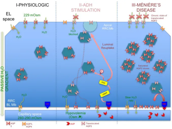
IV-5. Hypothetical model for endolymphatic sac pathology
We describe in Figure 5 a hypothetical model for the pathology of Ménière’s Disease involving AQP2 in the endolymphatic sac epithelium. Other aquaporin channels are expressed on the ELS, but were omitted for simplicity and because AQP2 is the only expressed
aquaporin under hormonal control. Physiologic expression of AQP2 is on the luminal
membrane of RRCs (low expression density) to allow passive movement of endolymph across the epithelial cell. The vast majority of AQP2 is stored in endosomal vesicles. Overexpression of AQP2, whether by hyperactive V2R or constitutive expression (AVP-V2R independent), resembles a state of constant AVP stimulation in favor of endolymph volume preservation. Normal variations of pAVP may have a more potent effect on the hypersensitive RRCs. Stress levels of pAVP further exacerbate this phenomenon. Luminal AQP2 is chronically trapped in endosomes by this process, reducing membrane permeability to water and limiting the EL efflux from the ELS. The result is slow endolymphatic volume build up, followed eventually by retrograde flow into the vestibule and cochlea, causing vertigo spells only when critical volume is reached and perhaps Reissner’s membranes rupture.
IV-6. Study limitations and future research
The present study is not without evident limitations. First of all, obtaining endolymphatic sac samples without cellular contamination from neighboring dura matter is no easy task, as these structures are in close proximity. In the case of MD patients undergoing endolymphatic
duct blockage surgery, only a small biopsy was obtained from the lateral portion of the main body of the sac, due to limited dissection in order to preserve the posterior semi-circular canal integrity. This translates into less epithelial tissue when compared to the intact lateral
endolymphatic sac samples obtained from controls, as suggested by total epithelial surface areas described earlier. However, the algorithm used by the Visiomorph® software takes this difference into account and generates weighted means. Furthermore, during microtome sectioning of paraffin-embedded samples, some epithelial structures were sloughed off, thus reducing our effective sample of epithelial cells. Finally, the quantitative protein expression analysis performed in this study does not reflect the functional status of investigated channels. Future research should focus on establishing a clear mechanism for AQP2 translocation in the inner ear, as precise membrane localization was only demonstrated in one experiment on guinea pig ELS 41. Replication studies are needed, preferably on human ELS samples or organotypic cell cultures. Other sites of AQP2 expression (Stria vascularis, Reissner’s membrane) should also be studied more extensively. Another topic of interest should be the composition of human endolymph in vivo, particularly in the endolymphatic sac, to better understand osmotic gradients across the ELS epithelium. Experimental setups that study the effect of AVP stimulation, NKCC2 blockade (by loop diuretics such as furosemide) and V2R blockade (OPC-31260) on ELS endolymph composition are also warranted and would
improve our understanding of MD’s pathology. Finally, genetic research is needed to better understand familial and sporadic forms of Ménière’s disease, with particular attention to epigenetic processes, promoter regions, splice sites and other posttranscriptional phenomena involving inner ear aquaporins, as well as functional and regulatory sites of the V2R receptor.
V. Conclusion
In this study, we have demonstrated an increased expression of AQP2 in the endolymphatic sac epithelium of patients affected by Ménière’s disease, which constitutes strong evidence for involvement of this aquaporin in the pathophysiology of MD. We could not detect an altered protein expression of V2R, NKCC2 or TRPV4 on the ELS. We have not observed plasma vasopressin variations that could be attributed to Ménière’s disease.
VI.REFERENCES
1. Ménière P. Sur une forme de surdité grave dépendant d’une lésion de l’oreille interne.
Gaz Méd de Paris. 1861;16:29.
2. Stahle J, Stahle C, Arenberg IK. Incidence of Meniere's disease. Arch Otolaryngol. 1978;104(2):99-102.
3. Wladislavosky-Waserman P, Facer GW, Mokri B, Kurland LT. Meniere's disease: a 30-year epidemiologic and clinical study in Rochester, Mn, 1951-1980. Laryngoscope. 1984;94(8):1098-1102.
4. Shaver EF, Jr. Allergic management of Meniere disease. Arch Otolaryngol. 1975;101(2):96-99.
5. Arenberg IK, Cabriac G, Marks S, Arenberg JG, Pfeiffer PR, Murray RS. Cytomegalovirus antibodies in endolymphatic sac biopsies of patients with
endolymphatic hydrops and Meniere's disease. Annals of the New York Academy of
Sciences. 1997;830:314-318.
6. Wackym PA, Linthicum FH, Jr., Ward PH, House WF, Micevych PE, Bagger-Sjoback D. Re-evaluation of the role of the human endolymphatic sac in Meniere's disease.
Otolaryngol Head Neck Surg. 1990;102(6):732-744.
7. Soliman AM. A subpopulation of Meniere's patients produce antibodies that bind to endolymphatic sac antigens. The American journal of otology. 1996;17(1):76-80. 8. Yoo TJ, Shea J, Jr., Ge X, et al. Presence of autoantibodies in the sera of Meniere's
disease. The Annals of otology, rhinology, and laryngology. 2001;110(5 Pt 1):425-429. 9. Derebery MJ, Rao VS, Siglock TJ, Linthicum FH, Nelson RA. Meniere's disease: an
immune complex-mediated illness? Laryngoscope. 1991;101(3):225-229.
10. Lopez-Escamez JA, Saenz-Lopez P, Gazquez I, et al. Polymorphisms of CD16A and CD32 Fcgamma receptors and circulating immune complexes in Meniere's disease: a case-control study. BMC medical genetics. 2011;12:2.
11. Rauch SD. Clinical hints and precipitating factors in patients suffering from Meniere's disease. Otolaryngol Clin North Am. 2010;43(5):1011-1017.
12. Ren H, Yin T, Lu Y, Kong W, Ren J. Intratympanic dexamethasone injections for refractory Meniere' s disease. Int J Clin Exp Med. 2015;8(4):6016-6023.
13. Pullens B, Verschuur HP, van Benthem PP. Surgery for Meniere's disease. Cochrane
Database Syst Rev. 2013(2):CD005395.
14. Saliba I, Gabra N, Alzahrani M, Berbiche D. Endolymphatic duct blockage: a randomized controlled trial of a novel surgical technique for Meniere's disease treatment. Otolaryngol Head Neck Surg. 2015;152(1):122-129.
15. Andersen HC. Passage of trypan blue into the endolymphatic system of the labyrinth.
Acta Otolaryngol. 1948;36(3-4):273-283.
16. Salt AN, Ohyama K, Thalmann R. Radial communication between the perilymphatic scalae of the cochlea. I: Estimation by tracer perfusion. Hear Res. 1991;56(1-2):29-36. 17. Salt AN, Ohyama K, Thalmann R. Radial communication between the perilymphatic
scalae of the cochlea. II: Estimation by bolus injection of tracer into the sealed cochlea.
Hear Res. 1991;56(1-2):37-43.
18. Salt AN, DeMott JE. Endolymph volume changes during osmotic dehydration measured by two marker techniques. Hear Res. 1995;90(1-2):12-23.
19. Eckhard A, Muller M, Salt A, Smolders J, Rask-Andersen H, Lowenheim H. Water permeability of the mammalian cochlea: functional features of an aquaporin-facilitated water shunt at the perilymph-endolymph barrier. Pflugers Arch. 2014;466(10):1963-1985.
20. Kimura RS. Experimental blockage of the endolymphatic duct and sac and its effect on the inner ear of the guinea pig. A study on endolymphatic hydrops. The Annals of
otology, rhinology, and laryngology. 1967;76(3):664-687.
21. Kimura R, Schuknecht H. Membranous hydrops in the inner ear of the guinea pig after obliteration of the endolymphatic sac. ORL. 1965;27(6):343-354.
22. Manni JJ, Kuijpers W, van Wichem P. Experimental endolymphatic hydrops in the rat.
Archives of otolaryngology--head & neck surgery. 1986;112(4):423-427.
23. Shinomori Y, Kimura R, Adams J. Changes in immunostaining for Na+, K+, 2Cl-cotransporter 1, taurine and c-Jun N-terminal kinase in experimentally induced endolymphatic hydrops. Paper presented at: ARO Abstr2001.
24. Salt AN, Thalmann R. Cochlear fluid dynamics. In: Physiology of the Ear. Raven Press New York; 1988:341-357.
25. Semaan MT, Alagramam KN, Megerian CA. The basic science of Meniere's disease and endolymphatic hydrops. Curr Opin Otolaryngol Head Neck Surg. 2005;13(5):301-307.
26. Rauch SD, Merchant SN, Thedinger BA. Meniere's syndrome and endolymphatic hydrops. Double-blind temporal bone study. The Annals of otology, rhinology, and
laryngology. 1989;98(11):873-883.
27. Merchant SN, Adams JC, Nadol JB, Jr. Pathophysiology of Meniere's syndrome: are symptoms caused by endolymphatic hydrops? Otol Neurotol. 2005;26(1):74-81. 28. Wu Q, Dai C, Zhao M, Sha Y. The correlation between symptoms of definite
Meniere's disease and endolymphatic hydrops visualized by magnetic resonance imaging. Laryngoscope. 2016;126(4):974-979.
29. Ishiyama G, Lopez IA, Ishiyama A. Aquaporins and Meniere's disease. Curr Opin
Otolaryngol Head Neck Surg. 2006;14(5):332-336.
30. Li J, Verkman A. Impaired hearing in mice lacking aquaporin-4 water channels.
Journal of Biological Chemistry. 2001;276(33):31233-31237.
31. Eckhard A, Dos Santos A, Liu W, et al. Regulation of the
perilymphatic-endolymphatic water shunt in the cochlea by membrane translocation of aquaporin-5.
Pflugers Arch. 2015;467(12):2571-2588.
32. Celestino D, Iannetti G. Meniere's disease and plasmatic hyperosmolarity. J Laryngol
Otol. 1973;87(3):229-234.
33. Celestino D, Ralli G. Plasmatic osmolality in Meniere's disease. J Laryngol Otol. 1981;95(3):273-277.
34. Bichet DG. Lithium, cyclic AMP signaling, A-kinase anchoring proteins, and
aquaporin-2. Journal of the American Society of Nephrology : JASN. 2006;17(4):920-922.
35. Conner AC, Bill RM, Conner MT. An emerging consensus on aquaporin translocation as a regulatory mechanism. Mol Membr Biol. 2013;30(1):1-12.
36. Couloigner V, Berrebi D, Teixeira M, et al. Aquaporin-2 in the human endolymphatic sac. Acta Otolaryngol. 2004;124(4):449-453.
37. Fukushima K, Takeda T, Kakigi A, et al. Effects of lithium on endolymph homeostasis and experimentally induced endolymphatic hydrops. ORL J Otorhinolaryngol Relat
Spec. 2005;67(5):282-288.
38. Kumagami H, Loewenheim H, Beitz E, et al. The effect of anti-diuretic hormone on the endolymphatic sac of the inner ear. Pflugers Arch. 1998;436(6):970-975.
39. Takeda T, Takeda S, Kitano H, Okada T, Kakigi A. Endolymphatic hydrops induced by chronic administration of vasopressin. Hear Res. 2000;140(1-2):1-6.
40. Takeda T, Sawada S, Takeda S, et al. The effects of V2 antagonist (OPC-31260) on endolymphatic hydrops. Hearing Research. 2003;182(1-2):9-18.
41. Maekawa C, Kitahara T, Kizawa K, et al. Expression and translocation of aquaporin-2 in the endolymphatic sac in patients with Meniere's disease. J Neuroendocrinol. 2010;22(11):1157-1164.
42. Gamba G, Miyanoshita A, Lombardi M, et al. Molecular cloning, primary structure, and characterization of two members of the mammalian electroneutral sodium-(potassium)-chloride cotransporter family expressed in kidney. J Biol Chem. 1994;269(26):17713-17722.
43. Mori N, Ninoyu O, Morgenstern C. Cation transport in the ampulla of the semicircular canal and in the endolymphatic sac. Archives of oto-rhino-laryngology.
1987;244(1):61-65.
44. Kakigi A, Nishimura M, Takeda T, Taguchi D, Nishioka R. Expression of aquaporin1, 3, and 4, NKCC1, and NKCC2 in the human endolymphatic sac. Auris Nasus Larynx. 2009;36(2):135-139.
45. Nishimura M, Kakigi A, Takeda T, Takeda S, Doi K. Expression of aquaporins, vasopressin type 2 receptor, and Na+(-)K+(-)Cl(-) cotransporters in the rat endolymphatic sac. Acta Otolaryngol. 2009;129(8):812-818.
46. Moller MN, Kirkeby S, Vikesa J, Nielsen FC, Caye-Thomasen P. Gene expression in the human endolymphatic sac: the solute carrier molecules in endolymphatic fluid homeostasis. Otol Neurotol. 2015;36(5):915-922.
47. Akiyama K, Miyashita T, Matsubara A, Mori N. The detailed localization pattern of Na+/K+/2Cl- cotransporter type 2 and its related ion transport system in the rat endolymphatic sac. J Histochem Cytochem. 2010;58(8):759-763.