Université de Montréal
The influence of cell size on cytokinesis in situ and genomic
interrogation of human cell size regulation
par Karine Gauvin Bourdages
Programme de biologie moléculaire Faculté de médecine
Thèse présentée
en vue de l’obtention du grade de doctorat en biologie moléculaire
option biologie des systèmes
Décembre, 2017
Résumé
La cellule est l’élément fondamental de la vie. Plus d’une vingtaine de trillions de cellules forment les organes et tissus de notre corps. Ces cellules sont de taille spécifique puisqu’elles ont des fonctions précises au sein de leur tissu respectif. Dans la plupart des cas, les cellules doivent proliférer en se divisant pour se renouveler et ainsi assurer le bon fonctionnement d’un organisme. La dernière étape de la division cellulaire, la cytokinèse, est exécutée par la contraction d’un anneau contractile d’actomyosine, nécessaire pour effectuer la séparation physique de la cellule en deux cellules filles. La première partie des travaux décrits dans cet ouvrage portent sur la caractérisation de la cytokinèse en utilisant, comme modèle in vivo, les cellules précurseur de la vulve (VPCs) du nématode C. elegans. Notre étude révèle que plusieurs aspects de l’anneau d’actomyosine s’ajustent en fonction de la taille de la cellule. Entre autres, la largeur de l’anneau contractile, juste avant sa constriction, s’ajuste en fonction de la longueur des VPCs. De plus, la rapidité avec laquelle l’anneau se contracte dépend de la circonférence de la cellule. Ces découvertes nous ont amené à nous demander comment la cellule régule sa taille? Les cellules en prolifération maintiennent leur taille en homéostasie en équilibrant leur taux de croissance et de division cellulaire. Afin d’interroger les gènes impliqués dans le maintien de la taille cellulaire du mammifère, nous avons utilisé la technologie CRISPR/Cas9, afin d’éliminer par délétion tous les gènes humains, à raison d’un par cellule, pour identifier ceux qui causent une augmentation ou une diminution de la taille cellulaire. Cette étude nous a permis d’identifier plusieurs gènes déjà connus régulant la croissance cellulaire. De plus, nous avons identifié un groupe de gènes, incluant TLE4 un corépresseur de la transcription que nous avons caractérisé, n’ayant jamais été associé avec une fonction de contrôle de la taille cellulaire chez les mammifères. En somme, nos travaux ont contribué à l’approfondissement des connaissances sur la division cellulaire, plus précisément la cytokinèse, et des gènes impliqués dans le maintien de la taille cellulaire. Une meilleure connaissance du fonctionnement de ces deux évènements cellulaires est essentielle puisque leur dérégulation peut entrainer plusieurs pathologies, incluant le cancer.
Mots-clés: Cytokinèse, taille cellulaire, C. elegans, VPCs, CRISPR/Cas9, criblage, mTORC1,
Abstract
Cells are the fundamental building blocks of life. The human body contains over twenty trillion cells that make up the different tissues and organs of our bodies. Cells within organs are of specific sizes to perform their specialized functions. In most cases, these cells must divide to proliferate and replenish the population of cells essential for proper organism function. The final stage of cellular division, termed cytokinesis, entails the assembly and constriction of a contractile ring that drives the dramatic cell shape changes required to physically partition the cell into two daughter cells. The first part of the work presented in this thesis addresses the characterization of cytokinesis in the epithelial vulval precursor cells (VPCs) of the nematode worm C. elegans. This study principally revealed that several aspects of cytokinesis scale with cell size. For instance, I observed that the breadth of the actomyosin ring scaled with VPC length. In addition, the speed of contractile ring constriction scaled with the circumference of VPCs. These scaling events raised the more general question as to how cells regulate their size. Proliferating cells attain cell size homeostasis by balancing cell growth and cell division. In order to define the molecular regulators of size in human cells a genome-wide approach was taken. Recently developed CRISPR/Cas9 technology was used to perform the first pooled knockout screens for human cell size regulators in the NALM-6 pre-B lymphocytic cell line. These screens revealed many genes that affect the size of NALM-6 cells, a number of which were previously known to be involved in growth regulation. In addition, these screens revealed the identity of many genes with no previously established functions associated with cell size regulation. Amongst the previously unknown regulators, I characterized the function of a co-repressor of transcription, TLE4, which I showed functions as a regulator of the B-cell lineage. This work contributes to the knowledge of the mechanics of cytokinesis in C. elegans epithelial cells and of the genes that coordinate cell size in humans. These results provide insights into cell growth and division in normal cells and how these processes may be perturbed in cancer and other diseases.
Table of Contents
Résumé ... i
Abstract... ii
Table of Contents ... iii
List of Figures and Table ... vii
List of Abbreviations ... viii
Acknowledgements ... xii
Introduction... 1
1 General concepts ... 2
1.1 The intriguing question of size ... 2
1.1.1 Enormous size scale... 2
1.1.2 Cell size de-regulation associated with human diseases... 4
1.2 Cytokinesis... 5
1.2.1 Cytokinesis and cancer ... 7
1.3 Scaling unifies cytokinesis and cell size... 8
1.3.1 Cell size influences cell division processes including cytokinesis ... 8
2 The events of cytokinesis ... 10
2.1 Division site specification... 10
2.1.1 Cues from the mitotic apparatus ... 10
2.1.2 Formation of the spindle midzone ... 12
2.1.3 Cell-type specific requirements for division plane establishment ... 17
2.1.4 Ect2-dependent RhoA activation at the equatorial cortex ... 18
2.2 Contractile ring assembly ... 20
2.2.1 Structural components of the contractile ring... 20
2.2.2 Molecular constituents required for contractile ring assembly... 23
2.2.3 The scaffolding protein Anillin... 24
2.3.1 Asymmetric cytokinesis... 28
2.4 Mechanistic insights into cytokinesis in epithelial cells... 30
2.4.1 Molecular composition of intercellular junctions... 30
2.4.2 The C. elegans apical junctions ... 31
2.4.3 The mechanics of cytokinesis in Drosophila epithelia... 32
2.5 The C. elegans vulva as a model epithelium to study cytokinesis... 34
2.5.1 The model organism C. elegans... 35
2.5.2 Seminal discoveries using C. elegans... 36
2.5.3 The C. elegans vulva... 37
2.5.4 The C. elegans vulval precursor cells (VPCs)... 38
2.5.5 The characterization of VPC cytokinesis... 40
3 Results Part 1 - Article 1 ... 41
Quantitative analysis of cytokinesis in situ during C. elegans postembryonic development ... 41
Author contributions ... 41
3.1 Abstract... 43
3.2 Introduction... 43
3.3 Results... 46
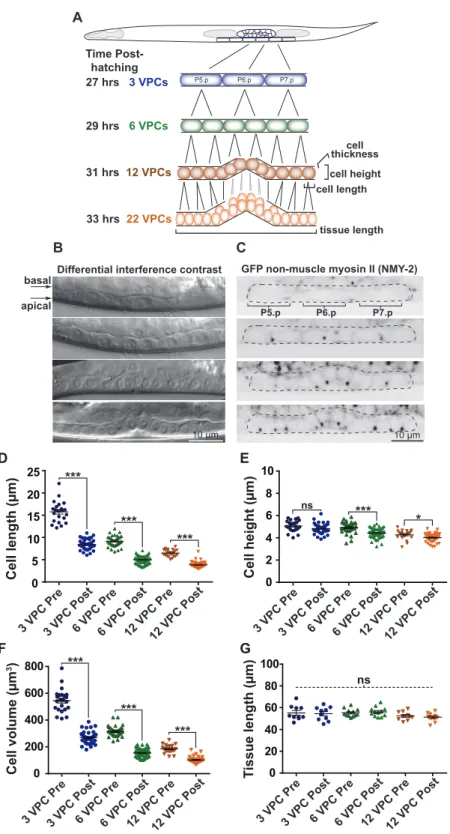
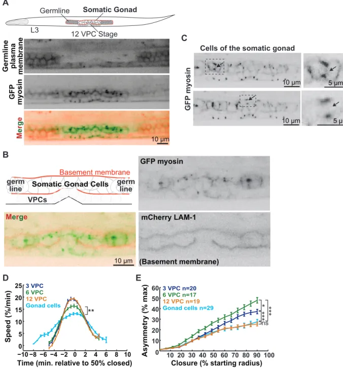
3.3.1 Visualization of the vulval precursor cells (VPCs) in situ... 46
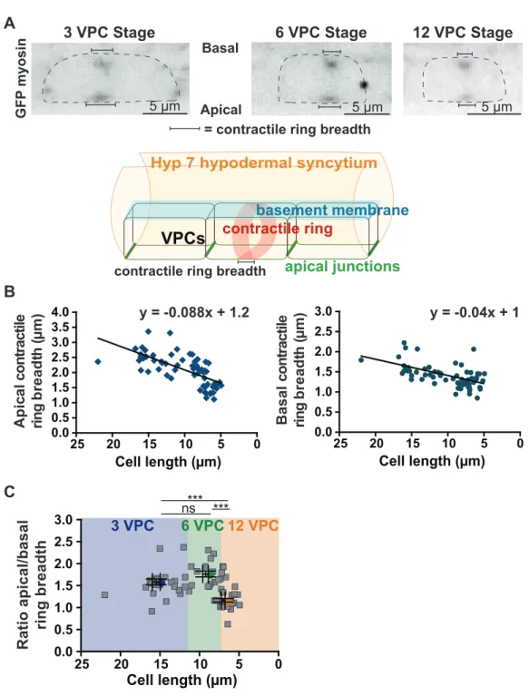
3.3.2 Contractile ring dimensions scale with cell size... 47
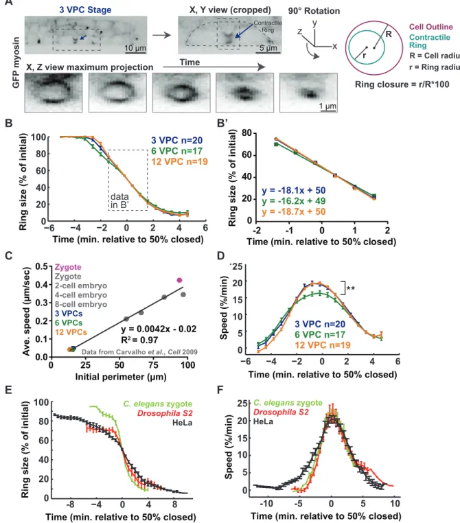
3.3.3 Furrowing speed scales with division plane dimensions ... 48
3.3.4 Contractile ring closure occurs via acceleration and deceleration... 49
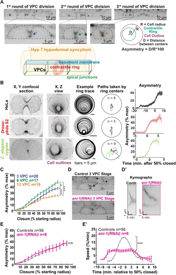
3.3.5 Asymmetric furrowing occurs towards the apical membrane of VPCs... 49
3.3.6 Epithelial organization influences the kinetics of cytokinesis... 51
3.4 Discussion... 51
3.5 Figures and legends... 55
3.6 Supplemental Figure and legend... 65
3.7 Materials and methods ... 67
3.8 Acknowledgements... 70
4.1 Cell size scales with ploidy... 71
4.2 The existence of a cell size threshold ... 72
4.2.1 A critical cell size in yeasts... 74
4.2.2 A cell size threshold in animal cells ... 75
4.3 Cell size regulation in budding yeast... 75
4.3.1 Environmental conditions modulate cell growth ... 76
4.3.2 Identification of the first cell size mutants in yeast ... 76
4.3.3 Start architecture ... 77
4.3.4 Systematic screens for size regulators in yeast... 78
4.4 Mammalian cell size regulation... 79
4.4.1 Mammalian cell growth ... 79
4.4.2 The target of rapamycin (TOR) growth regulatory network ... 80
4.4.3 Signaling through mTORC1... 81
4.4.4 MYC participates in cell growth regulation... 85
4.4.5 Metazoan cell cycle control ... 86
4.5 Reconciling the coordination of growth with division ... 87
4.5.1 Measuring growth rates... 88
4.6 Knowledge gaps in mammalian cell size regulation... 89
5 The CRISPR/Cas9 technology... 90
5.1 CRISPR/Cas9 for precise gene editing... 90
5.2 Genome-wide CRISPR/Cas9 pooled screens ... 93
5.3 Applications of CRISPR/Cas9... 95
6 Results Part 2 - Article 2 ... 98
A genome-wide CRISPR/Cas9 knockout screen reveals that TLE4 participates in the maintenance of pre-B cell size homeostasis ... 98
Author contributions ... 98
6.1 Abstract... 99
6.2 Introduction... 99
6.3 Results... 102
6.3.2 Many hits were identified in the cell size screens including the strong candidate
gene TLE4... 103
6.3.3 The cell size phenotype conferred by loss of TLE4 function and several other candidates was validated... 105
6.3.4 Transcripts associated with B cell-specific functions are up-regulated following TLE4 knockout ... 107
6.3.5 CD19 differentiation factor is over-expressed following TLE4 deletion ... 109
6.4 Discussion... 110
6.5 Figures and Legends ... 113
6.6 Materials and Methods... 122
6.7 Acknowledgements... 126
7 Discussion and perspectives ... 128
7.1 Future directions in cytokinesis studies ... 128
7.2 Global analysis of genetic regulators of mammalian cell size... 131
7.3 Do cells sense their size? ... 133
7.4 Mammalian cell size; parts list and conservation ... 134
Concluding remarks ... 135
List of Figures and Table
FIGURE 1.1.1 EXAMPLES OF THE CELL SIZE DIVERSITY OBSERVED IN NATURE ... 4 FIGURE 1.2 AN OVERVIEW OF CYTOKINESIS ... 6 FIGURE 2.1.2 MOLECULAR CONSTITUENTS OF THE SPINDLE MIDZONE ... 13 FIGURE 2.2.1 STRUCTURAL COMPONENTS OF THE CONTRACTILE RING ... 22 FIGURE 2.5.1 ANATOMY OF AN ADULT C. ELEGANS WORM... 36 FIGURE 3.5.1 THE C. ELEGANS VULVAL PRECURSOR CELLS (VPCS) INSIDE THE LIVING AND DEVELOPING ANIMAL…... 55 FIGURE 3.5.2 CONTRACTILE RING DIMENSIONS SCALE WITH THE LENGTH OF VPCS ... 57 FIGURE 3.5.3 QUANTITATIVE ANALYSIS OF THE KINETICS OF CONTRACTILE RING CLOSURE IN THE VPCS ... 59 FIGURE 3.5.4 STRONG INTERCELLULAR ADHESION LEADS TO ROBUST ASYMMETRIC CONTRACTILE RING CLOSURE IN THE VPCS ... 61 FIGURE 3.5.5 TISSUE GEOMETRY INFLUENCES THE KINETICS OF CYTOKINESIS IN THE CELLS OF THE SOMATIC GONAD…... 63 FIGURE 3.6.1 SUPPLEMENTAL FIGURE ... 65 FIGURE 4.2 CELL GROWTH IN G1 TO REACH THE CELL SIZE THRESHOLD AND TRIGGER S PHASE ENTRY... 73 FIGURE 4.4.3 THE MOLECULAR REGULATORS OF CELL GROWTH THROUGH MTORC1 SIGNALING ... 85 FIGURE 5.1 THE CRISPR/CAS9 SYSTEM... 93 FIGURE 6.5.1 OUTLINE OF GENOME-WIDE CRISPR/CAS9 CELL SIZE SCREENS IN NALM-6 PRE-B LYMPHOCYTES…. ... 113 FIGURE 6.5.2 MAMMALIAN CELL GROWTH REGULATORS ASSOCIATED WITH MTORC1 IDENTIFIED IN CELL SIZE SCREENS... 115 FIGURE 6.5.3 CELL SIZE ASSAYS CONFIRM PHENOTYPES CONFERRED BY SEVERAL CANDIDATES FROM THE SCREENS... 117 FIGURE 6.5.4 THE SMALL CELL SIZE PHENOTYPE RESULTING FROM TLE4 DELETION SEEMS NALM-6-SPECIFIC….. ... 118 FIGURE 6.5.5 GENES ASSOCIATED WITH B CELL-SPECIFIC FUNCTIONS ARE UP REGULATED UPON TLE4 KNOCKOUT ... 119 FIGURE 6.5.6 THE LOSS OF TLE4 FUNCTION LEADS TO AN INCREASE IN CD19 EXPRESSION ... 120 TABLE TOP SCORING GENES SHOWING TRANSCRIPTS ENRICHMENT IN TLE4 KNOCKOUT CELLS ... 121List of Abbreviations
4E-BP1: Eukaryotic translation initiation factor 4E-binding protein 1 AC: Anchor cell
AH: Anillin Homology
AJM-1: Apical junction molecule 1 AJs: Adherens junctions
ALL: Acute lymphoblastic leukemia C. elegans: Caenorhabditis elegans CD: Cluster of differentiation Cdc: Cell division cycle
CDKs: Cyclin-dependent kinases CeAJs: C. elegans apical junctions
Chip-Seq: Chromatin immunoprecipitation sequencing CPC: Chromosomal passenger complex
CRISPR: Clustered regularly interspaced short palindromic repeats CRISPRi: CRISPR interference
crRNA: CRISPR RNA
CyanRing: Cytokinesis analysis of the contractile ring DIC: Differential interference contrast
DLG-1: Discs Large 1 E. coli: Escherichia coli Egl: Egg-laying defective
eIF4E: Eukaryotic translation initiation factor 4E EKO: Extended knockout
F-actin: Filamentous actin
FACS: Fluorescence-activated cell sorting GeCKO: Genome-scale CRISPR/Cas9 KO GFP: Green fluorescent protein
HDR: Homology-directed repair HMP: Humpback HMR: Hammerhead HR: Homologous recombination Hyp7: hypodermis IgM: Immunoglobulin M
INCENP: Inner centromere protein Indels: Insertions or deletions L1-4: Larval stage 1 through -4 LAM-1: Laminin-1
MKLP1: Mammalian kinase-like protein 1 MKLP2: Mammalian kinase-like protein 2 mTOR: Mechanistic target of rapamycin
mTORC1: Mechanistic target of rapamycin complex 1 mTORC2: Mechanistic target of rapamycin complex 2 Muv: Multiple vulvae
NHEJ: Non-homologous end joining NMY-II: Non-muscle myosin II NSCs: Neuronal stem cells PAM: Protospacer adjacent motif PLC: Phospholipase C
PLK1: Polo-like kinase 1
PRC1: Protein regulating cytokinesis 1 Pre-BCR: Pre-B cell receptor
Pvl: Protruding vulva
RANKS: Robust analytics and normalization for knockout screens Rb: Retinoblastoma
rMLC: Regulatory myosin light chain RNA-Seq: RNA sequencing
RNAi: RNA interference ROCK: Rho-dependent kinase
S6K1: S6 kinase 1
sgRNA: Single guide or synthetic guide RNA shRNA: short hairpin RNA
siRNA: Small interfering RNA SJs: Septate junctions
TALENS: Transcription activator-like effector nucleases TJs; Tight junctions
TKO: Toronto knockout
TLE4: Transducin like enhancer of split 4 TOR: Target of rapamycin
tracrRNA: Trans-activating CRISPR RNA TSC: Tuberous sclerosis complex
VPCs: Vulval precursor cells Vul: Vulvaless
Je dédie cet ouvrage à mes parents, qui m’ont transmis leur amour pour les sciences, particulièrement à ma mère là-haut.
Acknowledgements
I want to begin by expressing how thankful I am to everyone I had the pleasure of working with over the course of my graduate studies. First and foremost, I want to thank both of my supervisors, Dr. Amy Maddox and Dr. Mike Tyers, who gave me the opportunity to work on exciting projects. Thank you, Amy, for teaching me to be rigorous and detailed oriented in science. You were a great mentor and always believed in me. Mike, I want to thank you for sharing your passion and exciting ideas about science and for the warm welcome to your lab. You were an excellent mentor, who allowed me to bring my own ideas and be more independent. I also want to thank Dr. Paul Maddox and Dr. Lea Harrington, with whom I shared insightful scientific discussions. Thank you for making this journey even more enjoyable by bringing labs together for scientific exchanges. I also want to thank the members of my thesis committee, Dr. Sébastien Carréno, Dr. Brian Wilhelm and Dr. Christian Rocheleau for their support and helpful comments throughout the years.
I want to thank all members of both Maddox labs. It was an immense pleasure to work with you all. I will always remember the fun we had on our ASCB trips to San Francisco and New Orleans! Benjamin, you were an incredible mentor who taught me everything I needed to know when I arrived in the Maddox lab. You were always there to encourage me and support my work in the lab. I also want to thank Carlos and Jonas for their contribution to the scientific publication we brought to completion with Ben and Amy. It was a great pleasure to work with such incredible colleagues. I also want to say thank you to my colleagues of the Labbé lab. It was great to talk about C. elegans and share numerous moments of laughter. Eugénie, I thank you for being such an exceptional friend. A special thank you to Valérie, it was a pleasure to work with you during the Maddox times and to renew our friendship when you came back to IRIC. I thank you for all the great advice you gave me over the years.
I want to thank all members of the Tyers lab. You kindly welcomed me and made the transition to your lab easy for me. I am grateful to have had the chance to work with such knowledgeable colleagues. Thierry, I thank you for your incredible mentorship. You taught me everything I needed to know and shared your passion for research. Thank you for all the tips you gave me and for our insightful conversations. A special thank you to Luisa, you were
always there to help me. Your guidance was key to the realization of this work. You taught me so such much about science and life in general and for that I am very grateful. Jasmin, thanks for your contribution to the project, without you this work wouldn’t have been feasible. Almer, Susan and Andrew, I thank you all for the animated scientific discussions we had over the years. I also thank you for all the great moments we shared over lunch and during office chats. I also want to thank Sylvain for his help with thesis-related work. I thank Samuel, Jing, Lily, Ghada, Linnea, Daniel, Corinne, Yahya, Manon, Caroline and Roger for being incredible colleagues.
I thank the members of IRIC platforms for their technical help on experiments. Thank you, Christian Charbonneau, for your help with microscopes and for training with the Illustrator software. I want to say thank you to Gaël Dulude and Danièle Gagné for technical assistance with FACS experiments. I also want to thank all members of the high-throughput screening facility and genomics platform at IRIC. Also, thank you to Dr. Mader and members of her lab with whom I did my master’s degree rotation. I want to say thanks to everyone at the academic affairs of IRIC for their help over all these years. A special thank to Julie for answering all my questions and for your help in preparation of the next chapter.
Finally, I would not have been able to make it through my doctoral studies without the support from my family. I want to thank my dad and sister for having been there when I needed encouragement and advice. You were always there for me so thank you. I want to thank my grandparents, Gérald and Marie-Claire, who gave me the inspiration to write this thesis at their lake house. As well, I thank my grandmother Marie-Berthe for her encouragements. I thank my aunt Diane and my uncle Tom, who supported me during my doctoral studies and especially while writing my thesis. I also thank all other members of my family. Finally, I want to say thanks to my significant other, Jean-Mathieu, for his ongoing support and his patience during the past four years. Thank you for believing in me.
1 General concepts
1.1 The intriguing question of size
Cells are the fundamental structures of life in all living organisms. Across species, cell shape and size variations are remarkable (Maniloff and Morowitz, 1972; Smith et al., 1992). However, within an organism, cells of the same tissue type show very little variation in size, including cells that make up the different organs in our body (Ginzberg et al., 2015). How cells achieve this uniformity in size despite experiencing changing environmental conditions and physiological stress represents a fundamental question in biology. This question of how cells precisely regulate their size has remained an enigma from the early days on modern cell biology. Haldane stated: “The most obvious differences between different animals are differences of size, but for some reason the zoologists have paid singularly little attention to them” (Haldane, 1985). Even though most differences of size are attributed to differences in cell number, cell size variations are important in body size determination (Conlon et al., 2001). Nonetheless, it is fascinating how such a striking observation in nature has remained underappreciated and little studied over nearly one hundred years. In recent decades, studies on cell size regulation began to emerge (Amodeo and Skotheim, 2016; Cook and Tyers, 2007; Ginzberg et al., 2015; Jorgensen and Tyers, 2004; Turner et al., 2012). New molecular genetic approaches developed in the post-genomic era have enabled the long-standing question of how cells regulate their size to be addressed. In the following sections, the progress made in identifying the molecular regulators of cell size will be summarized for unicellular organisms to mammals, including humans.
1.1.1 Enormous size scale
The astonishing range in size of living organisms is perhaps the most obvious feature of life on earth (Bonner, 2011). The smallest bacteria, such as Pelagibacter ubique and mycoplasma species are only 0.2 µm and 0.3 to 1 µm in diameter, respectively (Maniloff and Morowitz, 1972; Rappe et al., 2002). At the other end of the spectrum lies the largest mammal on earth, the blue whale that can reach 100 feet in length and weigh 200 tons (Marshall et al.,
2012). The diversity in species size is further underscored by the incredibly small size of nano-organisms, approximately 6 aL in volume, compared to the clonal Armillaria fungi that can span thousands of acres (Smith et al., 1992; Uwins et al., 1998). This remarkable range in organism size is largely attributed to variations in cell number and to a lesser extent to differences in cell size. For instance, the large difference in body size between a human adult and a mouse is the result of a 3000-fold difference in cell number (Conlon and Raff, 1999). Even though cell number variations are predominant, differences in cell size are also important to consider. For instance, cells of tetraploid salamanders are twice the size of that of diploid animals whilst body sizes are the same (Fankhauser, 1945).
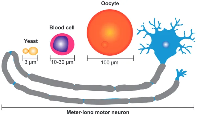
Cell size also spans a vast range. Small unicellular eukaryotes, such as yeast cells of only 3 µm in length, are dwarfed when compared to the largest unicellular eukaryote, the giant Caulerpa taxifolia alga that can span several meters in length (Ranjan et al., 2015). Within the human body cells span a substantial range of sizes, from small blood cells of only 10 to 30 µm in diameter to meter-long motor neurons to large oocytes of 100 µm in diameter (Figure 1.1.1) (Guertin and Sabatini, 2006). In contrast, cells of the same type, such as pancreatic cells or columnar cells of the epidermis show hardly any variation in size (Ginzberg et al., 2015). Evolution has shaped this diversity in cell size to accomplish specialized functions (Guertin and Sabatini, 2006).
Meter-long motor neuron
3 µm 10-30 µm 100 µm
Yeast
Blood cell
Oocyte
Figure 1.1.1 Examples of the cell size diversity observed in nature
Schematic representing a meter-long motor neuron (blue) compared to a budding yeast S. cerevisiae (yellow) approximately 3 µm in length (yellow), a blood cell (pink) 10 to 30 µm in diameter and compared to a human oocyte (orange) 100 µm in diameter. Adapted from (Guertin and Sabatini, 2006).
1.1.2 Cell size de-regulation associated with human diseases
Even though there is a broad diversity in cell size observed in nature, cell-type specific homogeneity in size is achieved within a cell population. Cell size homeostasis, an equilibrium state between cell growth and cell division, needs to be attained for proper cell function. Diseases can arise when cells fail to achieve size homeostasis. For instance, cardiac hypertrophy results when cardiac myocytes enlarge to compensate for anomalies of the heart (Heineke and Molkentin, 2006). Individuals with gigantism, due to growth hormone over-secretion, present severe organ overgrowth and are usually of abnormally large heights due to increased cell size and cell numbers (Melmed, 2009). In tuberous sclerosis (TSC), where affected patients develop benign tumors mainly in the brain, kidney, lungs and skin, giant cells are characteristic of brain tumors (Goto et al., 2011). These giant cells are the result of uncontrolled cell growth caused by loss-of-function mutations in either TSC1 or TSC2 genes, which negatively regulate the mTOR growth control network (Goto et al., 2011). In another
example, abnormally large cancer cells can arise upon entosis (cell engulfment) or aberrant cell division, including cytokinesis failure, which will be discussed in the following sections (Lacroix and Maddox, 2012).
Other examples of cell size plasticity include the active modulation of pathogen cell size to avoid immune detection by the host. For instance, the pathogenic yeast C. neoformans increase in size and become “titan” cells to escape immune surveillance (Zaragoza and Nielsen, 2013). Cell size de-regulation has also been observed in metabolic and mitochondrial disorders, aging and Parkinson’s Disease (Herrera et al., 2015; Maciak et al., 2014). Thus, cell size de-regulation is associated with various diseases reflecting the importance of understanding how cells achieve cell size homeostasis.
1.2 Cytokinesis
Individual proliferating cells achieve size homeostasis by balancing cell growth and cell division. The size of a cell is dictated by the size of the precursor (mother) cell it originates from and the amount of growth it accomplishes. Cell size uniformity for a particular cell type depends on a tight coordination between cell growth and cell division for each individual cell in the population. Therefore, it is important to understand how both cell growth and cell division are governed. In part, my studies have focused on the final step of cell division termed cytokinesis.
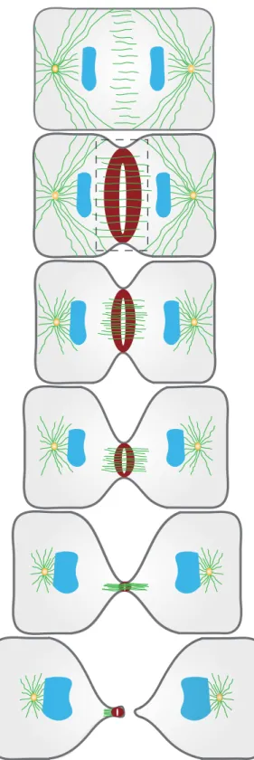
Cytokinesis is the final stage of cellular division that physically partitions the cytoplasm of a cell into two daughter cells. Cytokinesis is intricately coordinated in space and time. Cytokinesis begins in anaphase, following chromosome segregation to opposite spindle poles. Cytokinesis is robust to physical perturbations and drives cell shape changes in the form of membrane curves that ingress to bisect the cell. In the early events of cytokinesis, the spindle midzone specifies the site of division. At this precise location, a contractile ring is assembled beneath the plasma membrane and constricts to drive cell shape changes (Figure 1.2). Membrane ingression driven by the contractile ring proceeds until only a structure called the midbody remains (Figure 1.2). The midbody is resolved during abscission, involving membrane scission to render the newly formed daughter cells topologically distinct (Figure
1.2). The proper coordination of these events is essential, as cytokinesis failure can lead to diseases including cancer (Lacroix and Maddox, 2012).
Schematic representation of a cell undergoing cytokinesis. A contractile ring (red) assembles between segregated chromosome masses (blue). The contractile ring constricts and drives membrane ingression (dark grey). Contractile ring closure progresses until the midbody remains, a structure cleaved during abscission. This process marks the physical separation of a cell into two newly formed daughter cells. Adapted from (Green et al., 2012).
1.2.1 Cytokinesis and cancer
In the event of cytokinesis failure, cells become tetraploid, containing four copies of the genome. In most cases, tetraploidy results in activation of the p53 tumor suppressor gene and thus cell cycle arrest in G1 that can lead to cell death (Ganem and Pellman, 2007). However, under certain conditions including loss of p53, tetraploid cells can further proliferate. Upon division, these cells can fail to assemble a bipolar spindle due to their supernumerary microtubules organizing centers (Fujiwara et al., 2005). This can lead to merotelic chromosome attachments and chromosome segregation errors resulting in genetic instability (Ganem et al., 2009; Silkworth et al., 2009). Chromosome instability is manifested in several ways including mutations, deletions, chromosomal rearrangements and fusion, gene amplification and aneuploidy, an important hallmark of cancer (Lacroix and Maddox, 2012; Williams and Amon, 2009). Experiments in mice showed that cells that fail a round of cytokinesis are more prone to tumorigenesis (Fujiwara et al., 2005). However, it is important to mention that while cytokinesis failure may lead to cancer, it can also cause cell death upon p53 activation (Ganem and Pellman, 2007). How the decision between these two outcomes is made remains unknown. Thus, it is important to establish a deep understanding of the mechanisms ensuring the proper execution of cytokinesis.
Cytokinesis failure can be viewed as a double-edged sword. For certain cells, cytokinesis failure can cause genetic instability leading to diseases, as previously described. However, other cell types purposely fail cytokinesis to perform specialized functions (Lacroix and Maddox, 2012). For these cells, the completion cytokinesis can lead to pathological states (Lacroix and Maddox, 2012). These specialized cell types include hepatocytes, megakaryocytes and cardiomyocytes that reach high levels of ploidy by either failing cytokinesis or an earlier step in mitosis (Lacroix and Maddox, 2012). This re-enforces the importance of understanding the fundamental principles governing cytokinesis.
1.3 Scaling unifies cytokinesis and cell size
“Everywhere Nature works true to scale and everything has its proper size accordingly.” This citation by D’Arcy Wentworth Thompson nicely illustrates the fundamental biological principle of scaling according to size (D’Arcy Wentworth, 1945). In biology, scaling describes the ability of an organelle or any cellular component to adjust according to changes in cell size (Reber and Goehring, 2015). Adequate scaling events are essential for proper cell, organ and consequently organism function (Levy and Heald, 2012). This important question of how molecular structures scale with cell size for proper function rapidly generated interests in the biological science community. However, the lack of tools to precisely measure small structures in the micrometer range of most cells and organelles prevented progress in the early days of the scaling field. High-resolution microscopy and other imaging techniques, have allowed intracellular scaling to be examined at unprecedented resolution (Levy and Heald, 2012; Reber and Goehring, 2015). The following sections describe scaling events important for cell division.
1.3.1 Cell size influences cell division processes including cytokinesis
Cell size and cytokinesis are intimately related. Several examples illustrate the impact of cell size on intracellular structures governing cell division processes. Early observations by Schroeder demonstrated scaling of contractile rings filaments in cells of different sizes (Schroeder, 1972). Larger amphibian eggs possessed filaments of increased dimensions compared to smaller somatic cells (Schroeder, 1972). In addition, other subcellular structures are scaled to cell size as it reduces during early embryonic development, as the embryo undergoes several rounds of rapid cleavage in the near absence of growth. In both C. elegans and Xenopus embryos, the size of the cell influences mitotic spindle length. As cells get smaller during rapid cleavages so does the length of the mitotic spindle (Brown et al., 2007; Hara and Kimura, 2009; Loughlin et al., 2011). Experiments performed in C. elegans embryos showed that both spindle length and the speed of spindle elongation scaled with cell size (Hara and Kimura, 2009; Marshall et al., 2012). Elegant work in C. elegans embryos also showed that chromosome length scaled with cell size (Ladouceur et al., 2015). Altogether, these findings demonstrate that cell size influences the molecular architecture of the cell.
Several other examples illustrate the influence cell size has on the proper orchestration of cytokinesis events. For instance, my work demonstrated that several aspects of cytokinesis scale with cell size (Chapter 3). Using high-resolution microscopy, I characterized cytokinesis occurring in the vulval precursor cells (VPCs) of the nematode Caenorhabditis elegans (C. elegans). As the VPCs decrease in length from one round of division to the next, we observed that the breadth of the contractile ring scaled with cell length (Chapter 3). Quantitative assessments of the speed of contractile ring closure in the C. elegans VPCs revealed that it scaled with cell circumference and thus cell dimensions (Chapter 3). This observation supported previous findings in the C. elegans embryo and filamentous fungi Neurospora crassa that illustrated the conserved nature of this property (Calvert et al., 2011; Carvalho et al., 2009). This work is described in detail in Chapter 3. An understanding of how both cytokinesis and cell size are coordinated and influence each other should reveal general principles of scaling in biology.
2 The events of cytokinesis
Cytokinesis proceeds by a series of sequential events, each described in the following sections. The beginning of cytokinesis is marked by re-organization of the mitotic spindle to specify the site of contractile ring assembly. Molecular signals emanating from microtubules of the spindle midzone reach the above cortex to promote the recruitment of contractile ring components. This event triggers the assembly of a contractile ring beneath the plasma membrane and primarily composed of actin filaments, the motor non-muscle myosin II and scaffolding proteins including septins and anillin. Molecular motors of the contractile ring drive cell shape changes. Membrane ingression proceeds until the midbody remains. The final event of cytokinesis is termed abscission and involves midbody severing to physically separate the two daughter cells.
2.1 Division site specification
The initial event in cytokinesis is the establishment of the division plane. The maintenance of genome integrity requires that cell partitioning occurs at a precise location that is between segregated chromosome masses. Pioneering work in the cytokinesis field provided insights on the molecular structures implicated in this early event of cytokinesis. Later studies provided a detailed map of the molecular players involved in the specification of the division plane. Lastly, distinct cues provide different contributions to division site establishment depending on the organism.
2.1.1 Cues from the mitotic apparatus
More than 50 years ago, Ray Rappaport performed several elegant micromanipulation experiments that led to the identification of the mitotic spindle as the general structure responsible for positioning the cleavage furrow. In a first experiment, Ray pressed a glass rod into the middle of a sand dollar egg, such that the first cleavage did not completely divide the egg but generated a binucleate doughnut-shaped cell (Rappaport, 1961). During the next division, two mitotic spindles juxtaposed to the rod were formed and each induced a cleavage furrow (Rappaport, 1961). Surprisingly, an additional furrow appeared between astral
microtubules emanating from the two opposing spindles (Rappaport, 1961). This demonstrated that the mitotic spindle and its emanating astral microtubules could specify the division plane, independently of intervening chromosomes.
In another experiment, Rappaport deformed echinoderm eggs in a capillary tube. Sideways movements of the tube provoked displacement of the mitotic spindle along the tubular cell. Cleavage furrow initiation occurred to bisect the spindle (Rappaport, 1985). Upon displacement of the spindle, the cleavage furrow regressed from its initial location and reformed to bisect the spindle at this new site (Rappaport, 1985). Altogether, Rappaport’s observations suggested that the mitotic spindle signals the overlying cortex to specify the division plane. The first experiment suggested that the anaphase spindle and not the metaphase plate dictated the location of the cleavage furrow. Rappaport’s later experiment illustrated the dynamicity of the signal sent by the mitotic spindle.
Subsequent studies supported Rappaport’s early findings. In another micromanipulation experiment, a microneedle was used to create small perforations in adherent epithelial cells in culture (Cao and Wang, 1996). When the perforation was made adjacent to chromosome masses before anaphase onset, the cell failed to induce a cleavage furrow next to the perforation (Cao and Wang, 1996). This result further supports the role of the mitotic spindle in specifying the division plane.
Another group imaged cytokinesis in sea urchin embryos placed in chambers with adjustable atmospheric pressure. Increased pressure disrupted the structure of cortical cytoskeletal components and prevented astral microtubule elongation resulting in failure of cleavage furrow induction (Salmon and Wolniak, 1990). Restoring normal pressure in the chamber prior to the second round of division allowed the cell to proceed with the assembly of two mitotic spindles and induce four cleavage sites during cytokinesis (Salmon and Wolniak, 1990). This experiment indicates that astral microtubules play a role in determining the site of division.
Eckley and colleagues performed a similar experiment to that of Salmon and Wolniak but in somatic cells. In their experiment, they fused two cells together generating a v-shaped cell (Eckley et al., 1997). When dividing, the two mitotic spindles were oriented in a diagonal
(v-shape). In most cases, they observed induction of two cleavage furrows adjacent to both mitotic spindles (Eckley et al., 1997). However, in rare instances they saw a third furrow between microtubule asters of the opposing spindles (Eckley et al., 1997). Thus, in somatic cells, the mitotic spindle seems to play a more prominent role in division plane specification. These results also support the contribution from astral microtubules in determining the site of division. Altogether, these elegant micromanipulation experiments by Rappaport and others well illustrate the participation of the mitotic spindle and astral microtubules in establishing the site of division.
2.1.2 Formation of the spindle midzone
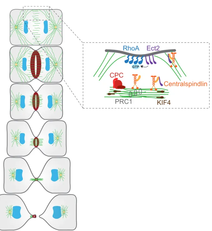
The mitotic spindle elongates during anaphase to pull apart sister chromatids towards opposing poles of the cell. Following faithful genome partitioning, re-organization of the mitotic spindle occurs. During this critical step, microtubules accumulate between chromosome masses to form a cellular structure known as the spindle midzone (Figure 2.1.2). The spindle midzone is composed of a bundle of overlapping antiparallel microtubules with their plus-ends oriented towards the cell center (Mastronarde et al., 1993). This specific region presents a small overlap of microtubules (Hu et al., 2011). The kinesin motor protein KIF4 is essential to restrict the microtubule overlap to delimit the division plane (Hu et al., 2011). In addition, microtubules of the midzone are much more stable than microtubules emanating from the spindle poles (Canman et al., 2003). These stable microtubules deliver signaling molecules to the equatorial cortex and membrane via plus-end directed molecular motors localized along these microtubules (Foe and von Dassow, 2008; Odell and Foe, 2008). The more dynamic astral microtubules near the cell poles act negatively to prevent accumulation of signaling molecules that promote contractile ring assembly (Foe and von Dassow, 2008; Odell and Foe, 2008). The identification of this specialized structure and its characteristics was key to the understanding of the mechanisms governing division site specification.
RhoA
Centralspindlin
CPC
Ect2
KIF4
PRC1
GTPFigure 2.1.2 Molecular constituents of the spindle midzone
The CPC (red), centralspindlin complex (orange), PRC1 (light grey) and KIF4 (brown) are recruited on microtubules of the midzone. The centralspindlin complex recruits Ect2 (purple) at the equatorial membrane for activation of RhoA (blue). Adapted from (Green et al., 2012).
The active participation of the spindle midzone in determining the site of division is well established (Cao and Wang, 1996; Foe and von Dassow, 2008; Odell and Foe, 2008; Rappaport, 1961; Rappaport, 1985). Several elegant imaging and inhibitory experiments performed in different organisms identified the conserved molecules required for spindle midzone assembly. Establishment of the spindle midzone requires three major components; protein regulating cytokinesis 1 (PRC1), the chromosomal passenger complex (CPC) and the centralspindlin complex. Described below are the molecular constituents of each complex that form an intricate signaling network that contributes to proper positioning of the cleavage furrow.
PRC1 directly interacts with microtubules of the midzone and promotes their bundling (Figure 2.1.2) (Jiang et al., 1998; Mollinari et al., 2002; Neef et al., 2003). PRC1 also recruits the kinesin motor protein KIF4 to the overlapping bundles of microtubules (Bieling et al., 2010). KIF4 in turn binds to the plus-end of microtubules and inhibits their elongation once proper overlap has been achieved (Figure 2.1.2) (Bieling et al., 2010; Hu et al., 2011). Upon KIF4 depletion, the spindle midzone over elongates resulting in a broadened zone of division place specification (Hu et al., 2011). Thus, PRC1 acts as a cross-linker of midzone microtubules, while KIF4 provides negative feedback to inhibit this process upon completion.
Spindle midzone formation also requires the presence of the Chromosomal Passenger Complex. The CPC includes inner centromere protein (INCENP), Aurora B kinase, Survivin and Borealin (Earnshaw and Bernat, 1991; Earnshaw and Cooke, 1991). The CPC forms a communication bridge between chromosomes and microtubules of the midzone. At anaphase onset, the CPC translocates from mitotic chromosomes to microtubules of the spindle (Figure 2.1.2) (Gruneberg et al., 2004). During animal cytokinesis, the mammalian kinase-like protein 2 (MKLP2) mediates this process (Gruneberg et al., 2004). Conversely, in C. elegans embryos, the localization of the Aurora B orthologue AIR-2 at the spindle midzone is dependent on INCENP (ceICP-1) (Kaitna et al., 2000).
The presence of Aurora B at the spindle midzone is required for proper cytokinesis (Terada et al., 1998). Early studies in mammalian cells revealed high levels of multinucleation due to cytokinesis failure upon Aurora B overexpression (Terada et al., 1998). C. elegans
embryos depleted of Aurora B (ceAIR-2) initiate furrow ingression but the furrow rapidly regresses to form a binucleated cell (Kaitna et al., 2000). Similar cytokinesis defects were observed following the depletion of INCENP (ceICP-1) (Kaitna et al., 2000).
Aurora B and INCENP of the CPC participate in signal transmission to the cell cortex for division plane establishment (Lewellyn et al., 2011). Early work revealed the presence of INCENP and Aurora B at both the spindle midzone and cell cortex at the future site of division (Earnshaw and Cooke, 1991). At the spindle midzone, Aurora B promotes the recruitment of the centralspindlin complex (Kaitna et al., 2000; Severson et al., 2000).
The centralspindlin complex is composed of a heterotetramer of two molecules of the kinesin-6 motor MKLP1 and two MgcRacGAP/CYK-4 molecules (Figure 2.1.2) (Mishima et al., 2002; Pavicic-Kaltenbrunner et al., 2007; Somers and Saint, 2003). Work in C. elegans embryos supported the role of Aurora B (ceAIR-2) in recruiting the centralspindlin complex to the midzone. Experiments led by Severson showed that AIR-2 interacts with MKLP1 (ceZEN-4) to promote ZEN-4 localization at the spindle mizdone (Severson et al., 2000).
The centralspindlin complex is required for proper cytokinesis and contributes to midzone microtubule bundling. The activity of the centralspindlin complex is regulated in a precise temporal fashion. The master cell cycle regulator, Cdk1-cyclin B, phosphorylates MKLP1 (ceZEN-4) and MgcRacGAP/CYK-4 (ceCYK-4) during mitosis (Mishima et al., 2004). After anaphase onset, MKLP1 (ceZEN-4) dephosphorylation by CDC-14 promotes its localization to the spindle midzone and motor activities (Gruneberg et al., 2002; Mishima et al., 2004). In addition, work in C. elegans embryos and mammalian cultured cells, has demonstrated that MKLP1 (ceZEN-4) alone or together with MgcRacGAP/CYK-4 (CYK-4) function to bundle microtubules in vitro (Jantsch-Plunger et al., 2000; Mishima et al., 2002; Mishima et al., 2004). Furthermore, C. elegans embryos depleted of both components independently exhibited similar cytokinesis defects. During the first cleavage, embryos showed furrow initiation and ingression but failed to complete membrane ingression (Powers et al., 1998; Raich et al., 1998). Work in mammalian cells also demonstrated the essential role of this complex during cytokinesis. HeLa cells overexpressing MgcRacGAP/CYK-4 were for the most part multinucleated (Hirose et al., 2001).
Another important function of the centralspindlin complex is to recruit the RhoGEF Ect2 to the spindle midzone (Figure 2.1.2). Experiments in HeLa cells showed that both MKLP1 and CYK-4 are necessary for Ect2 localization at the spindle midzone and equatorial cortex (Nishimura and Yonemura, 2006; Yuce et al., 2005). Direct interaction between CYK-4 and Ect2 was demonstrated in a yeast two-hybrid assay performed in Drosophila S2 cells (Somers and Saint, 2003). This interaction was later reported in HeLa cells by co-immunoprecipitation (Yuce et al., 2005). The interaction between CYK-4 and Ect2 at the spindle midzone is cell-cycle dependent requiring Ect2 dephosphorylation after anaphase onset (Yuce et al., 2005). This ensures spatiotemporal coordination of division plane specification following faithful chromosome segregation. Altogether, these elegant studies in various organisms illustrate the importance of the centralspindlin complex in the early events of cytokinesis.
An additional molecular component of the spindle midzone and required for cytokinesis is polo-like kinase 1 (PLK1). In Drosophila, polo kinase (plk) associates with the MKLP1 fly orthologue Pavarotti (pav) (Adams et al., 1998). This interaction is required for the establishment of the cleavage furrow and membrane ingression (Adams et al., 1998). Conversely, during mammalian cytokinesis MKLP2 recruits PLK1 to the spindle midzone (Neef et al., 2003). The presence of PLK1 at the midzone further enhances the activity of MKLP2 (Neef et al., 2003). Thus, the synergistic relationship between PLK1 and MKLP2 is important for cytokinesis in mammalian cells.
In addition to spindle midzone assembly, PLK1 is required for furrowing formation (Brennan et al., 2007). The development of PLK1 chemical inhibitors allowed for functional characterization of PLK1 during cytokinesis. In mammalian cultured cells, PLK1 inhibition resulted in the absence of furrow ingression and prevented anaphase spindle elongation (Brennan et al., 2007). Cells treated with PLK1 inhibitors failed to recruit RhoA and its GEF Ect2 at the cell equator (Brennan et al., 2007). Furthermore, PLK1 mediates the interaction between the centralspindlin complex and Ect2 required for subsequent targeting of Ect2 at the cell equator for contractile ring assembly (Kim et al., 2014). These results demonstrate that PLK1 coordinates different aspects of cytokinesis.
2.1.3 Cell-type specific requirements for division plane establishment
Even though the molecular machinery operating at the spindle midzone is conserved, these components provide different contributions depending on the organism. For instance, Drosophila cytokinesis is highly sensitive to the localization of Pavarotti (pav; MKLP1) at the spindle midzone. Pav mutant fly embryos showed abnormal spindle midzones and failed to establish a cleavage furrow (Adams et al., 1998). Conversely, mammalian cell cytokinesis is strongly dependent on MKLP2 function, only found in mammals. MKLP2 depletion leads, in most cases, to cytokinesis failure due to a failure in recruitment of Aurora B and PLK1 to the spindle midzone (Neef et al., 2003). Altogether, these findings suggest that components of the spindle midzone play a prominent role in establishing the site of division in both mammals and Drosophila.
The contribution from molecular players at the spindle midzone differs during C. elegans embryonic cleavage. In C. elegans zygotes depleted of the centralspindlin components CYK-4 and ZEN-4 independently, the site of cleavage is properly defined and membrane ingression proceeds (Jantsch-Plunger et al., 2000; Powers et al., 1998; Raich et al., 1998). Micromanipulation experiments showed that ablating part of the spindle midzone did not prevent furrowing (Bringmann and Hyman, 2005). In another study, PRC1 (ceSPD-1) depletion prevented formation of the spindle midzone but cytokinesis proceeded to completion (Verbrugghe and White, 2004). Thus, in C. elegans embryos the spindle midzone is dispensable for cleavage plane specification.
In C. elegans embryos, signals from astral microtubules play a prominent role in determining the site of cleavage. In an experiment, Lewellyn and colleagues restricted spindle elongation by genetic manipulations and observed an increase in cortical contractility leading to the apparition of multiple furrows (Lewellyn et al., 2010). The shorter astral microtubules failed to reach the cell cortex to restrict the site of division between segregated chromosome masses (Lewellyn et al., 2010). In another study, Zanin and colleagues also demonstrated the importance of astral microtubules in restricting signals to the equatorial cortex for division plane establishment. Upon nocodazole treatment, HeLa cells showed hypercontractility at the poles of the cell and a broadening of RhoA accumulation at the equatorial cortex (Zanin et al.,
2013). In sum, these results support the role of astral microtubules in restricting molecular components required for furrowing at the equatorial cortex.
A different scenario was observed in Drosophila neuroblasts depleted of centrioles, where astral microtubules emanate. In most cases, cells without centrioles were able to assemble a functional contractile ring and complete cytokinesis (Basto et al., 2006). This indicates that in flies, astral microtubules are dispensable for establishing the division site and for proper execution of cytokinesis (Basto et al., 2006). Altogether, these findings demonstrate that astral microtubules and the spindle midzone make different contributions to specify the site of furrowing, depending on the organism.
2.1.4 Ect2-dependent RhoA activation at the equatorial cortex
Once the site of division has been determined, the information is relayed to the equatorial cortex. A central player during cytokinesis and enriched at the equatorial cortex is the GTPase RhoA (Figure 2.1.2). Yüce and colleagues used florescent imaging to show that RhoA localizes at the equatorial cortex and is enriched throughout furrow ingression (Yuce et al., 2005). RhoA enrichment at the cell equator is essential for the cell to proceed with later events of cytokinesis in all systems studied. For instance, RhoA inhibition by either the C3 enzyme or its inhibitory protein RhoGDI prevented furrow formation in Xenopus embryos (Kishi et al., 1993). C. elegans embryos depleted of RhoA also failed to assemble a contractile ring (Jantsch-Plunger et al., 2000). In human cells, RhoA depletion by RNA interference (RNAi) prevented furrow ingression (Yuce et al., 2005). These studies illustrate the conserved nature of RhoA and its crucial role in cytokinetic furrow formation.
RhoA is activated by the exchange of GDP to GTP mediated by the RhoGEF Ect2 (Figure 2.1.2) (Prokopenko et al., 1999; Tatsumoto et al., 1999; Yuce et al., 2005). The first evidence that Ect2 promoted RhoA activity came from work in Drosophila, where it was demonstrated that Rho1 (RhoA) interacts with Pebble (Drosophila Ect2) in vivo (Prokopenko et al., 1999). The Rho1 and Pebble interaction was observed in a yeast two-hybrid experiment (Prokopenko et al., 1999). In addition, both proteins localized at the ingressing furrow and Rho1 mutant flies presented binucleated cells resulting from cytokinesis failure (Prokopenko et al., 1999). In mammalian cells, expression of a dominant negative form of Ect2 resulted in
cytokinesis failure (Tatsumoto et al., 1999). HeLa cells depleted of Ect2 failed to present RhoA enrichment at the equatorial cortex (Yuce et al., 2005). These studies supported the role of Ect2 in promoting local RhoA activation.
Other studies were directed at the identification of the GAP protein responsible for RhoA inactivation. Minoshima and colleagues first demonstrated that MgcRacGAP/CYK-4 processed a GAP activity towards RhoA (Minoshima et al., 2003). This group showed that Aurora B phosphorylated MgcRacGAP/CYK-4 in vitro and this event stimulated the inactivating GAP activity of MgcRacGAP/CYK-4 towards RhoA (Minoshima et al., 2003). In a later study, Miller and coworkers demonstrated the importance of the GAP activity of MgcRacGAP/CYK-4 for RhoA enrichment during cytokinesis. Xenopus laevis embryos injected with MgcRacGAP/CYK-4 harboring defective GAP domains showed a broadening of RhoA enrichment at the equatorial cortex (Miller and Bement, 2009). Completely removing the GAP domain of MgcRacGAP/CYK-4 led to broader RhoA enrichment accompanied by furrow oscillation causing cytokinesis failure (Miller and Bement, 2009). These results supported the Rho GTPase flux model. This model proposes that a constant flux of RhoA activation by its GEF Ect2 and inactivation by the GAP domain of MgcRacGAP/CYK-4 is required for proper enrichment of RhoA at the equatorial cortex and the subsequent stages of cytokinesis (Miller and Bement, 2009).
Work in C. elegans zygotes and human cells illustrated variations to this model. The identification of MP-GAP (ceRGA-3 and ceRGA-4) as a GAP acting to inactivate RhoA included a new molecular regulator of RhoA activity (Zanin et al., 2013). Unlike MgcRacGAP/CYK-4, MP-GAP depletion caused cortical hypercontractility but did not lead to a broadening of the RhoA zone at the equatorial cortex (Zanin et al., 2013). Only when astral microtubule function was abolished by nocodazole treatment did MP-GAP participate in restricting RhoA enrichment at the cell equator (Zanin et al., 2013). Therefore, MP-GAP also contributes to the RhoA GTPase flux model and acts as a fail-safe mechanism for proper RhoA enrichment at the equatorial cortex.
Molecular regulators at the spindle midzone also contribute to local RhoA enrichment. MKLP1, member of the centralspindlin complex, participates in restricting RhoA enrichment at the cell equator (Yuce et al., 2005). In HeLa cells, MKLP1 depletion results in broadening
of the RhoA zone at the cell equator. This group also showed that depleting MgcRacGAP/CYK-4 prevented RhoA enrichment at the cell equator altogether (Yuce et al., 2005). In addition, Aurora B was recently implicated in RhoA recruitment at the furrowing site, since it promotes centralspindlin localization at the equatorial membrane, a requirement for the activation of RhoA (Basant et al., 2015). Aurora B and the centralspindlin complex contribute to RhoA enrichment at the site of contractile ring assembly.
Bement and colleagues showed that the breadth of the RhoA zone at the cell equator scaled with diameter of both urchin and frog embryos (Bement et al., 2005). They also showed that local RhoA enrichment preceded the recruitment of contractile ring components (Bement et al., 2005). Later work revealed the important role of RhoA in targeting several downstream effectors required for contractile ring assembly (Su et al., 2011). Thus, the breadth of RhoA enrichment at the equatorial cortex is important for subsequent assembly of a contractile ring scaling with cell size. The work presented in Chapter 3 will address this property of the contractile ring (Bourdages et al., 2014).
2.2 Contractile ring assembly
In the next stage of cytokinesis, structural components are recruited to the equatorial cortex and assemble to form a contractile ring (Figure 2.2.1A). In the following section, principal constituents of the contractile ring are described along with their requirements for contractile ring assembly. The contractile ring is a robust yet dynamic structure that constricts to mechanically separate a cell into two. How this is achieved will be addressed in a later section. Finally, high-temporal resolution of contractile rings revealed an interesting property of contractile ring closure. Across metazoans, the contractile ring closes asymmetrically as will be discussed in the ending section.
2.2.1 Structural components of the contractile ring
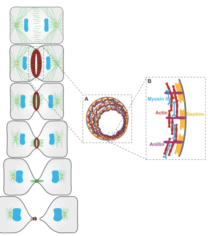
Structural analysis of contractile rings began with the advent of electron and fluorescence microscopy techniques. Early observations uncovered the invariable nature of the basic architecture of contractile rings (Schroeder, 1970; Schroeder, 1972). The contractile ring assembles beneath the plasma membrane as a very thin layer (0.1-0.2 µm) as measured by
electron microscopy (Schroeder, 1972). In general, contractile rings are 5-10 µm wide once assembled and when viewed in two dimensions (Schroeder, 1972).
Actin filaments (F-actin) and non-muscle myosin II are the major constituents of contractile rings (Figure 2.2.1B). F-actin and non-muscle myosin II present in contractile rings assemble as mini-filaments (Figure 2.2.1B) (Cao and Wang, 1990; Otto and Schroeder, 1990; Sanger and Sanger, 1980; Zhou and Wang, 2008). Upon contractile ring assembly, F-actin and non-muscle myosin II overlap at the equatorial cortex where they assemble into a ring and remain enriched throughout constriction (Cao and Wang, 1990; Otto and Schroeder, 1990; Zhou and Wang, 2008). Actin microfilaments of different orientations form bundles arranged circumferentially beneath the plasma membrane (Kamasaki et al., 2007; Mabuchi et al., 1988). Imaging adherent cells beneath the surface revealed the presence of myosin II mini-filaments accompanying F-actin (Zhou and Wang, 2008).
Actin filaments and non-muscle myosin II are the molecular drivers of contractile ring ingression, further described in a later section. When cells are treated with actin or myosin inhibitors, such as cytochalasin B or blebbistatin, contractile ring constriction is completely blocked (Mabuchi et al., 1988). Thus, actin and non-muscle myosin II mini-filaments assemble into a ring around the cell equator that constricts to physically partition the mother cell into two daughter cells.
Myosin II Actin Anillin Septins A B
Figure 2.2.1 Structural components of the contractile ring
(A) Schematic representing contractile ring constriction over time. (B) Magnified view on the contractile ring in A, depicting the principal structural components, non-muscle myosin II (blue), actin filaments (red), septins (yellow) and Anillin (purple). Adapted from (Green et al., 2012).
2.2.2 Molecular constituents required for contractile ring assembly
Additional components have been implicated in contractile ring assembly and ensure proper spatiotemporal coordination of contractile ring closure. As previously mentioned, activated RhoA at the equatorial cortex targets several effectors necessary for contractile ring assembly (Su et al., 2011). This includes formins that bind to GTP-bound RhoA and promote the assembly of actin filaments (Piekny et al., 2005). Formins drive actin nucleation and polymerization to generate actin filaments (Piekny et al., 2005). This process is also mediated by profilin, that binds to G-actin monomers and facilitates nucleation and polymerization by formins (Piekny et al., 2005).
Contractile ring assembly also requires the activation of the motor protein non-muscle myosin II. The RhoA effector Rho-dependent kinase (ROCK) is the primary kinase responsible for myosin II activation. ROCK phosphorylates the regulatory light chain (rMLC) of myosin II and promotes the assembly of myosin filaments (Kosako et al., 2000). ROCK also stimulates the inhibition of myosin phosphatases to prevent myosin II inactivation (Piekny et al., 2005). Upon ROCK inhibition cleavage furrow ingression is delayed (Kosako et al., 2000). This illustrates the importance of ROCK-dependent myosin II activation in the temporal control of cytokinesis. Other kinases, including Citron-kinase, are known to be implicated in myosin II activation (Yamashiro et al., 2003), but their contributions to cytokinesis are less well defined.
Another molecular constituent of the contractile ring is septin (Figure 2.2.1B). Mammalian septin complexes were found to co-localize with actin filaments and contribute to their bundling in vitro (Kinoshita et al., 2002). A later study confirmed the bundling activity of septins towards F-actin in Drosophila embryo furrow canals (Mavrakis et al., 2014). In addition, Drosophila septin (Peanut) was able to induce curvature of actin filament bundles (Mavrakis et al., 2014). Septin also interacts with lipids of the plasma membrane, suggesting that it plays a role in linking the underlying actin cytoskeleton to the membrane during cytokinesis (Tanaka-Takiguchi et al., 2009). A further role for septin and specific to C. elegans embryos is to promote asymmetric ingression of the contractile ring together with Anillin, as further described in a later section (Maddox et al., 2007). In sum, the recruitment of septins is required for proper contractile ring assembly.
2.2.3 The scaffolding protein Anillin
Anillin is a key structural component of the contractile ring. It functions as a scaffolding protein of contractile rings assembled during cytokinesis. Anillin is a multi-domain protein that binds several structural components of the cortical cytoskeleton, including actin filaments, myosin II and septin amongst others (Figure 2.2.1B) (Piekny and Maddox, 2010; Zhang and Maddox, 2010). Thus, Anillin serves diverse functions required for the proper spatiotemporal coordination of contractile ring closure. Characterization studies of Anillin across eukaryotes revealed its conserved nature (Field and Alberts, 1995; Field et al., 2005; Maddox et al., 2007; Oegema et al., 2000; Straight et al., 2005; Sun et al., 2015). In the following section, the numerous activities of Anillin during cytokinesis are addressed in detail. Anillin was first isolated from Drosophila embryo extracts (Field and Alberts, 1995). Early work with Drosophila provided important insights into the function of Anillin during cytokinesis. In vitro, Anillin was identified as an actin filament binding protein and a minimal domain of it was subsequently shown to be sufficient to bundle F-actin (Field and Alberts, 1995). In vivo studies showed that Anillin localized to contractile structures, including the contractile ring of Drosophila cultured cells and furrow canals (Field and Alberts, 1995; Field et al., 2005). Anillin co-localized with actin, myosin II and septin of Drosophila contractile furrow canals (Field et al., 2005). This work with Drosophila laid the foundation for functional characterization studies of Anillin.
Later work in mammalian cells supported the idea of a conserved role for Anillin during cytokinesis. Oegema and colleagues observed translocation of human Anillin from the nucleus in interphase to the contractile ring of dividing HeLa cells (Oegema et al., 2000). Functional analysis of the Anillin protein revealed its large multi-domain identity. The C-terminal region of Anillin comprises an Anillin Homology (AH) domain and a PH domain (Piekny and Maddox, 2010). Anillin recruits septins to the contractile ring via its PH domain (D'Avino et al., 2008; Hickson and O'Farrell, 2008; Oegema et al., 2000). Additional studies showed that Anillin interacted with PIP2 component of the plasma membrane via its PH domain (Liu et al., 2012; Sun et al., 2015). In mammalian cells, this interaction is required for the recruitment of Anillin to the contractile ring (Liu et al., 2012). This was previously demonstrated by introducing mutations in the PH domain of Anillin that resulted in failure to
recruit Anillin to the contractile ring (Oegema et al., 2000). These studies also suggested that Anillin provided a link between the plasma membrane and the underlying furrow (Liu et al., 2012).
Mechanistic insights regarding the recruitment of Anillin to the division plane were obtained via the expression of tagged truncations in mammalian cultured cells (Piekny and Glotzer, 2008). In this elegant study in mammalian cells, Anillin was depleted by RNAi and various Anillin constructs harboring different domain truncations and tagged with the green fluorescent protein (GFP) were re-introduced in the cell (Piekny and Glotzer, 2008). Truncation of the PH domain prevented Anillin enrichment at the contractile ring as previously demonstrated. Cytokinesis failure was observed upon truncation of both the actin and myosin II binding domains located at the N-terminal region of the protein. Finally, removal of the AH domain caused defects during furrow ingression, as Anillin translocated at the cell poles leading to furrow oscillation (Piekny and Glotzer, 2008). Furthermore, the authors observed the co-localization of Anillin and RhoA at the cytokinetic furrow (Piekny and Glotzer, 2008). These proteins interacted in vitro via Anillin’s AH domain. Cells depleted of either RhoA or its GEF activator Ect2 failed to accumulate Anillin at the equatorial cortex (Hickson and O'Farrell, 2008; Piekny and Glotzer, 2008). Thus, RhoA is responsible for recruiting Anillin to the equatorial cortex and the PH domain of Anillin is essential for its enrichment at the contractile ring (Hickson and O'Farrell, 2008; Piekny and Glotzer, 2008).
Anillin possesses additional interacting partners during cytokinesis suggesting that it serves other activities. However, it is important to note that these interactions are not as well characterized. Anilin interacts with the F-actin polymerization protein formin (Watanabe et al., 2010). Anillin participates in the recruitment of formin to the equatorial cortex required for actin nucleation and polymerization (Watanabe et al., 2010). In Drosophila Anillin directly interacts with the centralspindlin complex component CYK-4 (Drosophila RacGAP50C) suggesting a role for Anillin in division plane establishment (D'Avino et al., 2008; Hickson and O'Farrell, 2008). Thus, Anillin performs numerous activities during cytokinesis.
Anillin is an essential structural component of the contractile ring ensuring proper contractile ring closure. As previously mentioned, truncations of either the myosin II or the actin-binding domain of Anillin results in furrow oscillation and contractility at the poles,